cell division lecture
DESCRIPTION
all about cell division interphasemitosisprophase metaphaseanaphase telophaseTRANSCRIPT

Cell Division

Cell division
Is a process where a cell (parent cell) divides into two/four cell (daughter cell)
In mitotic division, the daughter cells can become parent cells and undergo subsequent division
In meiotic division, the daughter cells will be permanently transformed and cannot divide again
For unicellular organisms such as the Amoeba, one cell division reproduces an entire organism
Cell division can create progeny from multi-cellular organisms, such as plants that grow from cuttings

Cell division also enables sexually reproducing organisms to develop from one-celled zygote, which itself was produced by cell division from gametes
Cell division allows for continual renewal and repair of the organism

Prokaryotes cell division
Bacteria divide by binary fission.-the single, circular bacterial chromosome is
replicated-replication begins at the origin of replication
and proceeds bidirectionally-new chromosomes are partitioned to opposite
ends of the cell-a septum forms to divide the cell into 2 cells

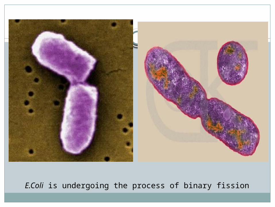
E.Coli is undergoing the process of binary fission

Eukaryotes
Eukaryotes must divide their nucleus (and other organelles such as mitochondria) in preparation for cell division (mitosis or meiosis)
Before the nucleus divides, the genetic material replicates

Eukaryotic Chromosomes
Eukaryotic chromosomes – -linear chromosomes-every species has a different number of
chromosomes-composed of chromatin – a complex of DNA
and proteins-heterochromatin – not expressed-euchromatin – expressed regions

Chromosomes are very long and must be condensed to fit within the nucleus.
-nucleosome – DNA wrapped around a core of 8 histone proteins
-nucleosomes are spaced 200 nucleotides apart along the DNA
-further coiling creates the 30-nm fiber or solenoid

The solenoid is further compacted:-radial loops are held in place by scaffold
proteins-scaffold of proteins is aided by a complex of
proteins called condensin
karyotype: the particular array of chromosomes of an organism

Chromosomes must be replicated before cell division.
-Replicated chromosomes are connected to each other at their centromeres
-cohesin – complex of proteins holding replicated chromosomes together
-sister chromatids: 2 copies of the chromosome within the replicated chromosome

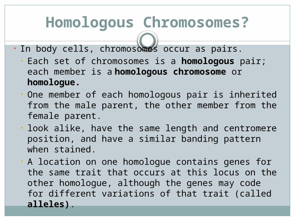
Homologous Chromosomes?
• In body cells, chromosomes occur as pairs. • Each set of chromosomes is a homologous pair;
each member is a homologous chromosome or homologue.
• One member of each homologous pair is inherited from the male parent, the other member from the female parent.
• look alike, have the same length and centromere position, and have a similar banding pattern when stained.
• A location on one homologue contains genes for the same trait that occurs at this locus on the other homologue, although the genes may code for different variations of that trait (called alleles).


Cell cycle
Series of events that take place in a eukaryotic cell leading to its duplication through cell division
Length of a complete cell cycle varies among cell types
5 main phases G1 (Growth Phase 1) S (Synthesis) G2 (Growth Phase 2) M (Mitosis) C (Cytokinesis)
Interphase

G1 Phase
The first phase within interphase, from the end of the previos M phase till the beginning of DNA synthesis is called G1
During this phase, the biosynthetic activities of the cell, which had been considerably slowed down during M phase, resume at high rate
This phase is marked by synthesis of various enzymes that are required in S phase, mainly those needed for DNA replication
Duration of G1 is highly variable, even among different cells of the same species

S Phase
Starts when DNA synthesis commences: when it is complete, all of the chromosomes have been replicated, eg: each chromosome has two (sister) chromatids)
The amount of DNA in the cell has effectively doubled, though the ploidy of the cell remains the same
Rates of RNA transcription and protein synthesis are very slow during this phase
An exception to this is histone production, most of which occurs during the S phase
The duration of S phase is relatively constant among cells of the same species

G2 Phase
Cell undergoes a period of rapid growth to prepare for mitosis
Significant protein synthesis occurs during this phase, mainly involving the production of microtubules, which are required during the process of mitosis
Inhibition of protein synthesis during G2 phase prevents the cell from undergoing mitosis

Control of the Cell Cycle
• The cell cycle is controlled by both internal and external signals.
• A signal is a molecule that either stimulates or inhibits a metabolic event.
• Growth factors are external signals received at the plasma membrane.

Cell Cycle Checkpoints
• There appear to be three checkpoints where the cell cycle either stops or continues onward, depending on the internal signals it receives.
• Researchers have identified a family of proteins called cyclins, internal signals that increase or decrease during the cell cycle.
• Cyclin must be present for the cell to move from the G1 stage to the S stage, and from the G2 stage to the M stage.

• The cell cycle stops at the G2 stage if DNA has not finished replicating; stopping the cell cycle at this stage allows time for repair of possible damaged DNA.
• Also, the cycle stops if chromosomes are not distributed accurately to daughter cells.
• DNA damage also stops the cycle at the G1 checkpoint by the protein p53; if the DNA is not repaired, p53 triggers apoptosis

Mitosis
Method of eukaryotic cell division that produces two genetically identical cells
All cells in an organism except sperm and eggs are produced by mitosis
Basis of asexual reproduction5 stages;
Prophase Metaphase Anaphase Telophase Cytokinesis
Interphase
The chromosomes are extended long threads that cannot be visibly identified. The DNA of the chromosomes is replicated during this phase, resulting in duplication of the genetic material


Prophase
The duplicated chromosomes condense and become visible as distinct sister chromatids
The nuclear envelope breaks down and the centrosomes move toward the poles of the cell
The mitotic spindle attached to a specialized structure called the kinetochore, located at the centromere of each replicated chromosome

Prophase
Pair of centriole
s
Aster
Chromosome consisting of 2
sister chromatids
Centromere

Metaphase
The replicated chromosomes align at the equator (metaphase plate) of the cell

Anaphase
The sister chromatids separate and are moved toward opposite poles of the cell by the spindle.
The cell begins to elongate towards the poles


Telophase
The cell continues to elongate throughout telophase
The mitotic spindle breaks downA new nuclear envelope forms at each end of
the cellThe chromosomes within begin to unfold into
chromatin


Cytokinesis
The cytoplasm and organelles are evenly divided between the two new cells during cytokinesis, completing the processs of cell division
Cytokinesis differs skightly in animals and plants. Animal: a ring of microfilaments contracts in the
center of the elongated cell, producing a cleavage furrow that eventually pinches off the two cells
Plant: a cell plate is formed as vesicles containing cell membrane materials fuse together along the equator of the cell. Once the cell has fused with the plasma membrane and the two cells are completely divided, cellulose is secreted to form the cell wall.


What is the purpose of all of this?
Mitosis is the way cells (within tissues and organisms) grow and repair
GrowthRepair

Meiosis
Method of cell division that takes place in sexually reproducing organisms specifically for the creation of gametes (egg and sperm cells)
Results in the production of 4 haploid cells, each is genetically different
To produce 4 cells, meiosis requires two rounds of cell division Meiosis 1 Meiosis 2

Meiosis I
Homologous pairs of each chromosome join and might exchange genetic material
The homologous chromosome are pulled to opposite poles in the cell, at which the cell separates, resulting in two cells
Each chromosome remains in the duplicated state and is made up of two sister chromatids

Occurs in 5 stage similar to mitosis Prophase 1 Metaphase 1 Anaphase 1 Telophase 1 Cytokinesis 1
During the S phase of interphase (prior to start of meiosis I), each chromosome replicates to produce two sister chromatids
The 2 genetically homologous sister chromatids remain attached at their centromeres

Prophase 1
Synapsis and crossing over occurred Synapsis – two homologous chromosome condense
and combine to form complexes called tetrads Crossing over – exchange of genetic material that
takes place between these homologous chromosomes along several junctions known as chiasmata. Result of crossing over – cells are genetically variable
Other events is similar to those occurred during prophase in mitosis

Metaphase 1
The tetrads align along the metaphase plate of the cells

Anaphase 1
The homologous chromosome of each tetrads separate and are pulled toward opposite poles of the cell by the spindle
The side of the cell toward which a homologous chromosome is pulled is random, depending only on the orientation of the tetrad.
Telophase 1
Identical to telophase in mitosisCell continues to elongate and the mitotic
spindle breaks downA new nuclear envelope forms at each end of
the cell and the chromosomes within unfold into chromatin

Cytokinesis 1
Identical to cytokinesis in mitosisEnd of this stage, 2 genetically different
haploid cells have been producedEach chromosome is still in the dulpicated
state and is made up of two sister chromatidsFor human; starts with one cell containing 46
chromosomes (23 pairs) to 2 cells containing 23 chromosomes
Result of crossing over- each chromosome is the mixture of the original homologous


Meiosis II
Follows similar steps as meiosis I in the creation of two or more cells
Chromosome do not replicate between Meiosis I and Meiosis II.
The result is 4 haploid cells genetically different from one another

Prophase II
The chromosome within the haploid cell condense
The spindle attaches to the kinetochore of each chromosome
The nuclear envelope breaks down and the centrosomes move toward the poles of the cell

Metaphase II
The chromosome align along the center of the metaphase plate

Anaphase II
The sister chromatids separate and are moved toward opposite poles of the cell by the spindle
Cell begins to elongate towards the poles

Telophase II
The cell continues to elongate and the mitotic spindle breaks down
A new nuclear envelope forms at each end of the cell

Cytokinesis II
The cytoplasm and organelles are divided between the two cells, completing the process of cell division
4 genetically different haploid cells have been produced


How can a male (diploid; 46 chromosomes) and a female (diploid; 46 chromosomes) produce a child with 46 chromosomes?
Why do you look somewhat similar - but distinctly different - from your brothers and sisters, and from your parents, if you all basically have the same chromosomes? Each meiosis and fertilization is literally like a 'roll of a dice' - so no two individuals are alike!

1. Crossing Over: The chromosomes you receive from your mom's egg and your dad's sperm and NOT the same chromosomes that your mom and dad have. They are new, 'shuffled' versions of their chromosomes - and you only get ONE of them from each parental unit....
2. Independent assortment: Homologues line up or "shuffle" randomly on the metaphase plate in Meiosis I. With 23 chromosomes assorting independently, there are 2^23, or 8 million, possible assortments of chromosomes inherited for every cell!!
3. Random fertilization: The ovum has 8 million possible chromosome combinations, so does the sperm cell. 8 million x 8 million = 64 trillion possible diploid combinations in EACH AND EVERY zygote! WOW!!