cell apoptosis induced by -elemene in colorectal
TRANSCRIPT

hon p.1 [100%]
1403
e-mail: yangwei0719@163.comFig. 1. Chemical Structure of b-Elemene, g-Elemene and d-
Elemene
1403YAKUGAKU ZASSHI 129(11) 1403―1413 (2009) 2009 The Pharmaceutical Society of Japan
―Regular Articles―
Cell Apoptosis Induced by d-Elemene in Colorectal AdenocarcinomaCells via a Mitochondrial-mediated Pathway
Chun-Ying XIE,a Wei YANG,c Ming LI,e Jun YING,c
Shu-Juan TAO,d Karen LI,e Jin-Hua DONG,d and Xi-Sha WANG,b
aThe Sixth A‹liated Hospital, SUN YAT-SEN University, bCollege of Pharmacy, Guang Dong PharmaceuticalUniversity, No. 280 Waihuan Rd., Higher Education Mega Center of Guangzhou, Guangdong, China,
cCenter for Drug Non-clinical Research and Evaluation of Pharmaceutical Industrial ResearchInstitute in Guangzhou, dDepartment of Chemical Pharmaceutics, Shenyang Pharmaceutical
University, and ePrince of Wales Hospital, Chinese University of Hong Kong
(Received April 21, 2009; Accepted July 6, 2009)
The chemical compound d-elemene, isolated from the Chinese herbal medicine plant Curcuma Wenyujin, has beenknown to exert antitumor activity. In this study we demonstrated that apoptotic cell death induced by d-elemene inDLD-1 cells was concentration-and time-dependent, and had little inhibition of the normal human liver cell lineWRL-68. Apoptosis was further conˆrmed and quantiˆed by DNA fragmentation ELISA, Annexin V (AnV) binding ofexternalized phosphatidylserine and the mitochondrial probe JC-1 using ‰ow cytometry. The rapid increase in intracel-lular reactive oxygen species (ROS) levels was involved in the mechanism of cell death. Western blot analysis demon-strated that d-elemene activated the caspase-signaling pathway, leading to the proteolysis conversion of pro-caspase-3 toactivate caspase-3, and the subsequent cleavage of the caspase substrate PARP. In the process of the induction of apop-totic cell death, Bax translocated into mitochondria, a reduction in Dwm was observed and a release of cytochrome c andapoptosis inducing factor (AIF) from mitochondria into the cytosol occurred, indicating that cell death induced by d-elemene was through a mitochondrial-mediated pathway.
Key words―d-elemene; apoptosis; mitochondria; human colorectal adenocarcinoma
INTRODUCTION
Elemene is a naturally occurring compound thatcan be isolated from the traditional Chinese medicinalherb Curcuma Wenyujin, which is used to treattumors in Chinese folk medicine.1,2) Elemene exists asan essential oil mixture of b-, g- and d-elemene (Fig.1). The major antitumor active component, b-ele-mene, possesses broad-spectrum clinical activity intreatment of various tumors and is known to induceapoptosis.3) d-elemene is another isomeric compoundof b-elemene with a diŠerent site of double bond. Ourprevious study was shown that d-elemene exerts an-titumor activity by inducing apoptosis in Hela cells,4)
and possesses no signs of bone marrow cells and nor-mal liver cell lines WRL-685) suppression.
Apoptosis, deˆned as programmed cell death, canbe initiated by a death receptor-mediated pathway orby a mitochondria-mediated pathway.6) The deathreceptor pathway is initiated when a speciˆc ligandbinds to a tumor necrosis factor (TNF) receptor su-
perfamily member. The mitochondrial pathway is in-itiated by the translocation of Bax into mitochon-dria.7) This is followed by an increase in mitochondri-al membrane permeability (Dwm) and a release ofcytochrome c (cyto c) and apoptosis inducing factor(AIF) from mitochondria into the cytosol. In thecytosol, cyto c activates post-mitochondrial caspasecascades, caspase-3 and caspase-9, which leads toapoptotic cell death.8) AIF is a caspase-independentapoptotic eŠector which, on translocating from thecytosol to the nucleus, causes chromatin condensa-tion and large scale DNA fragmentation, leading toapoptotic cell death.9)

hon p.2 [100%]
14041404 Vol. 129 (2009)
In this communication, the antitumor activity andthe potential mechanisms of d-elemene were investi-gated. Reactive oxygen species (ROS) are constantlygenerated under normal conditions as a consequenceof aerobic metabolism.10) There is a growing consen-sus that oxidative stress and the redox state of a cellplay a pivotal role in regulating apoptosis.11,12) Oxy-gen free radical formation is associated with thegeneration and development of certain tumors. Ele-mene has an inhibitory eŠect on oxygen free radicalformation in tumor cells.13) Mitochondria are therichest source of ROS in the cell, converting 12% ofreduced oxygen into superoxide.14) Inhibition of themitochondrial electron transport chain, resulting insubsequent release of ROS, is an early event in manyforms of apoptosis.15,16) Oxidative stress induces anumber of downstream events in apoptosis, includingaspartate-speciˆc cysteine protease (caspase) activa-tion and DNA fragmentation. The caspase cascadeappears to be the main pathway by which cellulardeath is orchestrated. Poly (ADP-ribose) polymerase(PARP) is a nuclear enzyme selectively activated byDNA strand damage to participate in DNA repair.17)
In this study we explored the intracellular mecha-nism of d-elemene induced cell death in DLD-1 cells,human colorectal adenocarcinoma cell line. We inves-tigated the roles and relationships of ROS, mitochon-drial in DLD-1 cells treated with d-elemene. Ourresults suggest that d-elemene induced apoptotic celldeath in DLD-1 cells is mediated by an increase inROS generation and the activation of caspase-3 andmitochondria-mediated apoptosis pathways.
MATERIALS AND METHODS
Cell Lines Human colorectal adenocarcinomacell line DLD-1 and normal human liver embryonicWRL-68 line were purchased from American TypeCulture Collection (ATCC, #CRL221, #CL48, MD,USA). DLD-1 cells were cultured in RPMI-1640medium supplemented with 10% fetal bovine serum(FBS; GIBCO, Invitrogen), 100 U/ml of penicillin,and 100 mg/ml of streptomycin (GIBCO, Grand Is-land, NY, USA). WRL-68 cells were cultured in a-Minimum Essential Medium (GIBCO BRL, Rock-ville, MD, USA) with 10% FBS. Cells were incubatedin a humidiˆed atmosphere of 5% CO2 in air at 37°C.
Reagent and Cell Culture d-elemene and b-ele-mene were isolated from the essential oil of CurcumaWenyujin using the method reported in reference1)
(assay>97%, GC).Determination of Anti-proliferation Activity Using
MTT on DLD-1 Cell Lines Survival of cells wasassessed using 3-(4,5-dimethylthiazol-2-yl)-2,5-diphen-yltetrazolium bromide (MTT) (thiazolyl blue, Sig-ma, MO, USA) as previously described.18) Brie‰y,DLD-1 cells were cultured to 80% con‰uence andthen exposed to the indicated concentrations of d-ele-mene or b-elemene at 200 mM for 12, 24, 48 h. Twentymicroliters of 5 mg/ml stock solution of MTT wereadded to each well, and incubated at 37°C for 4 h, af-ter which, 150 ml of dimethyl sulfoxide (DMSO) wasadded to each well. The absorbance was measured us-ing a 96-well micro-titer plate reader at 570 nm. Thepercentage of cell growth inhibition was calculated asfollows:
Inhibition(%)=[A570(control)-A570(drug)]/A570(control)×100%
Assessment of Cell DNA Fragmentation DNAfragmentation was determined by a DNA fragmenta-tion ELISA kit (Roche Molecular Biochemicals,Mannheim, Germany). DLD-1 cells were cultured to80% con‰uence and then exposed to diŠerent concen-trations of d-elemene for the periods of time indicat-ed. L-Glutathione (GSH) and z-DEVD-fmk were ad-ded 1 or 2 h prior to the treatment with d-elemene.
Flow Cytometric Detection of Annexin-V (AnV)
Apoptosis was measured using ‰ow cytometry toquantify the levels of detectable phosphatidylserine(PS) externalization on the outer membrane of apop-totic cells.19) Brie‰y, DLD-1 cells were plated at 5×10⁴/ml in RPMI1640. Flasks were incubated with 200mM d-elemene. GSH was added 1 h prior to the treat-ment with d-elemene. Cells were harvested, suspend-ed at 1×10⁶/ml, and washed two times with ice-coldPBS after 12, 24, 48 h. The cells were suspended in300 ml of dilute binding buŠer from an Annexin-VFITC kit (Alexis, Lausen, Switzerland), then 5 ml ofpropidium iodide (PI) and 5 ml of Annexin-V-FITCwere added. The tubes were gently mixed and kept inthe dark on ice for 10 min before analysis by ‰owcytometry (FACScan, Becton Dickinson, San Jose,CA, USA). Data were analyzed using LYSIS IIsoftware. (AnV+)PI- cells were considered earlyapoptotic, and (AnV+)PI+ cells were considered lateapoptotic and necrotic.
Measurement of Generation of Reactive OxygenSpecies Hydroperoxide and superoxide produc-tion was determined using 2',7'-dichloro‰uorescein

hon p.3 [100%]
14051405No. 11
and dihydroethidium (CM-H2DCFDA; MolecularProbes Inc., Eugene, OR, USA.) as described byCurtin and Donovan,20) with some modiˆcations.Brie‰y, after the treatment of cells with 200 mM ofd-elemene for 6, 12 and 24 h, DLD-1 cells were col-lected by centrifugation and suspended in 200 ml ofRPMI medium, after which the cells were loaded withCM-H2DCFDA (10 mM) for 45 min at 37°C and thenwashed with PBS to remove the CM-H2DCFDA. Thelevels of ROS were measured by ‰ow cytometry bydetermining the ‰uorescence intensity relative to thatof the control group. GSH was added 1 h prior to thetreatment with d-elemene for 3 h.
Mitochondrial Transmembrane Potential Measure-ment The mitochondrial transmembrane poten-tial (Dwm) was evaluated by JC-1 assay. In the treat-ment of cells with 200 mM of d-elemene for 6, 12 and24 h, DLD-1 cells were suspended in 200 ml of RPMImedium, after which JC-1 (300 nM, MolecularProbes) was added for 15 min incubation at 37°C inthe dark. The cells were then harvested by trypsin(0.25%) and washed once with PBS. Followingresuspension in PBS, the cells were subjected to ‰owcytometry analysis. GSH was added 1 h prior to thetreatment with d-elemene for 3 h.
Preparation of Proteins in the Mitochondrial andCytosolic Fractions Preparation of proteins wasperformed as previously described10) with minormodiˆcations. The cells were washed twice in ice-coldPBS and resuspended in ˆve volumes of ice-cold ex-tract buŠer (20 mM Western blotting analysis Hepes-KOH, 1.5 mM MgCl2, 1 mM EDTA, 1 mM EGTA, 1mM DTT, and 0.1 mM phenylmethanesulfonyl ‰uo-ride (PMSF), pH 7.5). The resuspended cells werehomogenized with 10 strokes of a Te‰on homogeniz-er. The homogenates were centrifuged twice at 750 gfor 10 min at 4°C. The supernatants were centrifugedat 10 000 g for 15 min at 4 to obtain the mitochondri-al pellets. Cytosolic fractions were obtained after fur-ther centrifugation at 100 000 g for 1 h at 4°C. Theprotein concentrations of the resulting supernatantsand mitochondrial fractions were measured andstored at -70°C.
Western Blotting Analysis Brie‰y, 10 mg ofprotein from the mitochondrial or cytosolic fractionand 40 mg of total protein from treated cells wereloaded onto l2% sodium dodecyl sulfate (SDS)-polyacylamide gels, and the separated proteins trans-ferred to nitrocellulose membranes (Amersham Bio-
sciences, Piscataway, NJ). To detect the levels ofBax, cyto c and AIF a mouse-anti-human monoclonalBax antibody (Santa Cruz Biotechnology, SantaCruz, CA), a mouse-anti-human monoclonal cyto cantibody (BioVision Research Products, MountainView, CA) and a mouse-anti-human monoclonal AIFantibody (Santa Cruz Biotechnology) were employedwith a horseradish peroxidase-conjugated goat-anti-mouse IgG antibody (Santa Cruz Biotechnology).The level of cleaved caspase-3 was detected by using agoat polyclonal caspase-3 antibody (Santa CruzBiotechnology) as the primary antibody and a poly-clonal mouse-anti-goat IgG antibody as the secondaryantibody both from Santa Cruz Biotechnology. Thelevel of 85 kDa-cleaved PARP was detected using arabbit polyclonal PARP antibody (Promega, Madis-on, WI) as the primary antibody and a polyclonalgoat-anti-rabbit IgG antibody as the secondary an-tibody (Santa Cruz Biotechnology). A goat-anti-hu-man actin antibody was used to detect the levels of ac-tin, which was used as a control for equal loading.Detection of the target protein signal was achieved us-ing an ECL system (Amersham Bioscences).
Statistical Analysis All results were obtained inat least three independent experiments. Data were ex-pressed as means±S.D. Representative data wereanalyzed for statistical signiˆcance by softwareSPSS11.5. DiŠerences among groups were analyzedby One-Way ANOVA, multiple comparisons usedLSD and SNK's test for homogeneity of variance andDunnett's T3-test for heterogeneity of variance. p<0.05 was considered statistically signiˆcant.
RESULTS
Inhibition of Growth of DLD-1 Cells Was Doseand Time Dependent When DLD-1 cells in cul-ture were treated with various concentrations of d-ele-mene (0-400 mM) for 24 h (Fig. 2a) or with 200 mMd-elemene for indicated periods (Fig. 2b), the growthof these cells was signiˆcantly inhibited in time anddose-dependent manners. This inhibitory action wasclosely similar to that of b-elemene, and there was nosigniˆcant diŠerence between b-elemene and d-ele-mene treatment (Fig 2a). The inhibitory concentra-tion, 50% (IC50) value of d-elemene and b-elemenefor DLD-1 cells was 222.4 mM and 206.4 mM, respec-tively, when treated for 24 h. The IC50 of DLD-1 cellsat 12, 24 and 48 h was 308.4, 222.4 and 159.8 mM,respectively (Fig. 2b). However, the viability of nor-

hon p.4 [100%]
1406
Fig. 2. Dose- and Time-dependent Inhibition of Proliferationof DLD-1 Cells by d-Elemene Treatment(a) DLD-1 cells were treated with various doses of d-elemene and com-
pared with b-elemene for 24 h. (b) Cells treated with 200 mM d-elemene forvarious time periods. (c) EŠect of d-elemene in DLD-1 cells and WRL-68cells for 24 h. The results shown are representative of 3 independent experi-ments.
Fig. 3. EŠect of d-Elemene on DNA Fragmentation inDLD-1 Cells
Cells were treated with various concentrations of d-elemene (0200 mM)for 24 h (a) or with 200 mM of d-elemene for indicated periods (b). TheDNA fragmentation was determined by cellular DNA fragmentation ELISAassay. In the experiment using GSH and z-DEVD-fmk, cells were pretreatedwith 1 mM GSH or with 2 mM z-DEVD-fmk 1 or 2 h prior to the treatmentby 200 mM of d-elemene for 24 h (c). p<0.01 vs. control group; #p<0.05,##p<0.01 vs. 200 mM of d-elemene for 24 h.
1406 Vol. 129 (2009)
mal human cell line WRL-68 was almost unaŠecttedat 200 mM after treatment with d-elemene (Fig. 2a).
DNA Fragmentation in DLD-1 Cells Treated withd-Elemene d-Elemene is able to signiˆcantly in-duce DNA fragmentation, and the amount of frag-mentation was increased in a dose-dependent manner;there was 3.8 fold increase in 200 mM d-elemene for24 h with vector control cells (Fig. 3c. p<0.01 vs.control). The time course study showed that theamount of DNA fragmentation was elevated aftertreatment for 12 h (2.5 fold that of the vector con-trol), and it peaked at 48 h (4.0 fold that of the vec-
tor control) after treatment with 200 mM d-elemene(Fig. 3b). Furthermore, the DNA fragmentation inDLD-1 cells treated with d-elemene was inhibited byGSH or z-DEVD-fmk (Fig. 3c, #p<0.05, ##p<0.01vs. 200 mM d-elemene) for 24 h.
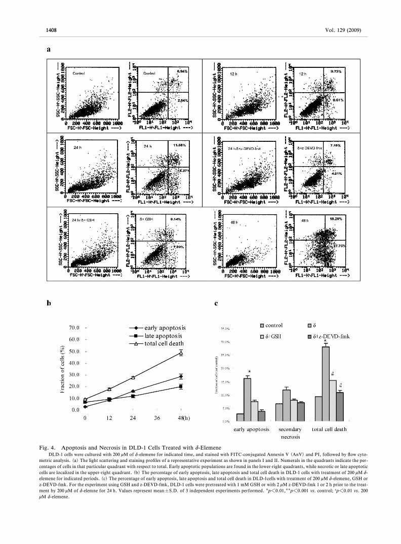
Cell Death Induced by d-Elemene Was in Mannerby Apoptosis Results showed that apoptosis wastriggered after treatment for 12 h (Fig. 4a). The per-centage of early apoptosis represented by (AnV)+
PI- cells was signiˆcantly increased by d-elemenetreatment in a time-dependent manner with 29% at 48h (Fig. 4b). Total cell death, which comprised earlyapoptotic and late apoptotic cells, calculated by the

hon p.5 [100%]
14071407No. 11
summation of the (AnV)+PI- and (AnV)+PI+ cellpopulations, was also increased at 24 h. Comparedwith the untreated control, more death events wereobserved in d-elemene treated cells for 48 h. Further-more, the rate of early apoptosis in DLD-1 cells treat-ed with d-elemene was inhibited by GSH or z-DEVD-fmk (Fig. 4c, #p<0.01, ##p<0.001 vs. 200 mM d-ele-mene) for 24 h.
Cell Death Induced by d-Elemene Was Associatedwith an Increase in ROS Generation ROS genera-tion in DLD-1 cells treated with d-elemene was ana-lyzed by ‰ow cytometry. The treatment of DLD-1cells with 200 mM d-elemene resulted in a time-depen-dant increase in ROS generation (Fig. 5a). As shownin Fig. 5b, there was approximately 7.8, 18.2 or 21.8fold increase in generation of ROS in 200 mM d-ele-mene for 1, 3 or 6 h compared with vector controlcells, a statistically signiˆcant increase (p<0.001and p<0.01). Furthermore, the change of ROS in-duced by d-elemene was inhibited by GSH to a level(3.46%) that was almost as low as the untreated con-trol (2.11%).These results indicate that the apoptosispathway in DLD-1 cells by d-elemene was associatedwith the generation of ROS.
Mitochondria Pathway Was Involved in CellApoptosis Induced by d-Elemene 1. Reduction inDwm of DLD-1 cells treated with d-elemene: To exa-mine whether d-elemene could induce a change ofDwm, we measured Dwm in DLD-1 cells treated withthis compound. Following treatment with d-elemene,a reduction in Dwm of the cells was observed (Fig.6a). The Dwm started to decrease at 6 h followingtreatment with d-elemene and the reduction occurredin a time-dependent manner. Compared with controlcells, DLD-1 cells with 200 mM d-elemene treatmentfor 6 h exhibited green JC-1 ‰uorescence, which isconsistent with a loss of mitochondrial membranepolarization. As shown in Fig. 6b, the trends of in-creasing incidences of compromised Dwm for 6, 12 or24 h were 2.24 ± 0.12, 2.85 ± 0.33, 5.11 ± 0.24 foldwith respect to the untreated control, respectively(Fig. 6b, p<0.05, p<0.01, respectively, vs. un-treated control, n=3).
2. Translocation of Bax to mitochondria in DLD-1cells treated with d-elemene: Translocation of Bax tomitochondria can alter the outer mitochondrial mem-brane permeability. It allows the release of pro-apop-totic proteins such as cyto c and AIF into the cytosol.To test whether the apoptosis-inducing eŠect of d-ele-
mene was related to the translocation of Bax intomitochondria, we determined the level of Bax in themitochondrial fraction of DLD-1 cells treated with d-elemene by Western blot. As shown in Fig. 7, the levelof Bax in mitochondria of DLD-1 cells treated with d-elemene increased in a time dependent manner, im-plying that the translocation of Bax into mitochon-dria was involved in cell death induced by d-elemene.
3. Release of cyto c and AIF from mitochondriainto the cytosol in d-elemene-treated DLD-1 cells:DLD-1 cells were treated with 200 mM of d-elemenefor various determined periods of time (Fig. 7).Analysis of cyto c and AIF in the cytosol by Westernblot than showed that the d-elemene levels in thesecells had increased in a time dependent manner (Fig.7). This implies that after d-elemene treatment, therelease of cyto c and AIF from mitochondria into thecytosol was involved in cell death induced by d-ele-mene.
Activation of Caspase-3 and Cleavage of PARPWere Involved in Cell Death Induced by d-Elemene The caspase signaling pathway has been demon-strated to play a central role in cellular apoptosis. Thereduction of procaspase-3 indicates the activation ofcaspase-3. Two hundred mM of d-elemene was able tocleave procaspase-3 (32 kDa) to activate caspase-3after treatment for 12 h in Fig. 8a. PARP, acting in-tranuclearly to repair damaged DNA, is one of thesubstrates of activated caspase. As shown in Fig. 8a,200 mM d-elemene was able to cleave intact PARP(116 kDa) into an 85 kDa fragment after treatmentfor 6 h in a time-dependent manner. The reduced lev-el of procaspase-3 and elevated level of cleaved PARPwere much more signiˆcant than that of the untreatedcells in Fig. 8.
DISCUSSION
Recently, the exploration for non-cytotoxic an-titumor drug has been a major trend in the interna-tional medicine area. The cytotoxicity of elemene hasbeen determined in various tumor cell lines invitro.21,22) The IC50 of elemene for normal peripheralblood leukocytes is 1.244 mM.23) The present studyshowed that d-elemene inhibited DLD-1 cell growthin a time- and dose- dependent manner. The IC50
values of d-elemene and b-elemene in DLD-1 cellswere 222.4 mM and 206.4 mM, respectively, and weresimilar to the IC50 (182.4 mM) of elemene for DLD-1cells.24) In this study the eŠect of d-elemene was com-
hon p.6 [100%]
1408
Fig. 4. Apoptosis and Necrosis in DLD-1 Cells Treated with d-ElemeneDLD-1 cells were cultured with 200 mM of d-elemene for indicated time, and stained with FITC-conjugated Annexin V (AnV) and PI, followed by ‰ow cyto-
metric analysis. (a) The light scattering and staining proˆles of a representative experiment as shown in panels I and II. Numerals in the quadrants indicate the per-centages of cells in that particular quadrant with respect to total. Early apoptotic populations are found in the lower-right quadrants, while necrotic or late apoptoticcells are localized in the upper-right quadrant. (b) The percentage of early apoptosis, late apoptosis and total cell death in DLD-1 cells with treatment of 200 mM d-elemene for indicated periods. (c) The percentage of early apoptosis, late apoptosis and total cell death in DLD-1cells with treatment of 200 mM d-elemene, GSH orz-DEVD-fmk. For the experiment using GSH and z-DEVD-fmk, DLD-1 cells were pretreated with 1 mM GSH or with 2 mM z-DEVD-fmk 1 or 2 h prior to the treat-ment by 200 mM of d-elemne for 24 h. Values represent mean±S.D. of 3 independent experiments performed. p<0.01,p<0.001 vs. control; #p<0.01 vs. 200mM d-elemene.
1408 Vol. 129 (2009)

hon p.7 [100%]
1409
Fig. 5. The Increase in ROS Generation Related to Cell Apoptosis Induced by d-Elemene in DLD-1 CellsCells were treated with 200 mM of d-elemene for 1, 3 and 6 h. After treatment, cells were stained with 10 mM CM-H2DCFDA for 45 min at 37°C and analyzed by
‰ow cytometry (a). The horizontal axis shows the relative ‰uorescence intensity, and the vertical axis shows cell numbers. In the experiment of inhibition by GSH onthe generation of ROS, DLD-1 cells were pretreated with 1 mM GSH 1 h prior to the treatment with 200 mM of d-elemene for 3 h. The results shown are representa-tive of three independent experiments. The data represent a mean of three independent experiments.p<0.001 and p<0.01, compared with control.
1409No. 11
pared between several tumor cell lines and a normalhuman liver embryonic cell by MTT assays. It ap-pears that the growth inhibitory eŠect of d-elemeneon DLD-1 cells is much stronger than that on normalliver cells.5) d-elemene has no signiˆcant inhibitoryaction on normal cells, and it is suggested that d-ele-mene may be a new non-cytotoxic compound.
DNA fragmentation, PS externalization and theloss of Dwm are indication that the DLD-1 cell deathwith d-elemene are in an apoptosis manner.
ROS production frequently occurs in response to
various stimuli. In many experimental situations, theinduction of apoptosis is accompanied by a rise in in-tracellular ROS.25) Furthermore, the observed inhibi-tion of apoptosis by diŠerent antioxidants such asNAC,25,26) ascorbate27) and a-tocopherol27) suggeststhat ROS production plays an important role in apop-tosis in diverse cell lines. Our results showed that ROSwere involved in apoptosis in DLD-1 cells by d-ele-mene. Further, it was noted that the DNA fragmenta-tion in DLD-1 treated with d-elemene was inhibitedby GSH (Fig. 3c), that GSH could attenuate early

hon p.8 [100%]
1410
Fig. 6. Reduction in Dwm of DLD-1 Cells Treated with d-ElemeneCells were treated with 200 mM of d-elemene for 6, 12, and 24 h. After treatment, cells were stained with JC-1 for 15 min at 37°C and analyzed by ‰ow cytomet-
ry. (a) After d-elemene treatment, depolarization of Dwm became evident as indicated by increased cell population in R2. Cells found in region R1 had few scatter-ing properties typical of apoptosis. (b) The mean and SD of the results were obtained from three independent experiments. Mitochondrial damage was most markedat 24 h post d-elemene treatment. (p<0.05, p<0.01).
1410 Vol. 129 (2009)
apoptosis and induced late apoptosis (Fig. 4c), andthe elevation of ROS level could be inhibited by GSH(Fig. 5b). Moreover, d-elemene-induced apoptosiswas reduced to a level almost as low as the control byGSH, indicating that ROS play a critical role in apop-tosis in DLD-1 cells by d-elemene.
Caspases involved in apoptosis are generally divid-ed into two categories: the initiator caspases, whichinclude caspase-2,-8,-9 and -10, and the eŠectorcaspases, which include caspase-3,-6, and -7.28) Ac-tive caspase-3 consists of 17 and 12 kDa subunitswhich are derived from a 32 kDa proenzyme (pro-
caspase-3). The caspase-3 antibody recognizes boththe 32 kDa pro-caspase-3 and the 17 kDa subunit ofactive caspase-3.29) In the present study, d-elemenewas able to activate caspase-3 in a time-dependentmanner by cleaving pro-caspase-3 (32 kDa) to activecaspase-3 (17 kDa).
AIF is a caspase-independent death eŠector which,in the induction of apoptosis, translocates from themitochondrial intermembrane to the nucleus via thecytosol and causes chromatin condensation and frag-mentation of DNA into 50 kb fragments.9) In con-trast to cyto c, AIF does not appear to require the

hon p.9 [100%]
1411
Fig. 7. EŠect of d-Elemene on Bax, Cytochrome c and AIF Release from Mitochondria in DLD-1 CellsTranslocation of Bax into mitochondria and the release of cyto c and AIF from mitochondria into the cytosol in DLD-1 cells treated with d-elemene. DLD-1
cells were treated with 200 mM of d-elemene for indicated periods up to 24 h. After treatment, mitochondrial and cytosolic fractions were extracted. (a)The levels ofBax in the mitochondrial fraction and the levels of cyto c and AIF in the cytosolic fractions were analyzed by Western blot analysis. (b) The mean and S.D. of theresults were obtained from three independent experiments. (p<0.05, p<0.01).
1411No. 11
presence of further cytosolic factors to induce apop-totic features in the nucleus.9,30,31) In the presentstudy, we found that both AIF and cyto c werereleased from mitochondria into the cytosol in DLD-1cells treated with d-elemene. These ˆndings suggestedthat d-elemene induced apoptosis in both caspase de-pendent and caspase-independent pathways ensuringe‹cient DNA breakdown. On the other hand, AIFmigrates into the nucleus and induces high-molecular-mass DNA fragmentation and marginal chromatincondensation. Therefore, the apoptotic phenotypeand cell death observed in DLD-1 cells apparentlyresults from the combined action of caspases andAIF.
Mitochondria respond to multiple death stimuli in-cluding those in which pro-apoptotic Bcl-2 family
proteins such as Bax and Bak induce mitochondrialmembrane permeabilization to cause the release ofapoptotic molecules into the cytosol.32,33) Our studyshowed that d-elemene elevated the level of Bax inmitochondria, and that this was followed by a releaseof cyto c and AIF from motochondria into thecytosol, and by a reduction in the mitochondria mem-brane potential. Our study also showed that apoptoticcell death was induced when caspase-3 was activatedand PARP cleaved.
In summary, this study suggests that d-elemene is apotent antitumor compound which is able to inducethe death of human colorectal adenocarcinoma cells.The apoptosis induced by d-elemene was clearly con-ˆrmed by the features of cell death which are anti-proliferation, DNA fragmentation and PS externali-

hon p.10 [100%]
1412
Fig. 8. Time Course of Caspase-3 Activation, PARP Cleavage in DLD-1 CellsDLD-1 cells were treated with 200 mM d-elemene for indicated periods. (a) The levels of procaspase-3 and 85 kDa cleaved PARP fragment in total protein ex-
tract were analyzed by Western blot. (b) The mean and S.D. of the results were obtained from three independent experiments. (p<0.05, p<0.01).
1412 Vol. 129 (2009)
zation. Cell death induced by d-elemene is throughmitochondria caspase-dependent and caspase-in-dependent pathways. The increase in ROS generationand pro-caspase-3 activation and cleaved PARP arealso involved in the mechanism of d-elemene-inducedcell death. The potential application of this com-pound in cancer cell apoptosis warrants further eluci-dation.
REFERENCES
1) Guo Y. T., Wu X. Y., Chen Y. L., Bull. Chin.Med., 8, 3134 (1983).
2) Zheng S., Yang H., Zhang S., Wang X., J.Cell Biochem., 67, 106112 (1998).
3) Yang H, Wang X. P., Yu L. L., Chinese Jour-nal of Cancer Research, 9, 8388 (1997).
4) Wang X. S., Yang W., Tao S. J.,YAKUGAKU ZASSI, 126, 979990 (2006).
5) Wang X. S., Yang W., Wang M. W., Zhonghui yiyao zazhi, 6, 841843 (2006).
6) Gupta S., Life Sci., 69, 29572964 (2001).7) Gross A., McDonnell J. M., Korsmeyer S. J.,
Genes Dev., 13, 18991911 (1999).8) Eldering E, Mackus W. J., Derks I. A., Eur.
J. Immunol., 34, 19501960 (2004).9) Susin S. A., Lorenzo H. K., Zamzami N., Na-
ture, 397, 441446 (1999).10) Liu Z. M., Chen G. G., Vlantis. A. C., Apop-
tosis, 10, 13451356 (2005)11) Lafon C., Mathieu C., Guerrin M., Cell
Growth DiŠer., 7, 10951104 (1996).12) Bladier C., Wolvertang E. J., Cell Growth
DiŠer., 8, 589598 (1997).13) Xu H. S., Li J., Liu J., Chin. J. Clin. Oncol.,
23, 527529 (1996).14) Richter C., Gogvadze V., Biochim. Biophys.

hon p.11 [100%]
14131413No. 11
Acta, 1271, 6774 (1995).15) Hug H., Strand S. J. Biol. Chem., 272, 28191
28193 (1997).16) Castedo M., Macho A. Eur. J. Immunol., 25,
32773284 (1995).17) Chambon P., Weill J. D., Biochem. Biophys.
Res. Comm., 11, 3943 (1963).18) Zhang K., Mack P., Mol. Pharmacol., 59, 837
843 (2001).19) Evens A. M., Prachand S., Shi B., Clin. Can-
cer Res., 10, 14811491 (2004).20) Curtin J. F., Donovan M., J. Immunol.
Methods, 265, 4972 (2002).21) Hu J, Jin W, Yang P. M., Zhonghua Zhong
Liu Za Zhi 26, 268270 (2004).22) Zhou H. Y., Shen J. K., Ai Zheng 22, 959963
(2003).23) Tao L., Zhou L., Zheng L. Y., Cancer
Chemother. Pharmacol., 58, 2434 (2006).24) Wang X. W., Drugs of the Future, 23, 266
270 (1998).25) Lafon C, Mathieu C, Guerrin M. A., Cell
Growth DiŠer., 7, 10951104 (1996).26) Wu Y. J., Muldoon L. L., Neuwelt E. A., J.
Pharmacol. Exp. Ther., 312, 424431 (2005).27) Barroso M. P., Gomez-Diaz C., Lopez-Lluch
G., Arch. Biochem. Biophys., 343, 243248(1997).
28) Shi Y., Mol. Cell, 9, 459470 (2002).29) Krajewska M., Wang H. G., Cancer Res., 57,
16051613 (1997).30) Plesnila N., Zhu C., Culmsee C., Groger M.,
J. Cereb. Blood Flow Metab., 24, 458466(2004).
31) Cande C., Vahsen N., Kouranti I., Oncogene,23, 15141521 (2004).
32) Gross A, McDonnell J. M., Korsmeyer S. J.,Genes Dev, 13, 18991911 (1999).
33) RuŠolo S. C., Breckenridge D. G., NguyenM., Cell Death DiŠer., 7, 11011108 (2000).