cell adhesion molecules
TRANSCRIPT

CELL ADHESION MOLECULES Nahla Imbarak Teaching Assistant of Histology and Cell Biology
Faculty of Medicine- Suez Canal University

Introduction:
Cell adhesion is the ability of a single cell to stick to another cell or an extracellular matrix (ECM). Cell adhesion molecules are proteins that help cell stick to each other and to the surroundings.
Adhesion plays an integral role in cell communication and regulation, and is of fundamental importance in the development and maintenance of tissues.
Cell adhesion is involved in stimulating signals that regulate cell differentiation, cell cycle, cell migration, and cell survival.
According to the “cell adhesion model”, the more a cell sticks the more it shows the greater number of chemical bonds it has on its surface.

Introduction
• Changes in cell adhesion can be the defining event in a
wide range of diseases including arthritis, atherosclerosis,
osteoporosis, and cancer .
• Cell adhesiveness is generally reduced in human cancers.
Reduced intercellular adhesiveness allows cancer cells to
disobey the social order, resulting in destruction of
histological structure, which may be related to the invasive
and metastatic potential.

Integral membrane protein parts:
Integral membrane protein embedded in the phospholipid
bilayer , and has free major portions.
The portion of the protein that spends the membrane, called
trans- membrane region (TMR).
The extracellular domain of the protein (ECD).
The intracellular domain of the protein (ICD).

Types of cell adhesion molecules (CAMs) binding:
• Homophilic binding:
If the cell adhesion molecules between two neighboring cells are the identical. (Cadherin-Cadherin)
• Hetrophilic binding:
If the cell adhesion molecules between two neighboring cell are not identical. (Selectin-Mucin)

Hetrophilic binding:
If the cell adhesion molecules bind between a cell and the
extracellular matrix meshwork=
collagen fibers
fibronectin protein Dimer
polysaccharides (heparin sulfate)
Dense and wide binding

Cell adhesion molecules
1. Immunoglobulin super family cell adhesion
molecules
2. Integrin
3. Selectin
4. Cadherin

IMMUNOGLOBULIN SUPER FAMILY
CELL ADHESION MOLECULES

1. Immunoglobulin super family cell adhesion
molecules (IgSF CAMs):
Structure:
The trans-membrane region of all the IgSF-CAMs is a single
alpha helix span the membrane. (one protein)
Inside and outside we have extracellular domain.
IgSF CAMs

Example: 1. Intercellular adhesion molecule-1
(ICAM-1):
Its is expressed in the apical membrane of
endothelial cells (mainly capillary and post
capillary venues).
When there is an invading pathogen, the
endothelial cells become activated and will
express ICAM-1 on their surfaces.
ICAM-1 will bind to cell adhesion molecule
LFA-1 (Lymphocyte function associated -1,
Integrin molecule) expressed on monocytes.
Monocytes will move across the endothelial
cells to the interstitial space to differentiate to
macrophage.
IgSF CAMs

2. Vascular cell adhesion molecule-1
(VCAM-1):
Its is expressed in the apical membrane of
endothelial cells.
When there is an invading pathogen, the
endothelial cells become activated and will
express VCAM-1 on their surfaces.
VCAM-1 will bind to cell adhesion molecule
VLA-4 (very late antigen-4, Integrin molecule)
expressed on monocytes.
Monocytes will move across the endothelial cells
to the interstitial space to differentiate to
macrophage.
Heterophilic binding
IgSF CAMs

3. Platelet endothelial cell adhesion-1 (PCAM-1)= CD31:
Endothelial cells posses three types of junctions:
I. Tight junction
II. Adherent junction
III. Two PCAM-1 protein on the opposing endothelial cell membranes.
( Homophilic binding )
IgSF CAMs

Integrin

2. Integrin:
Structure:
The protein is formed of two subunits, α and β subunit.
(there are 18 α and 8 β subunit type, however, only 24 types
found in human body )
Has a large extracellular domain.
Single membrane spanning α helix.
A very small intracellular domain.
Integrin

Example:
1. Lymphocyte function-associated antigen–1(LFA-1):
LFA-1= α11 β2 = αL β2
Found on the surface of all leucocytes and mainly on the
monocyte.
In the inflammatory state, the endothelial cells starts to
express the ICAM-1 on its surface to bind to LFA-1 of the
monocytes > differentiate to macrophage in the interstitial
space.
2. Very late antigen-4 (VLA-4):
VLA-4 = α4 β1
Found on the surface of the monocyte.
In the inflammatory state, VLA-4 binds to VCAM-1.
Integrin

3. Integrin bind directly to collagen:
α1 β1
α2 β1
4. Integrin bind indirectly to collagen:
First bind to Fibronectin
α5 β1
Integrin
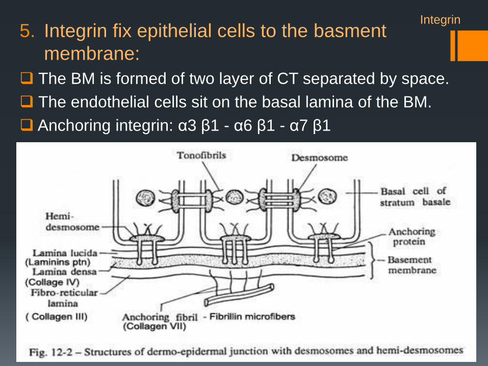
5. Integrin fix epithelial cells to the basment
membrane:
The BM is formed of two layer of CT separated by space.
The endothelial cells sit on the basal lamina of the BM.
Anchoring integrin: α3 β1 - α6 β1 - α7 β1
Integrin

Selectin

3. Selectins:
Example:
1. P- Selectin: Found on the endothelial cells to help the
recruitment of the neutrophils, P-Selectin
glycoprotein ligand 1, type 1
activation (induced by histamine and
induced in minutes)
2. E-Selectin:
Found on the endothelial cells and binds
to Sialyl-Lewis X found always on the
surface of the neutrophil type 2
activation (takes hours)

3. L-Selectin:
The high endothelial cells allows the naïve T lymphocytes to move from
the blood into lymph nodes and back out.
Naïve T-lymphocytes have on their
surface the L-Selectin that binds to
GLYCAM-1 on the high endothelial cells
movement of the lymphocyte to the
Lymph node.
The unusual cuboidal endothelial cells (EC) have
lighter-staining nuclei than the lymphocytes (L)
which are in the process of migrating from the
lumen (bloodstream) into the diffuse cortex (DC)
of the node.
Selectin
Cadherin

4. Cadherin superfamily:
Extra cellular cadherin domain range from (1-34) domains.
All cadherin are a trans membrane protein except T-cadherin.
Types of Cadherin:
1. Type I classical cadherin
2. Type II atypical cadherin
3. Truncated cadherin
4. Desmosomal cadherin
5. Flamingo cadherin
6. Proto cadherin
7. Others
Cadherin

1. T- cadherin is completely outside the cell membrane
and attached to it by a lipid called Glycosyl
phosphatidylinositol (GPI).
Types of Cadherin: Cadherin

2. Type I classical cadherin:
It produce cell to cell direct connection.
(Adherent Junction)
Example:
E- Cadherin: found in the epithelial cells
N - Cadherin: found in the neuron, muscle tissue
P - Cadherin : found in the placenta, epidermis
Structure:
They all have 5 extracellular cadherin domain ended
with amino terminal NH2.
The N terminal of the Extra cadherin domain will bind
to the neighboring N terminal of the EC domain.
The binding is dependent to the extra cellular Ca ions
Cadherin

The cytoplasmic terminal end binds to 2 proteins:
1. P120 catanin
2. β- catinin + α – catinin
α – catinin anchored the cadherin to the cytoskeleton actin filaments.
E-cadherin–catenin complex functions as a master molecule in regulating
not only cell adhesion but also polarity, differentiation, migration,
proliferation, and survival of epithelial cells.
Cadherin
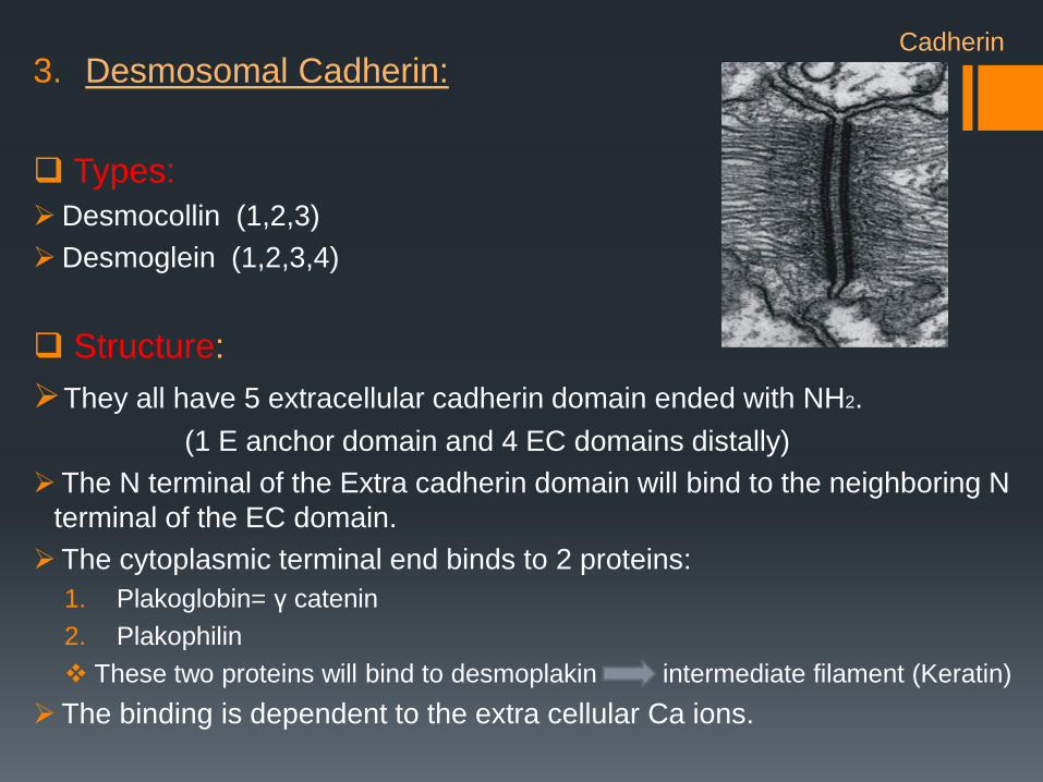
3. Desmosomal Cadherin:
Types:
Desmocollin (1,2,3)
Desmoglein (1,2,3,4)
Structure:
They all have 5 extracellular cadherin domain ended with NH2.
(1 E anchor domain and 4 EC domains distally)
The N terminal of the Extra cadherin domain will bind to the neighboring N
terminal of the EC domain.
The cytoplasmic terminal end binds to 2 proteins:
1. Plakoglobin= γ catenin
2. Plakophilin
These two proteins will bind to desmoplakin intermediate filament (Keratin)
The binding is dependent to the extra cellular Ca ions.
Cadherin

The extracellular core allows water and ions flow in between the cells.
E.g: Skin, Intestins
Cadherin

• Integrin and Ig Superfamily CAMs are Ca independent.
• Cadherin and Selectin are Ca dependent.
• Integrins participate in cell-matrix interaction while other
CAMs participate in cell-cell interaction.

TYPES OF CELL JUNCTIONS
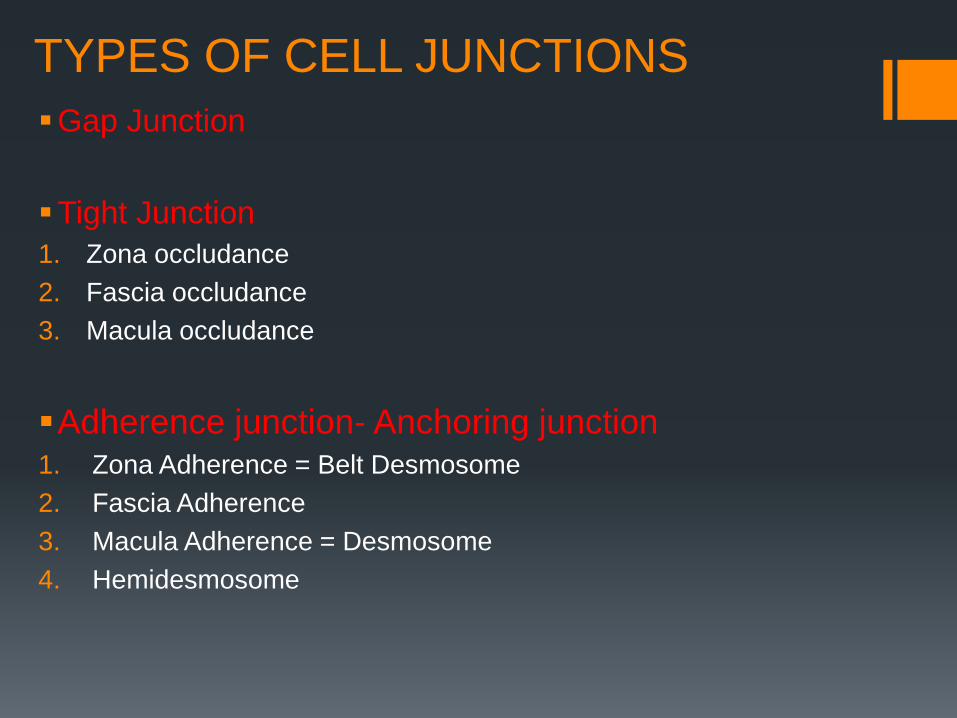
TYPES OF CELL JUNCTIONS
Gap Junction
Tight Junction
1. Zona occludance
2. Fascia occludance
3. Macula occludance
Adherence junction- Anchoring junction 1. Zona Adherence = Belt Desmosome
2. Fascia Adherence
3. Macula Adherence = Desmosome
4. Hemidesmosome

GAP JUNCTIONS= NEXUS= MACULA
COMMUNICATION It like a tunnel between two neighboring cells
It allows water and ions flow between this gab tunnels.
E.G: Cardiac Muscle cells, Neurons, smooth muscle

:TIGHT JUNCTIONS
It connects two cells together like a glue
It is a water tight seal: complete fluid barrier
E.g.: Urinary Bladder, Intestine and kidney

Freeze fracture technique:
When the plasma membrane is fractured at the site of the zonula occludens, the junctional proteins are observed on:
the P-face of the membrane, where they appear as ridgelike structures.
the E-face, reveals complementary grooves resulting from detachment of the protein particles from the opposing surface.
• P face: an anastomosing network of ridges
located on the fracture membrane surface.
• The E-face of the fractured membrane
would show a complementary pattern of
grooves.
Tight Junction

Thank you
Divergence of structural strategies
for homophilic E-cadherin binding