celiac artery avulsion secondary to blunt trauma: a case
TRANSCRIPT
Case Report J Med Cases • 2013;4(5):280-283
PressElmer
Articles © The authors | Journal compilation © J Med Cases and Elmer Press™ | www.journalmc.orgThis is an open-access article distributed under the terms of the Creative Commons Attribution License, which permits unrestricted use, distribution, and reproduction
in any medium, provided the original work is properly cited
Celiac Artery Avulsion Secondary to Blunt Trauma: A Case Report and Review of the Literature
Zachary Osbornea, b, Uretz J Oliphanta, John Aucara, Michelle M Olsona
Abstract
A 70-year-old male presented with a celiac artery avulsion follow-ing a motor vehicle collision. He was hemodynamically stable and complained of severe abdominal pain. A Computed Tomography (CT) scan demonstrated a central supramesocolic retroperitoneal hematoma. There was concern for discontinuity of the celiac artery. At laparotomy, using a left medial visceral rotation, the celiac artery was found completely disrupted and both ends were ligated. He was discharged on post-operative day eleven, without complications. Celiac artery avulsion is reported in 0.01-0.1% of patients sustain-ing abdominal vascular injuries. This is only the second reported case of blunt celiac artery avulsion.
Keywords: Celiac artery injuries; Blunt trauma; Blunt abdominal trauma
Introduction
Celiac Artery (CA) injuries are very rare, and often are as-sociated with a high mortality. These injuries are more com-monly associated with penetrating trauma. Our case high-lights a blunt trauma injury. The majority of the literature on blunt CA injuries is only been published recently, and only 8 articles have published injuries related to blunt CAs.
Case Report
A 72-year-old male driver, in a head-on motor-vehicle col-
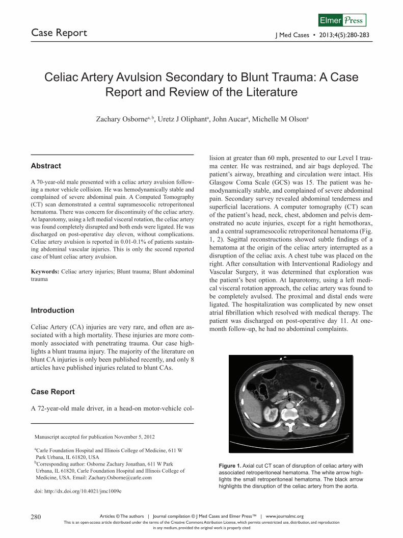
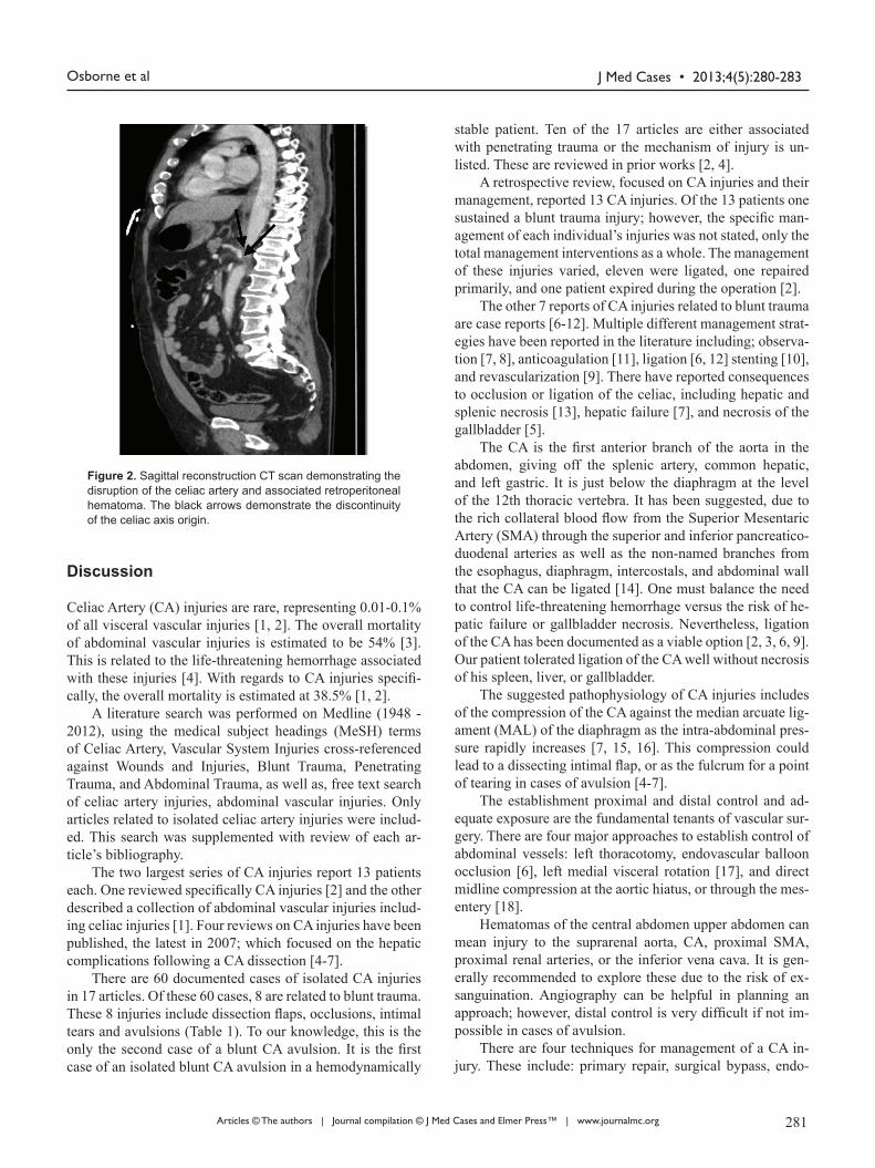
lision at greater than 60 mph, presented to our Level I trau-ma center. He was restrained, and air bags deployed. The patient’s airway, breathing and circulation were intact. His Glasgow Coma Scale (GCS) was 15. The patient was he-modynamically stable, and complained of severe abdominal pain. Secondary survey revealed abdominal tenderness and superficial lacerations. A computer tomography (CT) scan of the patient’s head, neck, chest, abdomen and pelvis dem-onstrated no acute injuries, except for a right hemothorax, and a central supramesocolic retroperitoneal hematoma (Fig. 1, 2). Sagittal reconstructions showed subtle findings of a hematoma at the origin of the celiac artery interrupted as a disruption of the celiac axis. A chest tube was placed on the right. After consultation with Interventional Radiology and Vascular Surgery, it was determined that exploration was the patient’s best option. At laparotomy, using a left medi-cal visceral rotation approach, the celiac artery was found to be completely avulsed. The proximal and distal ends were ligated. The hospitalization was complicated by new onset atrial fibrillation which resolved with medical therapy. The patient was discharged on post-operative day 11. At one-month follow-up, he had no abdominal complaints.
Manuscript accepted for publication November 5, 2012
aCarle Foundation Hospital and Illinois College of Medicine, 611 W Park Urbana, IL 61820, USAbCorresponding author: Osborne Zachary Jonathan, 611 W Park Urbana, IL 61820, Carle Foundation Hospital and Illinois College of Medicine, USA. Email: [email protected]
doi: http://dx.doi.org/10.4021/jmc1009e
280 281
Figure 1. Axial cut CT scan of disruption of celiac artery with associated retroperitoneal hematoma. The white arrow high-lights the small retroperitoneal hematoma. The black arrow highlights the disruption of the celiac artery from the aorta.
J Med Cases • 2013;4(5):280-283Osborne et al
Articles © The authors | Journal compilation © J Med Cases and Elmer Press™ | www.journalmc.org
Discussion Celiac Artery (CA) injuries are rare, representing 0.01-0.1% of all visceral vascular injuries [1, 2]. The overall mortality of abdominal vascular injuries is estimated to be 54% [3].This is related to the life-threatening hemorrhage associated with these injuries [4]. With regards to CA injuries specifi-cally, the overall mortality is estimated at 38.5% [1, 2].
A literature search was performed on Medline (1948 - 2012), using the medical subject headings (MeSH) terms of Celiac Artery, Vascular System Injuries cross-referenced against Wounds and Injuries, Blunt Trauma, Penetrating Trauma, and Abdominal Trauma, as well as, free text search of celiac artery injuries, abdominal vascular injuries. Only articles related to isolated celiac artery injuries were includ-ed. This search was supplemented with review of each ar-ticle’s bibliography.
The two largest series of CA injuries report 13 patients each. One reviewed specifically CA injuries [2] and the other described a collection of abdominal vascular injuries includ-ing celiac injuries [1]. Four reviews on CA injuries have been published, the latest in 2007; which focused on the hepatic complications following a CA dissection [4-7].
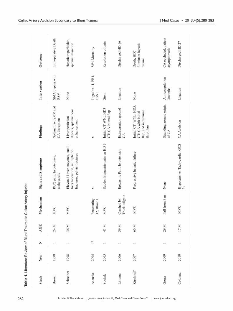
There are 60 documented cases of isolated CA injuries in 17 articles. Of these 60 cases, 8 are related to blunt trauma. These 8 injuries include dissection flaps, occlusions, intimal tears and avulsions (Table 1). To our knowledge, this is the only the second case of a blunt CA avulsion. It is the first case of an isolated blunt CA avulsion in a hemodynamically
stable patient. Ten of the 17 articles are either associated with penetrating trauma or the mechanism of injury is un-listed. These are reviewed in prior works [2, 4].
A retrospective review, focused on CA injuries and their management, reported 13 CA injuries. Of the 13 patients one sustained a blunt trauma injury; however, the specific man-agement of each individual’s injuries was not stated, only the total management interventions as a whole. The management of these injuries varied, eleven were ligated, one repaired primarily, and one patient expired during the operation [2].
The other 7 reports of CA injuries related to blunt trauma are case reports [6-12]. Multiple different management strat-egies have been reported in the literature including; observa-tion [7, 8], anticoagulation [11], ligation [6, 12] stenting [10], and revascularization [9]. There have reported consequences to occlusion or ligation of the celiac, including hepatic and splenic necrosis [13], hepatic failure [7], and necrosis of the gallbladder [5].
The CA is the first anterior branch of the aorta in the abdomen, giving off the splenic artery, common hepatic, and left gastric. It is just below the diaphragm at the level of the 12th thoracic vertebra. It has been suggested, due to the rich collateral blood flow from the Superior Mesentaric Artery (SMA) through the superior and inferior pancreatico-duodenal arteries as well as the non-named branches from the esophagus, diaphragm, intercostals, and abdominal wall that the CA can be ligated [14]. One must balance the need to control life-threatening hemorrhage versus the risk of he-patic failure or gallbladder necrosis. Nevertheless, ligation of the CA has been documented as a viable option [2, 3, 6, 9]. Our patient tolerated ligation of the CA well without necrosis of his spleen, liver, or gallbladder.
The suggested pathophysiology of CA injuries includes of the compression of the CA against the median arcuate lig-ament (MAL) of the diaphragm as the intra-abdominal pres-sure rapidly increases [7, 15, 16]. This compression could lead to a dissecting intimal flap, or as the fulcrum for a point of tearing in cases of avulsion [4-7].
The establishment proximal and distal control and ad-equate exposure are the fundamental tenants of vascular sur-gery. There are four major approaches to establish control of abdominal vessels: left thoracotomy, endovascular balloon occlusion [6], left medial visceral rotation [17], and direct midline compression at the aortic hiatus, or through the mes-entery [18].
Hematomas of the central abdomen upper abdomen can mean injury to the suprarenal aorta, CA, proximal SMA, proximal renal arteries, or the inferior vena cava. It is gen-erally recommended to explore these due to the risk of ex-sanguination. Angiography can be helpful in planning an approach; however, distal control is very difficult if not im-possible in cases of avulsion.
There are four techniques for management of a CA in-jury. These include: primary repair, surgical bypass, endo-
280 281
Figure 2. Sagittal reconstruction CT scan demonstrating the disruption of the celiac artery and associated retroperitoneal hematoma. The black arrows demonstrate the discontinuity of the celiac axis origin.
J Med Cases • 2013;4(5):280-283 Celiac Artery Avulsion Secondary to Blunt Trauma
Articles © The authors | Journal compilation © J Med Cases and Elmer Press™ | www.journalmc.org
Stud
yYe
arN
AG
EM
echa
nism
Sign
s and
Sym
ptom
sFi
ndin
gsIn
terv
entio
nO
utco
me
Bro
wn
1998
124
MM
VC
RU
Q p
ain,
hyp
oten
sive
, ta
chyc
ardi
cSp
leni
c La
c, S
MV
and
C
A d
isru
ptio
nSM
A b
ypas
s with
R
SVIn
traop
erat
ive
Dea
th
Schr
eibe
r19
981
36 M
MV
CEl
evat
ed L
iver
enz
ymes
, sm
all
liver
lace
ratio
n, m
ultip
le ri
b fr
actu
res,
pelv
ic fr
actu
res
Live
r per
fusi
on
defe
cts,
sple
nic
poor
en
hanc
emen
t
Non
eH
epat
ic re
perf
usio
n,
sple
nic
infa
rctio
n
Ase
nsio
2005
13Pe
netra
ting
12, B
lunt
1x
xLi
gatio
n 11
, PR
1,
ExS
138
% M
orta
lity
Such
ak20
051
41 M
MV
CSu
dden
Epi
gast
ric p
ain
on H
D 3
Initi
al C
T:W
NL
HD
3 C
T: C
A in
timal
flap
Sten
tR
esol
utio
n of
pai
n
Linu
ma
2006
139
MC
rush
ed b
y Tr
uck
tailg
ate
Epig
astri
c Pa
in, h
ypot
ensi
onEx
trava
satio
n ar
ound
C
ALi
gatio
nD
isch
arge
d H
D 1
6
Kirc
hhof
f20
071
66 M
MV
CPr
ogre
ssiv
e he
patic
failu
reIn
itial
CT:
WN
L, H
D5:
C
T: C
A w
ith in
itial
fla
p, a
nd in
tram
ural
th
rom
bus
Non
eD
eath
, HD
7 fu
lmin
ant h
epat
ic
failu
re
Gor
ra20
091
29 M
Fall
from
9 m
Non
eSt
rand
ing
arou
nd o
rigin
of
CA
Ant
icoa
gula
tion
3mon
ths
CA
occ
lude
d, p
atie
nt
asym
ptom
atic
Col
onna
2010
117
M M
VC
Hyp
oten
sive
, Tac
hyca
rdic
, GC
S 3t
CA
Avu
lsio
nLi
gatio
nD
isch
arge
d H
D 2
7
Tabl
e 1.
Lite
ratu
re R
evie
w o
f Blu
nt T
raum
atic
Cel
iac
Arte
ry In
jurie
s
282 283

J Med Cases • 2013;4(5):280-283Osborne et al
Articles © The authors | Journal compilation © J Med Cases and Elmer Press™ | www.journalmc.org
vascular stenting or embolization, or ligation. Primary repair has been advocated in the literature as the preferred ap-proach; however ligation is an acceptable [1, 2, 14]. Surgical bypass with an interposition graft of vein or prosthetic mate-rial is undertaken if the other major vessels of the abdomen have been injured [3, 9].
Conclusion
The current case represents the second reported CA avulsion related to blunt trauma. It is the first case in the literature of an isolated blunt CA avulsion in a hemodynamically stable patient. The decision to operatively explore this patient was based on the fact that this was central supramesocolic he-matoma and there was some question if the proximal left re-nal artery was involved. Consultation with vascular surgery and interventional radiology was obtained. We performed a left medial rotation of the viscera. Ligation was the selected treatment on the basis of adequate collateral circulation. The patient had no complications related to ligation of the CA including liver injury or necrosis of his gallbladder.
Disclaimer All authors have no conflict of interest in this report.
References
1. Graham JM, Mattox KL, Beall AC, Jr., DeBakey ME. Injuries to the visceral arteries. Surgery. 1978;84(6):835-839.
2. Asensio JA, Petrone P, Kimbrell B, Kuncir E. Lessons learned in the management of thirteen celiac axis inju-ries. South Med J. 2005;98(4):462-466.
3. Asensio JA, Chahwan S, Hanpeter D, Demetriades D, Forno W, Gambaro E, Murray J, et al. Operative man-agement and outcome of 302 abdominal vascular injuries. Am J Surg. 2000;180(6):528-533; discussion 533-524.
4. Asensio JA, Forno W, Roldan G, Petrone P, Rojo E, Ce-ballos J, Wang C, et al. Visceral vascular injuries. Surg Clin North Am. 2002;82(1):1-20, xix.
5. Kavic SM, Atweh N, Ivy ME, Possenti PP, Dudrick SJ. Celiac axis ligation after gunshot wound to the ab-domen: case report and literature review. J Trauma. 2001;50(4):738-739.
6. Linuma Y, Yamazaki Y, Hirose Y, Kinoshita H, Kum-agai K, Tanaka T, Miyajima M, et al. A case of isolated celiac axis injury by blunt abdominal trauma. J Trauma. 2006;61(2):451-453.
7. Kirchhoff C, Stegmaier J, Krotz M, Muetzel Rauch E, Mutschler W, Kanz KG, Heindl B. Celiac dissection af-ter blunt abdominal trauma complicated by acute hepatic failure: case report and review of literature. J Vasc Surg. 2007;46(3):576-580.
8. Schreiber JP, Angle JF, Matsumoto AH, Young JS, Hag-spiel KD, Spinosa DJ. Acute visceral ischemia occur-ring subsequent to blunt abdominal trauma: potential culpability of median arcuate ligament compression. J Trauma. 1998;45(2):404-406.
9. Brown DB, Singh H, Atnip RG, Cardella JF, Waybill PN. Blunt traumatic injury to the superior mesenteric ar-tery and celiac axis. J Vasc Interv Radiol. 1998;9(5):783-785.
10. Suchak AA, Reich D, Ritchie W. Traumatic isolated dissection of the celiac artery. AJR Am J Roentgenol. 2007;189(6):W373-374.
11. Gorra AS, Mittleider D, Clark DE, Gibbs M. Asymp-tomatic isolated celiac artery dissection after a fall. Arch Surg. 2009;144(3):279-281.
12. Colonna AL, Enniss TM, Meredith JW, Hildreth AN. Celiac artery avulsion and right atrial rupture after blunt multisystem trauma. Am Surg. 2010;76(7):E83-85.
13. Francque S, Condat B, Asselah T, Vilgrain V, Durand F, Moreau R, Valla D. Multifactorial aetiology of hepatic infarction: a case report with literature review. Eur J Gastroenterol Hepatol. 2004;16(4):411-415.
14. Goaley TJ, and Feliciano DV, Abdominal Vascular In-juries, 410-415: In Asensio JA, Trunkey DD. Current Therapy of Trauma and Surgical Critical Care. Portland, Mosby Inc. 2008.
15. Lindner HH, Kemprud E. A clinicoanatomical study of the arcuate ligament of the diaphragm. Arch Surg. 1971;103(5):600-605.
16. Stoney RJ, Wylie EJ. Recognition and surgical man-agement of visceral ischemic syndromes. Ann Surg. 1966;164(4):714-722.
17. Mattox KL, McCollum WB, Beall AC, Jr., Jordan GL, Jr., Debakey ME. Management of penetrating injuries of the suprarenal aorta. J Trauma. 1975;15(9):808-815.
18. Feliciano DV: Abdominal vascular injury. Trauma, 5th ed. Moore EE, Feliciano DV, Mattox KL, Eds. McGraw-Hill, New York, 2004.
282 283