catalyst - marine biological...
TRANSCRIPT

CatalystA Beacon for ScienceInside the Whitman Center for Research and Discovery
Page 2
FALL 2011Volume 6, Number 2
Founded in 1888 as the Marine Biological Laboratory
I N T H I S I S S U E
4To Regenerate
or Not?
8The Visionaries:
Pioneers in Super-Resolution
Microscopy
10The Geometry
of Life

Dear Friends,
I first came to the MBL as a graduate student in 1965, officially to take a course but with the ulterior motive of gleaning information from certain visiting investigators. I knew the world’s leading biological scientists gather at the MBL each summer, and I wanted someone to show me how to isolate a component of cells, the mitotic spindle. Once here, I knocked on the doors of senior investigators including Lenny Rebhun and Bob Kane. They were very generous with their time, and they taught me much more than what I had come to learn.
My first MBL experience was key, not least because it was pivotal to our discovery of an important cellular protein, tubulin, which my University of Chicago colleagues and I purified later that year. So, after finishing my Ph.D. and joining the University of Wisconsin, I returned to the MBL as a summer investigator from 1972 to 1976. This was also a significant time in my scientific career, one in which I transitioned from biochemist to cell biologist. Because I wanted to extend my studies of cell division from the test tube to the living cell, I camped on the doorstep of MBL scientist Shinya Inoué so I could observe the living cell using the polarization microscope he had pioneered.
The rewards of those summers at the MBL were great. It was not just my interaction with many of the world’s best scientists, or the collaborative atmosphere, or the access to superb microscopy and instrumentation, or the beautiful Woods Hole setting. It was the unique mix of these things that made my years as a visiting investigator so productive and inspiring.
We hope this issue of MBL Catalyst illustrates that my experience was in no way unusual. The summer investigator program began when the MBL opened its doors in 1888, and is now called the Whitman Center for Research and Discovery after the MBL’s founding director, C.O. Whitman. The Center has enriched and propelled the research of generations of biological and biomedical scientists, and the dividends to them extend to all of society, in the form of a stellar record of innovation and discovery.
Many thanks to Bob Goldman of Northwestern University for his leadership of the Whitman Center, and for serving as guest science editor of this issue of MBL Catalyst. Bob knows as well as I do that the Whitman Center is a critical component of the MBL’s mission and success. To solidify its long-term strength, the MBL has launched a $2 million Whitman Center Endowment fundraising initiative, with an initial $500,000 matching commitment from the MBL. This endowment will support building enhancements to maintain Rowe Laboratory as a state-of-the-art facility for visiting investigators, and fellowship funds to continue to bring the world’s best researchers into the Whitman Center fold. By building a solid foundation for the Whitman Center, we will ensure the MBL’s continuing stature as the world’s crossroads for innovation and discovery in the biological, biomedical, and environmental sciences.
Catalyst
Gary BorisyPresident and Director
Fall 2011 Volume 6, Number 2
MBL Catalyst is published twice yearly by the Office of Communications at the Marine Biological Laboratory (MBL) in Woods Hole, Massachusetts. The MBL is dedicated to scientific discovery and improving the human condition through research and education in biology, biomedicine, and environmental science. Founded in 1888, the MBL is an independent, nonprofit corporation. Senior AdvisorsPresident and Director: Gary BorisyChief Academic and Scientific Officer: Joshua HamiltonDirector of External Relations: Pamela Clapp Hinkle
MBL Catalyst StaffEditor-in-Chief: Andrea EarlyManaging Editor/Senior Writer: Diana KenneyGuest Science Editor: Robert GoldmanDesigner: Beth Ready LilesContributors: Gina Hebert Amanda Rose Martinez Pamela Wilmot Photography: Inside cover: Rowe Laboratory (T. Kleindinst and P. Oberlander); Gary Borisy (E. Armstrong). P. 1: Rowe Laboratory (T. Kleindinst and P. Oberlander); Sea lamprey illustration (dreamstime.com); Whitman Investigator David Piston of Vanderbilt University (E. Armstrong); Microfabricated chambers holding sea urchin embryos (N. Minc, F. Chang and D. Burgess). Pp. 2–3, clockwise from top: Section of oral biofilm imaged using CLASI FISH (B. Rossetti); Whitman Investigators Tim Mitchison of Harvard Medical School and Ron Vale of HHMI/University of California-San Francisco (E. Armstrong); Charles Otis Whitman (MBL Archives); Whitman Investigators Amy Gladfelter of Dartmouth College and Christine Field of Harvard Medical School (E. Armstrong); Whitman Investigator Robyn Crook of University of Texas-Houston (E. Armstrong). Pp. 4-5: Sea lamprey illustration (dreamstime.com); Giant axons in the intact lamprey spinal cord (O. Bloom); (L to R) Ona Bloom, Joel Smith, Jennifer Morgan, and Joseph Buxbaum (T. Kleindinst). Pp. 6-7, clockwise from top left: Fluorescence LC-PolScope image of a living cell (MDCK) expressing septin molecules linked to GFP (R. Oldenbourg); Frog illustration (dreamstime.com); Beta cell (Wikipedia.com); Late-stage midshipman larvae (M. Marchaterre); dreamstime.com illustration. Pp. 8-9, clockwise from top left: Jim and Cathy Galbraith (T. Kleindinst); Eric Betzig (T. Kleindinst); Cellular structures as seen by Bessel beam plane illumination microscopy, clockwise from upper left: membrane ruffles in a monkey kidney cell; chromosomes (green) and golgi (magenta) in a dividing pig kidney cell; mitochondria in a living pig kidney cell; and microtubules (green) and nuclei (magneta) in a pair of human osteosarcoma cells (E. Betzig); A cultured neuron expressing homer-GFP, a postsynaptic marker. Bright fluorescent puncta along the dendrites show localization of synapses (P. Selvin); Microtubules labeled with CF633 secondary antibodies in fixed COS-7 cells, gSHRImP image (P. Selvin). Pp. 10-11: Flower of Life illustration (dreamstime.com); Microfabricated chambers (N. Minc, F. Chang and D. Burgess); David Burgess and Fred Chang (T. Kleindinst). Pp. 12-13: Rodolfo Llinás (T. Kleindinst); Synaptic transmission illustration (NIA-NIH/Wikipedia); Loligo pealei illustration (B. Harmon). P. 14, L to R: MBL course (T. Kleindinst); 3D rendering of cuttlefish skin cells, stained with acid fuchsin (S. Senft); MBL SES graduate Amy Townsend-Small in the Andes (courtesy of A. Townsend-Small). P. 15: Aequorea jellyfish (O. Shimomura); John Costello and Kakani Young (E. Klos) P. 16: Robert Goldman (T. Kleindinst). P. 17: Plate 6 from Ernst Haeckel, Kunstformen der Natur (1904) (MBL Archives). Back cover, L to R: Weddell seal (Census of Marine Life/Galatée Films); Sea cucumber (Census of Marine Life/L. Madin); P. helianthoides (Census of Marine Life/C. Debenham); Flamingo tongue snail (Census of Marine Life/K. Moody).
About the cover: Rowe Laboratory, home of the Whitman Center for Research and Discovery. Photo illustration by Tom Kleindinst and Paul Oberlander.
Online extras: For full image descriptions, supplemental materials, and other information related to this issue, visit:
www.mbl.edu/catalyst
Send correspondence to: MBL Communications Office7 MBL Street, Woods Hole, MA 02543508-289-7423, [email protected]
Fr o m t h e Di r e c t o r
MBL
De pa rt m e N t s
6 News & Notes
Recent publications from the Whitman Center for Research and Discovery.
12 mbl mo m e N t
How the Squid Offered Deep Insight into Alzheimer’s DiseaseRodolfo Llinás’s research on the Woods Hole squid leads him to a profound understanding of a neurodegenerative disorder.
14 Gi F t s & Gr a N t s
14 ac c o l a D e s
15 co o l to o l
Opening a Portal on the Behavior of JelliesSean Colin and John Costello dive in to discover how ocean currents affect the hunt for prey.
16 sc i e N t i s t ’s ey e Vi e w
Placing a Value on the Invaluable Robert Goldman honors the Whitman Center’s unique standing as a global “collaboratorium” for biology and biomedicine.
17 me m o r a b i l i a
Renewing the Passion for ScienceS. Meryl and Florence Rose inspired new generations toward a love of research and the MBL.
A Beacon for Science
Every year, hundreds of scientists move their labs to the MBL’s Whitman Center for Research and Discovery. Find out why they spend weeks or months in this international hot spot for science.
To Regenerate or Not? That is the Question
How do nerve cells recover and restore function after spinal cord injury? Whitman Investigators Jennifer Morgan, Ona Bloom, and Joseph Buxbaum seek the answer.
The Visionaries
The MBL has always been at the leading edge of microscopy and imaging. Last summer, the buzz was all about “super-resolution.”
The Geometry of Life
Fred Chang and David Burgess address a longstanding puzzle about dividing cells.
2
Fe at u r e s
10
iN t h i s i s s u e
8
4
Catalyst
1MBL Catalyst FALL 2011

“Scientists have different philosophies of why they come to the MBL. Mine is to do experiments that aren’t necessarily in the game plan in my home lab, to do something new. The spirit of MBL is to try things, to be willing to be adventurous, take risks, even if they don’t work out.”
— Ron Vale, Whitman Investigator from HHMI/University of California, San Francisco
“The MBL is a very creative and also peaceful environment for me. I can just work in the lab without any diversions. I enjoy bench work, but I also think better when I can work in peace and quiet. I work hard here. Some people ask me, why do you work like crazy? Do you want to get another Nobel Prize? No, my aim is not to get another Nobel Prize. My aim is to gain more understanding.”
— Avram Hershko, Whitman Investigator from Technion-Israel Institute of Technology and 2004 Nobel Laureate in Chemistry
Each spring, a distinct rustle of activity is heard in Woods Hole that is more than just the change in season. At first one by one, then in a steady stream, researchers from all over the world begin to arrive at the MBL. They bring their students, families, their ideas and ambitions. They set up a lab at the MBL, all the while greeting old friends or making new connections. By July Fourth, the MBL is humming with their energy and, once again, is an unmatched global hub of scientific discussion, exploration, and discovery.
These are the hundreds of investigators in the biological, biomedical, and environmental sciences who come every year to the Whitman Center for Research and Discovery. Some stay for a few weeks or months, while others return over the course of decades or their entire careers. They join the MBL’s vibrant community of year-round scientists, course faculty, and students.
If you ask the Whitman Investigators why they have come to Woods Hole, you’ll hear a variety of reasons. For some, the MBL is an ideal place to meet with collaborators from other institutions and try out the experiments they need or just really want to do. They believe no amount of e-mail can reap the rewards of working side-by-side with their collaborators at the MBL.
MBL
Inside The Whitman Center for Research and Discovery
A Beacon for Science
2 MBL Catalyst FALL 2011

“It is absolutely wonderful to be at the MBL, especially for someone like me who knows physics and is trying to learn biology. You walk down the hall and you meet the world’s experts in so many fields of neuroscience. And every time we talk we come up with new ideas and experiments to do.”
— Paul Selvin, Whitman Investigator from University of Illinois at Urbana-Champaign
“When you come into Woods Hole and the MBL, you feel the energy of research. A scientific ether hangs about the place and you can’t help but be intoxicated by it. It has always been there, and always will.”
— James Galbraith, Whitman Investigator from the National Institutes of Health
Other Whitman Investigators deeply appreciate having the time to work hard in the laboratory, free from the distractions of their home institutions. Some say they accomplish more in a few months at the MBL than they do the rest of the year.
The MBL has always been at the cutting edge of microscopy and imaging, which is a major draw for many Whitman Investigators. Others are drawn by the organisms—squid, surf clam, Xenopus (frogs) and many others—that are collected and maintained for research by MBL staff.
And for many Whitman Investigators, the MBL is where they renew their scientific knowledge and creativity. They have a standing invitation to an abundance of lectures by the world’s top researchers in cell and molecular biology, neuroscience, microbiology and numerous other fields. No matter where they are—in the lab, at dinner, in Lillie Auditorium, or at Stony Beach—scientists are talking science, and the insights and experimental ideas fly thick and fast. By joining the MBL conversation, Whitman Investigators rekindle their passion for scientific research. This is where the scientific and medical knowledge that benefits society begins.
“I get so much done at the MBL and then I keep going on that after I leave. I am still doing great experiments into the fall.”
— Christine Field, Whitman Investigator from Harvard Medical School
C.O. WhitmanFirst Director of the MBL
2011 Whitman Investigators
132 Principal Investigators231 Other Researchers
From 176 Institutions and 24 Countries
3MBL Catalyst FALL 2011

Watching a larval lamprey, a
jawless fish with an eel-like body, swim
around a tank is hardly impressive. But
knowing that only 11 weeks prior, the
animal sustained a severe spinal cord
injury, the lamprey’s simple motion
seems nothing short of remarkable.
Although scientists have reported
since the 1950s that the lamprey can
regenerate its spinal cord and recover
the ability to swim, the prevailing
question continues to be: How do they
do this? A team of MBL investigators,
aided by recent advances in DNA
sequencing technology and analysis,
is exploring a new approach that
may yield crucial insight into how the
lamprey’s regeneration process unfolds.
Whitman Center researchers Ona
Bloom, Jennifer Morgan, and Joseph
Buxbaum are collaborating to chart a
timeline of the cellular and molecular
changes that occur throughout the
lamprey’s healing process. “We’d like
to know what’s happening immediately
after the injury, say six or 24 hours
after, a week later, two weeks later—to
get a really full picture,” says Bloom.
The hope is that having such a detailed
series of molecular “snapshots”
could reveal the step-by-step genetic
changes that are occurring before their
effects are observable in the lamprey’s
behavior.
Above: Giant nerve fibers in the intact lamprey spinal cord are labeled in red by a fluorescent retrograde tracer.
Right: (L to R) Whitman Investigator Ona Bloom (Feinstein Institute for Medical Research and Hofstra North Shore - LIJ School of Medicine), MBL scientist Joel Smith, Whitman Investigator Jennifer Morgan (University of Texas at Austin), and Whitman Investigator Joseph Buxbaum (Mount Sinai School of Medicine).
To Regenerate or Not?
That is the Que tion
4 MBL Catalyst FALL 2011

his office on his first day,” says Bloom.
“He was really excited about the
project.”
Functional genomics gives you the
ability to look at “what’s causing those
genes to be expressed, meaning which
ones are turned on or not,” says Smith.
“Because, of course, what genes are
turned on or are being turned on gives
a particular cell its identity.” Once you
know what’s being expressed, you can
begin to isolate a set of genes that
may be causing the cell to behave a
certain way. The eventual goal, says
Smith, is to be able to alter the genetic
information in the RS neurons that die
such that they would act like the RS
neurons that survive and regrow.
As to what contribution this research
may one day make to spinal cord injury
treatment in humans, the researchers
are cautious to say. But they remain
hopeful.
“In terms of the basic locomotor
systems that drive the lamprey’s
swimming, they’re the same locomotor
systems that drive our walking,” says
Morgan. “And there’s a surprisingly
high degree of gene similarity between
lampreys and humans. So I think we
will find that the lamprey can be a very
good model to study some of these
basic molecular and cellular responses.
Of course, translating the information
to the mammalian nervous system is
much more complicated. Everyone
wants to work toward a treatment.
That’s the ultimate goal.”
• —ARM
Besides its regenerative ability, the
lamprey makes a great experimental
model because the very neurons
that are capable of regrowth, called
reticulospinal or RS neurons, are
relatively large and therefore easy to
study. (Just to give perspective, the
cell body of a RS neuron is 10 to 20
times larger in diameter than that of a
mammalian neuron.)
About 30 of these super-sized RS
neurons, the cell bodies of which reside
in the lamprey’s brain, have been
identified. “They look like clusters of
grapes, and they’re very visible without
any special magnification,” says
Morgan.
When a lamprey’s spinal cord is
transected, about half of its RS neurons
can regrow their nerve fibers (axons)
back through the injury site, restoring
movement. The other half can’t and
eventually die. This raises a core
question of regenerative biology that
the researchers hope to directly address:
What causes one cell to survive and
regenerate while another one doesn’t
and dies?
The researchers first extract the genetic
material from the lampreys’ RS neurons
at certain time points during the
animal’s recovery. Then, they sequence
it using a new technique called next-
generation sequencing. What they end
up with are lists of genes that are or are
not being expressed at each stage of
recovery. The challenge then becomes
to try and understand the relationship
among those genes and to identify
which genes are driving the changes in
the animal’s behavior and physiology as
it heals.
That’s where Joel Smith, an assistant
scientist at the MBL’s Eugene Bell Center
for Regenerative Biology and Tissue
Engineering, comes in. Smith, an expert
in functional genomics, joined the new
Bell Center in 2010 and the Whitman
team wasted no time in recruiting his
expertise. “We actually showed up at
A core question of
regenerative
biology that the
researchers hope to
directly address:
What causes
one cell to survive
and regenerate
while another one
doesn’t and dies?
5MBL Catalyst FALL 2011
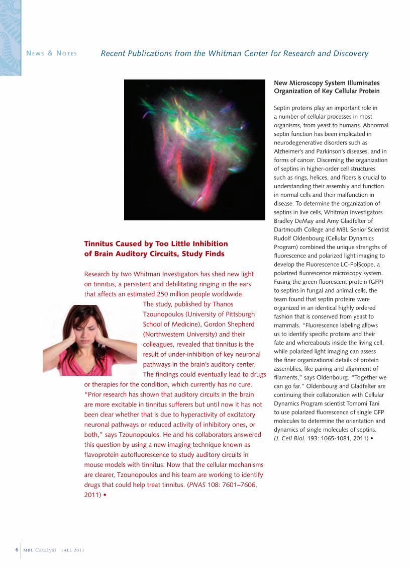
New Microscopy System Illuminates Organization of Key Cellular Protein
Septin proteins play an important role in a number of cellular processes in most organisms, from yeast to humans. Abnormal septin function has been implicated in neurodegenerative disorders such as Alzheimer’s and Parkinson’s diseases, and in forms of cancer. Discerning the organization of septins in higher-order cell structures such as rings, helices, and fibers is crucial to understanding their assembly and function in normal cells and their malfunction in disease. To determine the organization of septins in live cells, Whitman Investigators Bradley DeMay and Amy Gladfelter of Dartmouth College and MBL Senior Scientist Rudolf Oldenbourg (Cellular Dynamics Program) combined the unique strengths of fluorescence and polarized light imaging to develop the Fluorescence LC-PolScope, a polarized fluorescence microscopy system. Fusing the green fluorescent protein (GFP) to septins in fungal and animal cells, the team found that septin proteins were organized in an identical highly ordered fashion that is conserved from yeast to mammals. “Fluorescence labeling allows us to identify specific proteins and their fate and whereabouts inside the living cell, while polarized light imaging can assess the finer organizational details of protein assemblies, like pairing and alignment of filaments,” says Oldenbourg. “Together we can go far.” Oldenbourg and Gladfelter are continuing their collaboration with Cellular Dynamics Program scientist Tomomi Tani to use polarized fluorescence of single GFP molecules to determine the orientation and dynamics of single molecules of septins. (J. Cell Biol. 193: 1065-1081, 2011) •
Tinnitus Caused by Too Little Inhibition of Brain Auditory Circuits, Study Finds
Research by two Whitman Investigators has shed new light
on tinnitus, a persistent and debilitating ringing in the ears
that affects an estimated 250 million people worldwide.
The study, published by Thanos
Tzounopoulos (University of Pittsburgh
School of Medicine), Gordon Shepherd
(Northwestern University) and their
colleagues, revealed that tinnitus is the
result of under-inhibition of key neuronal
pathways in the brain’s auditory center.
The findings could eventually lead to drugs
or therapies for the condition, which currently has no cure.
“Prior research has shown that auditory circuits in the brain
are more excitable in tinnitus sufferers but until now it has not
been clear whether that is due to hyperactivity of excitatory
neuronal pathways or reduced activity of inhibitory ones, or
both,” says Tzounopoulos. He and his collaborators answered
this question by using a new imaging technique known as
flavoprotein autofluorescence to study auditory circuits in
mouse models with tinnitus. Now that the cellular mechanisms
are clearer, Tzounopoulos and his team are working to identify
drugs that could help treat tinnitus. (PNAS 108: 7601–7606,
2011) •
Recent Publications from the Whitman Center for Research and Discovery
6 MBL Catalyst FALL 2011
Ne w s & No t e s

Scientists Observe New Force During Cell Cycle
Animal cells change in shape and stiffness as they proceed through the cell cycle, the basic life process in which one cell divides into two. Most models posit that the driving force for these shape changes comes from the interaction of actin and myosin (two proteins that power contraction in a muscle fiber and are also present in most cells). Current models suggest that actomyosin at the cell’s edges creates tension that regulates the cell’s shape, much as surface tension governs the shape of a liquid droplet. These models typically ignore any role the fluid interior of the cell (the bulk cytoplasm) may play in cell shape. However, in a recent study, Whitman Investigators Christine Field, Martin Wühr, and Timothy Mitchison of Harvard Medical School and their colleagues find actomyosin forces at work in the bulk cytoplasm that may modulate cell shape. Working with extract from Xenopus (frog) eggs, they found that actomyosin in the bulk cytoplasm undergoes dramatic cell-cycle-regulated changes in organization, suggesting that it too contributes to shaping cells, and/or organizing their internal contents, in a cell-cycle regulated manner. More work is required to test the functions of this interior actomyosin activity. (J. Cell Sci. 124: 2086-2095, 2011) •
Study Provides Insight into Insulin Release from Pancreatic Beta Cells
After a meal is eaten, the sugar (glucose) level in the blood increases. In a non-diabetic person, the pancreatic beta cells respond by secreting insulin, which then moves the glucose from the blood into fat, liver or muscle cells, where it is metabolized or stored for energy. In diabetics, however, the pancreas does not make enough insulin to effectively lower the blood glucose level. At present, the mechanism underly-ing insulin release in response to elevated blood glucose is not fully understood. In a recent study, Whitman Investigator Joshua Gray (U.S. Coast Guard Academy), MBL scientist Emma Heart (Cellular Dynamics Program) and colleagues identified a novel metabolic pathway in pancreatic beta cells, called PMET (plasma membrane electron transport). While PMET plays an important role in a variety of cell types, this is the first study to demonstrate its presence in insulin-secreting cells. Gray and Heart found that PMET activity in beta cells is dependent on glucose concentration and also identified a protein, NQO1, as part of the beta-cell PMET network. These continuing studies will contribute to the understanding of beta cell function and insulin secretion and help in the design of effective strategies to treat diabetes type 2. (Am. J. Physiol. Endocrinol. Metab. 301: E113-E121, 2011) •
What Fish Tell Us About Vocal Communication
Many fish, the most ancient group of vertebrates, vocalize during courtship and territorial defense. Fish social calls are similar in their acoustic structure to vowels and consonants, the most basic units of human vocalization and speech. In 2008, a team of Whitman Investigators discovered a compartment in the developing brain of midshipman fish that produces nerve cells (neurons) that directly control muscles used for sound production. The brains of other vocal vertebrates, including primates, have this same vocal compartment, suggesting that the vocal basis for acoustic communication among all vertebrates, including humans, evolved from an ancestrally shared brain compartment originating with fishes. Following up on their earlier work, Whitman Investigators Andrew Bass of Cornell University and Robert Baker of New York University Langone Medical Center teamed up again, along with Boris Chagnaud of the University of Munich, to show what the neurons in this evolutionarily shared vocal compartment “do for a living” when they grow up. The team recently reported that separate populations of premotor neurons in the hindbrain code for frequency and duration, two of the most basic features of vocalizations among all vertebrates. “This new work provides a road map for understanding the fundamental working units of the brain that underlie the performance of vocal communication, one of the more complex social behaviors that vertebrates have evolved,” says Bass. (Nature Comm. 2: 346, 2011) •
7MBL Catalyst FALL 2011

P. S
elvi
nP.
Sel
vin
E. B
etzi
g
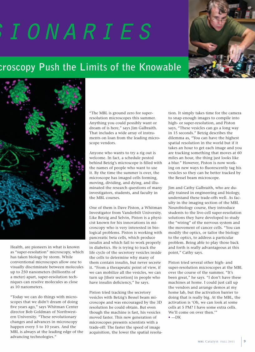
ric Betzig flips on a movie and soon one can hear a pin drop in the Loeb Laboratory room. On his computer screen, a three-dimensional clump of chromosomes is floating in black space. Suddenly the clump starts to pull into halves as the cell divides, and within 20 seconds it is done, the halves disen-tangled and pushing apart.
What’s so exciting is this movie is not a graphic artist’s simulation: It is a real (though sped up) process in a dividing cell. Betzig (top, right) made the movie with a microscope he co-developed that collects up to 200 images per second, piles them into 3D stacks, and renders the biological specimen in unprec-edented depth and detail. “There is no other microscope in the world that can [capture a 3D cellular process] nearly so fast,” Betzig says.
It’s this combination of three-dimen-sionality and speed that really knocks biologists out about the microscope, which Betzig calls the Bessel beam plane illumination. This is why Betzig has brought the microscope to the MBL for two consecutive summers from Howard Hughes Medical Institute’s Janelia Farm campus, where he is a group leader. The first time, in 2010, was when the microscope was brand new and “bleed-ing edge,” he says. “It was a really good time to bring it to the MBL, where all sorts of world-class cell biologists throw everything they can think of at it. We can learn through trial by fire what works and what doesn’t.”
Betzig is one of several physicists at the MBL this summer—experts in optics who rely on the MBL’s powerful conflu-ence of biologists for the feedback they
E
To see images comparing conventional to super-resolution microscopy, please visit www.mbl.edu/catalyst/index.html
The VisionariesPioneers in Super-Resolution Microscopy Push the Limits of the Knowable
need to optimize their inventions. “We need biologists to take these new microscopy techniques and run with them. And hopefully we will get some new biology from them, and eventu-ally something clinically relevant,” says physicist Paul Selvin of the University of Illinois at Urbana-Champaign. Selvin is helping fellow Whitman Investiga-tors get finely detailed images of the ion channels (proteins) that are funda-mental to learning and memory pro-cesses—and to neural disorders such as Alzheimer’s disease.
Selvin and Betzig, along with Whitman Investigators Jim and Cathy Galbraith (top left) of the National Institutes of
8 MBL Catalyst FALL 2011
“The MBL is ground zero for super-resolution microscopes this summer. Anything you could possibly want or dream of is here,” says Jim Galbraith. That includes a wide array of instru-ments on loan from the leading micro-scope vendors.
Anyone who wants to try a rig out is welcome. In fact, a schedule posted behind Betzig’s microscope is filled with the names of people who want to use it. By the time the summer is over, the microscope has imaged cells forming, moving, dividing, and dying, and illu-minated the research questions of many investigators, students, and faculty in the MBL courses.
One of them is Dave Piston, a Whitman Investigator from Vanderbilt University. Like Betzig and Selvin, Piston is a physi-cist known for his innovations in mi-croscopy who is very interested in bio-logical problems. Piston is working with pancreatic beta cells, which produce insulin and which fail to work properly in diabetics. He is trying to track the life cycle of the secretory vesicles inside the cells to determine why many of them contain insulin, but never secrete it. “From a therapeutic point of view, if we can mobilize all the vesicles, we can turn up [their secretion] in people who have insulin deficiency,” he says.
Piston tried tracking the secretory vesicles with Betzig’s Bessel beam mi-croscope and was encouraged by the 3D resolution he could obtain. But even though the machine is fast, his vesicles moved faster. This new generation of microscopes presents scientists with a trade-off: The faster the speed of image acquisition, the lower the spatial resolu-
The VisionariesPioneers in Super-Resolution Microscopy Push the Limits of the Knowable
tion. It simply takes time for the camera to snap enough images to compile into high- or super-resolution, and Piston says, “These vesicles can go a long way in 15 seconds.” Betzig describes the dilemma as, “You can have the highest spatial resolution in the world but if it takes an hour to get each image and you are tracking something that moves at 60 miles an hour, the thing just looks like a blur.” However, Piston is now work-ing on new ways to fluorescently tag his vesicles so they can be better tracked by the Bessel beam microscope.
Jim and Cathy Galbraith, who are du-ally trained in engineering and biology, understand these trade-offs well. As fac-ulty in the imaging section of the MBL Neurobiology course, they introduce students to the live-cell super-resolution solutions they have developed to study the “wiring” of the nervous system and the movement of cancer cells. “You can modify the optics, or tailor the biology to the optics, to address a particular problem. Being able to play those back and forth is really advantageous at this point,” Cathy says.
Piston tried several other high- and super-resolution microscopes at the MBL over the course of the summer. “It’s been great,” he says. “I don’t have these machines at home. I could just call up the vendors and arrange demos at my home lab, but the activation barrier to doing that is really big. At the MBL, the activation is ‘Oh, we can look at some cells at 5 PM? I have some extra cells. We’ll come on over then.’” • —DK
Health, are pioneers in what is known as “super-resolution” microscopy, which has taken biology by storm. While conventional microscopes allow one to visually discriminate between molecules up to 250 nanometers (billionths of a meter) apart, super-resolution tech-niques can resolve molecules as close as 10 nanometers.
“Today we can do things with micro-scopes that we didn’t dream of doing five years ago,” says Whitman Center director Bob Goldman of Northwest-ern University. “These revolutionary changes and advances in microscopy happen every 5 to 10 years. And the MBL is always at the leading edge of the advancing technologies.”
9MBL Catalyst FALL 2011

The GeomeTry of Life
10 MBL Catalyst FALL 2011
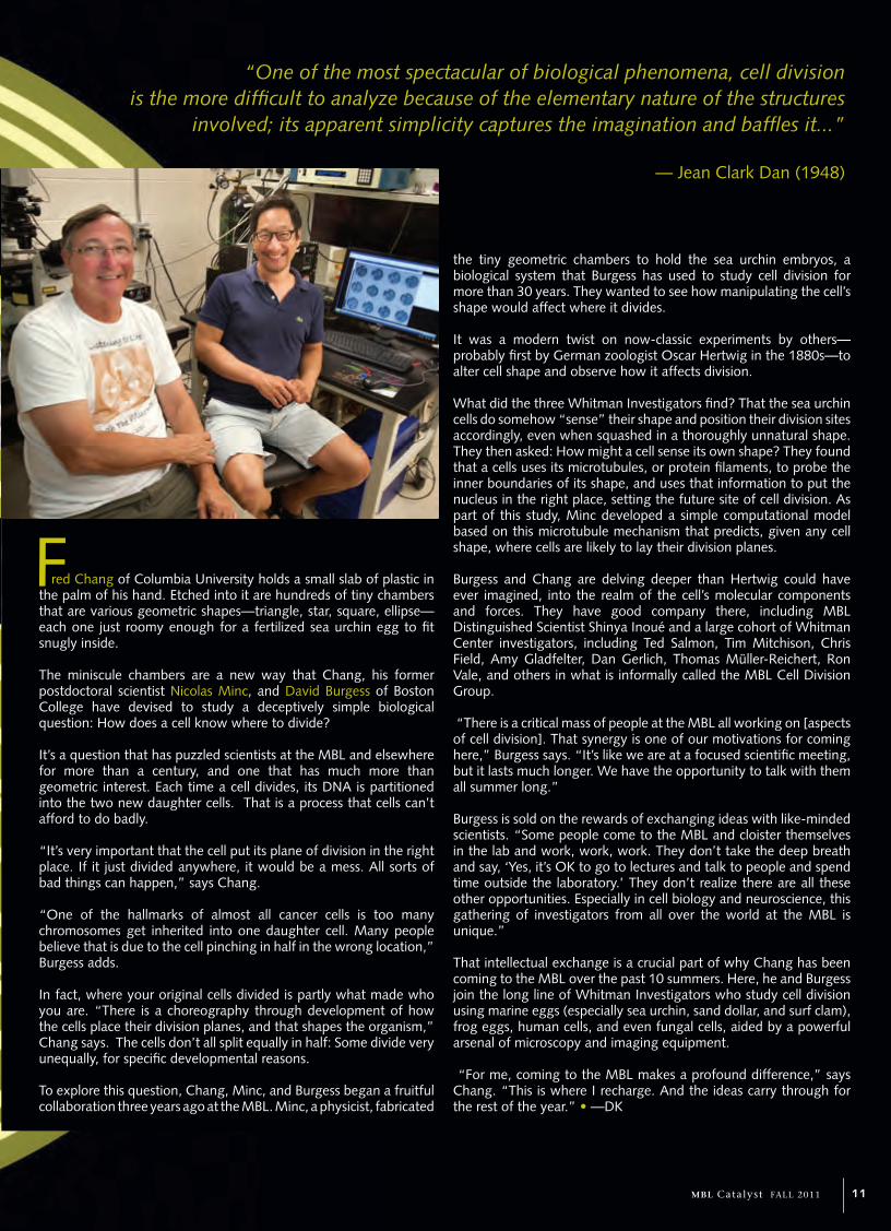
red Chang of Columbia University holds a small slab of plastic in the palm of his hand. Etched into it are hundreds of tiny chambers that are various geometric shapes—triangle, star, square, ellipse—each one just roomy enough for a fertilized sea urchin egg to fit snugly inside.
The miniscule chambers are a new way that Chang, his former postdoctoral scientist Nicolas Minc, and David Burgess of Boston College have devised to study a deceptively simple biological question: How does a cell know where to divide?
It’s a question that has puzzled scientists at the MBL and elsewhere for more than a century, and one that has much more than geometric interest. Each time a cell divides, its DNA is partitioned into the two new daughter cells. That is a process that cells can’t afford to do badly.
“It’s very important that the cell put its plane of division in the right place. If it just divided anywhere, it would be a mess. All sorts of bad things can happen,” says Chang.
“One of the hallmarks of almost all cancer cells is too many chromosomes get inherited into one daughter cell. Many people believe that is due to the cell pinching in half in the wrong location,” Burgess adds.
In fact, where your original cells divided is partly what made who you are. “There is a choreography through development of how the cells place their division planes, and that shapes the organism,” Chang says. The cells don’t all split equally in half: Some divide very unequally, for specific developmental reasons.
To explore this question, Chang, Minc, and Burgess began a fruitful collaboration three years ago at the MBL. Minc, a physicist, fabricated
the tiny geometric chambers to hold the sea urchin embryos, a biological system that Burgess has used to study cell division for more than 30 years. They wanted to see how manipulating the cell’s shape would affect where it divides.
It was a modern twist on now-classic experiments by others—probably first by German zoologist Oscar Hertwig in the 1880s—to alter cell shape and observe how it affects division.
What did the three Whitman Investigators find? That the sea urchin cells do somehow “sense” their shape and position their division sites accordingly, even when squashed in a thoroughly unnatural shape. They then asked: How might a cell sense its own shape? They found that a cells uses its microtubules, or protein filaments, to probe the inner boundaries of its shape, and uses that information to put the nucleus in the right place, setting the future site of cell division. As part of this study, Minc developed a simple computational model based on this microtubule mechanism that predicts, given any cell shape, where cells are likely to lay their division planes.
Burgess and Chang are delving deeper than Hertwig could have ever imagined, into the realm of the cell’s molecular components and forces. They have good company there, including MBL Distinguished Scientist Shinya Inoué and a large cohort of Whitman Center investigators, including Ted Salmon, Tim Mitchison, Chris Field, Amy Gladfelter, Dan Gerlich, Thomas Müller-Reichert, Ron Vale, and others in what is informally called the MBL Cell Division Group.
“There is a critical mass of people at the MBL all working on [aspects of cell division]. That synergy is one of our motivations for coming here,” Burgess says. “It’s like we are at a focused scientific meeting, but it lasts much longer. We have the opportunity to talk with them all summer long.”
Burgess is sold on the rewards of exchanging ideas with like-minded scientists. “Some people come to the MBL and cloister themselves in the lab and work, work, work. They don’t take the deep breath and say, ‘Yes, it’s OK to go to lectures and talk to people and spend time outside the laboratory.’ They don’t realize there are all these other opportunities. Especially in cell biology and neuroscience, this gathering of investigators from all over the world at the MBL is unique.”
That intellectual exchange is a crucial part of why Chang has been coming to the MBL over the past 10 summers. Here, he and Burgess join the long line of Whitman Investigators who study cell division using marine eggs (especially sea urchin, sand dollar, and surf clam), frog eggs, human cells, and even fungal cells, aided by a powerful arsenal of microscopy and imaging equipment.
“For me, coming to the MBL makes a profound difference,” says Chang. “This is where I recharge. And the ideas carry through for the rest of the year.” • —DK
F
“One of the most spectacular of biological phenomena, cell division is the more difficult to analyze because of the elementary nature of the structures
involved; its apparent simplicity captures the imagination and baffles it...”
— Jean Clark Dan (1948)
11MBL Catalyst FALL 2011

mBL mo m e N t
MBL: Why is the squid so useful for studying how nerve cells communicate?
RL: If you consider the nervous system, the brain, you find it is a conglomerate of single cells known as neurons, and glia. Neurons contact each other—they “shake hands,” if you will—at the synapses. For the most part, a synapse functions by very quickly releasing small molecules, called synaptic transmitters, which then activate the next cell in line. And so, it is a chemical event.
I have spent 45 summers at the MBL basically trying to understand synaptic transmission in the squid mantle, the muscular covering for the animal’s vital organs. This is the largest and, to me, the most beautiful synapse ever. While I have also worked with many other synapses, mostly in the mammalian central nervous system, there are very few that are as clear, as large, and as easy to address as that in the squid. We come to the MBL every summer for the squid, and also because the lab is beautiful, the facilities are superb, and the intellectual climate is second to none!
MBL: Over the course of your MBL research, you have literally “written the book” on neurotransmission in the squid. How did this lead to your current research on Alzheimer’s disease?
RL: All of our research seemed to suggest that the squid synapse is very similar to the human synapse not just from a biophysical point of view, but more interestingly, from a molecular point of view. So we—Drs. Mutsuyuki Sugimori, Herman Moreno and I—decided to inject molecules known to produce neural diseases in humans into the squid synapse. The advantage is tremendous in the squid because we can inject the substance directly into the presynaptic [nerve] terminal and see where the injection is located, how quickly it moves to a particular terminal and so on. We have perfect control of the location, and a very good idea of concentration. We decided to study two sets of molecules. The first was beta amyloid, a protein that is a necessary step in the generation of Alzheimer’s disease. When we injected beta amyloid into the squid synapse, we found that it blocked neurotransmission. But the real horror of Alzheimer’s is a protein called tau 42. Because tau 42 can generate Alzheimer’s, it is believed to be the mechanism for the neural
with …
Rodolfo LlinásMBL Whitman Investigator from New York University School of Medicine
Rodolfo R. Llinás is the Thomas and Suzanne Murphy Professor of Neuroscience and chairman of the Physiology and Neuroscience Department at NYU School of Medicine and a University Professor at NYU. He has spent nearly 45 summers conducting research at the MBL, and has also served on the faculty of the Neurobiology course (1971-1978), the Neural Systems and Behavior course (1982), and the Methods in Computational Neuroscience course (1996). Llinás received his M.D. from the Universidad Javeriana in Bogotá and completed his Ph.D. with Nobel Laureate John Eccles. He has received seven honorary doctorates and is an elected member of the U.S. National Academy of Sciences, the American Academy of Arts and Sciences, the American Philosophical Society, the French National Academy of Science, the Spanish Royal Academy of Medicine, and the Colombian National Academy of Medicine. Llinás has authored more than 400 scientific papers and 11 books, including The Squid Giant Synapse: A Model for Chemical Transmission (Oxford University Press, 1999); and was editor-in-chief of the journal Neuroscience from 1974-2000.
When Rodolfo Llinás first came to the MBL in 1962, he was a young
M.D. who had been interested in nervous system physiology since
childhood. That summer, he met distinguished senior investigators
who were among the greatest neuroscientists in history, as well
as several peers who were destined to become his lifelong research
colleagues. “The ‘high’ that followed that first MBL experience has
stayed with me ever since,” he later wrote. After completing his Ph.D.
in neurophysiology, Llinás came back to the MBL in 1965 and has
returned every summer since 1970. His abiding research interest has
been how squid nerve cells communicate with one another at the
synapses, their points of contact. His most recent collaborations at
the MBL have made major strides in illuminating the pathologies of
Alzheimer’s disease and related disorders. At present, a drug Llinás
co-developed to address neural degeneration in Alzheimer’s disease is
in clinical trials.
How the Squid Offered Deep Insight into Alzheimer’s Disease
12 MBL Catalyst FALL 2011
degeneration. So we injected human tau 42 into the squid synapse, and it blocked transmission extraordinarily powerfully and very quickly. We realized the squid synapse could, in fact, tell us a lot about the molecular mechanism for Alzheimer’s. This is the paper we published last spring. We were quite elated as it promised to be quite useful information.
MBL: If the synapses cannot release neurotransmitter, do the nerve cells die?
RL: No, the cells do not die immediately. Rather, they begin to disconnect from each other and retract or “die back.” This is extraordinarily intriguing and very important. The issue of the dying back of neurons arose in discussions with Scott Brady (an MBL Whitman Investigator from the University of Illinois College of Medicine at Chicago). Scott has a very important lab, studying the flow of molecules in the nerve fiber, or axon. He found that tau 42 modifies the movement of molecular motors in the axon. So now we know two issues concerning the effects of tau 42: Synapses cannot release as mentioned above, but also from the work of Scott, the molecular motors are gummed up. That is probably what is happening with Alzheimer’s: It is basically a disconnection syndrome. The cells don’t necessarily die; they just cease to talk to each other. They pull up their roots; they pull up their connectivity.
That being the case, the future is very interesting. If one can prevent cells from disconnecting, or even better make the cells grow connectivity back again, you would have, at the very least, a reduction
How the Squid Offered Deep Insight into Alzheimer’s Disease
of the speed of neural degeneration in Alzheimer’s, and, in the best of cases, you may regain functionality you lost. This is where we are, and it is extraordinarily exciting. We are beginning to understand seriously what the mechanism of Alzheimer’s is, thanks to the squid, to the MBL and to our colleagues. This could not have been done without the squid. Not only is it a beautiful animal, but it is certainly generous as far as allowing us to understand the nature of nervous system function.
MBL: Besides the abundance of squid available, how has being at the MBL enhanced your research?
RL: It is due to the interaction among neurologists, chemists, pharmacologists, physiologists, and computer scientists at the MBL that this level of science can be done. This is really extraordinarily important. Several of my close colleagues (Herman Moreno and Scott Brady, to name just two) are in different places in the winter, but in the summer we all come together at the MBL. We spend three months discussing and writing and agreeing and disagreeing and doing experiments. The importance of being able to get together at the MBL and having the squid to work with is seriously difficult to compare with any other situation. Not only do we have beautiful tools and much support, but we can meet and discuss in the middle of the night as in, ‘Well, you know, this molecule isn’t right. Why don’t we try something else?’ The MBL is a watering hole like no other, as far as science is concerned. • — DK
For a description of discoveries by Rodolfo Llinás and colleagues at the MBL, please visit www.mbl.edu/catalyst/index.html
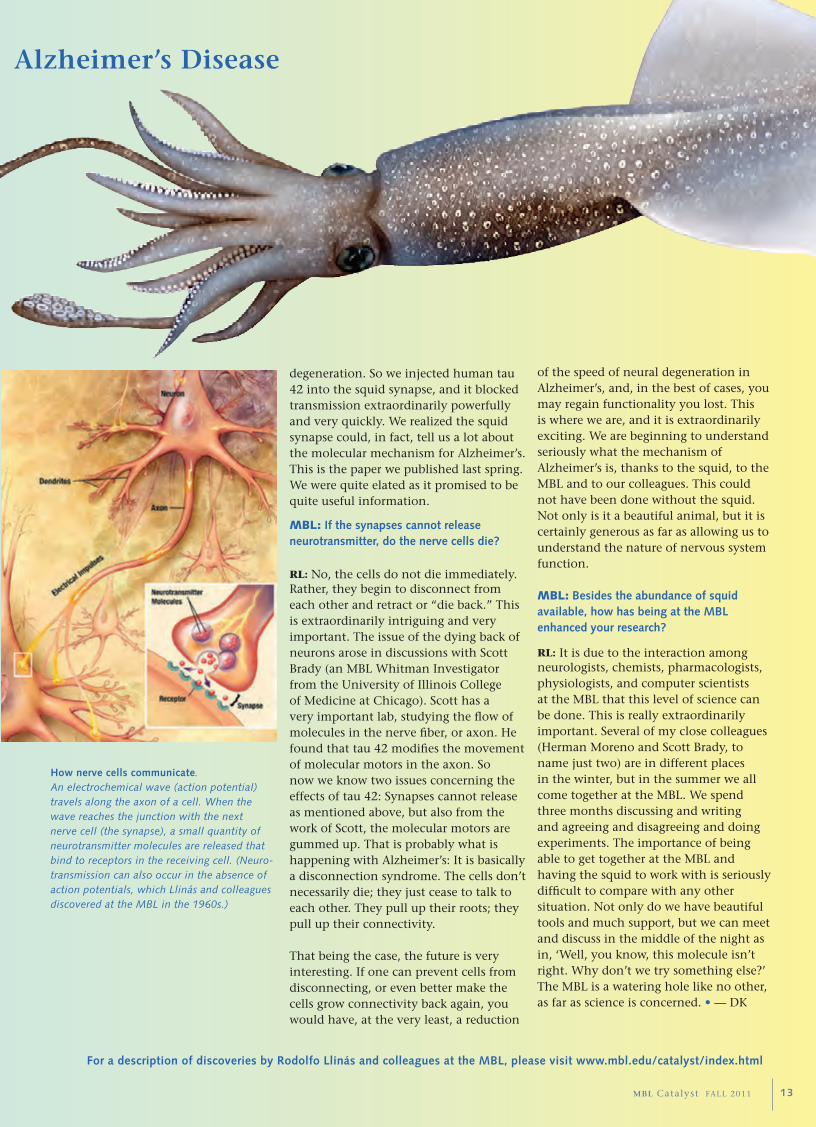
How nerve cells communicate. An electrochemical wave (action potential) travels along the axon of a cell. When the wave reaches the junction with the next nerve cell (the synapse), a small quantity of neurotransmitter molecules are released that bind to receptors in the receiving cell. (Neuro-transmission can also occur in the absence of action potentials, which Llinás and colleagues discovered at the MBL in the 1960s.)
13MBL Catalyst FALL 2011

Ac c o L A D e s
MBL Distinguished Scientist and 2008 Nobel Prize Laureate Osamu Shimomura was recognized at a ceremony at the State House in Boston for his “significant contribution to biomedical research in Massachusetts.” The honor was presented by the Massachusetts Society for Medical Research.
The Biotechnology and Biological Sciences Research Council (UK) named Jason Swedlow of the University of Dundee “Innovator of the Year” for his work on the Open Microscopy Environment, a revolutionary venture into open source software. Swedlow co-directs the MBL’s Analytical & Quantitative and Light Microscopy course.
Ecosystems Center Distinguished Scientist Jerry Melillo was named chairman of a joint public-private sector committee that will produce the next National Climate Assessment report for the United States.
Corporation member Thoru Pederson received a European Molecular Biology Organization medal in Prague for “distinction in cell biology” and for “longstanding support of Czech science.”
MBL President and Director Gary Borisy received the 2011 E.B. Wilson Medal from the American Society for Cell Biology. The medal is awarded for “far-reaching contributions to cell biology over a lifetime in science.”
The following members of the MBL community were elected to the National Academy of Sciences: John Eppig, faculty, Frontiers in Reproduction; John Heuser, faculty, Neurobiology, Biology of Parasitism, Microbial Diversity; Alexander Johnson, faculty, Embryology; Susan McConnell, faculty, Fundamental Issues in Vision Research; Carl Nathan, faculty, Biology of Parasitism; Michel Nussenzweig, alumnus, Physiology; Jisoon Ihm, alumnus, Compu-tational Neuroscience.
John Valois, longtime specimen collector and naturalist and former manager of the MBL Supply Department, was honored with the dedication of the John J. Valois Tank Room in the MBL Marine Resources Center.
Corporation member Arthur Pardee attended the 50th anniversary celebration of the discovery of the operonat Institut Pasteur in Paris. Pardee co-authored (with François Jacob and Jacques Monod) the seminal 1959 “PaJaMo Paper,” representing the culmination of findings that illuminated how gene expression could be controlled. Jacob and Monod won the 1965 Nobel Prize in Physiology or Medicine “for their discoveries concerning genetic control of enzyme and virus synthesis.”
The National Science Foundation awarded $2 million for a project titled “Fire in Northern Alaska: Effect of a Changing Disturbance Regime on a Regional Macrosystem.” Gaius Shaver is the principal investigator.
Paul R. Dupee, Jr. pledged $1.55 million to the MBL General Endowment and the Annual Fund.
The Office of Naval Research awarded $1.4 million for a project titled “Biologically Inspired Routes to Sense-and-Response in
Adaptive, Intelligent Metamaterials.” Roger Hanlon is the principal investigator.
The National Science Foundation awarded $1 million for a project titled “ABI Innovation: Collaborative Research: A Global Names Architecture as a Cyberinfrastructure for Biological Research.” David Patterson is the principal investigator.
The National Institutes ofHealth awarded $1 million for a project titled “Infrared Photoactivation of Inner-Ear Hair Cells.” Whitman Investigator and Adjunct MBL Scientist Richard Rabbitt of University of Utah is the principal investigator.
The Burroughs Wellcome Fund awarded $300,000 to support the MBL Molecular Mycology course.
Gerald and Ruth Fischbach pledged $200,000 to support the Fischbach Scholarship and Lecture and the Annual Fund.
The Simons Foundation pledged $250,000 to create The Fischbach Fellowship in honor of Dr. Gerald Fischbach.
The Davis Educational Foundation granted $263,994 to the Semester in Environmental Science program for equipment and operating expenses.
14 MBL Catalyst FALL 2011
Gi F t s & Gr A N t s
co o L to o L
on the laser spread the beam into a paper-thin sheet, which ensures that only one layer of particles is visible. When a jelly swims through the flattened beam, a high-definition video camera captures footage of what is essentially a cross-sec-tion of the animal, displacing the particles as it moves. Back in the lab, a software program uses the change in location of displaced particles over a series of video frames to calculate the animal’s speed as it glides through the water.
This past summer, the re-searchers used the SCUVA to explore the effects that natural turbulence has on the feeding behavior of the warty comb jelly, a predator in plentiful supply off the coast of Woods Hole. The comb jelly, however, is merely a model, serving as a means to address a more basic question of biological ocean-ography. “All of our exchange rates are based on laboratory work,” Costello says. “So, fundamentally, we’re asking: Is what we see in our laboratory experiments representative of what we’re seeing out in the ocean? If they’re not, we’ll try and find out how unrepresen-tative they are and how we might work to correct that.” • — ARM
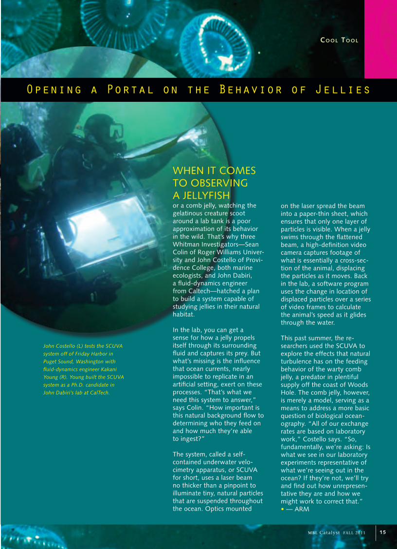
or a comb jelly, watching the gelatinous creature scoot around a lab tank is a poor approximation of its behavior in the wild. That’s why three Whitman Investigators—Sean Colin of Roger Williams Univer-sity and John Costello of Provi-dence College, both marine ecologists, and John Dabiri, a fluid-dynamics engineer from Caltech—hatched a plan to build a system capable of studying jellies in their natural habitat.
In the lab, you can get a sense for how a jelly propels itself through its surrounding fluid and captures its prey. But what’s missing is the influence that ocean currents, nearly impossible to replicate in an artificial setting, exert on these processes. “That’s what we need this system to answer,” says Colin. “How important is this natural background flow to determining who they feed on and how much they’re able to ingest?”
The system, called a self-contained underwater velo-cimetry apparatus, or SCUVA for short, uses a laser beam no thicker than a pinpoint to illuminate tiny, natural particles that are suspended throughout the ocean. Optics mounted
John Costello (L) tests the SCUVA
system off of Friday Harbor in
Puget Sound, Washington with
fluid-dynamics engineer Kakani
Young (R). Young built the SCUVA
system as a Ph.D. candidate in
John Dabiri’s lab at CalTech.
Opening a Portal on the Behavior of Jellies
WHEN IT COMES TO OBSERVING A JELLYFISH
15MBL Catalyst FALL 2011

Placing a Value on the Invaluable
By Robert GoldmanDirector, Whitman Center for Research and Discovery
“Summer research” at the MBL, now known as the Whitman Investigators program, is
entering its 123rd year. From a historical perspective, the scientific output of the Whitman
Center could hardly be more distinguished. Historians have established the crucial role
the MBL has played in setting the foundations of the basic biological sciences, from cell
and developmental biology to neuroscience, biochemistry, and biophysics. Whitman
Investigators have included 30 Nobel Laureates and many more trailblazers, from E.B.
Wilson and Nettie Stevens in the 1900s, who first proposed that sex is determined by
chromosomes, to Ron Vale and others in the 1990s, who identified a major class of
cellular proteins. One could point to hundreds of discoveries at the MBL that have
fundamentally shaped the modern life sciences, and provide the conceptual basis for
understanding human health and disease and developing medical therapeutics.
Given this record of accomplishment, one might ask what is it that makes the Whitman
Center successful and productive. I think there are several factors at play. One is that
the MBL provides the space and time for curiosity-driven research. It’s a place where
scientists can come and really think about their work and gain an undisturbed period
of time to explore new questions. While it may seem disruptive for them to relocate to
Woods Hole, they find the great payoff is a focused research period in a highly supportive,
stimulating environment.
Another factor is the MBL has always been at the leading edge of microscopy. As
a consequence, MBL researchers have pioneered whole new areas of investigation in
cell and molecular biology. For many years, the leading microscope companies have
cooperated to bring their latest equipment to the MBL, which is of enormous utility to
Whitman Investigators. They can often get enough information using MBL microscopes
to finish a publication that would have taken months or been impossible to do in their
home labs.
A third factor, I think, may be the most significant: The MBL has always been a gathering
place for many of the world’s best biomedical researchers. There is inestimable value to
bringing hundreds of researchers to the MBL every year to discuss science, in both formal
and informal ways. Sometimes they have heated disagreements about the interpretation
of data, but many more times they hear something that ignites a spark and they
realize, “Wow, that is exciting.” And then they develop synergistic relations and start
collaborations that really move science forward.
These kinds of de novo collaborations spring up at the Whitman Center all the time. And
this is the aspect of the MBL that is truly unique, and doesn’t happen anywhere else in
the world. Just one example is the collaboration between Rodolfo Llinás, a neuroscientist
from New York, and Scott Brady, a cell biologist from Chicago. They met at the MBL
years ago through their common use of the squid as a model system, and today, they are
making major progress in elucidating the molecular basis of Alzheimer’s disease.
The value of an “international laboratory” where scientists come together to argue,
discuss and try hands-on experiments, sometimes at all hours of the night, is not simple
to calculate. But as scientists well know, and as the MBL’s history plays testament, such a
place is invaluable as a catalyst for scientific progress and discovery. •
Robert Goldman first came to the MBL as a graduate student in 1963 and has returned every summer since 1977, both to conduct research and to serve the laboratory in a host of ways. He is a former MBL trustee (1986-1996), instructor and director of the Physiology Course (1981-1988); director and founder of the Science Writers Hands-on Laboratory Course (1990-1995); and co-director of the Science Journalism Program (1990-2008). He spends the academic year at Northwestern University Medical School, where he is the Stephen Walter Ranson Professor and Chairman of the Department of Cell and Molecular Biology. His research focuses on the structure and function of two important components of the cell: the cytoskeleton intermediate filaments (IFs) and the nuclear lamins; and he is currently using the MBL as a meeting ground for a National Institutes of Health program project grant on the IFs. Goldman received his Ph.D. from Princeton University. He has received numerous honors, including election to the American Association of the Advancement of Science; Merit Award from the National Institute of General Medical Science (1999-2009); Ellison Foundation Senior Scholar; and president of the American Society for Cell Biology (2008). He is chairman of the 2012 Gordon Research Conference on Intermediate Filaments.
16 MBL Catalyst FALL 2011
sc i e N t i s t ’s ey e Vi e w

T r e a s u r e s f r o m T h e m B L ’ s a r c h i v e s
Renewing the Passion for Science
When a scientist invites a student to work in his or her Whitman Center lab, often the student ends up feeling very lucky.
She finds herself welcomed into a “family” that is enriched by the culture of Woods Hole, one that often births a new
generation of distinguished investigators. One such lab was that of S. Meryl Rose and his wife, Florence. From the 1930s
to the 1970s, the Roses studied growth, differentiation, and regeneration at the MBL, and the students Meryl regularly
brought into their lab enjoyed rare access to inspirational mentors. One, Elizabeth D. Hay, later became the first female full
professor in a Harvard Medical School preclinical department. But while still navigating the challenges of her own medical
school training, “what saved me was the summers I went to Meryl and Florence’s laboratory in Woods Hole,” she later re-
called. The Roses often welcomed their students over for dinner and after they would play board games “or just sit around
talking science,” Hay said. The attention the Roses gave them was exceptional, “and it could only have been done, really,
in Woods Hole.” •
Tubularia, relatives of the jellyfish whose colonies grow profusely on docks in Woods Hole, are one of the historical mainstays of MBL research on regeneration, including by S. Meryl Rose and his student Kenyon Tweedell, who later became an MBL independent investigator. Illustration from Ernst Haeckel, Kunstformen der Natur (Leipzig und Wien: Verlag des Bibliographischen Instituts, 1904), plate 6: Tubulariae.
17MBL Catalyst FALL 2011
me m o r A B i L i A

iN t h e Ne x t MBL CAtA Ly s t
Printed with recycled materials.
Biodiversity: Living on Earth
From marine microbes to millions of species in the Encyclopedia of Life, MBL research extends far and wide into the animal and
plant kingdoms. In the next issue of MBL Catalyst, we highlight MBL projects that are furthering our understanding of living species
and their environments.
Smartphone users:
Scan QR code for
online content.
Catalyst
MBL7 MBL StreetWoods Hole, MA 02543 USA
NON-PROFIT ORG.
U.S. POSTAGE
PAID
Marshfield, MA
PERMIT NO. 51
www.mbl.edu