cases in acquired pediatric heart disease common … · cases in acquired pediatric heart disease...
TRANSCRIPT

ANAS ABU HAZEEM
CONSULTANT PEDIATRIC CARDIOLOGIST
KIDSHEART MEDICAL CENTER
Cases in Acquired Pediatric Heart Disease Common Cases With a Twist

9 year old Ali
Ali is a previously healthy 9 yr old boy
Referred to me via his pediatrician because of a heart murmur
Although asymptomatic from a cardiac standpoint, he has a neurologist’s appointment for newly started involuntary movements

Further Hx
There is no Hx of sore throat or viral illness
No skin rash or nodules, no joint pain or swelling
No fever, chills or rigors
No Hx of previous cardiac surgery
No similar family Hx

Examination
Obese child not in pain or distress
There is a 3/6 pan-systolic murmur at the LMSB, radiating to the axilla
You notice frequent non-purposeful movements of the arms and hands that the patient tries to suppress
Rest of the exam is unremarkable

Investigations
Blood count, renal and hepatic profiles were unremarkable
ESR and CRP were normal
ASO was negative
ECG shows biphasic and prolonged p wave in V1
Echocardiogram shows thickened mitral valve leaflets with moderate mitral regurgitation with LA dilation. Aortic valve leaflets are also thickened with mild regurgitation.

DIAGNOSIS?

Rheumatic fever, the modified Jones Criteria
Major criteria
1. Carditis
2. Polyarthritis
3. Erythema marginatum
4. Subcutaneous nodules
5. Sydenham’s chorea
Minor criteria:
1. Fever
2. Arthralgia
3. Elevated acute phase reactants
4. Previous history of RF
5. Prolonged PR interval
*Have to have evidence of antecedent Strep infection: ASO / Strep antibodies / Strep group A throat culture / Recent scarlet fever / anti-deoxyribonuclease B / anti-hyaluronidase/ rapid test ** Two Major criteria or one major and two minor

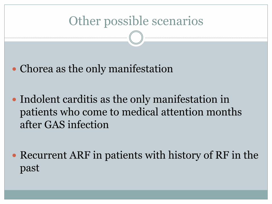
Other possible scenarios
Chorea as the only manifestation
Indolent carditis as the only manifestation in patients who come to medical attention months after GAS infection
Recurrent ARF in patients with history of RF in the past

Rheumatic Fever
Caused by Group A streptococcus pharyngitis
Age group 5-15 years are most affected
The body generates antibodies to fight the bug
Because of similarity (mimicry) between some bacterial and human antigens, the antibodies might result in damage to native tissue (Joints, heart, skin and nervous system)

Epidemiology
470,000 new cases, 233,000 deaths a year
Still endemic in our region
Overcrowding, poor hygiene and limited access to health care are risk factors

Natural History
Usually happens 2-4 weeks after throat infection with GAS but can happen with carrier patients with no history of pharyngitis.
Affects the heart, CNS, joints and skin
Heart disease results in most of the mortality and morbidity


Arthritis
Usually migrates affecting large joints in quick succession
Responds well to NSAIDs
Does not leave damage to the joints

Differentiating ARF from PSRA
Latent period shorter in PSRA (1-2 weeks)
Arthritis responds to ASA better in ARF
No carditis in PSRA
Acute phase reactants usually higher in ARF
Tenosynovitis or renal involvement more with PSRA

Carditis
Causes pancarditis (pericardium, epicardium, myocardium and endocardium)
Although significant damage can be caused by one episode, most of the damage is caused by recurrent episodes
Valves most affected: Mitral alone, both mitral and aortic, aortic alone (left sided always affected)

Erythema marginatum or annulare
Pink or faintly red non-pruritic rash on the trunk and sometimes the extremities but not the face
Extends to the outside with return of normal skin in the center
Not a frequent manifestation (5%)


Subcutaneous nodules
The least common manifestation of ARF
Firm, painless lesions.
Usually located over bony surfaces or tendons
Surface of the skin not inflamed, and is movable


Sydenham’s Chorea (St. Vitus dance)
Most common form of chorea in childhood
Can be a very late manifestation (months after GAS)
Non-rhythmic involuntary movements, muscular weakness and emotional disturbances
Usually improves during sleep

The Lines of Management
Eradication of GAS
Symptomatic relief of acute disease manifestations
Prophylaxis against future GAS infection to prevent recurrence

GAS Eradication
Treat with antibiotics even if pharyngitis not present now
Screen all family members, treat who test positive even if asymptomatic

Carditis and arthritis
Anti-HF meds in severe cases
High dose ASA (80-100 mg/kg/day) in children and 4-8 g/day in adults till symptoms are gone or inflammatory markers are normalized
Use of steroids is controversial (unlikely useful except for resistant arthritis)
Valve surgery

Chorea
Usually self-limited
Responds to treatment with haloperidol
Other modes of treatment: valproic acid, phenobarbital, diazepam, chlorpromazine, steroids, plasma exchange

Primary Prevention
Primary prevention: by identifying
patients with GAS pharyngitis
and treating them promptly
Can be difficult to achieve as one
third of ARF patients do not have
apparent infection
Can use Oral penicillin V, Amoxicillin,
single dose of IM penicillin G
benzathine, Cephalexin, azithromycin,
clarithromycin or clindamycin

Secondary prophylaxis
Can use Penicillin G benzathine every 3-4 weeks (best results)
Oral penicillin V twice daily
Sulafadiazine once daily
Azithromycin daily

Secondary prophylaxis duration
Secondary prevention: duration is unclear but depends on number of previous episodes and the presence of risk factors
1. ARF with carditis and residual heart disease: 10 years or
until 40 years of age or for life.
2. ARF with carditis but no residual disease: 10 years or until 21 years of age (whichever longer)
3. ARF without carditis: 5 years or until 21 years of age (whichever longer)

Late manifestations
Rheumatic heart disease is the most severe manifestation of ARF
Mitral valve is the most affected resulting in calcification, stenosis and +/- regurgitation
Rarely recurrent arthritis can lead to Jaccoud arthropathy
Take Home Message
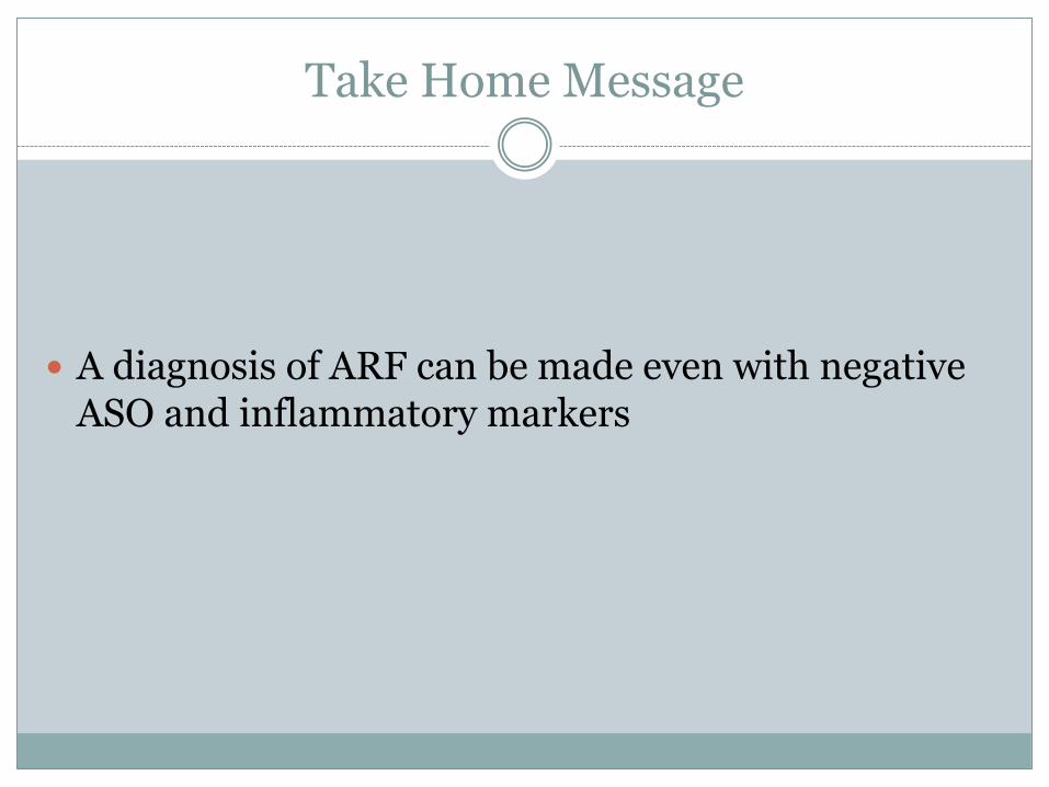
A diagnosis of ARF can be made even with negative ASO and inflammatory markers

9 month old Juri
Juri presented to our ER with fussiness, prolonged fever, pink eyes and swollen lips.
She also has runny nose and there are multiple sick contacts at home

Examination
Very difficult to examine
Febrile with 39.6 C. No rash seen
Teary red eyes without pus
No cervical LAP
Tongue is very red, lips are mildly swollen
Hands and feet are normal
She’s tachycardic and a flow murmur is heard.
Work-up
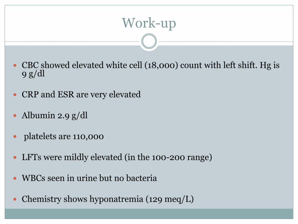
CBC showed elevated white cell (18,000) count with left shift. Hg is 9 g/dl
CRP and ESR are very elevated
Albumin 2.9 g/dl
platelets are 110,000
LFTs were mildly elevated (in the 100-200 range)
WBCs seen in urine but no bacteria
Chemistry shows hyponatremia (129 meq/L)
DIAGNOSIS?

Diagnostic criteria
Fever > 5 days (must have)
And 4 out of 5: 1. Polymorphous rash
2. Cervical lymphadenitis (>1.5 cm)
3. Changes in the lips and mucus membranes
4. Extremity skin changes (redness, swelling, peeling of the skin)
5. Non-purulent bulbar conjunctivitis

Incomplete Kawasaki disease
Following the strict criteria for Kawasaki disease resulted in missing 10-15% of patients who have Kawasaki disease.
Not a small percentage of those patients ended up developing coronary artery aneurysms.
So what to do in-order not to miss those patients?


Kawasaki Disease
Also called mucocutaneous lymph node syndrome
Likely caused by an abnormal immune system response to an infectious agent
Most common between 1-2 years of life
Cases below 3 months or more than 8 years are rare

Pathology
During the acute phase of the illness, microvasculitis occurs with predilection to the coronary arteries
Coronary artery aneurysms develop in 15-25% of untreated patients
Diffuse pancarditis can happen leading to cardiac dysfunction, AV valve regurgitation, conduction abnormalities and pericarditis.
During the late phases, fibrosis can result in narrowing of the coronary arteries leading to stenosis and possible myocardial infarctions
Elevated platelet count increases the risk of MI

Other manifestations
Sterile pyuria
Elevation in liver enzymes
Arthritis or arthralgia
Gallbladder hydrops

Acute Phase (First 10 days)
Abrupt onset of high fever and irritability. Conjunctivitis
resolves quickly. Redness of mucus membranes, fissuring, ulcerations, strawberry tongue. Rash can be of different kinds. Cervical LAP happens in 50% of patients. Fever duration 11-12 days average, but resolves quickly with treatment.
Leukocytosis, thrombocytosis, high inflammatory markers, sterile pyuria, elevated LFTs, lipid abnormalities.
Coronaries can get affected during this stage.
Subacute Phase
Desquamation and peeling of the fingers and toes
Rash, fever and LAP disappear
Most of cardiovascular manifestations occur in this phase
Worsening of thrombocytosis

Convalescent Phase
This phase lasts till all inflammatory markers and platelets return to normal levels
Beau’s lines appear during this phase

Complications
Cardiac manifestations dictate the prognosis
Coronary aneurysms, thrombosis, stenosis
Cardiac dysfunction, AV valve damage, dilation of the ascending aorta, effusion, heart block
Peripheral artery aneurysms and stenosis


Management
Admission
High dose ASA (30-100 mg/kg/day)
Anti-pyretics
IVIG (2 g/kg IV infusion) +/- oral steroids. Repeat IVIG if no
improvement
Pulse steroids IV for non-responders
Infliximab for resistant cases

Management
Switch ASA to low-dose before discharge, some
recommend not before at least 14 days of illness
Stop ASA after platelets are normal, or 6-8 weeks after illness, whatever longer
If coronary abnormalities persist, patient will be always on ASA
In case of aneurysms, clopidogrel or even warfarin can be added

Question
Would do you start our patient on steroids along with IVIG and ASA when the patient presented?

The Kobayashi score
Sodium less than 133 mmol/L (2 points)
AST >100 Units/L (2 points)
CRP >10 mg/dl (1 point)
Neutrophils >80% of WBC (2 points)
Platelets less than 300,000/mm3 (1 point)
Days of illness at initial treatment less than 5 (2 points)
Age less than 12 months (1 point)

Follow up
Depending on the level of coronary involvement

Take home messages
You don’t have to fulfill 4 criteria to diagnose Kawasaki disease
Infants less than 6 months with fever for more than 7 days, consider strongly treating as Kawasaki disease
In patients with risk of IVIG treatment failure, start steroids at the same time of IVIG and ASA

Michelle
16 yr old female previously healthy
Started having chest pain that is getting worse over the last couple of days
Sharp, worse with breathing and leaning forward
No SOB, palpitations or syncope
Examination
There is a friction rub, otherwise exam is normal
CXR showed mild cardiomegaly
ECG showed elevated ST segment in anterior precordial leads.
Echocardiogram showed small pericardial effusion.


DIAGNOSIS?

Follow up
Patient was started on Ibuprofen and given a very close follow up
After 1 week, the patient reported having SOB, fatigue and dizziness
CXR showed enlarging heart shadow
Echo showed large pericardial effusion


Follow up
The patient was admitted and underwent pericardiocentesis
Examination of the fluid showed elevated WBCs but cultures and viral studies were negative
Patient was given colchicine in addition to the ibuprofen and on follow up, patient continued to do well.

Pericarditis
The most common cause of CARDIAC chest pain in pediatrics
Esp in pediatrics, pericarditis is most commonly caused by a viral infection, but in many cases, the cause won’t be known.

Clinical manifestations
Chest pain, Fatigue, SOB, syncope
On exam, a friction rub might be heard
Pulsus paradoxus if there is cardiac tamponade.

Work up
Chest X-ray shows flask-shaped cardiomegaly in the presence of effusion
ECG shows diffuse ST segment elevation with depression in V1 and aVR
T-wave inversion can happen and usually persists with chronic pericarditis

Initial Work up
Blood samples for markers of inflammation
CBC, blood cultures (if febrile)
Cardiac enzymes can be positive in 32% of patients
CXR, ECG
Echocardiogram shows effusion

Additional work up in atypical cases
Tuberculin skin test
HIV titers
ANA
CT scan
MRI
Pericardial fluid sample if pericardiocentesis done for diagnostic or therapeutic purposes

Treatment of acute pericarditis
Activity restriction
Anti-inflammatory medications like ibuprofen
Colchicine to be initiated with NSAIDs
Steroids for resistant cases
Pericardiocentesis and drainage
Pericardial window, pericardiotomy or pericardioectomy for
resistant cases

Take Home message
You don’t have to wait for refractory pericarditis to start colchicine. You can start it at the same time as NSAIDs

Thank you