case report metastasis of renal cell carcinoma to ... j clin exp pathol 2016;9(8):8730-8735...
TRANSCRIPT
Int J Clin Exp Pathol 2016;9(8):8730-8735www.ijcep.com /ISSN:1936-2625/IJCEP0026741
Case Report Metastasis of renal cell carcinoma to hemangioblastoma of the spinal cord in von Hippel-Lindau disease: report of a case and review of literature
Guobin Zhang, Yingzhi Hou, Wenqing Jia, Jun Yang, Yulun Xu
Department of Neurosurgery, China National Clinical Research Center for Neurological Diseases (NCRC-ND), Center of Brain Tumor, Beijing Institute for Brain Disorders, Beijing Key Laboratory of Brian Tumor, Beijing Tiantan Hospital, Capital Medical University, Beijing 100050, P. R. China
Received February 26, 2016; Accepted May 22, 2016; Epub August 1, 2016; Published August 15, 2016
Abstract: Tumor-to-tumor metastasis is considered to be an unusual event. Here, we describe a patient with von Hippel-Lindau (VHL) syndrome with metastasis of renal cell carcinoma (RCC) to a hemangioblastoma (HAB) of the spinal cord. Histological examination revealed cells with clear cytoplasm arranged in alveolar or tubular pat-terns in most fields of view. In the other area, the tumor showed abundant thin-walled vessels and clear cells. Immunohistochemically, the tumor cells were positive for vimentin and CK-8/18, which indicated a metastatic le-sion of RCC clear cells. The diagnosis of RCC metastasis within an HAB might be overlooked because they have similar morphological characteristics. Detailed histological analyses and immunohistochemical stains should be helpful to determine whether HAB contain metastatic RCC, especially in patients with VHL syndrome.
Keywords: Von Hippel-Lindau (VHL) syndrome, renal cell carcinoma, hemangioblastoma, tumor-to-tumor metasta-sis
Introduction
Von Hippel-Lindau (VHL) disease is an autoso-mal dominant inherited disorder, characterized by multiple hemangioblastomas (HAB) of the central nervous system and cysts or tumors of various organs such as the kidney, adrenal gland and pancreas, which is now widely ackn- owledged as a hereditary neoplastic syndrome caused by germline mutations of the VHL tumor suppressor gene [1, 2]. Histologically, the pres-ence of clear cells and highly vascularized stro-ma are common to HAB and renal cell carcino-ma (RCC) of clear cell type, thus, metastatic lesions of RCC might be confused with HAB in patients with VHL disease [3]. Positive immu-nohistochemical reactions for cytokeratin and vimentin in the metastatic carcinomas facili-tate differentiation between these two lesions [4, 5].
Tumor-to-tumor metastasis is considered to be an uncommon event in the central nervous sys-
tem (CNS), which involves a pre-existing CNS tumor with metastatic deposits from a syste- mic neoplasm [6]. Theoretically, HAB is a good recipient of tumor-to-tumor metastasis because of its slow growth and abundant vascularization [7]. However, few cases of HAB complicated by metastatic tumor had been reported previously. We report a 34-year-old man with (VHL) syn-drome who had a HAB in the cervical cord com-plicated by a metastasis from RCC.
Materials and methods
A case of metastasis of RCC to HAB of the spi-nal cord in VHL disease was retrieved in the department of Neurosurgery, Beijing Tiantan Hospital. The tumor sample was fixed in 10% neutral buffered formalin, routinely processed, and embedded in paraffin. Hematoxylin-eosin stained sections and immunohistochemical staining for both CK8/18 and vimentin were reviewed independently by two experienced pathologists.
Metastasis of RCC to HAB of the spinal cord
8731 Int J Clin Exp Pathol 2016;9(8):8730-8735
DNA sequence analysis of the VHL gene was performed for the patient and his daughter. Venous blood was obtained, and genomic DNA was isolated from peripheral blood leukocytes. The VHL gene was amplified by polymerase chain reaction (PCR). The following primers were used for both PCR and sequencing: 5’-TGGAAATACAGTAACGAGTTGG-3’ and 5’-GG- CTTCAGACCGTGCTAT-3’, the forward and re- verse primers for exon 1, respectively; 5’-TGG- GATTACAGGTGTGGG-3’ and 5’-GAAGTCAGCAA- CAAATAAGC-3’, the forward and reverse prim-ers for exon 2, respectively; and 5’-GGTAGTT- GTTGGCAAAGC-3’ and 5’-TCAAAGAGGAGAAAG-
ACTTG-3’, the forward and reverse primers for exon 3, respectively. The PCR amplification pro-tocol included 35 cycles of 94°C for 60 s, 60°C for 60 s, and 72°C for 60 s. Sanger DNA sequencing was performed. Products were sequenced using the ABI 3700 DNA Genetic Analyzer, and then assessed using DNAMAN.
Results
Case report
In October 2015, a 34-year-old man was admit-ted to our hospital with multiple HAB in the cer-ebellum, medulla oblongata and cervical cord,
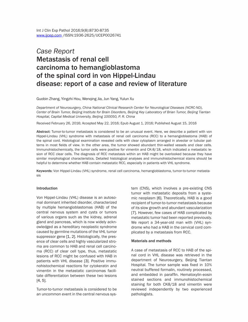
Figure 1. A. Preoperative sagittal T1-weighted contrast-enhanced MRI showing two enhancing nodule in the medulla oblongata and 5th cervical cord. B. Postoperative early stage sagittal T1-weighted contrast-enhanced MRI showing no residual mass lesion in cervical cord. C. An abdominal contrast-enhanced CT scan revealing multiple, cystic le-sions in the pancreas and cystic (red arrows). D. The abdominal contrast-enhanced CT scan showing the cystic and solid masses in left kidney (red arrows).
Metastasis of RCC to HAB of the spinal cord
8732 Int J Clin Exp Pathol 2016;9(8):8730-8735
who presented with a 4-month history of pro-gressive neck pain and numbness of the upper extremities. Upon examination, the patient had slight neck stiffness and disorders of shallow sensation. His father died from a cerebral hem-orrhage when he was still unborn. Between 2004 and 2009 he had undergone two surgical procedures for HAB in the cerebellum and medulla oblongata. A T1-weighted contrast-enhanced MRI disclosed two enhancing nodule in the medulla oblongata and 5th cervical cord
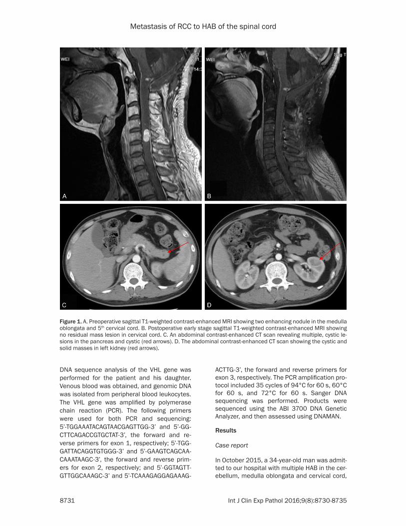
(Figure 1A). The abdominal contrast-enhanced CT scan showed multiple cystic masses in the pancreas (Figure 1C) and cystic and solid masses in left kidney but without any clinical symptoms (Figure 1D). The patient and his daughter accepted the genetic evaluation after informed consent and both of them were con-firmed as the missense mutation (c.500G>A (p.R167Q) and c.589G>C (p.D197H)) in exon 3 of the VHL gene (Figure 2). Intraoperatively, the tumor of the 5th cervical cord had a well-defined
Figure 2. Detection of the VHL gene mutation in the patient and his daughter. A. DNA sequence traces of the mis-sense mutation (c.500G>A (p.R167Q) and c.589G>C (p.D197H)) in exon 3 of the VHL gene from the patient’s pe-ripheral blood. B. DNA sequence traces of the missense mutation (c.500G>A (p.R167Q) and c.589G>C (p.D197H)) in exon 3 of the VHL gene from the peripheral blood by the patient’s daughter.
Metastasis of RCC to HAB of the spinal cord
8733 Int J Clin Exp Pathol 2016;9(8):8730-8735
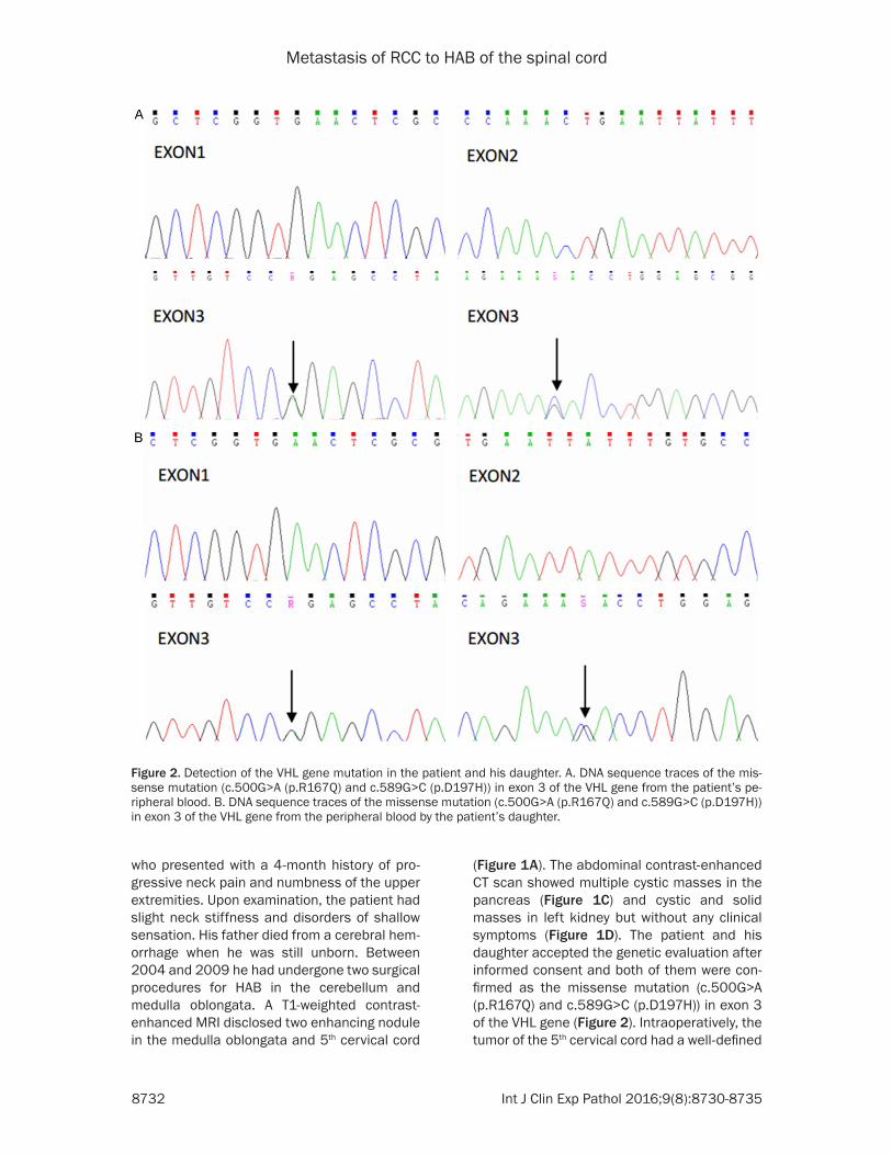
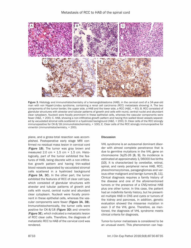
plane, and a gross-total resection was accom-plished. Postoperative early stage MRI con-firmed no residual mass lesion in cervical cord (Figure 1B). The tumor was gray brown and measured 2.0 cm × 1.5 cm × 1.5 cm. Histo- logically, part of the tumor exhibited the fea-tures of HAB, being discrete with a non-infiltra-tive growth pattern and having thin-walled blood vessels separated by vacuolated stromal cells scattered in a hyalinized background (Figure 3A, 3C). In the other part, the tumor exhibited the features of RCC of clear cell type, which consisted of glandular structures with alveolar and tubular patterns of growth and cells with round, central nuclei and abundant clear cytoplasm. Nucleoli were focally promi-nent in these epithelial cells, whereas the vas-cular components were fewer (Figure 3A, 3B). Immunohistochemically, the tumor cells were positive for CK-8/18 (Figure 3D) and vimentin (Figure 3E), which indicated a metastatic lesion of RCC clear cells. Therefore, the diagnosis of metastatic RCC to HAB of the cervical cord was rendered.
Discussion
VHL syndrome is an autosomal dominant disor-der with almost complete penetrance that is due to germline mutations in the VHL gene on chromosome 3q25-26 [8, 9]. Its incidence is estimated at approximately 1/36000 live births [10]. It is characterized by cerebellar, retinal, spinal, and rarely peripheral nerve HAB, RCC, pheochromocytomas, paragangliomas and var-ious other malignant and benign tumors [6, 11]. Clinical diagnosis requires a family history of the disease and one of the aforementioned tumors or the presence of a CNS/retinal HAB plus one other tumor. In this case, the patient had an indefinite family history, but he present-ed multiple HAB in CNS and cysts or tumors of the kidney and pancreas, in addition, genetic evaluation showed the missense mutation in exon 3 of the VHL gene. Therefore, as men-tioned, the diagnosis of VHL syndrome meets clinical criteria for diagnosis.
Tumor-to-tumor metastasis is considered to be an unusual event. This phenomenon can hap-
Figure 3. Histology and immunohistochemistry of a hemangioblastoma (HAB), in the cervical cord of a 34-year-old man with von Hippel-Lindau syndrome, containing a renal cell carcinoma (RCC) metastasis showing: A. The two components of the tumor border, the upper side, a HAB and the lower side, a RCC (H&E, × 40); B. RCC consisted of glandular structures with alveolar and tubular patterns of growth and cells with round, central nuclei and abundant clear cytoplasm. Nucleoli were focally prominent in these epithelial cells, whereas the vascular components were fewer (H&E, × 200); C. HAB, showing a non-infiltrative growth pattern and having thin-walled blood vessels separat-ed by vacuolated stromal cells scattered in a hyalinized background (H&E, × 200); D. Clear cells of the RCC strongly immunopositive for CK-8/18 (immunohistochemistry, × 100); E. Clear cells of the RCC strongly immunopositive for vimentin (immunohistochemistry, × 200).
Metastasis of RCC to HAB of the spinal cord
8734 Int J Clin Exp Pathol 2016;9(8):8730-8735
pen in tumor-bearing patients with concurrent or metachronous second malignant tumors. In 1984, Pamphlett defined the criteria for diag-nosis of tumor-to-tumor metastasis [12]. He established three criteria. 1) The metastatic nidus must be at least partially enclosed by a rim of histologically distinct primary tumor tis-sue. 2) The existence of the primary carcinoma must be proven. 3) The metastatic tumor must be demonstrated to be compatible with the pri-mary carcinoma by morphological or immuno-histochemical means [12]. RCC and meningio-ma are the most common visceral and intra- cranial recipients of metastasis, respectively, although the mechanism of tumor-to-tumor metastasis is poorly understood [13]. In theory, both HAB and meningioma are good recipients of RCC because they have abundant vascular-ization, slow growth and often remain unno-ticed for a long time [13]. Furthermore, it is pos-sible that RCC and HAB both represent attrac-tive sites for metastasis on the basis of their common VHL mutations and the effects of the protein product of the VHL gene (pVHL) muta-tions on the extracellular matrix [11]. Previous work has shown that a normal fibronectin matrix acts to prevent tumor cells from spread-ing into the extracellular matrix and then from spreading, thus the presence of a defective matrix may provide an environment that allows metastatic tumor cells to grow successfully [11].
Metastatic RCC develops in 40% of patients with VHL disease, and it is the cause of death in one third of them [3]. Earlier detection of RCC has an important impact on survival. Since high vascularization and clear cell morphology are common to both tumor types, these character-istics sometimes cause differential diagnostic problems between HAB and metastasis of clear cell RCC. Histologically, typical RCC has alveo-lar or tubular patterns, water-clear cytoplasm, and an absence of vacuolated stromal cells. HAB has prominent proliferations of stromal cells, and occasionally displays well defined lobules. However, it is often difficult to see detailed cell structure because of the abundant vascular tumor tissue and the little tissue avail-able for examination. Thus, morphology alone may not serve as a reliable diagnosis. Immuno-histochemical stains can be used to support this histological distinction. Immunohistoche- mical staining for cytokeratin, PAX-8, EMA,
CD10 and vimentin facilitates the differentia-tion of HAB and metastatic clear cell RCC, as HABs are negative for these epithelial markers [5]. Additionally, the MIB-1 labeling index (Ki-67 immunostain) is reported to be low in HAB (less than 5%) but moderate to high in RCC (more than 5%) [14].
Spinal HABs make up less than 2% of all spinal cord tumors and less than 7% of all central ner-vous system HABs [15]. Although HABs may occur sporadically, a spinal location is strongly associated with VHL disease [15]. Metastatic RCC to HAB of the spinal cord was first reported in 2001, in a patient with VHL disease. Since then, reports of 10 more cases have been pub-lished [16]. The current case is the first report that the metastasis of RCC to a spinal HAB in Chinese patients with VHL disease.
In conclusion, RCC is the most common meta-static tumor amongst those documented to metastasize to HAB and it usually occurs in the context of VHL syndrome. The diagnosis of RCC metastasis within an HAB might be overlooked because they have similar morphological char-acteristics. Detailed histological analyses and immunohistochemical stains should be helpful to determine whether HAB contain metastatic RCC, especially in patients with VHL syndrome.
Acknowledgements
This study was supported by National Key Technology Research and Development Pro- gram of the Ministry of Science and Technology of China (No. 2013BAI09B03) and Beijing Ins- titute for Brain Disorders (No. BIBD-PXM2013_ 014226_07_000084). Written informed con-sents were obtained from the patients for pub-lication of this case report and any accompany-ing images. A copy of the written consent is available for review by the editor-in-chief of this journal.
Disclosure of conflict of interest
None.
Address correspondence to: Yulun Xu, Department of Neurosurgery, China National Clinical Research Center for Neurological Diseases (NCRC-ND), Center of Brain Tumor, Beijing Institute for Brain Disorders, Beijing Key Laboratory of Brian Tumor, Beijing Tian- tan Hospital, Capital Medical University, 6 Tiantan
Metastasis of RCC to HAB of the spinal cord
8735 Int J Clin Exp Pathol 2016;9(8):8730-8735
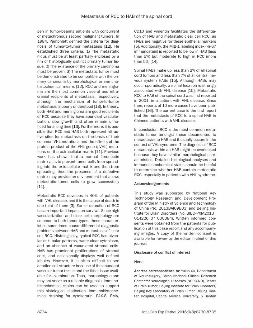
Xili, Chongwen District, Beijing 100050, P. R. China. Tel: +86 10 67098431; Fax: +86 10 67051337; E-mail: [email protected]
References
[1] Maher ER, Neumann HP and Richard S. von Hippel-Lindau disease: a clinical and scientific review. Eur J Hum Genet 2011; 19: 617-623.
[2] Nordstrom-O’Brien M, van der Luijt RB, van Rooijen E, van den Ouweland AM, Majoor-Krakauer DF, Lolkema MP, van Brussel A, Voest EE and Giles RH. Genetic analysis of von Hippel-Lindau disease. Hum Mutat 2010; 31: 521-537.
[3] Clelland CA and Treip CS. Histological differen-tiation of metastatic renal carcinoma in the cerebellum from cerebellar haemangioblasto-ma in von Hippel-Lindau’s disease. J Neurol Neurosurg Psychiatry 1989; 52: 162-166.
[4] Gouldesbrough DR, Bell JE and Gordon A. Use of immunohistochemical methods in the differ-ential diagnosis between primary cerebellar haemangioblastoma and metastatic renal car-cinoma. J Clin Pathol 1988; 41: 861-865.
[5] Polydorides AD, Rosenblum MK and Edgar MA. Metastatic renal cell carcinoma to hemangio-blastoma in von Hippel-Lindau disease. Arch Pathol Lab Med 2007; 131: 641-645.
[6] Mottolese C, Stan H, Giordano F, Frappaz D, Alexei D and Streichenberger N. Metastasis of clear-cell renal carcinoma to cerebellar he-mangioblastoma in von Hippel Lindau disease: rare or not investigated? Acta Neurochir (Wien) 2001; 143: 1059-1063.
[7] Hamazaki S, Nakashima H, Matsumoto K, Taguchi K and Okada S. Metastasis of renal cell carcinoma to central nervous system hemangioblastoma in two patients with von Hippel-Lindau disease. Pathol Int 2001; 51: 948-953.
[8] Feletti A, Anglani M, Scarpa B, Schiavi F, Boaretto F, Zovato S, Taschin E, Gardi M, Zanoletti E, Piermarocchi S, Murgia A, Pavesi G and Opocher G. Von Hippel-Lindau disease: an evaluation of natural history and functional disability. Neuro Oncol 2016; 18: 1011-20.
[9] Chittiboina P and Lonser RR. Von Hippel-Lindau disease. Handb Clin Neurol 2015; 132: 139-156.
[10] Wanebo JE, Lonser RR, Glenn GM and Oldfield EH. The natural history of hemangioblastomas of the central nervous system in patients with von Hippel-Lindau disease. J Neurosurg 2003; 98: 82-94.
[11] Abou-Hamden A, Koszyca B, Carney PG, Sandhu N and Blumbergs PC. Metastasis of renal cell carcinoma to haemangioblastoma of the spinal cord in von Hippel-Lindau disease: case report and review of the literature. Pathology 2003; 35: 224-227.
[12] Pamphlett R. Carcinoma metastasis to menin-gioma. J Neurol Neurosurg Psychiatry 1984; 47: 561-563.
[13] Caroli E, Salvati M, Giangaspero F, Ferrante L and Santoro A. Intrameningioma metastasis as first clinical manifestation of occult primary breast carcinoma. Neurosurg Rev 2006; 29: 49-54.
[14] Xiong J, Chu SG, Wang Y, Zhu JJ, Li C and Mao Y. Metastasis of renal cell carcinoma to a hae-mangioblastoma of the medulla oblongata in von Hippel-Lindau syndrome. J Clin Neurosci 2010; 17: 1213-1215.
[15] McEvoy AW, Benjamin E and Powell MP. Hae- mangioblastoma of a cervical sensory nerve root in Von Hippel-Lindau syndrome. Eur Spine J 2000; 9: 434-436.
[16] Fakih M, Schiff D, Erlich R and Logan TF. Intramedullary spinal cord metastasis (ISCM) in renal cell carcinoma: a series of six cases. Ann Oncol 2001; 12: 1173-1177.