case report macular amyloidosis: a rare case reportet_al.pdf · any atopic disease, drug intake,...
TRANSCRIPT
INTERNATIONAL JOURNAL OF CONTEMPORARY MEDICAL RESEARCH Volume 2 | Issue 2|
408 IJCMR
ABSTRACT Introduction: Amyloidosis is defined as deposition of abnormal extracellular proteins. Cutaneous amyloidosis is a rare entity. Macular amyloidosis is the most subtle form of cutaneous amyloidosis, characterized by asymptomatic reticulate hyperpigme- ntation or pruritic lichenoid papular lesions. Etiology however is still unknown. Case report: We reported a case of macular amyloidosis in a 32 year old male who presented with diffuse raised papules in upper torso. Conclusion: Owing to its wide spectra of presentation, rare occurrence and unknown etiology, this case provided an interesting insight into the disease. Keywords: Cutaneousamyloidosis, macular amyloidosis. How to cite this article: Sonia Chhabra, Pansi Gupta,Mansi Agarwal, Padam Parmar, Ashish Sharma, Rajeev Sen. Macular Amyloidosis: A Rare Case Report. International Journal Of Contemporary Medical Research. 2015;2(2):408-410 Source of Support: Nil Conflict of Interest: None
INTRODUCTION CutaneousAmyloidosis is a type of skin disorder characterized by extracellular deposition of a proteinaceous substance. Two types of cutaneous amyloidosis are observed. One of them is primary localized cutaneous amyloidosis (PLCA), without any deposits in the internal organs and the other
oneis known as secondary cutaneous amyloidosis.1 Primary cutaneous amyloidosis is a localized type of amyloidosis where amyloid is deposited in the skin and occurs as three variants- macular amyloidosis, lichen amyloidosis and nodular amyloidosis.2 Macular amyloidosis is the most common form of cutaneous amyloidosis but it has low prevalence.1 Macular amyloidosis is characterized by pruritic macules showing pigmentation with a rippled pattern. Upper back is fairly common site. Prolonged friction can be a cause. Both lichen and macular amyloidosis can be found in same patient. In such cases usually lichen appears in setting of macular amyloidosis due to scratching. Keratogenic origin of amyloid is proposed.
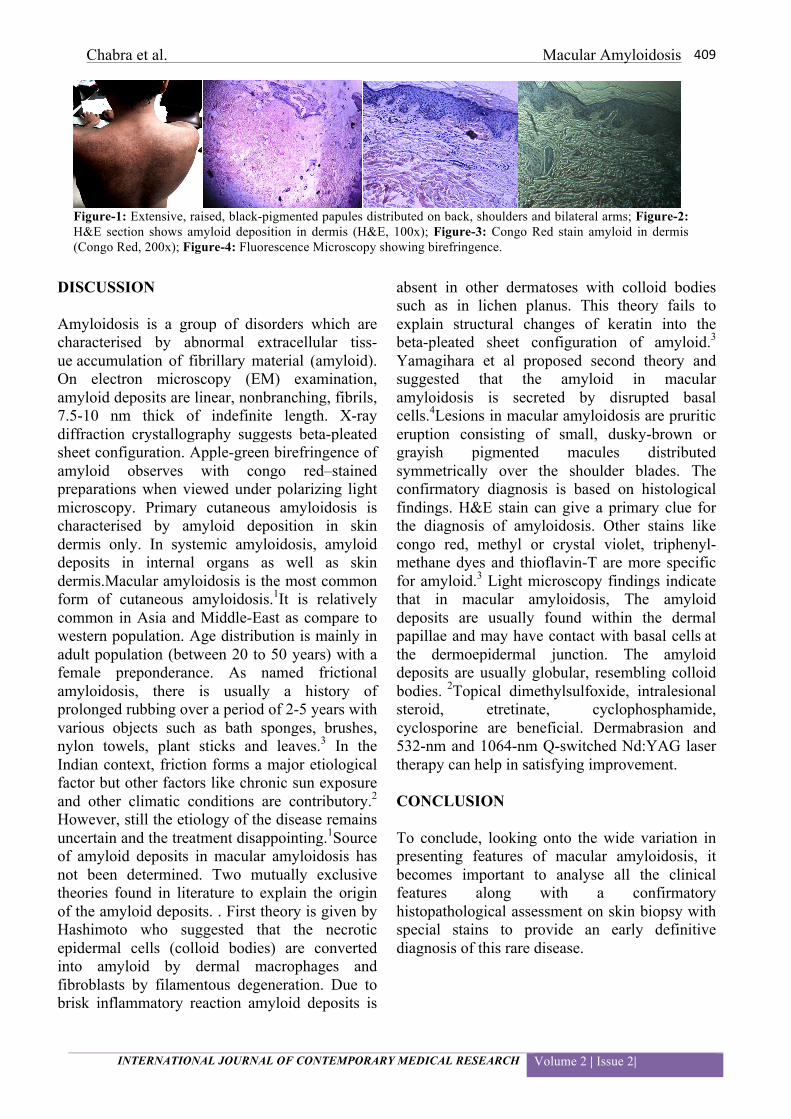
CASE REPORT A 32 year old man presented with generalised hyperpigmentation for 2 years. The patient gave history of photosensitivity, itching and seasonal variation. Dermatological examination revealed an extensive, raised, black-pigmented papules varying in size from 0.2- 0.4 cms distributed on back, shoulders and bilateral arms. The lesions had a characteristic rippled appearance. The lesions were intensely pruritic which on scratching leave hyperpigmented scar mark. There was no past history of infection at the site, any atopic disease, drug intake, chronic disease or any relevant family history. Diagnostic work-up revealed normal routine biochemical findings. A skin biopsy measuring 0.5x0.4x0.2cm was performed and sent to our histopathology department for hematoxylin-eosin and special stains. Microscopic examination of H&E stained section showed deposition of eosinophilc hyaline material in papillary dermis. Presence of amyloid was confirmed by congo red staining which gave the characteristic green birefringence in the papillary dermis. There were some melanophages and scant lymphocytes in the dermis.
CASE REPORT
Macular Amyloidosis: A Rare Case Report Sonia Chhabra1, Pansi Gupta2, Mansi Agarwal2, Padam Parmar2, Ashish Sharma2, Rajeev Sen3
1Professor, 2Resident, 3Senior Professor And Head Of Department Department Of Pathology, Pt.B.D.S.Post Grauate Institute Of Medical Sciences Rohtak , Haryana, India. Corresponding author: Dr. Pansi Gupta, Room No. 37, PG block, Girls Hostel, PGIMS, Rohtak, Haryana
Chabra et al. Macular Amyloidosis
INTERNATIONAL JOURNAL OF CONTEMPORARY MEDICAL RESEARCH Volume 2 | Issue 2|
409
DISCUSSION Amyloidosis is a group of disorders which are characterised by abnormal extracellular tiss- ue accumulation of fibrillary material (amyloid). On electron microscopy (EM) examination, amyloid deposits are linear, nonbranching, fibrils, 7.5-10 nm thick of indefinite length. X-ray diffraction crystallography suggests beta-pleated sheet configuration. Apple-green birefringence of amyloid observes with congo red–stained preparations when viewed under polarizing light microscopy. Primary cutaneous amyloidosis is characterised by amyloid deposition in skin dermis only. In systemic amyloidosis, amyloid deposits in internal organs as well as skin dermis.Macular amyloidosis is the most common form of cutaneous amyloidosis.1It is relatively common in Asia and Middle-East as compare to western population. Age distribution is mainly in adult population (between 20 to 50 years) with a female preponderance. As named frictional amyloidosis, there is usually a history of prolonged rubbing over a period of 2-5 years with various objects such as bath sponges, brushes, nylon towels, plant sticks and leaves.3 In the Indian context, friction forms a major etiological factor but other factors like chronic sun exposure and other climatic conditions are contributory.2
However, still the etiology of the disease remains uncertain and the treatment disappointing.1Source of amyloid deposits in macular amyloidosis has not been determined. Two mutually exclusive theories found in literature to explain the origin of the amyloid deposits. . First theory is given by Hashimoto who suggested that the necrotic epidermal cells (colloid bodies) are converted into amyloid by dermal macrophages and fibroblasts by filamentous degeneration. Due to brisk inflammatory reaction amyloid deposits is
absent in other dermatoses with colloid bodies such as in lichen planus. This theory fails to explain structural changes of keratin into the beta-pleated sheet configuration of amyloid.3
Yamagihara et al proposed second theory and suggested that the amyloid in macular amyloidosis is secreted by disrupted basal cells.4Lesions in macular amyloidosis are pruritic eruption consisting of small, dusky-brown or grayish pigmented macules distributed symmetrically over the shoulder blades. The confirmatory diagnosis is based on histological findings. H&E stain can give a primary clue for the diagnosis of amyloidosis. Other stains like congo red, methyl or crystal violet, triphenyl-methane dyes and thioflavin-T are more specific for amyloid.3 Light microscopy findings indicate that in macular amyloidosis, The amyloid deposits are usually found within the dermal papillae and may have contact with basal cells at the dermoepidermal junction. The amyloid deposits are usually globular, resembling colloid bodies. 2Topical dimethylsulfoxide, intralesional steroid, etretinate, cyclophosphamide, cyclosporine are beneficial. Dermabrasion and 532-nm and 1064-nm Q-switched Nd:YAG laser therapy can help in satisfying improvement. CONCLUSION To conclude, looking onto the wide variation in presenting features of macular amyloidosis, it becomes important to analyse all the clinical features along with a confirmatory histopathological assessment on skin biopsy with special stains to provide an early definitive diagnosis of this rare disease.
Figure-1: Extensive, raised, black-pigmented papules distributed on back, shoulders and bilateral arms; Figure-2: H&E section shows amyloid deposition in dermis (H&E, 100x); Figure-3: Congo Red stain amyloid in dermis (Congo Red, 200x); Figure-4: Fluorescence Microscopy showing birefringence.

Chabra et al. Macular Amyloidosis
INTERNATIONAL JOURNAL OF CONTEMPORARY MEDICAL RESEARCH Volume 2 | Issue 2|
410
REFERENCES
1. Krishna A, Nath B, Dhir G, Kumari R, Budhiraja V and Singh K. Study on epidemiology of cutaneous amyloidosis in northern India and effectiveness of dimethylsulphoxide in cutaneous amyloidosis. Indian Dermatol Online J 2012;3:182-6.
2. Razvi F, Kumar A. Clinical and histopathological study of primary cutaneous macular amyloidosis. J Med Allied Sci 2013;3:22-5.
3. Hashimoto K, Kobayashi H. Histogenesis of amyloid in the skin. Am J Dermatopathol. Summer 1980;2:165-71.
4. Yamagihara M, Kitajima Y, Yaoita H. Ultrastructural observation of the relationship between amyloid filaments and half desmosomes in macular amyloidosis. J Cutan Pathol. 1980;7:213.
5. Bhat Y, Manzoor S, Wani R. Macular amyloidosis in kashmiri females. J Basic Clin Sci 2012;1:43-6.