case report cytogenetic diagnosis of roberts sc phocomelia
TRANSCRIPT

CASE REPORT
Cytogenetic diagnosis of Roberts SC phocomelia
syndrome: First report from Kashmir
Tahir M. Malla, Arshad A. Pandith, Fayaz A. Dar, Mahrukh H. Zargar *
Advanced Centre for Human Genetics, Sher-i-Kashmir Institute of Medical Sciences, Srinagar 190011, India
Received 5 June 2015; accepted 18 June 2015
Available online 14 July 2015
KEYWORDS
Cytogenetics;
Roberts syndrome;
Premature centromeric
separation;
Premature chromatid
separation
Abstract There are several syndromes in which specific mitotic chromosomal abnormalities can be
seen, like premature centromere separation, premature (sister) chromatid separation, and somatic
aneuploidies. Identifications of such specific cytogenetic findings can be the key factor that
leads towards the diagnosis of syndromes like Roberts SC phocomelia. The case presented here
as Roberts SC phocomelia syndrome was identified as a child with multiple congenital anomalies
and dysmorphic features. Conventional cytogenetic analysis of the case revealed premature sister
chromatid separation. The premature centromeric separation was also confirmed by C banding
analysis of the child. It is the first and the only case of Roberts SC phocomelia diagnosed from this
part of the world. The present case report emphasizes the importance of conventional cytogenetics
in the diagnosis of such syndromes.� 2015 The Authors. Production and hosting by Elsevier B.V. on behalf of Ain Shams University. This is an
open access article under the CC BY-NC-ND license (http://creativecommons.org/licenses/by-nc-nd/4.0/).
1. Introduction
Roberts SC phocomelia syndrome is an autosomal recessivedevelopmental disorder characterized by pre and postnatal
growth retardation, microcephaly, craniofacial anomalies,mental retardation and tetraphocomelia in varying degreesof severity. Robert’s syndrome was initially reported byJohn Roberts in a male child bearing cleft lip and tetrapho-
comelia [1]. Later, in four individuals from two families ofEuropean descent, Herrmann et al. reported similar, butmilder malformations which were referred to as SC pho-
comelia [2]. These two syndromes had varying phenotypic
expression and were later concluded as the same entity becauseof resemblance of thalidomide embryopathy with Robert’ssyndrome and were therefore termed as Roberts SC pho-comelia syndrome [3]. The gene responsible for the syndrome
called ESCO2 gene is located at 8p21.1 and was discovered byHugo and Vega in 1995 [4]. This syndrome is rare withapproximately 100 cases described in the literature [5]. Ours
is the first report on Robert’s syndrome from Kashmir,North India.
1.1. Clinical description
The proband, a two year old baby girl, is a product of non-consanguineous marriage, second in birth order and born tothe parents of same geographical area of Kashmir valley.
The patient had characteristic dysmorphic facies with defective
* Corresponding author. Tel.: +91 01942 401013x2477.
E-mail address: [email protected] (M.H. Zargar).
Peer review under responsibility of Ain Shams University.
The Egyptian Journal of Medical Human Genetics (2016) 17, 137–140
HO ST E D BYAin Shams University
The Egyptian Journal of Medical Human Genetics
www.ejmhg.eg.netwww.sciencedirect.com
http://dx.doi.org/10.1016/j.ejmhg.2015.06.0061110-8630 � 2015 The Authors. Production and hosting by Elsevier B.V. on behalf of Ain Shams University.This is an open access article under the CC BY-NC-ND license (http://creativecommons.org/licenses/by-nc-nd/4.0/).
development of all four extremities that was the main con-stituent of malformation complex. The craniofacial abnormal-ities include small low set ears, prominent frontal bones,
prominent eyes, shallow orbits, hypertelorism, bilateral cleftlip and palate, micrognathia and short neck. She had severefixed flexion deformities of all limbs. The limbs were short with
hands and feet located closed to the body. The thumbs wereabsent bilaterally, with oligodactyly of upper and lowerextremities and flexion deformity at knee joints. There was
no visceromegaly and no cardio-vascular defects. The genitalswere normal.
2. Methodology
2.1. G banding analysis
For G banding analysis 72 h peripheral blood lymphocyte cul-tures were set using RPMI 1640 medium (Sigma, St Louis,USA) supplemented with phytohemagglutinin (Himedia
Labs, India) and fetal bovine serum (Himedia Labs,Mumbai, India). The cells were arrested in metaphase by col-cemid treatment 2 h prior to the hypotonic shock with
0.57 M KCl solution. The cells were finally fixed with prechilled Carnoy’s fixative and slides were prepared the nextday by air drop method [6]. GTG banding of one day old slides
was carried out and the slides were stained using 4% Giemsastain [7]. Thirty well spread metaphases were selected for theanalysis.
2.2. C banding analysis
For C banding analysis three day old slides were dipped for15 min in 0.2 N HCl and incubated in a saturated solution of
barium hydroxide at 60 �C for 8–10 min. The slides were rinsed
in distilled water for 30 s and excess barium hydroxide wasremoved by a rapid dip in 0.1 N HCl. The slides were reincu-bated in a coplin jar containing 2XSSC for one hour in a 60 �Cwater bath. The slides were briefly rinsed in distilled water andstained in 5% Giemsa stain [8].
3. Results
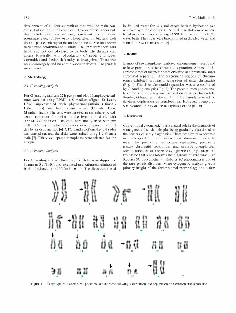
In most of the metaphases analyzed, chromosomes were foundto have premature sister chromatid separation. Almost all the
chromosomes of the metaphases observed had premature sisterchromatid separation. The centromeric regions of chromo-somes exhibited prominent separation of sister chromatids
(Fig. 1). The sister chromatid separation was also confirmedby C-banding analysis (Fig. 2). The parental metaphases ana-lyzed did not show any such separation of sister chromatids.
Besides, G-banding of the child and his parents revealed nodeletion, duplication or translocation. However, aneuploidywas recorded in 5% of the metaphases of the patient.
4. Discussion
Conventional cytogenetics has a crucial role in the diagnosis ofsome genetic disorders despite being gradually abandoned in
the new era of array diagnostics. There are several syndromesin which specific mitotic chromosomal abnormalities can beseen, like premature centromere separation, premature
(sister) chromatid separation, and somatic aneuploidies.Identifications of such specific cytogenetic findings can be thekey factor that leads towards the diagnosis of syndromes like
Roberts SC phocomelia [9]. Roberts SC phocomelia is one ofthe rare genetic disorders where cytogenetic analysis gives aprimary insight of the chromosomal morphology and a firm
Figure 1 Karyotype of Robert’s SC phocomelia syndrome showing sister chromatid separation and centromeric separation.
138 T.M. Malla et al.

diagnosis of the syndrome is made based on the presence ofearly centromeric separation of the sister chromatids.
At the cytogenetic level, chromosomes of Robert’s syn-drome present with a rod-like morphology resulting in a
‘railroad-track’ appearance due to the absence of the primaryconstriction at the centromeric regions [10–13]. This phe-nomenon known as premature centromere separation (PCS)
or heterochromatin repulsion (HR) constitutes the major diag-nostic marker for RS. In this case, the centromeric regions ofchromosomes exhibited prominent separation indicating the
absence of establishment of cohesion between their sister chro-matids. No direct diagnosis was made and chromosomal anal-ysis was performed on the peripheral blood lymphocytes of thepatient for the confirmatory diagnosis.
Aneuploidy has been found associated with PCS/HR mostlikely due to outlying, lagging or prematurely advancing chro-mosomes during mitosis [12]. The case in discussion not only
showed PCS/HR, but also somatic aneuploidies in 6 out of30 metaphases. Such somatic variegated aneuploidy has beenreported before in several patients with Roberts syndrome
[14,15] and is probably the direct consequence of the prema-ture separation resulting in mal-segregation of both chro-matids over the two daughter cells.
5. Conclusion
Recent advances in genetic diagnostics have enormous advan-
tages over traditional karyotyping and have increased the yieldof diagnoses in dysmorphology by approximately 15%.However, metaphase anomalies like PCS/HR and PSCS can-not be detected by techniques like array studies and therefore
milder forms of syndromes like those of Roberts SC pho-comelia may remain undetected. We therefore conclude fromthe present case study that routine cytogenetic analysis leads
to the accurate diagnosis of Robert’s phocomelia syndrome.
Therefore, it is recommended in children with pre-and postna-tal dysmorphic features and growth retardation.
Acknowledgment
The authors acknowledge the parents of the child for theircooperation.
References
[1] Herrmann J, Feingold M, Tuffli GA, Opitz JM. A familial
dysmorphogenetic syndrome of limb deformities, characteristic
facial appearance and associated anomalies: the ‘pseudothalido-
mide’ or ‘SC-syndrome’. Birth Defects Orig Art Ser 1969;V:81–9.
[2] Temtamy SA, Ismail S, Helmy NA. Roberts syndrome: study of
four new Rgyptian cases with comparison of clinical and
cytogenetic findings. Genet Couns 2006;17:1–13.
[3] Vega H, Waisfisz Q, Gordillo M, Sakai N, Yanagihara I, Yamada
M, et al. Roberts syndrome is caused by mutations in ESCO2, a
human homolog of yeast ECO1 that is essential for the establish-
ment of sister chromatid cohesion. Nat Genet 2005;37:468–70.
[4] Gordillo M, Vega H, Trainer AH, Hou F, Sakai N, Luque R,
et al. The molecular mechanism underlying Roberts syndrome
involves loss of ESCO2 acetyltransferase activity. Hum Mol
Genet 2008;17:2172–80.
[5] Judge C. A sibship with the pseudothalidomide syndrome and an
association with Rh incompatibility. Med J Aust 1973;2:280–1.
[6] Moorhead PS, Nowell PC, Mellman WJ, Battips DM,
Hungerford DA. Chromosome preparation of leukocytes cultured
from human peripheral blood. Exp Cell Res 1960;20:613–6.
[7] Seabright M. A rapid banding technique for human chromo-
somes. Lancet 1971;II:971–2.
[8] Schere JMJC. Production of C and T bands in human chromo-
somes after heat treatment at high pH and staining with ‘‘Stains-
All’’. Human Genet 1974;23:311–4.
[9] Gerkes Erica H, Anne-Marie F, van der Kevie-Kersemaekers,
Yakin M, Dominique FCM, et al. The importance of
Figure 2 C-banded metaphase of Robert’s SC phocomelia syndrome showing centromeric separation as indicated by arrows.
Cytogenetic diagnosis of Roberts SC phocomelia syndrome 139

chromosome studies in Roberts syndrome/SC phocomelia and
other cohesinopathies. Eur J Med Genet 2010;53:40–4.
[10] German J. Roberts’ syndrome I. Cytological evidence for a
disturbance in chromatid pairing. Clin Genet 1979;16:441–7.
[11] Tomkins D, Hunter A, Roberts M. Cytogenetic findings in
Roberts-SC phocomelia syndrome(s). Am J Med Genet
1979;4:17–26.
[12] Louie E, German J. Roberts’s syndrome. II. Aberrant Y-
chromosome behavior. Clin Genet 1981;19:71–4.
[13] Jabs EW, Tuck-Muller CM, Cusano R, Rattner JB. Studies of
mitotic and centromeric abnormalities in Roberts syndrome:
implications for a defect in the mitotic mechanism. Chromosoma
1991;100:251–61.
[14] Schulz S, Gerloff C, Ledig S, Langer D, Volleth M, Shirneshan K,
et al. Prenatal diagnosis of Roberts syndrome and detection of an
ESCO2 frameshift mutation in a Pakistani family. Prenat Diagn
2008;28:42–5.
[15] Van Den Berg DJ, Francke U. Roberts syndrome: a review of 100
cases and a new rating system for severity. Am J Med Genet
1993;47:1104–23.
140 T.M. Malla et al.