cytogenetic techniques in diagnosing genetic...
TRANSCRIPT
3
Cytogenetic Techniques in Diagnosing Genetic Disorders
Kannan Thirumulu Ponnuraj Universiti Sains Malaysia
Malaysia
1. Introduction
When the discovery of giant banded, salivary chromosomes in Drosophila was made by Painter in 1934, it gave a tremendous impact to the cytological work carried out in Drosophila. This made it possible to identify the chromosomes individually and also to discern the specific segments of the chromosome. Followed by this, cytogenetics bloomed with the establishment of chromosome number in man as 46 in the year 1956. Since then, lot of advancements and improvements have taken place over the years and combination of techniques have made cytogenetics as an undisputable source in diagnosing the various genetic disorders and now, human cytogenetics has completed its glorious 50 years after the discovery of chromosome number in normal human cells. This chapter provides an insight into the fundamentals of cytogenetics and its importance in the diagnosis of commonly occurring syndromes and disorders.
2. History of cytogenetics
When the genetic importance of polytene chromosomes of Diptera was rediscovered in the early thirties, almost every Drosophila geneticist started studying the salivary glands. Nageli, the Swiss botanist first described thread like structures in the nuclei of plant cells in the 1840s and called them “transitory cytoblasts”, which represented what now are called chromosomes. Later, the term “chromosome” was coined by Waldeyer in 1888 after staining techniques had been developed which made them better discernible (chromos = Greek for colour; soma = Greek for body). In 1909, Johannsen coined the term ‘gene’. This triggered the beginning of modern cytogenetics, but yet, the progress was moving at a snail’s pace. Still, attempts were going on to find the number of chromosomes, which became a serious issue and a matter of great concern among the various researchers. The quality of chromosomes were poor and the numbers varied each and every time. Even determining the diploid number of a mammalian species was considered a difficult accomplishment. The chromosomes were crowded in metaphase and considerations of biological function of the chromosomes and in particular, of modern genetics were beyond the scope of cytological research in the 19th century. It was quite cumbersome to obtain nice slides with good metaphase spreads for easy counting. However, in 1950s, there were advent of new techniques for chromosome preparations, like addition of colcemid and hypotonic treatment, led to the establishment of the diploid number of chromosomes in man as 46 (Tjio
www.intechopen.com
Advances in the Study of Genetic Disorders 46
& Levan, 1956) and the peripheral leucocyte culture method of Moorehead et al. (1960) was adopted by many cytogeneticists. Once the correct description of the normal human chromosome number was established, chromosome abnormalities were recognized to be clearly associated with specific congenital defects. It was possible to arrange the chromosomes in different groups based on their size and location of the centromere which enabled easy counting as well as detection of numerical chromosome aberrations like trisomy 21 in Down syndrome (Lejeune et al. 1959), 45X in Turner syndrome (Ford et al. 1959), 47XXY in Klinefelter syndrome (Jacobs & Strong, 1959), trisomy 13 (Patau et al. 1960) and trisomy 18 (Edwards et al. 1960), Philadelphia chromosome, a structural aberration involving chromosomes 9 and 22, was recognized in a patient with chronic myeloid leukemia (Nowell & Hungerford, 1960). The metaphase chromosomes were classified into 7 groups based on the Denver classification (1960), with revisions at the London Conference (Hamerton et al. 1963) and the Chicago Conference (1966). Karyotype is the normal nomenclature where the chromosomes are arranged in homologous pairs in a systematic manner to describe the normal or abnormal chromosomal complement of an individual, tissue or cell line (ISCN, 2005). Jau-hong Kao et al. (2008) described chromosome classification based on the band profile similarity along approximate medial axis. This was soon followed by amniocentesis to determine the chromosomal abnormalities in fetal cells in the amniotic fluid, which formed the core of prenatal genetic diagnosis (Steele & Breg, 1966). After the advent of these protocols and discoveries, the heyday of cytogenetics research appeared to be over (Hans Zellweger and Jane Simpson, 1977), the power of cytogenetics analysis improved with the development of staining protocols by Caspersson et al. (1968), that made chromosomes of the same group, which previously could not be distinguished from each other, discernible. This banding pattern was based on a fluorescent staining technique and the fluorescence intensity quickly quenched which made the technique less optimal for routine studies of patients. Hence, several other banding techniques were developed like G-, R-, C- and NOR banding each having their own specific properties and applications (Rooney, 2001). These banding patterns became the barcodes with which cytogeneticists could easily identify chromosomes, detect subtle deletions, inversions, insertions, translocations, fragile sites and other more complex rearrangements and refine breakpoints (Caspersson et al. 1970).
3. Cytogenetics
Cytogenetics is the study of the structure and properties of chromosomes, chromosomal behaviour during somatic cell division in growth and development (mitosis) and germ cell division in reproduction (meiosis), chromosomal influence on the phenotype and the factors that cause chromosomal changes (Hare & Singh, 1979). Discovery of new techniques, improvements of existing techniques or new combinations of well established techniques are often followed by progress in the biosciences. This is strikingly exemplified by the development of cytogenetics in the last 100 years.
3.1 Chromosomes and normal chromosome complement
Chromatins are dark staining materials present in the nucleus of a cell and in interphase, these chromatin materials are organised into a number of long, loosely coiled, irregular strands or threads called the chromatin reticulum. At the time of cell division, these chromatin bodies condense into shorter and thicker threads called chromosomes, that carry
www.intechopen.com
Cytogenetic Techniques in Diagnosing Genetic Disorders 47
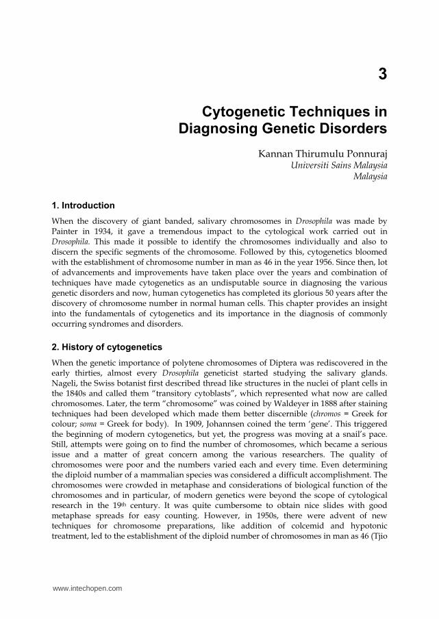
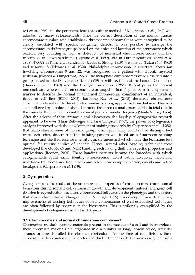
the genes and functions in the transmission of hereditary information. In a normal diploid cell, there are 46 chromosomes (23 chromosome pairs), where one of each pair is derived from the father and the other from the mother of the individual. The first 22 pairs are called the autosomes (non-sex chromosomes) and the 23rd pair is called the sex-chromosomes. In males, the 23rd pair is XY and in females, it is XX. The X-chromosome is maternally derived and the Y-chromosome is paternally derived. The karyotypes of a normal male (Figure 1) and female (Figure 2) are presented. Except in the case of mosaic individuals (where they have two or more populations of cells which differ in chromosome number), all the cells of an individual have the same chromosome complement in their diploid cells. In the case of gametic cells (sperm and ovum), or otherwise called haploid cells, they have only single chromosome from each pair.
Fig. 1. A karyotype of a normal male (46,XY) (Reproduced courtesy of Human Genome Centre, Universiti Sains Malaysia, Malaysia)
3.2 Cytogenetic analysis of chromosomes 3.2.1 Whole blood culture
The main advantage of whole blood culture is that blood is one of the most and easily accessible human tissues. Also, it has a very good growth potential after mitogenic stimulation. They have a cell cycle which is well characterized and the results can be obtained after a culture duration of 3 days.
3.2.1.1 Short term culture
The most commonly used technique for preparation of chromosomes is peripheral blood culture. The materials and reagents needed for culture are as below. 1. Sodium heparin – which is used as an anti-coagulant 2. Culture medium (E.g. RPMI 1640, TC 199 etc.) – provides nutrients and amino acids
needed for the growth of the cells
www.intechopen.com
Advances in the Study of Genetic Disorders 48
3. Fetal bovine serum – contains a rich variety of proteins that enhances the growth of cells 4. Antibiotics – suppreses the growth of contaminants 5. Mitogen – (E.g. Phytohaemagglutinin) – induces the cells to undergo mitosis 6. Colchicine or its synthetic derivative, Colcemid – arrests cell division 7. Potassium chloride solution (hypotonic treatment) – induces swelling of cells through
osmosis 8. Methanol: Acetic acid – for fixation of cells
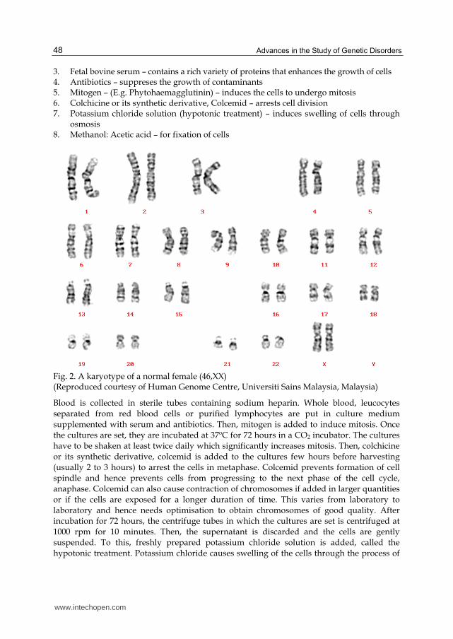
Fig. 2. A karyotype of a normal female (46,XX) (Reproduced courtesy of Human Genome Centre, Universiti Sains Malaysia, Malaysia)
Blood is collected in sterile tubes containing sodium heparin. Whole blood, leucocytes separated from red blood cells or purified lymphocytes are put in culture medium supplemented with serum and antibiotics. Then, mitogen is added to induce mitosis. Once the cultures are set, they are incubated at 37ºC for 72 hours in a CO2 incubator. The cultures have to be shaken at least twice daily which significantly increases mitosis. Then, colchicine or its synthetic derivative, colcemid is added to the cultures few hours before harvesting (usually 2 to 3 hours) to arrest the cells in metaphase. Colcemid prevents formation of cell spindle and hence prevents cells from progressing to the next phase of the cell cycle, anaphase. Colcemid can also cause contraction of chromosomes if added in larger quantities or if the cells are exposed for a longer duration of time. This varies from laboratory to laboratory and hence needs optimisation to obtain chromosomes of good quality. After incubation for 72 hours, the centrifuge tubes in which the cultures are set is centrifuged at 1000 rpm for 10 minutes. Then, the supernatant is discarded and the cells are gently suspended. To this, freshly prepared potassium chloride solution is added, called the hypotonic treatment. Potassium chloride causes swelling of the cells through the process of
www.intechopen.com
Cytogenetic Techniques in Diagnosing Genetic Disorders 49
osmosis and hence proper dispersion of chromosomes. The hypotonic treatment is achieved by incubating the centrifuge tubes in a CO2 incubator at 37ºC for about 30 minutes. Then, the process of centrifugation is repeated followed by addition of 3:1 methanol:acetic acid, which acts as a fixative. Methanol in the fixative denatures and precipitates the proteins by dehydration under acid conditions and acetic acid coagulates the nucleoproteins and casues swelling of cells thus counteracting the shrinking caused by methanol. The fixative penetrates the cells rapidly, preserves the chromosome structure and to a large extent, strips cytoplasmic proteins from cells. The fixative washes are repeated as many times as necessary until a clear cell button is obtained at the bottom of the tube. Then, the chromosomes are prepared by dropping the cell suspension on a clean, grease free slide, where, the drop spreads out and the chromosomes get fixed on the slides. Once the slides are prepared, suitable staining techniques are carried out as needed for the diagnosis of chromosomal disorders.
3.2.1.2 Bone marrow culture
The bone marrow culture is used to identify chromosome anomalies in hematopoietic cells, especially for hematological disorders like pre-leukemia and leukemia. Bone marrow aspirate of about 0.5 to 2.0 ml is collected in a heparinized syringe. Strict aseptic techniques are a must right from the beginning of collection until the final process is completed. Bone marrow is collected in transport media and mixed thoroughly. Then, the samples are spun at around 900 rpm for 10 minutes followed by pipetting off the supernatant. This is then followed by addition of about 1ml of sample to complete culture media (medium + Fetal bovine serum + L-glutamine + Antibiotics). After about 45 minutes, colcemid is added to this and mixed thoroughly. Then, the cultures are incubated at 37ºC in a CO2 incubator for 24 hours. This is followed by the routine hypotonic and fixative treatments as for the whole blood culture. The chromosomes are prepared on clean grease free slides, stained and examined under microscope for analysis.
3.2.2 Banding techniques
The different banding techniques allow precise identification of each chromosome as well as to detect structural chromosomal rearrangements. A combination of several banding techniques also help in obtaining the information necessary for chromosomal analysis.
3.2.2.1 Q-banding
This banding technique does not require any prior treatment of the chromosomes but requires a fluorescent microscope for analysis. Caspersson et al. (1970) discovered one of the first chromosome banding techniques, which involves staining chromosomes with a fluorochrome, such as quinacrine mustard or quinacrine dihydrochloride, and examining them with fluorescence microscopy. The Q-bands appear along each chromosome in alternating bright and dull bands with varying intensity. However, Q-banding does not permit permanent preparations. Certain antibiotics like anthracyclines produce fluorescent bands similar to Q-bands and are more stable than those produced by quinacrine.
3.2.2.2 G-banding
G-bands are produced by staining the chromosomes with a stain, Giemsa. This is done by treating the chromosomes with substances (usually trypsin), that alters the structure of
www.intechopen.com
Advances in the Study of Genetic Disorders 50
proteins followed by staining with a Giemsa solution (Rowley, 1973). It is the most common method of banding, as it produces the same banding pattern as quinacrine with even greater resolution; it allows permanent preparations and does not necessitate the use of fluorescence microscopy. Thus, G-band patterns can be used to pair and identify each of the human chromosomes accurately.
3.2.2.3 R-banding
R-bands are just the reverse of G-bands, which can be produced by a variety of methods. A modification of method of Dutrillaux and Lejeune (1971) involves thermic denaturation in Earle’s balanced salt solution (at 87ºC), which is the most common method. Since the staining ability of the chromosomes is somewhat lost due to heating, the use of phase contrast objectives gives a better contrast of the chromosomes for analysis.
3.2.2.4 C-banding
C-bands localize the heterochromatic regions of chromosomes. Pardue & Gall (1970) first reported C-bands in 1970 when they discovered that the centromeric region of mouse chromosomes is rich in repetitive DNA sequences and stains dark with Giemsa. The original method of Arrighi and Hsu (1971) involves treating the slides with 0.2 N hydrochloric acid followed by treatment with RNAse and sodium hydroxide. Many chromosomes have regions that differ among individuals but have no pathological importance. These polymorphic regions can be visualized optimally with C-band methods and are most often seen on acrocentric chromosomes, the centromeric region of chromosomes 1, 9, and 16, and the distal portion of the Y chromosome. C-banding is also useful to show chromosomes with multiple centromeres, to study the origin of diploid molar pregnancies and true hermaphroditism and to distinguish between donor and recipient cells in bone marrow transplantation.
3.2.2.5 T-banding
This method involves staining the telomeric (end) regions of the chromosomes. Dutrillaux (1973) treated the slides with either phosphate buffer or Earle’s balanced salt solution and then stained using mixed Giemsa solution to produce the T-bands.
3.2.2.6 CT-banding
Scheres, (1974) developed a method to stain both the centromeric heterochromatin as well as the telomere of chromosomes. He treated the slides with barium hydroxide to produce the CT-bands. Chamla & Ruffie (1976) obtained complete C- and T-bands by incubating the slides in Hank’s balanced salt solution.
3.2.2.7 Nucleolar Organizing Region-banding
Nucleolar organizing region (NOR)–banding is a technique that stains NORs of chromosomes (Matsui & Sasaki, 1973). These regions are located in the satellite stalks of acrocentric chromosomes and house genes for ribosomal RNA. NOR-bands may represent structural non-histone proteins that are specifically linked to NOR and bind to ammoniacal silver. Goodpasture et al. (1976) developed a simple silver nitrate staining technique that is now used widely. NOR-banding is useful in clinical practice to study certain chromosome polymorphisms, such as double satellites. This method is also helpful to identify satellite stalks that are occasionally seen on non-acrocentric chromosomes.
www.intechopen.com

Cytogenetic Techniques in Diagnosing Genetic Disorders 51
3.2.2.8 The choice of banding technique
For routine analysis, the banding technique using trypsin and Giemsa became the most accepted worldwide (Seabright, 1971). Since the banding pattern enabled the detection of various structural aberrations like translocations, inversions, deletions, and duplications next to the already well-known numerical aberrations, not only potentially unbalanced cases (patients) could be studied but also healthy individuals as possible carriers of a balanced aberration. For instance, healthy family members of already known carriers and couples suffering from repetitive spontaneous abortions were cytogenetically investigated (Dominique FCM Smeets, 2004).
3.2.2.9 High resolution banding
Despite the above banding patterns, resolution of chromosome studies remained relatively limited with an approximate count of 500 bands per haploid genome (resolution ≈ 6 million base pairs ≈ 50 genes per band) because the total number of bands produced on metaphase chromosomes are less and it is difficult to detect rearrangements involving small portions of chromosomes due to excessive condensation. This was improved by the development of so-called high-resolution banding by Yunis (1976) which was achieved by synchronizing the lymphocyte cultures and obtaining more number of cells in pro-metaphase or even prophase (increasing resolution from 500 to over 1000 bands in a haploid genome). High resolution cytogenetics provides precision in the delineation of chromosomal breakpoints and assignment of gene loci, greater than with earlier techniques, since analysis of late prophase sub-banding reveals more than twice the number of bands seen at metaphase (Sawyer & Hozier, 1986). By applying this technique, several already well-known clinical syndromes like Prader Willi and Angelman syndrome with a deletion at the proximal long arm of chromosome 15, Smith-Magenis and Miller-Dieker syndrome with (different) deletions in the short arm of chromosome 17 and DiGeorge/Velo Cardio Facial (VCF) syndrome with deletions in the long arm of chromosome 22 could be linked to small chromosome aberrations and the concept of the micro-deletion or contiguous gene syndrome was born (Schmickel, 1986).
3.2.2.10 Sex chromatin analysis
The number of sex chromatin bodies is one less than the number of X chromosomes in the chromosome complement. This is obtained by taking buccal smears on a clean slide followed by fixing them in ethanol, air drying, hydrolysing in hydrochloric acid, washing in distilled water to remove the acid and then finally staining using cyrstal violet. The presence of a chromatin mass, called the “Barr body” indicates a chromatin positive cell.
3.3 Specialized techniques to visualize chromosomes 3.3.1 Sister Chromatid Exchange (SCE)
SCE staining is accomplished in cell cultures by incorporating BrdU (bromodeoxyuridine) (in place of thymidine) into replicating cells for 2 cell cycles. As a result of semi-conservative DNA replication, chromosomes have one chromatid with BrdU in one strand of DNA and the other chromatid has BrdU in both strands of DNA. This produces an acridine fluorescence pattern in which one chromatid fluoresces more brightly than the other chromatid. Sister chromatid exchanges appear as an interchange between sister chromatids
www.intechopen.com
Advances in the Study of Genetic Disorders 52
of brightly and dully fluorescent segments. The biologic importance of SCEs is uncertain, but some mutagens and carcinogens increase their frequency (Perry & Evans, 1975).
3.3.2 Fragile sites and chromosome breakage
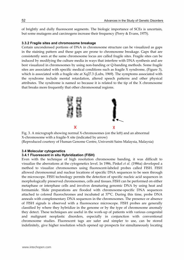
Certain uncondensed portions of DNA in chromosome structure can be visualized as gaps in the staining pattern and these gaps are prone to chromosome breakage. Gaps that are consistently seen at the same chromosome locus are called fragile sites. Fragile sites can be induced by modifying the culture media in ways that interfere with DNA synthesis and are best visualized in chromosomes by using non-banding or Q-banding methods. Some fragile sites are associated with specific medical conditions such as fragile X syndrome, (Figure 3), which is associated with a fragile site at Xq27.3 (Lubs, 1969). The symptoms associated with the syndrome include mental retardation, altered speech patterns and other physical attributes. The syndrome is named so because it is related to the tip of the X chromosome that breaks more frequently that other chromosomal regions.
Fig. 3. A micrograph showing normal X-chromosomes (on the left) and an abnormal X-chromosome with a fragile-X site (indicated by arrow) (Reproduced courtesy of Human Genome Centre, Universiti Sains Malaysia, Malaysia)
3.4 Molecular cytogenetics 3.4.1 Fluorescent in situ Hybridization (FISH)
Even with the technique of high resolution chromosome banding, it was difficult to visualize the aberrations at the cytogenetics level. In 1986, Pinkel et al. (1986a) developed a method to visualize chromosomes using fluorescent-labeled probes called FISH. FISH allowed chromosomal and nuclear locations of specific DNA sequences to be seen through the microscope. FISH technology permits the detection of specific nucleic acid sequences in morphologically preserved chromosomes, cells and tissues. FISH can be performed on either metaphase or interphase cells and involves denaturing genomic DNA by using heat and formamide. Slide preparations are flooded with chromosome-specific DNA sequences attached to colored fluorochromes and incubated at 37°C. During this time, probe DNA anneals with complementary DNA sequences in the chromosomes. The presence or absence of FISH signals is observed with a fluorescence microscope. FISH probes are generally classified by where they hybridize in the genome or by the type of chromosome anomaly they detect. These techniques are useful in the work-up of patients with various congenital and malignant neoplastic disorders, especially in conjunction with conventional chromosome studies. Fluorescent tags are safer and simpler to use, can be stored indefinitely, give higher resolution which opened up prospects for simultaneously locating
www.intechopen.com
Cytogenetic Techniques in Diagnosing Genetic Disorders 53
several DNA sequences in the same cell by labelling them with different fluorochromes (Barbara J Trask, 2002). Using FISH, cytogeneticists could detect chromosomal abnormalities that involved small segments of DNA. Even more importantly, FISH opened up the nuclei of non-dividing cells to karyotype analysis. Using FISH and chromosome-specific probes, cytogeneticists could enumerate chromosomes, simply by counting spots in each nucleus (Pinkel et al. 1986b).
3.4.2 Spectral Karyotyping (SKY) and Multicolour FISH (M-FISH)
After the advent of FISH, where a single copy gene could fluoresce, a more powerful technology called SKY or M-FISH was developed. M-FISH allows all the 24 human chromosomes to be painted in different colours. By making use of various combinations and concentrations of fluorescent dyes, it is even possible to give every single chromosome a different color (SKY) which can be of particular use when dealing with complex aberrations often associated with various types of solid tumors. SKY or M-FISH enables production of chromosome-specific ‘paints’: combines fluorochromes to produce 24 colour combinations, one for each chromosome (Ried et al. 1992) and hence multicolour analyses. SKY paints the entire chromosome in the same colour, whereas in the case of M-FISH, various fluorescence dyes to represent different painting probes at the same time are used. This offers the simultaneous presentation of all 24 different human chromosomes with a single hybridization. The unequivocal colour signature for each chromosome enables the analysis of hidden or complex chromosome aberrations as well as the composition of marker chromosomes. These imaging systems can be programmed to classify each chromosomal segment automatically and they offer the first real hope of automated karyotype analysis. SKY and M-FISH have proved to be extremely useful in detecting translocations and other complex chromosomal aberrations.
3.4.3 Comparative Genomic Hybridization (CGH)
While FISH investigations have proved to be advantageous in many ways, it also has demerits. Like all probes, it has to be hybridized and later microscopically analyzed. Moreover such procedures were time-consuming and difficult to automate. This led to the development of technique of FISH called CGH (Kallioniemi et al. 1992). Later, a further improved technique was developed which was an array based on comparative genomic hybridization (Sabina Solinas-Toldo et al. 1997; Albertson & Pinkel, 2003). In contrast to analysis carried out on banded chromosomes, CGH does not require preparation of metaphase chromosomes from the cells. Instead of hybridizing a labeled probe to human chromosomes on a slide, we now have the potential to print thousands of different and well-characterized probes on a glass slide. Subsequently, complete isolated and fragmented DNA from the patient is labeled in a certain color and mixed with exactly the same amount of DNA of a normal control (or a mix of controls) which is labeled in a different color. This DNA mix is then hybridized to the denatured probe DNA on the glass slide. After several washing steps, the fluorescence pattern of each spot can be analyzed and the ratio of test (patient) over reference (control) is measured. The array-CGH is even more promising than the conventional CGH (Pinkel et al. 1998). Array-CGH is the equivalent of conducting thousands of FISH experiments at once and provides better quantification of copy number and more precise information on the breakpoints of segments that are lost or gained than
www.intechopen.com
Advances in the Study of Genetic Disorders 54
does conventional CGH. These techniques will tell us much more about changes and variation within the human genome.
4. Prenatal genetic diagnosis
The term prenatal diagnosis refers broadly to a number of different techniques and procedures that can be performed during a pregnancy to provide information about the health of a developing fetus. Prenatal diagnosis of chromosomal aberrations requires cytogenetic analysis of amniotic fetal cells (Verma et al. 1998). Amniocentesis is an invasive, well-established, safe, reliable, and accurate procedure performed during pregnancy to detect chromosomal abnormalities as well as other specific genetic diseases. Fuchs and Riis (1956) reported the first use of amniotic fluid examination in the diagnosis of genetic disease in 1956 in their seminal article in "Nature". The determination of fetal sex led to the prenatal management of patients with Haemophilia in 1960 and Duchenne muscular dystrophy in 1964. Steele and Breg very importantly demonstrated in their seminal paper in the Lancet in 1966 that cultured amniotic fluid cells were suitable for karyotyping (Steele & Breg, 1966). Cytogenetic investigation of spontaneous pregnancy losses provides the basic information for accurate genetic counseling (Neus Baena et al. 2001). The prenatal genetic diagnosis is necessary in cases where the sonographic findings leads one to doubt on the chromosomal disorders, especially the syndromes associated with various trisomies. It is also warranted in individuals with a high risk of trisomic pregnancies based on pedigree analysis for chromosomal disorders to know the family history of trisomy, increased maternal age, and increased incidence of meiotic or mitotic non-disjunction and couples who are suspected or known to be carriers of inherited genetic disorders.
4.1 Amniocentesis and amniotic fluid culture
Amniocentesis is an invasive test during pregnancy that removes a small amount of fluid from the sac around the baby to look for birth defects and chromosomal problems. A reliable quality of preparations is important in amniocentesis as repeated removal of amniotic fluid and chorionic villi increases the risk of fetal loss. However, with good ultrasound scanning, samples can be obtained safely and reliably. Since, the cells in amniotic fluid consists of cells derived from skin, kidney, bladder, gut as well as from other fetal tissues, it is better to collect samples from multiple sites. A proper collection of sample along with proper culture technique leads to a proper interpretation of the results. Amniocentesis is done from 12 to 15 weeks of gestation for chromosomal analysis. There are basically two methods of culturing the cells; one is culturing and processing on coverslips, which retains the individual colonies of the cells and the other is culturing in flasks, removing the cells with trypsin, which mixes all the colonies in the flask. After the amniotic sample is received in the laboratory, the sample is centrifuged at 750 rpm for 10 minutes. The amniotic fluid is then carefully decanted from the cell pellet into a sterile test tube and then the cell pellet is re-suspended in amniotic fluid. Then, suitable medium supplemented with fetal bovine serum, L-glutamine and antibiotics are added and the cultures are incubated at 37ºC in 5% CO2 incubator. The cells are harvested at 8-10 days after culture, subjected to routine hypotonic and fixative treatments as for whole blood culture and the chromosomes are analyzed.
www.intechopen.com
Cytogenetic Techniques in Diagnosing Genetic Disorders 55
5. Syndromes associated with chromosomal abnormalities
5.1 Down syndrome (Trisomy 21)
Down syndrome represents one of the better-known cytogenetic diseases. In most of the cases, this is due to trisomy of chromosome 21 (where the chromosome 21 appears thrice). Various types of chromosome +21 anomalies can cause this syndrome. The extra chromosome results in abnormalities of the body and brain development. The physical development is slower and may also have delayed mental development. The symptoms of Down syndrome vary from one person to another ranging from mild to severe. Symptoms
• Nose is flattened • Small ears and mouth • Upward slanting of the eyes • Flat face (hypoplastic maxilla) • Decreased muscle tone at birth • Single palmar crease of the hand • Rounded inner corner of the eyes • Wide, short hands with short fingers • Abundant nuchal skin at the nape of the neck • Head smaller than normal and abnormally shaped • Separated joints between the sutures of the skull bone • Brushfield spots (white spots on the coloured part of the eye)
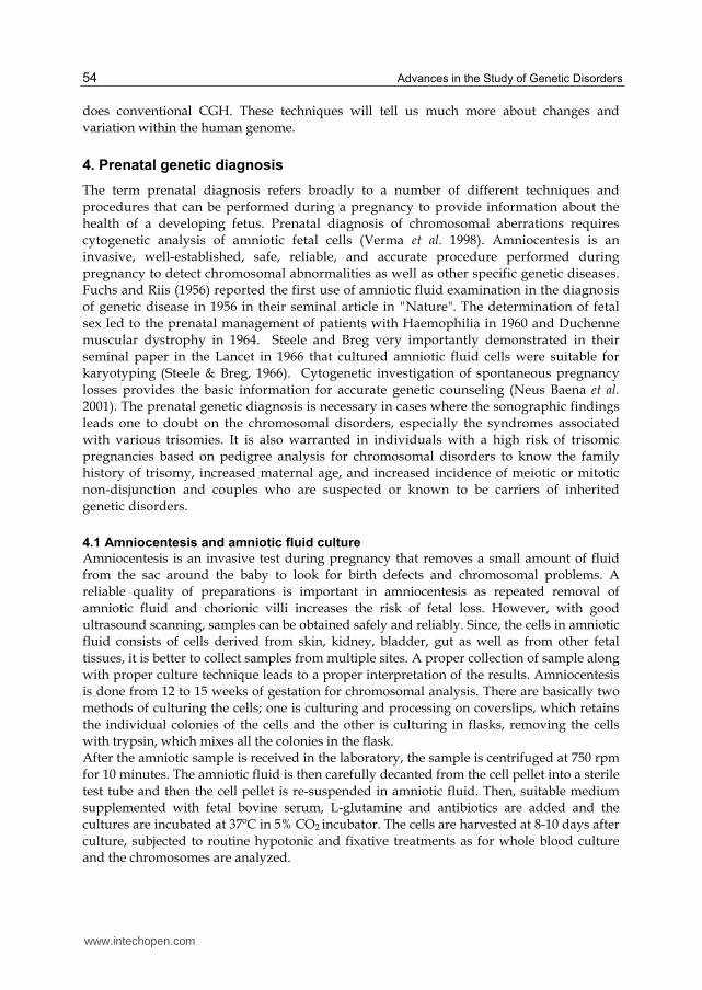
Fig. 4. A karyotype of a Down syndrome patient (47, XX,+21) (Reproduced courtesy of Human Genome Centre, Universiti Sains Malaysia, Malaysia)
Other medical conditions may also be noticed in Down syndrome people like birth defects of heart (atrial septal defect or ventricular septal defect), dementia, problems related to eye
www.intechopen.com
Advances in the Study of Genetic Disorders 56
(cataract), hearing problem, dysplastic pelvis, sleep apnea and hypothyroidism. Currently, there is no known treatment for Down syndrome. However, certain defects require surgery like heart problems etc. The risk is higher among women aged 35 years and above and couples having a Down syndrome baby have an increased risk of having another baby with the condition. A typical karyotype of a Down syndrome patient is given in Figure 4.
5.2 Edwards syndrome (Trisomy 18)
Edwards syndrome is a rare genetic chromosomal syndrome where the child has an extra third copy of chromosome 18. Most of the fetuses abort before term and is more severe than Down syndrome. This syndrome results in mental retardation and various physical defects which causes mortality of the infants at an early stage. Delayed psychomotor development as well as pre and post natal growth failure are the most common findings associated with this syndrome. Sometimes, only some of the body cells have an extra copy of chromosome 18. Hence, there is a mixed population of cells in the individual (called mosaicism). If the individual is a mosaic, then the individual exhibits fewer abnormalities compared to the typical Edwards syndrome features. Symptoms
• Small face • Low set ears • Omphalocele • Upturned nose • Arthrogryposis • Cleft lip/palate • Cryptorchidism • Ptosis of eyelids • Prominent occiput • Overlapping fingers • Small jaw and mouth • Limited hip abduction • Drooping upper eyelids • Developmental retardation • Clubfoot or rocker bottom feet • Malformations of heart and kidney • Webbing of the second and third toes • Widely spaced small eyes with narrow eyelid folds
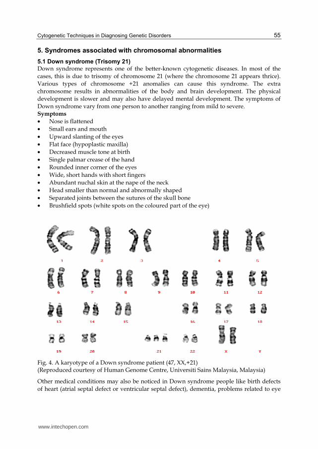
Fig. 5. A micrograph of trisomy 18 (Edwards syndrome) in comparison with its corresponding normal chromosomes (Reproduced courtesy of Human Genome Centre, Universiti Sains Malaysia, Malaysia)
www.intechopen.com
Cytogenetic Techniques in Diagnosing Genetic Disorders 57
The Edwards syndrome is untreatable but treatment can be provided for certain symptoms of the disease. Proper attention should be paid on providing proper nutrition as well as to keep them clean as they are more prone to infections. The survival rate is very low in the Edwards syndrome as half of them die while in the womb. Of those born, fifty percent die within two or three months of their birth, while others die by the time they enter their first year. A typical karyotype of an Edwards syndrome patient is given in Figure 5.
5.3 Patau syndrome (Trisomy 13)
Patau syndrome is a genetic disorder in which a person has three copies of chromosome 13, instead of the usual two copies. Rarely, the extra material may be attached to another chromosome (translocation). Trisomy 13 can appear as complete trisomy 13 or as mosaic or as partial trsiomy 13. Symptoms
• Hernias • Coloboma • Small eyes • Hypotonia • Polydactyly • Low set ears • Micrognathia • Microcephaly • Hypertonicity • Cleft lip/ palate • Epicanthal folds • Clenched hands • Single palmar crease • Skeletal abnormalities • Developmental retardation • Close-set eyes (eyes may actually fuse together into one) The infants who are born often have congenital heart disease (atrial septal defect, patent ductus arteriosus, ventricular septal defect). Most of the children with trisomy 13 die in the first month of their life. The patients with trisomy 13 also have other complictions like breathing difficulty, deafness, feeding problems, seizures and vision problems. Hence, treatment involves case by case basis. A typical karyotype of a Patau syndrome patient is given in Figure 6.
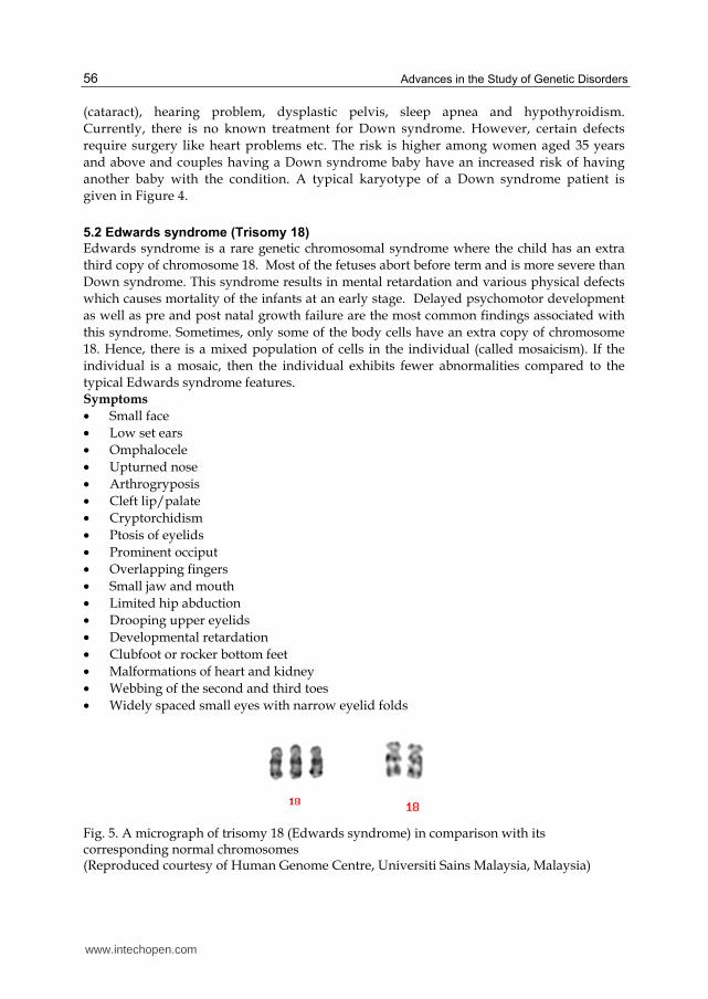
Fig. 6. A micrograph of trisomy 13 (Patau syndrome) in comparison with its corresponding normal chromosomes (Reproduced courtesy of Human Genome Centre, Universiti Sains Malaysia, Malaysia)
www.intechopen.com
Advances in the Study of Genetic Disorders 58
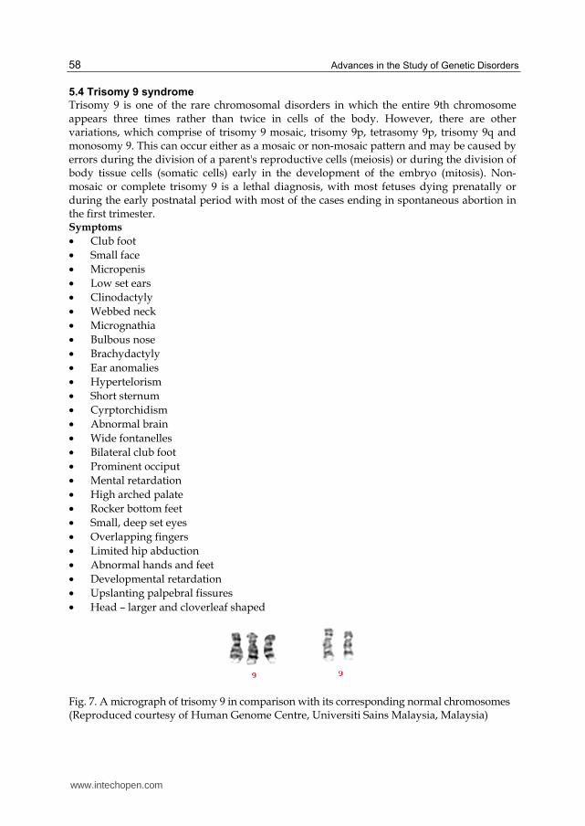
5.4 Trisomy 9 syndrome
Trisomy 9 is one of the rare chromosomal disorders in which the entire 9th chromosome appears three times rather than twice in cells of the body. However, there are other variations, which comprise of trisomy 9 mosaic, trisomy 9p, tetrasomy 9p, trisomy 9q and monosomy 9. This can occur either as a mosaic or non-mosaic pattern and may be caused by errors during the division of a parent's reproductive cells (meiosis) or during the division of body tissue cells (somatic cells) early in the development of the embryo (mitosis). Non-mosaic or complete trisomy 9 is a lethal diagnosis, with most fetuses dying prenatally or during the early postnatal period with most of the cases ending in spontaneous abortion in the first trimester. Symptoms
• Club foot • Small face • Micropenis • Low set ears • Clinodactyly • Webbed neck • Micrognathia • Bulbous nose • Brachydactyly • Ear anomalies • Hypertelorism • Short sternum • Cyrptorchidism • Abnormal brain • Wide fontanelles • Bilateral club foot • Prominent occiput • Mental retardation • High arched palate • Rocker bottom feet • Small, deep set eyes • Overlapping fingers • Limited hip abduction • Abnormal hands and feet • Developmental retardation • Upslanting palpebral fissures • Head – larger and cloverleaf shaped
Fig. 7. A micrograph of trisomy 9 in comparison with its corresponding normal chromosomes (Reproduced courtesy of Human Genome Centre, Universiti Sains Malaysia, Malaysia)
www.intechopen.com
Cytogenetic Techniques in Diagnosing Genetic Disorders 59
The infants who are born have congenital heart defects, kidney anomalies, musculoskeletal, genital and/or additional abnormalities. Most of those individuals that survive to be born at term are mosaics. Infants with non-mosaic trisomy 9 are more severely affected than those with mosaicism. The incidence and severity of malformations and mental deficiency correlate with the percentage of trisomic cells in the different tissues. A typical karyotype of a Trisomy 9 syndrome patient is given in Figure 7.
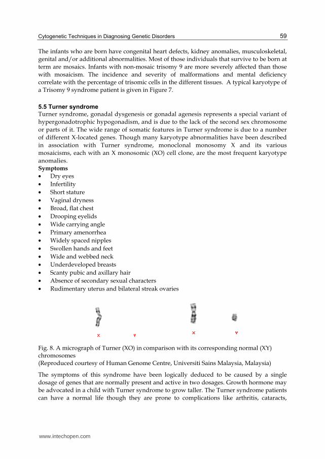
5.5 Turner syndrome
Turner syndrome, gonadal dysgenesis or gonadal agenesis represents a special variant of hypergonadotrophic hypogonadism, and is due to the lack of the second sex chromosome or parts of it. The wide range of somatic features in Turner syndrome is due to a number of different X-located genes. Though many karyotype abnormalities have been described in association with Turner syndrome, monoclonal monosomy X and its various mosaicisms, each with an X monosomic (XO) cell clone, are the most frequent karyotype anomalies. Symptoms
• Dry eyes • Infertility • Short stature • Vaginal dryness • Broad, flat chest • Drooping eyelids • Wide carrying angle • Primary amenorrhea • Widely spaced nipples • Swollen hands and feet • Wide and webbed neck • Underdeveloped breasts • Scanty pubic and axillary hair • Absence of secondary sexual characters • Rudimentary uterus and bilateral streak ovaries
Fig. 8. A micrograph of Turner (XO) in comparison with its corresponding normal (XY) chromosomes (Reproduced courtesy of Human Genome Centre, Universiti Sains Malaysia, Malaysia)
The symptoms of this syndrome have been logically deduced to be caused by a single dosage of genes that are normally present and active in two dosages. Growth hormone may be advocated in a child with Turner syndrome to grow taller. The Turner syndrome patients can have a normal life though they are prone to complications like arthritis, cataracts,
www.intechopen.com
Advances in the Study of Genetic Disorders 60
diabetes, heart defects, high blood pressure, renal problems, ear infections, obesity etc. A typical karyotype of a Tunrner syndrome patient is given in Figure 8.
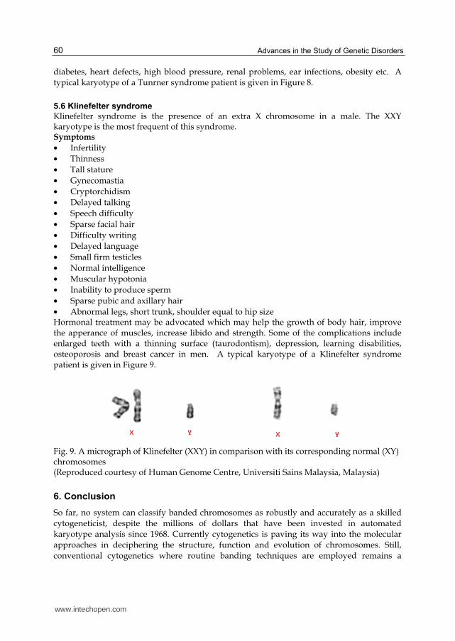
5.6 Klinefelter syndrome
Klinefelter syndrome is the presence of an extra X chromosome in a male. The XXY karyotype is the most frequent of this syndrome. Symptoms
• Infertility • Thinness • Tall stature • Gynecomastia • Cryptorchidism • Delayed talking • Speech difficulty • Sparse facial hair • Difficulty writing • Delayed language • Small firm testicles • Normal intelligence • Muscular hypotonia • Inability to produce sperm • Sparse pubic and axillary hair • Abnormal legs, short trunk, shoulder equal to hip size Hormonal treatment may be advocated which may help the growth of body hair, improve the apperance of muscles, increase libido and strength. Some of the complications include enlarged teeth with a thinning surface (taurodontism), depression, learning disabilities, osteoporosis and breast cancer in men. A typical karyotype of a Klinefelter syndrome patient is given in Figure 9.
Fig. 9. A micrograph of Klinefelter (XXY) in comparison with its corresponding normal (XY) chromosomes (Reproduced courtesy of Human Genome Centre, Universiti Sains Malaysia, Malaysia)
6. Conclusion
So far, no system can classify banded chromosomes as robustly and accurately as a skilled cytogeneticist, despite the millions of dollars that have been invested in automated karyotype analysis since 1968. Currently cytogenetics is paving its way into the molecular approaches in deciphering the structure, function and evolution of chromosomes. Still, conventional cytogenetics where routine banding techniques are employed remains a
www.intechopen.com
Cytogenetic Techniques in Diagnosing Genetic Disorders 61
simple and popular technique to get an overview of the human genome as a whole (Thirumulu Kannan Ponnuraj & Zilfalil Alwi, 2009). Routine banded karyotype analysis can now be combined with M-FISH and other molecular techniques leading to more precise detection of various syndromes in children. Through the analysis of chromosome banding patterns, thousands of chromosomal abnormalities have been associated with inherited or de novo disorders, generating many leads to the underlying molecular causes of these disorders and today, when high resolution genetic linkage analysis can be conducted easily, the discovery of a patient whose disorder is caused by a gross chromosomal abnormality is heralded as a valuable resource for locating the disease gene. Solid tumors also present a myriad of complex chromosomal aberrations and each is a possible clue to tumor initiation and progression. The challenge is to navigate from the visible morphological alteration to the DNA sequence level. In other words, chromosomal abnormalities exist as nature’s guide to the molecular basis of many unexplained human disorders. Hence, cytogenetics continue to remain as indispensable tools for the diagnosis of various genetic disorders which gives an overall picture of the whole genome for analysis. This could possibly also pave a way for treatment and management related to chromosomal disorders.
7. References
Albertson, D. & Pinkel, D. (2003). Genomic microarrays in human genetic disease and cancer. Human Molecular Genetics 12: 145–152.
Arrighi, F.E. & Hsu, T.C. (1971). Localization of heterochromatin in human chromosomes. Cytogenetics 10: 81-86.
Barbara J Trask. (2002). Human cytogenetics: 49 chromosomes, 46 years and counting. Nature 3: 769-778.
Caspersson, T., Farber, S., Foley, G.E., Kudynowski, J., Modest, E.J., Simonsson, E., Wagh, U. & Zech, L. (1968). Chemical differentiation along metaphase chromosomes. Experimental Cell Research 49: 219-222.
Caspersson, T., Zech, L. & Johansson, C. (1970). Differential binding of alkylating fluorochromes in human chromosomes. Experimental Cell Research 60: 315-319.
Chamla, Y. & Ruffie, M. (1976). Production of C and T bands in human mitotic chromosomes after treatment. Human Genetics 34: 213-216.
Chicago Conference, (1966). Standardization in Human Cytogenetics. Birth defects: Original Article Series, 11:2, New York, The National Foundation.
Denver Conference. (1960). The identification of individual chromosomes especially in man. American Journal of Human Genetics 12: 384–389.
Dominique FCM Smeets. (2004). Historical prospective of human cytogenetics: from microscope to microarray. Clinical Biochemistry 37: 439–446.
Dutrillaux, B. & Lejeune, J. (1971). Sur une nouvelle technique d’analyse du caryotype humain. C.R. Acad. Sci. Paris 272: 2638-2640.
Dutrillaux, B. (1973). Noveau susteme de marquage chromosomique: les bandes T. Chromosoma 41: 395-402.
Edwards, J.H., Harnden, D.G., Cameron, A.H., Crosse, V.M. & Wolff, O.H. (1960). A new trisomic syndrome. Lancet 1: 787–790.
www.intechopen.com
Advances in the Study of Genetic Disorders 62
Ford, C.E., Jones, K.W., Polani, P.E., De Almeida, J.C. & Briggs, J.H. (1959). A sex chromosome anomaly in a case of gonadal dysgenesis (Turner’s syndrome). Lancet 1: 711-713.
Fuchs, F. & Riis, P. (1956). Antenatal sex determination. Nature 177: 330. Goodpasture, C., Bloom, S.E., Hsu, T.C. & Arrighi, F.E. (1976). Human nucleolus
organizers: the satellites or the stalks? American Journal of Human Genetics 28: 559-566.
Hans Zellweger & Jane Simpson. (1977). Chromosomes of Man. William Heinemann Medical Books Ltd. J.B.Lippincott Co. Philadelphia.
Hamerton, J.L., Klinger, H.P., Mutton, D.E. & Lang, E.M. (1963). The London Conference on the normal human karyotype, 28th-30th August, 1963. Cytogenetics 25: 264-268.
Hare, W.C.D. & Singh, E.L. (1979). Cytogenetics in Animal Reproduction. Commonwealth Agricultural Bureaux, UK.
ISCN, 2005. An international system for human cytogenetics nomenclature (2005): recommendations of the International Standing Committee on Human Cytogenetic Nomenclature / editors, Lisa G. Shaffer, Niels Tommerup Basel ; Farmington, CT : Karger.
Jacobs, P.A. & Strong, J.A. (1959). A case of human intersexuality having a possible XXY sex-determining mechanism. Nature 183: 302–303.
Jau-hong Kao, Jen-hui Chuang. & TsaipeiWang. (2008). Chromosome classification based on the band profile similarity along approximate medial axis. Pattern Recognition 41: 77 – 89.
Johannsen, W. (1909). Elemente der exakten Erblichkeitslehre. Gustav Fischer, Jena. Kallioniemi, A., Kallioniemi, O.P., Sudar, D., Rutovitz, D., Gray, J.W., Waldman, F. & Pinkel,
D. (1992). Comparative genomic hybridization for molecular cytogenetic analysis of solid tumors. Science 258: 818–821.
Lejeune, J., gautier, M. & Turpin, R. (1959). Etude des chromosomes somatiques de neuf enfants mongliens. Comptes Rendus Hebd Seances Acad Sci 248 (11) : 1721–1722.
Lubs, H.A. (1969). A marker X chromosome. American Journal of Human Genetics 21: 231-244.
Matsui, S. & Sasaki, M. (1973). Differential staining of nucleolus organisers in mammalian chromosomes. Nature 246: 148-150.
Moorehead, P.S., Nowell, P.C., Mellman, W.J., Battips, D.M. & Hungerford, D.A. (1960). Chromosome preparations of leukocytes cultured from human peripheral blood. Experimental Cell Research 20: 613-616.
Neus Baena., Miriam Guitart., Joan Carles Ferreres., Elisabet Gabau., Manuel Corona., Francisco Mellado., Josep Egozcue & Maria Rosa Caballin. (2001). Fetal and placenta chromosome constitution in 237 pregnancies. Annales de Genetique 44: 83-88.
Nowell, P.C. & Hungerford, D.A. (1960). A minute chromosome in human chronic granulocytic leukemia. Science 132: 1497–1501.
Painter, T.S. (1934). A new method for the study of chromosome aberrations and the plotting of chromosome maps in Drosphila melanogaster. Genetics, 19: 175-188.
Pardue, M.L. & Gall, J.G. (1970). Chromosomal localization of mouse satellite DNA. Science
168: 1356-1358.
www.intechopen.com
Cytogenetic Techniques in Diagnosing Genetic Disorders 63
Patau, K., Smith, D.W., Therman, E., Inhorn, S.L. & Wagner, H.P. (1960). Multiple congenital anomaly caused by an extra autosome. Lancet 1: 790–793.
Perry, P. & Evans, H.J. (1975). Cytological detection of mutagen-carcinogen exposure by sister chromatid exchange. Nature 258: 121-125.
Pinkel, D., Seagraves, R., Sudar D., Clark, S., Poole, I., Kowbel, D., Collins, C., Kuo, W.L., Chen, C., Zhai, Y., Dairkee, S.H., Ljung, B.M., Gray, J.W. & Albertson, D.G. (1998). High resolution analysis of DNA copy number variation using comparative genomic hybridization to microarrays. Nature Genetics 20: 207–211.
Pinkel, D., Gray, J.W., Trask, B., van den Engh, G., Fuscoe, J. & van Dekken, H. (1986a). Cytogenetic analysis by in situ hybridization with fluorescently labeled nucleic acid probes. Cold Spring Harbor Symposia on Quantitative Biology 51: 151-157.
Pinkel, D., Straumem T. & Gray, J.W. (1986b). Cytogenetic analysis using quantitative, high-sensitivity, fluorescence hybridization. Proceedings of the National Academy of
Sciences USA 83: 2934-2938. Ried, T., Landes, G., Dackowski, W., Klinger, K. & Ward, D.C. (1992). Multicolor
fluorescence in situ hybridization for the simultaneous detection of probe sets for chromosomes 13, 18, 21, X and Y in uncultured amniotic fluid cells. Human
Molecular Genetics 1: 307–313. Rooney, D.E. (2001). Human cytogenetics: constitutional analysis. New York: Oxford Univ.
Press. Rowley, J.D. (1973). A new consistent chromosomal abnormality in chronic myelogenous
leukaemia identified by quinacrine fluorescence and Giemsa staining. Nature 273: 290–293.
Sabina Solinas-Toldo., Stefan Lampel., Stephan Stilgenbauer., Jeremy Nickolenko., Axel Benner., Hartmut Döhner., Thomas Cremer. & Peter Lichter. (1997). Matrix-based comparative genomic hybridization: biochips to screen for genomic imbalances. Genes Chromosomes Cancer 20: 399–407.
Sawyer, J.R. & Hozier, J.C. (1986). High resolution of Mouse chromosomes: Banding conservation between man and mouse. Science 232: 1632-1639.
Scheres, J.M.J.C. 1974. Production of C and T bands in human chromosomes after heat treatment at high ph and staining with “stains-all”. Human Genetics 23: 311-314.
Schmickel, R.D. (1986). Contiguous gene syndromes: a component of recognizable syndromes. Journal of Pediatrics 109: 231–241.
Seabright, M. (1971). A rapid banding technique for human chromosomes. Lancet 2: 971-972.
Steele, M.W. & Breg, W.R. (1966). Chromosome analysis of human amniotic-fluid cells. Lancet 1: 383–385.
Thirumulu Ponnuraj Kannan & Zilfalil Alwi. (2009). Cytogenetics: Past, Present and future. Malaysian Journal of Medical Sciences 16(2): 4-9.
Tjio, J.H. & Levan, A. (1956). The chromosome number in man. Hereditas. 42: 1-6. Verma, L., MacDonald, F., Leedham, P., McConachie, M., Dhanjal, S. & Hulten, M. (1998).
Rapid and simple prenatal DNA diagnosis of Down’s syndrome. The Lancet 352: 9- 12.
www.intechopen.com
Advances in the Study of Genetic Disorders 64
Waldeyer, W. (1888). Über Karyokinese und ihre Beziehungen zu den Befruchtungsvorgängen. Archiv für mikroskopische Anatomie und Entwicklungsmechanik. 32: 1-122.
Yunis, J.J. (1976). High resolution of human chromosomes. Science 191: 1268–1270.
www.intechopen.com
Advances in the Study of Genetic DisordersEdited by Dr. Kenji Ikehara
ISBN 978-953-307-305-7Hard cover, 472 pagesPublisher InTechPublished online 21, November, 2011Published in print edition November, 2011
InTech EuropeUniversity Campus STeP Ri Slavka Krautzeka 83/A 51000 Rijeka, Croatia Phone: +385 (51) 770 447 Fax: +385 (51) 686 166www.intechopen.com
InTech ChinaUnit 405, Office Block, Hotel Equatorial Shanghai No.65, Yan An Road (West), Shanghai, 200040, China
Phone: +86-21-62489820 Fax: +86-21-62489821
The studies on genetic disorders have been rapidly advancing in recent years as to be able to understand thereasons why genetic disorders are caused. The first Section of this volume provides readers with backgroundand several methodologies for understanding genetic disorders. Genetic defects, diagnoses and treatments ofthe respective unifactorial and multifactorial genetic disorders are reviewed in the second and third Sections.Certainly, it is quite difficult or almost impossible to cure a genetic disorder fundamentally at the present time.However, our knowledge of genetic functions has rapidly accumulated since the double-stranded structure ofDNA was discovered by Watson and Crick in 1956. Therefore, nowadays it is possible to understand thereasons why genetic disorders are caused. It is probable that the knowledge of genetic disorders described inthis book will lead to the discovery of an epoch of new medical treatment and relieve human beings from thegenetic disorders of the future.
How to referenceIn order to correctly reference this scholarly work, feel free to copy and paste the following:
Kannan Thirumulu Ponnuraj (2011). Cytogenetic Techniques in Diagnosing Genetic Disorders, Advances inthe Study of Genetic Disorders, Dr. Kenji Ikehara (Ed.), ISBN: 978-953-307-305-7, InTech, Available from:http://www.intechopen.com/books/advances-in-the-study-of-genetic-disorders/cytogenetic-techniques-in-diagnosing-genetic-disorders