case report an atypical presentation of kawasaki disease ... · considered in the differential...
TRANSCRIPT
CASE REPORT
An atypical presentation of Kawasaki disease: a 10-year-old boy with acute exudative tonsillitis andbilateral cervical lymphadenitisChiew-Yee Yap,I Lung-Huang Lin,I,II Nan-Koong WangI,II
I Cathay General Hospital, Department of Pediatrics, Taipei, Taiwan. II Fu-Jen Catholic University, College of Medicine, Hsinchuang, Taipei, Taiwan.
Email: [email protected]
Tel.: 886-0983701395
INTRODUCTION
Kawasaki disease (KD) is typically a self-limiting condi-tion that is a common cause of pediatric vasculitis (1) andthe leading cause of pediatric acquired heart disease (2). Thedisease most frequently occurs in children aged between 6months and 5 years (3) and is often associated with coronaryartery aneurysms (4). Indeed, findings from an epidemio-logic survey conducted in Japan revealed that only 1% ofKD cases occurred in children aged $9 years (5).
There is currently no laboratory test for diagnosing KD.Rather, diagnosis is performed with reference to establishedclinical criteria (6). Unfortunately, atypical manifestations ofKD appear to be on the rise (1), decreasing the likelihood oftimely diagnosis and appropriate treatment. Herein, wereport an unusual case of KD: a 10-year-old boy whopresented with acute exudative tonsillitis and bilateralcervical lymphadenitis.
CASE DESCRIPTION
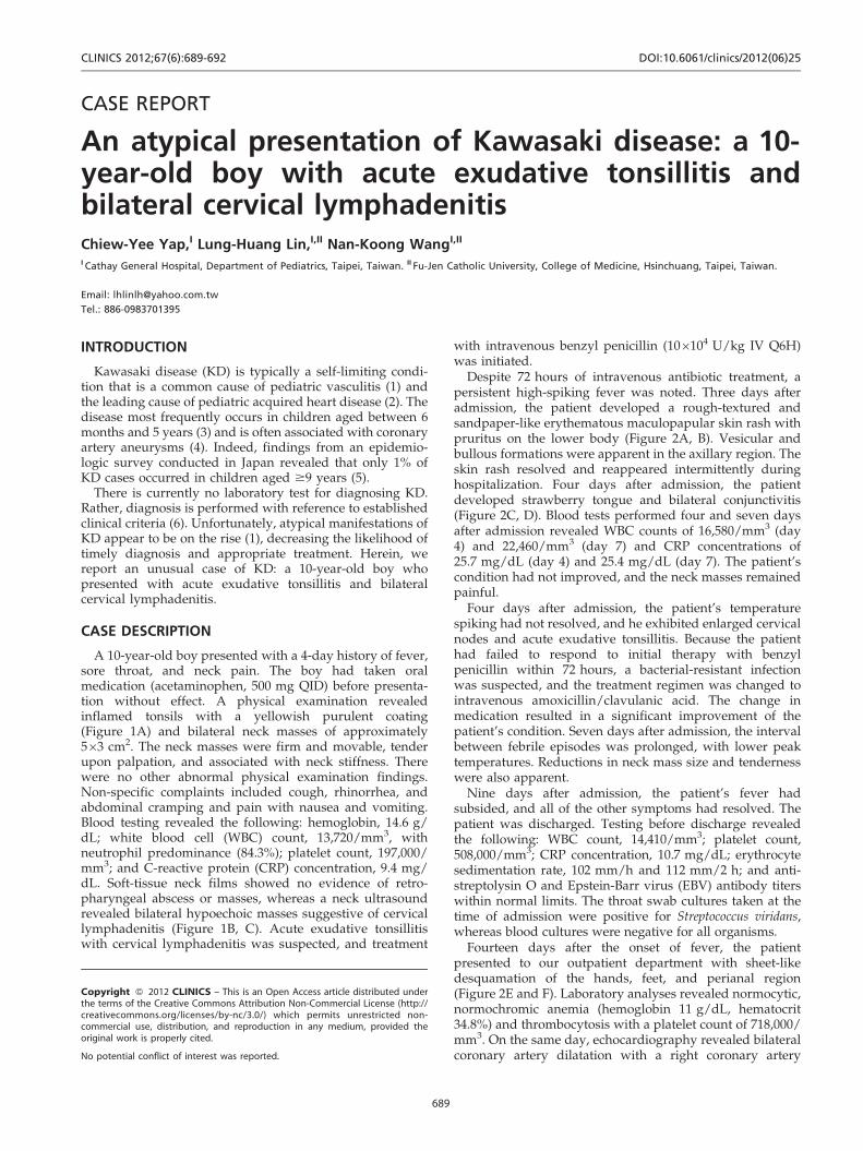
A 10-year-old boy presented with a 4-day history of fever,sore throat, and neck pain. The boy had taken oralmedication (acetaminophen, 500 mg QID) before presenta-tion without effect. A physical examination revealedinflamed tonsils with a yellowish purulent coating(Figure 1A) and bilateral neck masses of approximately563 cm2. The neck masses were firm and movable, tenderupon palpation, and associated with neck stiffness. Therewere no other abnormal physical examination findings.Non-specific complaints included cough, rhinorrhea, andabdominal cramping and pain with nausea and vomiting.Blood testing revealed the following: hemoglobin, 14.6 g/dL; white blood cell (WBC) count, 13,720/mm3, withneutrophil predominance (84.3%); platelet count, 197,000/mm3; and C-reactive protein (CRP) concentration, 9.4 mg/dL. Soft-tissue neck films showed no evidence of retro-pharyngeal abscess or masses, whereas a neck ultrasoundrevealed bilateral hypoechoic masses suggestive of cervicallymphadenitis (Figure 1B, C). Acute exudative tonsillitiswith cervical lymphadenitis was suspected, and treatment
with intravenous benzyl penicillin (106104 U/kg IV Q6H)was initiated.
Despite 72 hours of intravenous antibiotic treatment, apersistent high-spiking fever was noted. Three days afteradmission, the patient developed a rough-textured andsandpaper-like erythematous maculopapular skin rash withpruritus on the lower body (Figure 2A, B). Vesicular andbullous formations were apparent in the axillary region. Theskin rash resolved and reappeared intermittently duringhospitalization. Four days after admission, the patientdeveloped strawberry tongue and bilateral conjunctivitis(Figure 2C, D). Blood tests performed four and seven daysafter admission revealed WBC counts of 16,580/mm3 (day4) and 22,460/mm3 (day 7) and CRP concentrations of25.7 mg/dL (day 4) and 25.4 mg/dL (day 7). The patient’scondition had not improved, and the neck masses remainedpainful.
Four days after admission, the patient’s temperaturespiking had not resolved, and he exhibited enlarged cervicalnodes and acute exudative tonsillitis. Because the patienthad failed to respond to initial therapy with benzylpenicillin within 72 hours, a bacterial-resistant infectionwas suspected, and the treatment regimen was changed tointravenous amoxicillin/clavulanic acid. The change inmedication resulted in a significant improvement of thepatient’s condition. Seven days after admission, the intervalbetween febrile episodes was prolonged, with lower peaktemperatures. Reductions in neck mass size and tendernesswere also apparent.
Nine days after admission, the patient’s fever hadsubsided, and all of the other symptoms had resolved. Thepatient was discharged. Testing before discharge revealedthe following: WBC count, 14,410/mm3; platelet count,508,000/mm3; CRP concentration, 10.7 mg/dL; erythrocytesedimentation rate, 102 mm/h and 112 mm/2 h; and anti-streptolysin O and Epstein-Barr virus (EBV) antibody titerswithin normal limits. The throat swab cultures taken at thetime of admission were positive for Streptococcus viridans,whereas blood cultures were negative for all organisms.
Fourteen days after the onset of fever, the patientpresented to our outpatient department with sheet-likedesquamation of the hands, feet, and perianal region(Figure 2E and F). Laboratory analyses revealed normocytic,normochromic anemia (hemoglobin 11 g/dL, hematocrit34.8%) and thrombocytosis with a platelet count of 718,000/mm3. On the same day, echocardiography revealed bilateralcoronary artery dilatation with a right coronary artery
Copyright � 2012 CLINICS – This is an Open Access article distributed underthe terms of the Creative Commons Attribution Non-Commercial License (http://creativecommons.org/licenses/by-nc/3.0/) which permits unrestricted non-commercial use, distribution, and reproduction in any medium, provided theoriginal work is properly cited.
No potential conflict of interest was reported.
CLINICS 2012;67(6):689-692 DOI:10.6061/clinics/2012(06)25
689
aneurysm (Figure 3). The diameters of the dilated vesselswere as follows: left main coronary artery (LMCA) 5.2 mm;left anterior descending artery 5.5 mm; left circumflex artery4.4 mm; and right coronary artery (RCA) 6.5 mm. Thepatient was diagnosed with KD based on the echocardio-graphic findings and was readmitted. Immediate treatmentwith intravenous gammaglobulin was initiated (1 g/kg for 2days), and the patient was subsequently discharged with aprescription for ongoing treatment with aspirin (5 mg/kg/day) and dipyridamole (6 mg/kg/day). Testing beforedischarge revealed the following: WBC count, 8,540/mm3;neutrophils, 55.5%; platelet count, 270,000/mm3; anderythrocyte sedimentation rate, 8 mm/h and 10 mm/2 h.These findings indicated that the inflammation associatedwith the disease was controlled.
Follow-up echocardiography examinations during a 6-month period after discharge revealed diffuse coronaryartery dilatation and aneurysms as follows: LMCA 4.8 mmand RCA 6.7 mm (1 month after discharge); LMCA 4.1 mm
and RCA 6.2 mm (two months after discharge); and LMCA3.9 mm and RCA 4.6 mm (six months after discharge).
DISCUSSION
The clinical manifestations of KD can be diverse. Indeed,recent case reports have highlighted atypical cases of KDinvolving patients presenting with symptoms such as severeshock (7) and pancreatitis (8). A recent case series has alsohighlighted the existence of KD in adults, indicating that ageis not a defining characteristic of the disease (9). It isimportant that clinicians are made aware of atypical cases tofacilitate prompt diagnosis and appropriate treatment.
Not all patients with KD have symptoms that meet theclassic diagnostic criteria. Children with KD exhibitingfever and fewer than four of the other characteristicsymptoms, so-called atypical or incomplete KD, maydevelop coronary artery aneurysms. In the case describedin this paper, a 10-year-old boy presented with fever, acute
Figure 1 - The clinical presentation of a 10-year-old boy before treatment. Bilateral injected tonsils with a yellowish purulent coating(A). Ultrasound images of the bilateral neck masses suggested cervical lymphadenitis (B, C).
Atypical Kawasaki diseaseYap C-Y et al.
CLINICS 2012;67(6):689-692
690

exudative tonsillitis, and lymphadenopathy. Althoughfever and lymphadenopathy are well-known symptomsof KD, the patient’s age and the lack of other initialsymptoms consistent with KD led us to consider otherdiagnoses, such as group A beta-hemolytic streptococcalinfection or EBV infection, which also cause prolongedfever, lymphadenopathy, and maculopapular skin rashafter treatment with antibiotics. To our knowledge, onlyone other similar case has been described in the literature,which also involved an older child (8 years) who presentedwith fever, lymphadenopathy, and tonsillitis (10). Therehave been several other reports describing patients whopresented with tonsillar or peritonsillar symptoms in theabsence of lymphadenopathy (11-13).
Clinicians need to be aware of the existence of KD andatypical KD and that early treatment is most likely moreeffective than late treatment. Patients with atypical KD maydevelop coronary artery damage prior to treatment. Indeed,the atypical presentation of our patient resulted in thedelayed diagnosis and initiation of appropriate treatment.Our patient did not meet the classical criteria for thediagnosis of KD until eight days after the onset of fever.Coronary artery aneurysms are relatively common inchildren with KD who are untreated (14). Our case clearlyhighlights the importance of early treatment in cases of KDand the need for ongoing echocardiographic monitoring todetect cardiac abnormalities, particularly if the initiation ofappropriate treatment has been delayed. Follow-up labora-tory testing should also be performed.
Atypical KD is most common in young infants, who areunfortunately at the greatest risk of developing coronarydisease. Identifying such cases can be quite challenging, andfatal outcomes have been reported (5). KD should beconsidered in the differential diagnosis of prolonged feverin infants. Many physicians who are experienced in thediagnosis and treatment of KD at large medical centers haveencountered patients in whom prolonged fever wasvirtually the sole manifestation of KD (15).
In summary, a diagnosis of KD should be considered inchildren presenting with fever, tonsillitis, and cervicallymphadenopathy, particularly in older children and ifempiric antibiotics have proven ineffective. Patients withsymptoms suggestive of atypical KD should be referred to apediatric cardiologist who is experienced in diagnosing thiscondition.
AUTHOR CONTRIBUTIONS
Yap CY and Wang N-K were responsible for the patient’s treatment and
clinical data collection. Lin LH was responsible for the patient’s treatment
and manuscript preparation.
REFERENCES
1. Cimaz R, Sundel R. Atypical and incomplete Kawasaki disease. BestPract Res Clin Rheumatol. 2009;23(5):689-97, http://dx.doi.org/10.1016/j.berh.2009.08.010.
2. De Rosa G, Pardeo M, Rigante D. Current recommendations for thepharmacologic therapy in Kawasaki syndrome and management of itscardiovascular complications. Eur Rev Med Pharmacol Sci. 2007;11(5):301-8.
3. Falcini F, Capannini S, Rigante D. Kawasaki syndrome: an intriguingdisease with numerous unsolved dilemmas. Pediatr Rheumatol Online J.2011;9:17, http://dx.doi.org/10.1186/1546-0096-9-17.
4. Newburger JW, Takahashi M, Gerber MA, Gewitz MH, Tani LY, BurnsJC, et al. Diagnosis, treatment, and long-term management of Kawasakidisease: a statement for health professionals from the Committee onRheumatic Fever, Endocarditis and Kawasaki Disease, Council onCardiovascular Disease in the Young, American Heart Association.Circulation. 2004;110(17):2747-71, http://dx.doi.org/10.1161/01.CIR.0000145143.19711.78.
5. Yanagawa H, Yashiro M, Nakamura Y, Kawasaki T, Kato H.Epidemiologic pictures of Kawasaki disease in Japan: from the nation-wide incidence survey in 1991 and 1992. Pediatrics. 1995;95(4):475-9.
6. Kawasaki T, Kosaki F, Okawa S, Shigematsu I, Yanagawa H. A newinfantile acute febrile mucocutaneous lymph node syndrome (MLNS)prevailing in Japan. Pediatrics. 1974;54(3):271-6. Epub 1974/09/01.
7. Thabet F, Bafaqih H, Al-Mohaimeed S, Al-Hilali M, Al-Sewairi W,Chehab M. Shock: an unusual presentation of Kawasaki disease.Eur J Pediatr. 2011;170(7):941-3, http://dx.doi.org/10.1007/s00431-011-1426-5.
8. Prokic D, Ristic G, Paunovic Z, Pasic S. Pancreatitis and atypicalKawasaki disease. Pediatr Rheumatol Online J. 2010;8:8, http://dx.doi.org/10.1186/1546-0096-8-8.
9. Gomard-Mennesson E, Landron C, Dauphin C, Epaulard O, Petit C,Green L, et al. Kawasaki disease in adults: report of 10 cases. Medicine(Baltimore). 2010;89(3):149-58.
10. Hathursinghe HR, Patel S, Uppal HS, Ray J. Acute tonsillitis: an unusualpresentation of Kawasaki syndrome: a case report and review of theliterature. Eur Arch Otorhinolaryngol. 2006;263(4):336-8, http://dx.doi.org/10.1007/s00405-005-1015-1.
11. Korkis JA, Stillwater LB. An unusual otolaryngological problem–mucocutaneous lymph node syndrome (Kawasaki’s syndrome) casereport. J Otolaryngol. 1985;14(4):257-60.
12. Rothfield RE, Arriaga MA, Felder H. Peritonsillar abscess in Kawasakidisease. Int J Pediatr Otorhinolaryngol. 1990;20(1):73-9, http://dx.doi.org/10.1016/0165-5876(90)90336-P.
13. Ravi KV, Brooks JR. Peritonsillar abscess - An unusual presentation ofKawasaki disease. J Laryngol Otol. 1997;111(1):73-4.
14. Suzuki A, Kamiya T, Kuwahara N, Ono Y, Kohata T, Takahashi O, et al.Coronary arterial lesions of Kawasaki disease: cardiac catheterizationfindings of 1100 cases. Pediatr Cardiol. 1986;7(1):3-9, http://dx.doi.org/10.1007/BF02315475.
15. Muise A, Tallett SE, Silverman ED. Are children with Kawasaki diseaseand prolonged fever at risk for macrophage activation syndrome?Pediatrics. 2003;112(6 Pt 1):e495, http://dx.doi.org/10.1542/peds.112.6.e495",-1,"xxx/6.e495.
Figure 2 - Symptoms during treatment. Maculopapular skin rashwith pruritus distributed over the buttocks and lower extremitieswas apparent 7 days after fever onset (A, B). Strawberry tongueand non-purulent acute conjunctivitis were apparent 8 days afterfever onset (C, D). Periungual desquamation of the fingers wasapparent 14 days after fever onset (E, F).
CLINICS 2012;67(6):689-692 Atypical Kawasaki diseaseYap C-Y et al.
691

Figure 3 - Echocardiography 20 days after fever onset. A bilateral coronary artery dilatation with a right coronary artery aneurysm isshown: (A) left main coronary artery, 5.2, mm and left anterior descending artery, 5.5 mm; (B) left circumflex artery, 4.4 mm; (C) rightcoronary artery, 6.5 mm.
Atypical Kawasaki diseaseYap C-Y et al.
CLINICS 2012;67(6):689-692
692