case presentation - stellenbosch university · case presentation amal alabdulla ... • lead to...
TRANSCRIPT

Case Presentation
Amal AlAbdullaDivision of Division of OtorhinolaryngologyOtorhinolaryngology
Faculty of Health SciencesFaculty of Health SciencesTygerbergTygerberg Campus, University of StellenboschCampus, University of Stellenbosch
Case History
• 44-year old male• Hoarseness• Stridor• Weight loss• Odynophagia• Dysphagia for solids
• Haemoptysis• Smoker• Alcohol• TB on Rx
Physical Examination
• 41kg, • Chachetic• Clubbing • RR 18, P 60, T 36.7,
Bp 100/60• Stridor severe pt
cannot lie flat• Chest reduced A/E
bilaterally
• Large hard, immobile neck mass extending into thoracic inlet
• Trachea displaced to L• Scope left pyriform
fossa fullness and a large defect in the vallecula
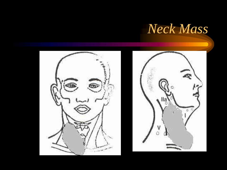
Neck Mass
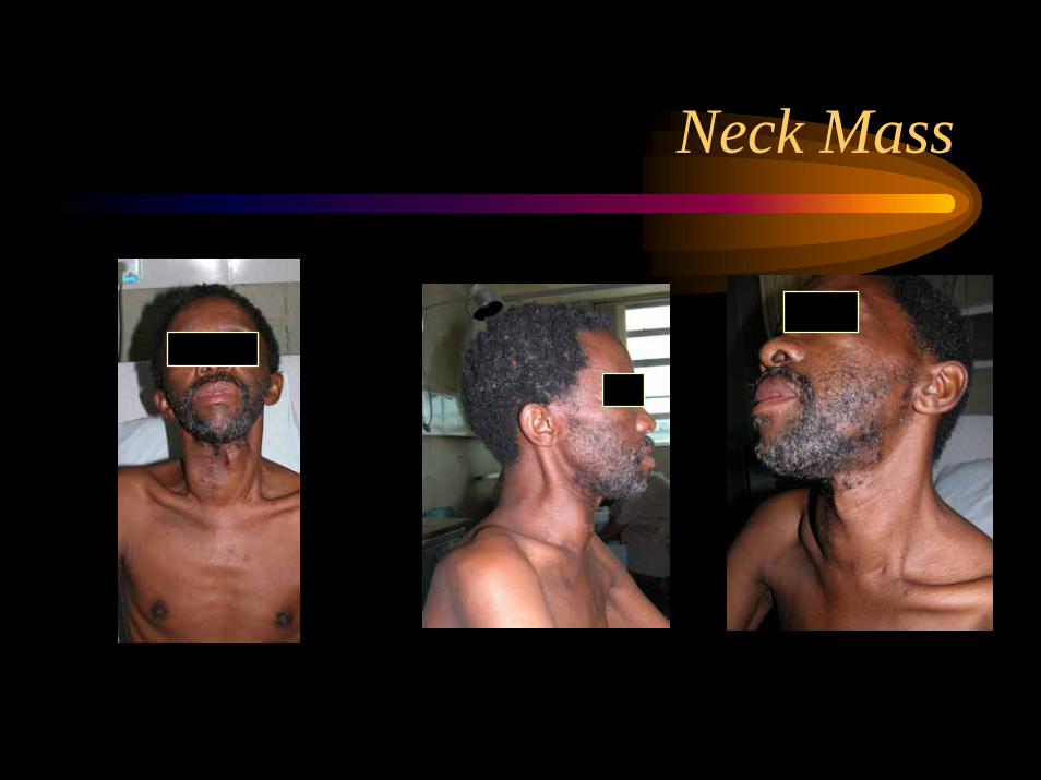
Neck Mass
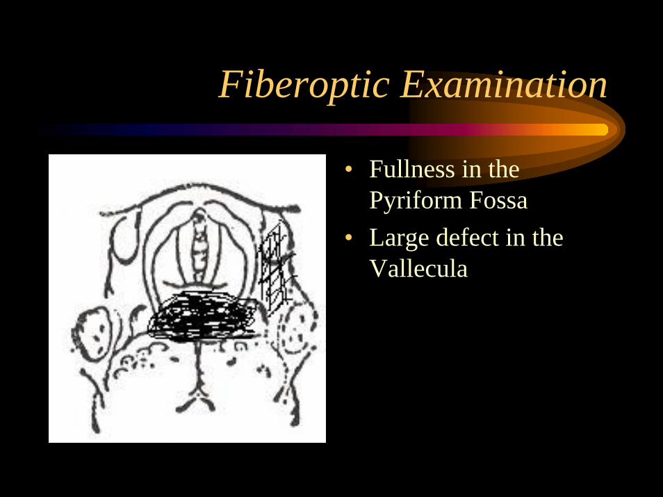
Fiberoptic Examination
• Fullness in the Pyriform Fossa
• Large defect in the Vallecula
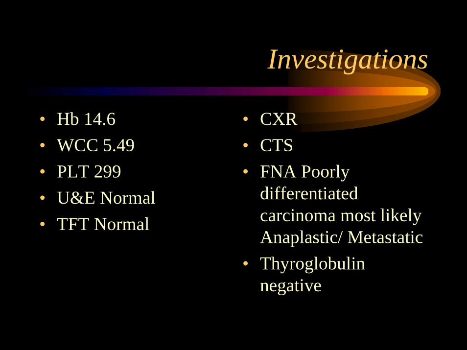
Investigations
• Hb 14.6• WCC 5.49• PLT 299• U&E Normal• TFT Normal
• CXR• CTS• FNA Poorly
differentiated carcinoma most likely Anaplastic/ Metastatic
• Thyroglobulin negative

Anatomy of the Thyroid Gland
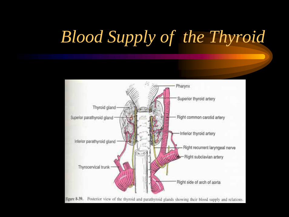
Blood Supply of the Thyroid
Lymphatic Drainage of Thyroid
Major• Middle Jugular lymph nodes (level III)• Lower Jugular lymph nodes (level IV)• Posterior triangle nodes (level V)Minor• Pretracheal and Paratracheal nodes (level VI)• Superior Mediatinal nodes (level VII)

Normal Histology
• Follicle separated from interstitium by a complete basement membrane
• 20-30 follicles organized into lobules separated by a thin layer of fibrous tissue
• Low cuboidal shape• Colloid is deeply
eosinophilic • Follicular cells abundant
eosinophilic cytoplasm Hurthle
• C-cells Calcitonin

Incidence of Thyroid Cancer
• Wide geographic variation in incidence• UK annual incidence 2-3/100 000 • Switzerland higher rate due to Iodine deficiency
mortality rate 10 times that in UK and Wales• F:M 3:1• Any age - predominantly in elderly• Young adults and adolescence well differentiated
papillary type

Aetiology of Thyroid Cancer
• Over stimulation by elevated TSH• Solitary thyroid nodule• ionizing radiation• Genetic factors• Chronic lymphocytic thyroiditis
Iodine deficiency
• Natural diet is deficient in Iodine• Deficient production of T3 and T4• Pt may become overtly hypothyroid • In both cases there is an increase in TSH • Lead to enlargement of the gland/goitre• Prolonged TSH stimulation

Solitary Thyroid Nodule
• There is a past history of ionizing radiation• It occurs in a patient with a family history
of thyroid cancer• There is a history of previous thyroid cancer• It is enlarging (particularly on suppressive
doses of thyroxin)• The nodule develops in a person <14 or >65• The patient is male
ionizing radiation
• 8.3/1000 cases of thyroid cancer in irradiated pts after a follow up of 20 years
• Latent period before cancer occurs is 20-30 years after exposure
• Thyroid nodule occurs in 30-40% of pts, 60% of those are benign
• FNAC is recommended as the initial investigation rather than proceed to surgery in every case
Genetic Factors
• A definitive tendency of hyperthyroidism, goitreand thyroid cancer to occur in the same family
• MEN II a (Autosomal dominant) - thyroid medullary ca, phaeochromocytoma and hyperparathyroidism
• MEN II b - medullary thyroid ca, phaeochromocytoma, marfinoid appearance with multiple mucosal Neuroma of lips, tongue and oropharynx, Ganglioneuromas of the GIT
• Familial non-MEN MTC is recognized
Chronic lymphocytic thyroiditis
• Thyroid lymphoma most often occurs against a background of
- Autoimmune lymphocytic thyroiditis- Hashimoto’s disease
Benign Thyroid Tumors
Follicular cell adenomaHurthle cell adenomaTeratoma

Malignant Thyroid tumors
Primary• Papillary ca 80%• Follicular ca 10%• Hurthle cell ca• Medullary ca 5%• Anaplastic ca• Lymphoma• Sarcoma• SCC
Secondary• Kidney• Colon• Lung• Breast
Benign Thyroid Tumors
Adenoma is the most common type• Presents as solitary thyroid nodule or
dominant nodule in multinodular gland• Encapsulated• Middle aged women• Not premalignant and rarely toxic• Microscopically: Follicular, Microfollicular,
Hurthle cell and Teratoma
Papillary Adenocarcinoma
• 80% of all thyroid malignancy
• 40-49 years of age• Presents as a nodule,
unencapsulated, well circumscribed
• Multicentric involves both lobes
• LN involvement 60%
Extent • Minimal/micro ca
<0.1cm• Intrathyroidal >0.1cm• Extrathyroidal:Beyond
gland capsule and/or LN metastases

Follicular Adenocarcinoma
• Older age group 50-59 years• 10-20% of all thyroid malignancy• Solitary thyroid nodule• Bone/Lung involvement 20-30%• LN involvement 10%• Well defined capsule• Malignancy vascular and capsular invasion
Hurthle Cell tumors
• Extremely uncommon• Malignant vascular and capsular invasion• LN metastases common

Medullary thyroid carcinoma
• 5% of all cases• As part of MEN IIA, MEN IIB,
Familial non-MEN, Sporadic• In MEN usually bilateral 90% and multifocal• Unifocal in sporadic cases• LN 25-50%• Arise from parafollicular or C cells

Lymphoma
• <5% of all cases of lymphoma• Rapidly increasing mass in the neck• Elderly women• Arises on a background of autoimmune
thyroiditis, extends outside the capsule• Majority high grade B-cell/ NHL
Anaplastic
• Elderly• Women• Over a long standing thyroid enlargement• Rapidly enlarges• Referred otalgia, hoarseness• Aggressively malignant with high metastatic
potential• Rapidly invade larynx, pharynx, oesophagus• Poor prognosis, Treatment ineffective• Pts die in 1 year

Presenting symptoms of thyroid tumors
• Solitary nodule• Cervical lymphadenopathy• Rapidly enlarging goitre• Pain in the neck• Stridor due to tracheal compression• Dysphagia due to oesophageal compression• Hoarseness due to vocal cord palsy• Metastases
Examination
• Thyroid examined particularly for hardness• Mobile, move with swallowing?• Get below the mass in the midline?• Retrosternal invasion? Neck extended to see if the
tumor comes up into the neck• Papillary/Medullary hard rubber nodules• Anaplastic hard, fixed• Lymphomas diffuse• Neck and Axilla for LNs• Fiberoptic examination
Investigations
• CXR: tracheal deviation, mediastinal extension or lymphadenopathy, pulmonary metastases
• U/S: tumor size, cystic/solid• CTS/MRI poorly specific and sensitive in the
diagnoses of thyroid cancer• CTS/MRI difficult in pts with compromised
airways for whom lying flat is uncomfortable• Radionuclide Scan I-123 cold nodule (adenoma)
hot nodule (malignant)• Ga 67 Scan can be useful in detecting lymphoma
Investigations
• TSH, T3, T4• FNA• Excision biopsy if preoperative diagnosis is
unreliable on FNA and Imaging

Prognostic factors
• Pt factors: age, sex• Tumor factors: size, histology, LN, local
invasion, distant metastases• Management factors: delay in therapy,
extent of surgery, experience of surgeon, thyroid hormone therapy, treatment with postop radiation

Treatment Modalities for Thyroid Cancer
• Surgery• Radioactive iodine• External beam radiotherapy• Thyroxin therapy• Chemotherapy