cardiovascular & respiratory systems nestor t. hilvano, m.d., m.p.h. (images copyright discover...
TRANSCRIPT

Cardiovascular & Respiratory Systems
Nestor T. Hilvano, M.D., M.P.H.(Images Copyright Discover Biology, 5th ed., Singh-Cundy and Cain,
Textbook, 2012.)
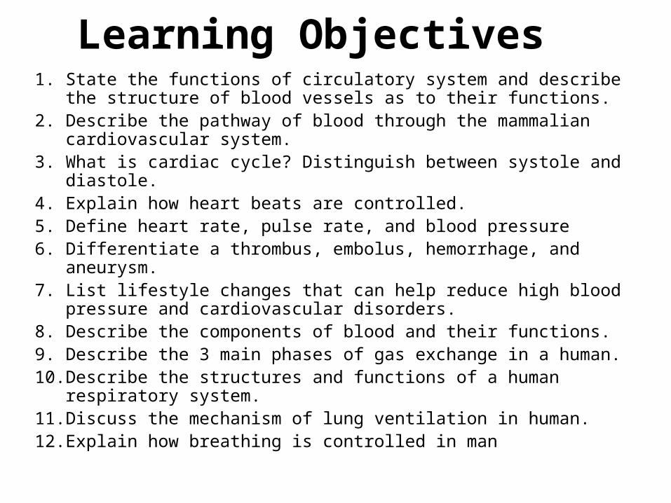
Learning Objectives 1. State the functions of circulatory system and describe the structure
of blood vessels as to their functions.2. Describe the pathway of blood through the mammalian
cardiovascular system.3. What is cardiac cycle? Distinguish between systole and diastole.4. Explain how heart beats are controlled.5. Define heart rate, pulse rate, and blood pressure6. Differentiate a thrombus, embolus, hemorrhage, and aneurysm. 7. List lifestyle changes that can help reduce high blood pressure and
cardiovascular disorders. 8. Describe the components of blood and their functions. 9. Describe the 3 main phases of gas exchange in a human.10. Describe the structures and functions of a human respiratory
system. 11. Discuss the mechanism of lung ventilation in human.12. Explain how breathing is controlled in man

Cardiovascular System • Functions = transport of nutrients and remove waste • Many invertebrates have open circulatory system – blood
is pumped by heart through open-ended vessels and flows out among the cells (tissues).
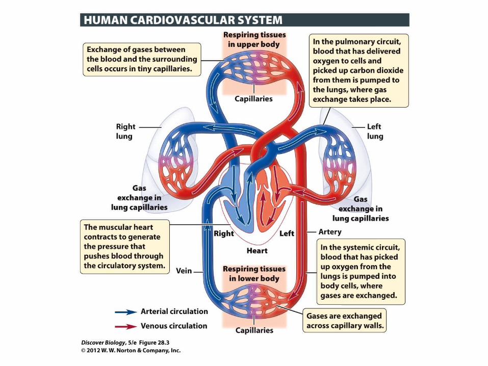
• Vertebrates have closed circulatory system (CVS) – blood is confined to vessels (heart →artery → arteriole → capillary (exchange of gases/nutrients) → venule → vein→ heart).
• Both systems have three basic components:
___ - a circulatory fluid a. blood vessels
___ - a set of tubes b. heart
___ - a muscular pump c. blood
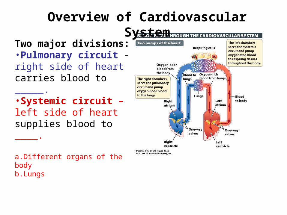
Overview of Cardiovascular System
Two major divisions:•Pulmonary circuit - right side of heart carries blood to _____. •Systemic circuit – left side of heart supplies blood to ____.
a.Different organs of the bodyb.Lungs
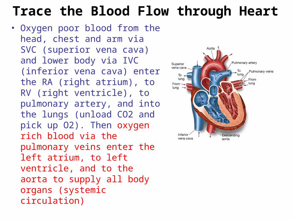
Trace the Blood Flow through Heart• Oxygen poor blood from the
head, chest and arm via SVC (superior vena cava) and lower body via IVC (inferior vena cava) enter the RA (right atrium), to RV (right ventricle), to pulmonary artery, and into the lungs (unload CO2 and pick up O2). Then oxygen rich blood via the pulmonary veins enter the left atrium, to left ventricle, and to the aorta to supply all body organs (systemic circulation)

Figure 23.3B
To lung
Right atrium
From lung
Semilunarvalve
Atrioventricular(AV) valve
To lung
Left atrium
From lung
Semilunarvalve
Atrioventricular(AV) valve
Left ventricle
Right ventricle
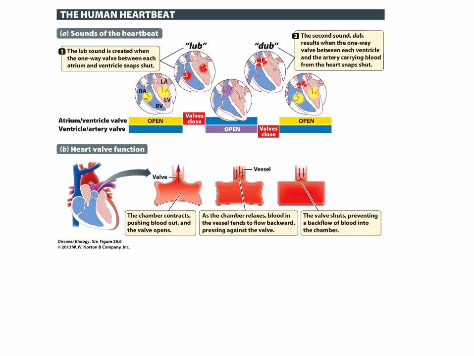
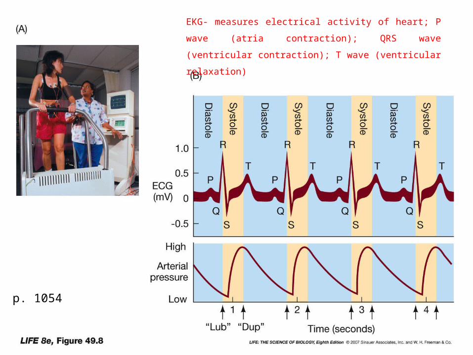
AV valve close = lub (S1 = 1st sound)Ventricle- vessel (semilunar valves) close = dub (S2 = 2nd sound)
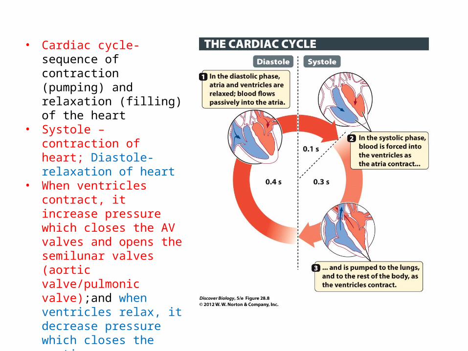
• Cardiac cycle- sequence of contraction (pumping) and relaxation (filling) of the heart
• Systole – contraction of heart; Diastole- relaxation of heart
• When ventricles contract, it increase pressure which closes the AV valves and opens the semilunar valves (aortic valve/pulmonic valve);and when ventricles relax, it decrease pressure which closes the aortic valve/pulmonic valve, and opens AV valves.
• Pressure wave (rush of blood) in the blood vessels (arteries) is felt as pulse (pulse rate)
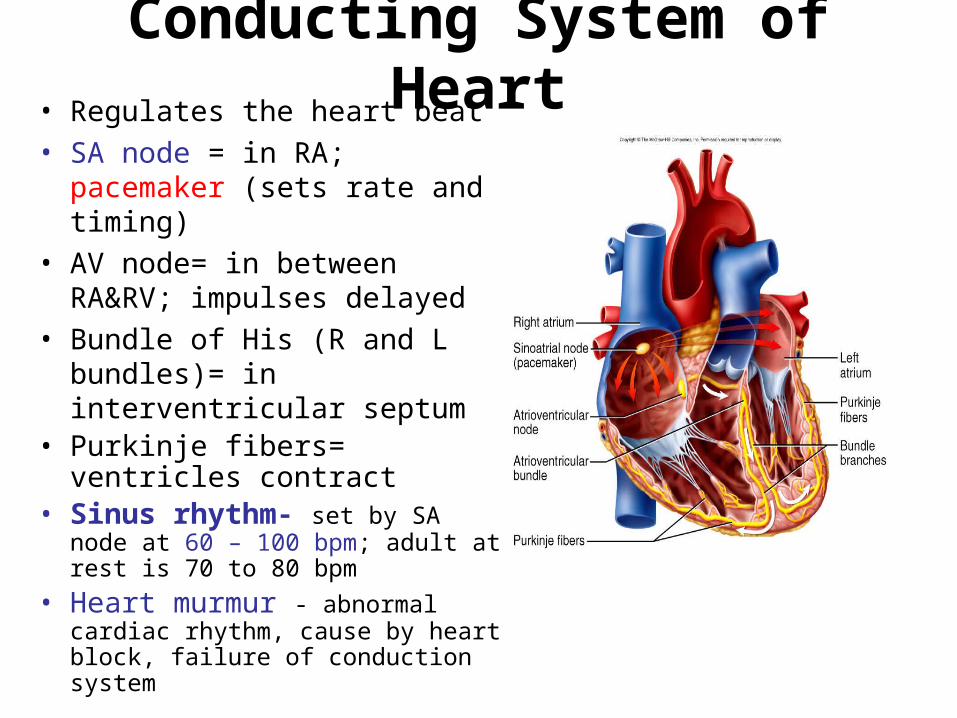
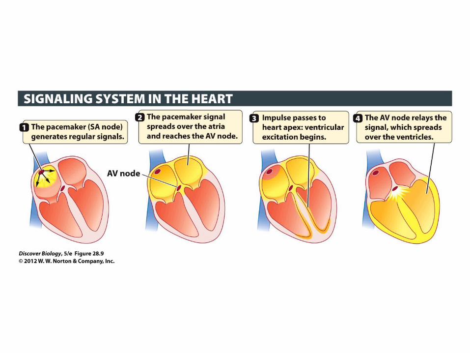
Conducting System of Heart• Regulates the heart beat• SA node = in RA; pacemaker
(sets rate and timing)• AV node= in between RA&RV;
impulses delayed• Bundle of His (R and L
bundles)= in interventricular septum
• Purkinje fibers= ventricles contract
• Sinus rhythm- set by SA node at 60 – 100 bpm; adult at rest is 70 to 80 bpm
• Heart murmur - abnormal cardiac rhythm, cause by heart block, failure of conduction system
p. 1054
EKG- measures electrical activity of heart; P wave (atria
contraction); QRS wave (ventricular contraction); T wave
(ventricular relaxation)
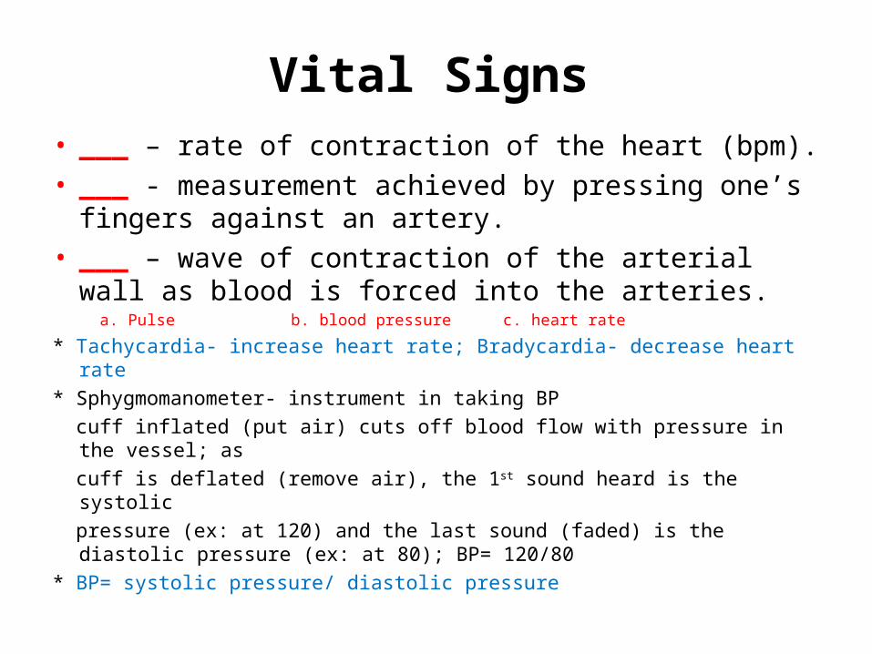
Vital Signs • ___ – rate of contraction of the heart (bpm). • ___ - measurement achieved by pressing one’s
fingers against an artery.• ___ – wave of contraction of the arterial wall as
blood is forced into the arteries. a. Pulse b. blood pressure c. heart rate
* Tachycardia- increase heart rate; Bradycardia- decrease heart rate
* Sphygmomanometer- instrument in taking BP
cuff inflated (put air) cuts off blood flow with pressure in the vessel; as
cuff is deflated (remove air), the 1st sound heard is the systolic
pressure (ex: at 120) and the last sound (faded) is the diastolic pressure (ex: at 80); BP= 120/80
* BP= systolic pressure/ diastolic pressure
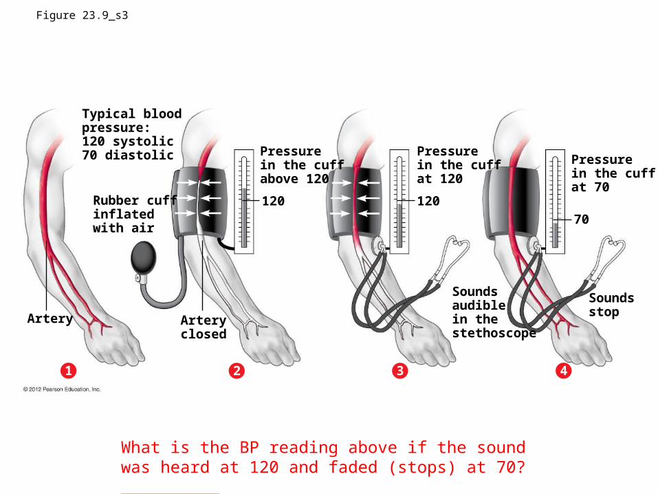
Figure 23.9_s3
Typical bloodpressure:120 systolic70 diastolic
Rubber cuffinflated with air
Artery Arteryclosed
120 12070
Pressure in the cuffabove 120
Pressure in the cuffat 120
Pressure in the cuffat 70
Soundsaudiblein thestethoscope
Soundsstop
21 3 4
What is the BP reading above if the sound was heard at 120 and faded (stops) at 70? __________

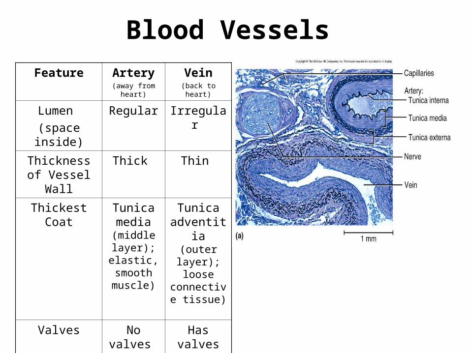
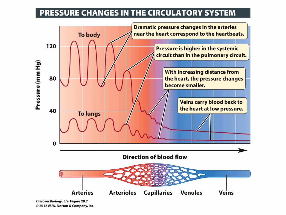
Blood Vessels • ___ – carry blood away
from the heart• ___ – return blood to
the heart• ___ – thin walled and
smallest vessels; supply cells/tissues; exchange of gases
a. Vein b. Capillaryc. Artery
Blood Vessels
Feature Artery(away from
heart)
Vein(back to heart)
Lumen
(space inside)
Regular Irregular
Thickness of Vessel Wall
Thick Thin
Thickest Coat Tunica media (middle layer); elastic, smooth muscle)
Tunica adventitia
(outer layer); loose connective
tissue)
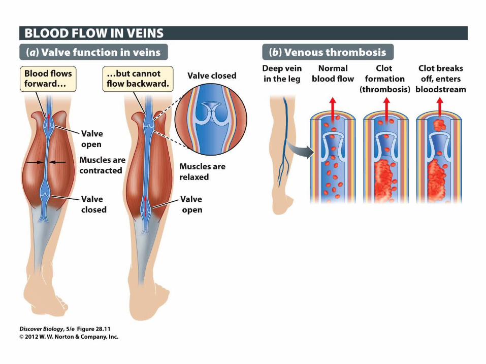
Valves No valves Has valves
(one way)

Disorders of Clotting Mechanism• ___ – blood clot within a vessel.• ___ – a thrombus that breaks free and is carried
in the bloodstream.• ___ – blood loss • ___ - abnormal bulge in the wall of blood vessel.
a. hemorrhage b. thrombus c. aneurysm c. embolus

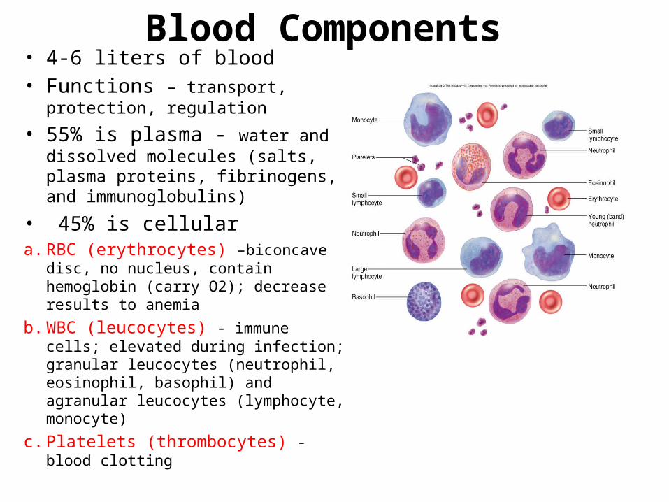
Blood Components • 4-6 liters of blood• Functions – transport, protection,
regulation
• 55% is plasma - water and dissolved molecules (salts, plasma proteins, fibrinogens, and immunoglobulins)
• 45% is cellulara. RBC (erythrocytes) –biconcave disc,
no nucleus, contain hemoglobin (carry O2); decrease results to anemia
b. WBC (leucocytes) - immune cells; elevated during infection; granular leucocytes (neutrophil, eosinophil, basophil) and agranular leucocytes (lymphocyte, monocyte)
c. Platelets (thrombocytes) - blood clotting
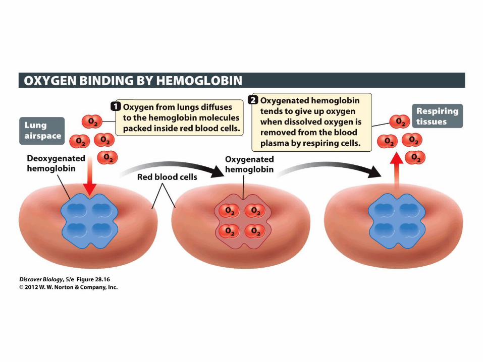
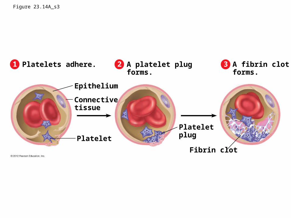
Figure 23.14A_s3
321 Platelets adhere. A platelet plugforms.
A fibrin clotforms.
Epithelium
Connectivetissue
Platelet
Plateletplug
Fibrin clot
Respiratory System: 3 Phases of Gas Exchange
1. Breathing (ventilation) – 1st phase, involves inhaling O2 and exhaling CO2
2. Transport of gases – 2nd phase, involves diffusion into and transport by hemoglobin in RBC
3. Exchange of gases – 3rd phase, cells take up O2 from the blood and release CO2 to the blood.
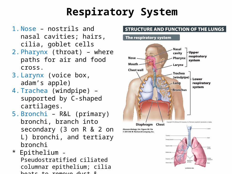
Respiratory System
1. Nose – nostrils and nasal cavities; hairs, cilia, goblet cells
2. Pharynx (throat) – where paths for air and food cross.
3. Larynx (voice box, adam’s apple)4. Trachea (windpipe) – supported
by C-shaped cartilages.5. Bronchi – R&L (primary) bronchi,
branch into secondary (3 on R & 2 on L) bronchi, and tertiary bronchi
* Epithelium - Pseudostratified ciliated columnar epithelium; cilia beats to remove dust & foreign particles; goblet cells trap particles & digest; cartilages keep airway open in upper respiratory tract (conducting portion).
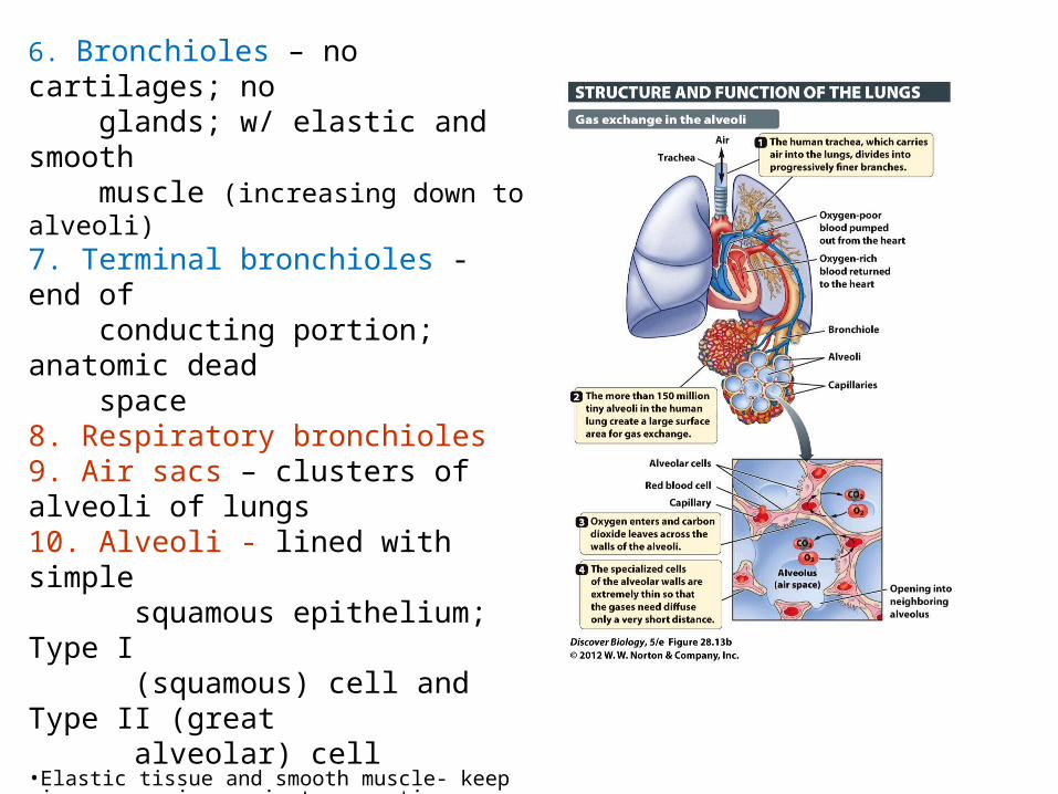
6. Bronchioles – no cartilages; no glands; w/ elastic and smooth muscle (increasing down to alveoli) 7. Terminal bronchioles - end of conducting portion; anatomic dead space8. Respiratory bronchioles 9. Air sacs – clusters of alveoli of lungs10. Alveoli - lined with simple squamous epithelium; Type I (squamous) cell and Type II (great alveolar) cell•Elastic tissue and smooth muscle- keep airway open in respiratory portion.
___ What cells produce surfactants which lower the surface tension of the lungs to prevent collapse?
* RDS in newborn- deficiency of surfactant, lungs collapse.
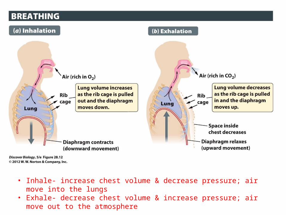
• Inhale- increase chest volume & decrease pressure; air move into the lungs• Exhale- decrease chest volume & increase pressure; air move out to the
atmosphere
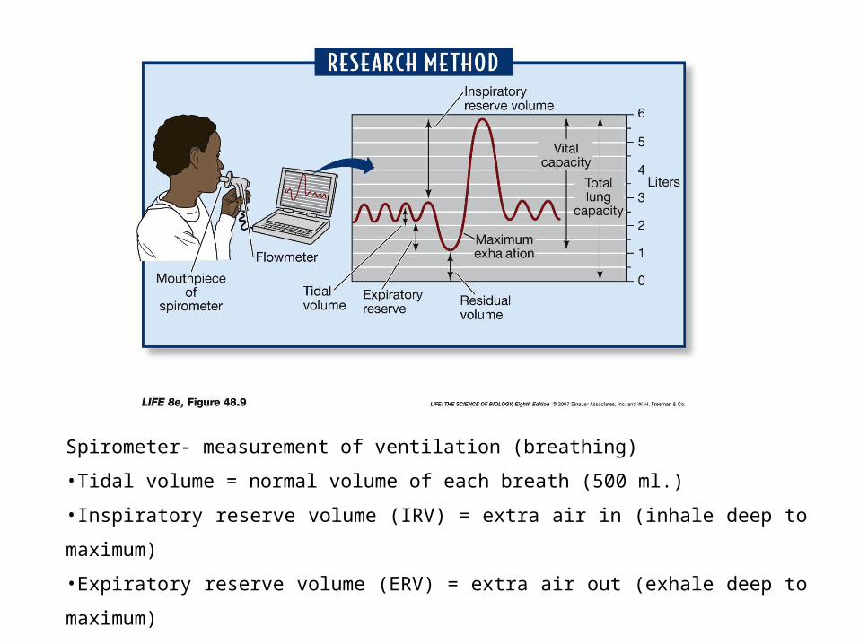
Spirometer- measurement of ventilation (breathing)
•Tidal volume = normal volume of each breath (500 ml.)
•Inspiratory reserve volume (IRV) = extra air in (inhale deep to maximum)
•Expiratory reserve volume (ERV) = extra air out (exhale deep to maximum)
•Vital capacity = TV + IRV + ERV (maximum volume that one can inhale and exhale)
•Total lung capacity (TLC) = VC + Residual volume (dead space; amount that is left)
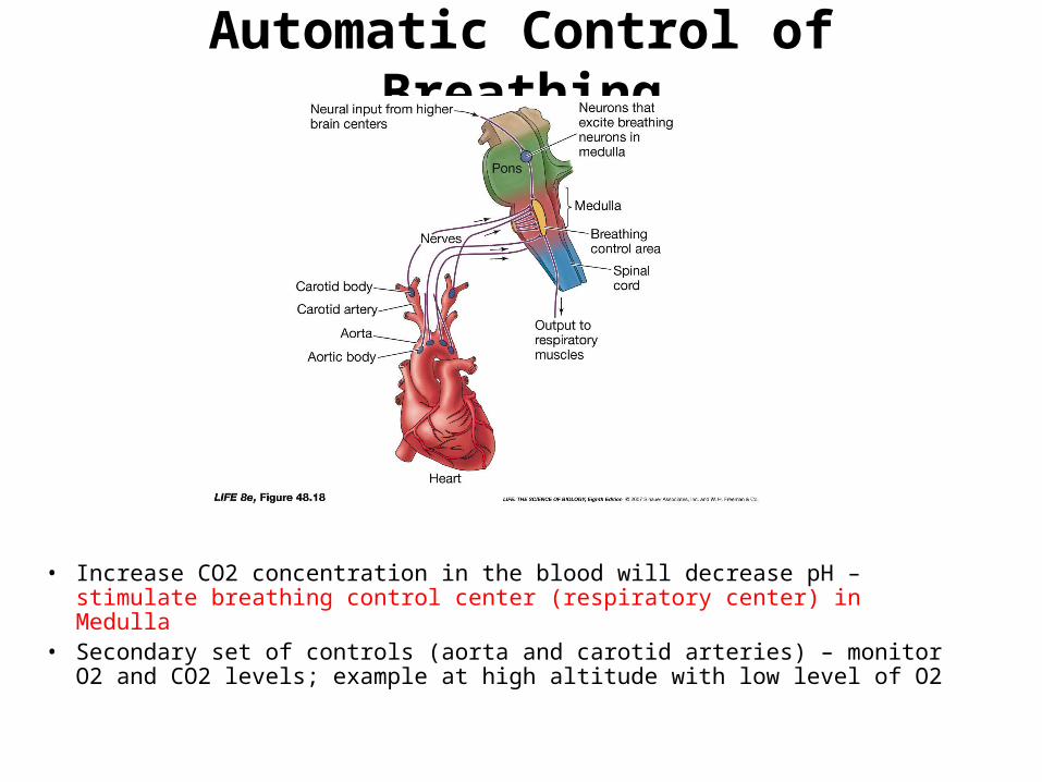
Automatic Control of Breathing
• Increase CO2 concentration in the blood will decrease pH – stimulate breathing control center (respiratory center) in Medulla
• Secondary set of controls (aorta and carotid arteries) – monitor O2 and CO2 levels; example at high altitude with low level of O2
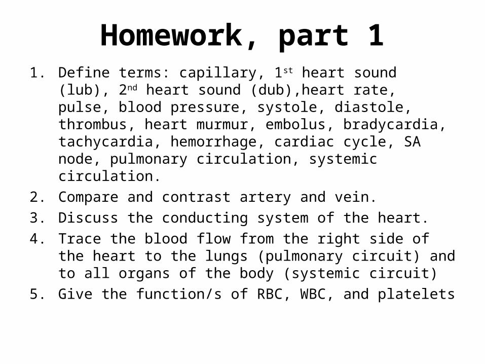
Homework, part 11. Define terms: capillary, 1st heart sound (lub), 2nd heart
sound (dub),heart rate, pulse, blood pressure, systole, diastole, thrombus, heart murmur, embolus, bradycardia, tachycardia, hemorrhage, cardiac cycle, SA node, pulmonary circulation, systemic circulation.
2. Compare and contrast artery and vein.
3. Discuss the conducting system of the heart.
4. Trace the blood flow from the right side of the heart to the lungs (pulmonary circuit) and to all organs of the body (systemic circuit)
5. Give the function/s of RBC, WBC, and platelets
Homework, part 2 1. Define terms: ventilation, tidal volume, vital
capacity, pharynx, larynx, trachea, respiratory distress syndrome in newborn, and type II (great alveolar) cell.
2. Give the function of cilia, goblet cell, and cartilages in the respiratory tract.
3. Describe inhalation and exhalation as to air flow and the movement of the diaphragm and chest wall.
4. Discuss how breathing is controlled in human.5. Trace the passage of air from the nose to the
alveoli of the lungs.