cardiomyopathy is associated with ribosomal protein - genetics
TRANSCRIPT

INVESTIGATION
Cardiomyopathy Is Associated with RibosomalProtein Gene Haplo-Insufficiency in
Drosophila melanogasterMichelle E. Casad,* Dennis Abraham,† Il-Man Kim,† Stephan Frangakis,* Brian Dong,* Na Lin,†,1
Matthew J. Wolf,† and Howard A. Rockman*,†,‡,2
*Department of Cell Biology, †Department of Medicine, and ‡Department of Molecular Genetics and Microbiology, DukeUniversity Medical Center, Durham, North Carolina 27710
ABSTRACT The Minute syndrome in Drosophila melanogaster is characterized by delayed development, poor fertility, and shortslender bristles. Many Minute loci correspond to disruptions of genes for cytoplasmic ribosomal proteins, and therefore the phenotypehas been attributed to alterations in translational processes. Although protein translation is crucial for all cells in an organism, it isunclear why Minute mutations cause effects in specific tissues. To determine whether the heart is sensitive to haplo-insufficiency ofgenes encoding ribosomal proteins, we measured heart function of Minute mutants using optical coherence tomography. We foundthat cardiomyopathy is associated with the Minute syndrome caused by haplo-insufficiency of genes encoding cytoplasmic ribosomalproteins. While mutations of genes encoding non-Minute cytoplasmic ribosomal proteins are homozygous lethal, heterozygousdeficiencies spanning these non-Minute genes did not cause a change in cardiac function. Deficiencies of genes for non-Minutemitochondrial ribosomal proteins also did not show abnormal cardiac function, with the exception of a heterozygous disruption ofmRpS33. We demonstrate that cardiomyopathy is a common trait of the Minute syndrome caused by haplo-insufficiency of genesencoding cytoplasmic ribosomal proteins. In contrast, most cases of heterozygous deficiencies of genes encoding non-Minute ribo-somal proteins have normal heart function in adult Drosophila.
THE Minute phenotype in Drosophila melanogaster ischaracterized by delayed development, poor fertility,
short slender bristles, and smaller body size (Lambertsson1998; Marygold et al. 2007). The distribution of Minute locithroughout the genome and the uniformity of the phenotypehave been of great interest to geneticists for nearly a century.Many Minute loci correspond to cytoplasmic ribosomalgenes, suggesting that the basis of the Minute phenotypesinvolves disruption of ribosomal function (Hart et al. 1993;Andersson et al. 1994; Mckim et al. 1996; Saeboe-Larssenand Lambertsson 1996; Schmidt et al. 1996; Saeboe-Larssen
et al. 1997; Van Beest et al. 1998; Torok et al. 1999;Fauvarque et al. 2001; Marygold et al. 2005; Alexanderet al. 2006; Tyler et al. 2007).
Eukaryotic cells contain cytoplasmic ribosomes andmitochondrial ribosomes, each containing many subunits.While all of these subunits are translated from nucleargenes, cytoplasmic ribosomes translate proteins in thecytosol and on the endoplasmic reticulum, whereas mito-chondrial ribosomes synthesize proteins from mitochondrialgenes in the mitochondrial matrix. Recent genetic andbioinformatic efforts have shown that Drosophila mela-nogaster have 88 genes encoding 79 different cytoplasmicribosomal proteins (CRPs) and 75 nuclear-encoded mito-chondrial ribosomal proteins (MRPs) (Marygold et al. 2007).While mutations in many CRP genes result in a Minute phe-notype, mutations in MRP genes are not associated with theMinute syndrome (Marygold et al. 2007).
Drosophila CRPs correspond with all 79 human CRPs,while MRPs are more divergent between species (Marygold
Copyright © 2011 by the Genetics Society of Americadoi: 10.1534/genetics.111.131482Manuscript received June 8, 2011; accepted for publication August 22, 20111Present address: Institute of Molecular Medicine, Peking University, Beijing, China,100871.
2Corresponding author: Department of Medicine, Duke University MedicalCenter, DUMC 3104, 226 CARL Bldg., Research Dr., Durham, NC, 27710.E-mail: [email protected]
Genetics, Vol. 189, 861–870 November 2011 861
Dow
nloaded from https://academ
ic.oup.com/genetics/article/189/3/861/6063873 by guest on 21 D
ecember 2021

et al. 2007). Mutations in human CRP genes as well as genesfor other proteins involved in ribosomal biogenesis havebeen shown to cause a variety of syndromes (reviewed inFreed et al. 2010), most notably CRP gene mutations in Di-amond Blackfan anemia, characterized by congenital ane-mia and a variety of developmental abnormalities. SeveralMRP genes also map to loci associated with human disordersas well (reviewed in O’brien et al. 2005).
Since ribosomal proteins are important in every cell of anorganism, we tested whether abnormal heart function ispart of the Minute syndrome. We addressed this questionusing optical coherence tomography (OCT) (Wolf et al.2006; Kim and Wolf 2009; Kim et al. 2010; Yu et al.2010) to examine in vivo cardiac size and function in adultDrosophila with ribosomal deficiency. We show that haplo-insufficiency of RpS15Aa, a predicted Minute gene, results incharacteristic short, slender bristles as well as significantdilation and dysfunction of the Drosophila heart. By exam-ining the heart phenotype of many other deficiencies acrossCRP and MRP genes we also show that cardiac abnormali-ties are common in Minute flies, indicating that the heart is
sensitive to deletion of a single copy of some ribosomalgenes.
Materials and Methods
Drosophila stocks
All ribosomal deficiency stocks were obtained from theBloomington Drosophila Stock Center. All UAS-RNAi stockswere obtained from the Vienna Drosophila RNAi Center(Dietzl et al. 2007): 19198 (RpS15Aa), 106321 (RpS3),36060 (RpS5), 105182 (mRpS33).
wy1;+/+;p{tinC-Gal4}/p{tinC-Gal4} stock was providedby Manfred Frasch (Yin and Frasch 1998). wy1;p{tubP-GAL80ts}/p{tubP-GAL80ts};p{tinC-Gal4}/p{tinC-Gal4} stockwas created as described (Kim and Wolf 2009; Yu et al.2010). All stocks were maintained on standard media atroom temperature.
Custom genomic deficiencies
PBac{WH}f08066, PBac{WH}f07791, PBac{RB}e00904, andP{XP}d02459 insertions stocks were obtained from the
Figure 1 Identification of a dele-tion on the X chromosome caus-ing dilated cardiomyopathy. (A)Schematic of the Df(1)Exel6245deletion containing 14 genes onthe X chromosome. Df(1)f08066-f07791 represents the flp-medi-ated recombination deletionmade with transposons f08066and f07791, covering 3 genes in-cluding RpS15Aa. (B) Represen-tative M-mode recordings ofDrosophila cardiac functionreflecting the change in cardiacchamber dimension over time.Control w1118 and representativedilated images of Df(1)Exel6245/FM7c and Df(1)f08066-f07791/FM7c are shown. End-diastolicand end-systolic positions are de-lineated with vertical, white lines.(C) Summary of cardiac functionfor w1118, Df(1)Exel6245/FM7c,and Df(1)f08066-f07791/FM7c.Mean 6SEM for each group,n ¼ 16–25 per group shown inparentheses. *P , 0.01 com-pared to w1118, one-wayANOVA.
862 Michelle E. Casad et al.
Dow
nloaded from https://academ
ic.oup.com/genetics/article/189/3/861/6063873 by guest on 21 D
ecember 2021

Exelixis Collection at Harvard Medical School. The genomicdeletion Df(1)f08066-f07791 was generated using PBac{WH}f08066 and PBac{WH}f07791 stocks, and Df(1)e00904-d02459 was generated using PBac{RB}e00904 andP{XP}d02459 stocks according to previously establishedprotocols (Parks et al. 2004). The phenotype of these customdeletions are described in the Results for Df(1)f08066-f07791 and shown in Table 1 for both Df(1)f08066-f07791and Df(1)e00904-d02459.
OCT measurement of adult cardiac function
In vivo adult Drosophila cardiac function was measured in7-day-old awake female flies as described (Wolf et al. 2006;Kim and Wolf 2009; Kim et al. 2010; Yu et al. 2010), usingan OCT microscopy system (Bioptigen, Inc., Durham, NC).
The cardiac chamber in the first abdominal segment wasfirst visualized using two-dimensional B-mode OCT, thenrecorded as one-dimensional line scans (M-mode images)that represent systolic and diastolic changes in cardiacchamber size as a function of time. OCT M-modes wereanalyzed using ImageJ software and a 125-mm standard.End-diastolic dimension (EDD) and end-systolic dimension(ESD) were calculated from three consecutive beats perM-mode trace. Fractional shortening (FS) was calculated as(EDD 2 ESD)/EDD · 100.
Images of bristles
Bristles were directly visualized using a Leica M165FCstereomicroscope with a DFC310x camera.
Statistical analysis
Data are expressed as mean 6SEM. Comparison of differ-ence in heart dimensions was tested by either a student’st-test for two samples or an analysis of variances (ANOVA)
for multiple comparisons. GraphPad statistical software (Graph-Pad Software Inc.) was used for analyses. P , 0.05 was con-sidered significant.
Results
Df(1)Exel6245 causes dilated cardiomyopathy
We used OCT to measure cardiac function in a screen ofgenomic deficiency mutants from the Exelixis and DrosDelcollections (Parks et al. 2004; Thibault et al. 2004; Ryderet al. 2007). OCT allows assessment of in vivo heart functionin awake adult Drosophila in a manner similar to that ofechocardiography in mammals (Wolf et al. 2006; Kim andWolf 2009; Kim et al. 2010; Yu et al. 2010). EDD measuresthe heart chamber in the fully relaxed state, and ESD meas-ures the fully contracted state during each beat. Fractionalshortening reflects the level of cardiac function of the heartand is calculated as the difference between EDD and ESDdivided by EDD. When compared to w1118 controls,Df(1)Exel6245/FM7c adult females had increased EDD andESD and a decrease in fractional shortening (Figure 1).These changes in cardiac parameters were consistent withan enlarged heart and reduced cardiac function.
The deficiency corresponding to Df(1)Exel6245 spans 95kb from cytology region 11E11 to 11F4 and encodes 14genes on the X chromosome (Figure 1A). Due to homozy-gous lethality, the cardiac phenotype of Df(1)Exel6245 wasobserved in heterozygous females maintained with an Xchromosome balancer. The abnormal cardiac phenotype per-sisted even after removal of the balancer chromosome bycrossing into a w1118 genetic background (data not shown).To narrow the candidate interval within Df(1)Exel6245, weengineered custom deletions using transposons containingFRT sites from the Exelixis collection (Parks et al. 2004).
Figure 2 Heart-specific knockdown of RpS15Aaseverely impairs cardiac function in adult Dro-sophila. (A) Representative images of bristles inw1118 control, Df(1)Exel6245/FM7c, and Df(1)f08066-f07791/FM7c. Both deletions have char-acteristically Minute bristles that are shorter andthinner than wild-type bristles. (B) Heart-specifictinC-Gal4 driving expression of RNAi againstRpS15Aa causes a severe cardiac phenotypeand severely reduced eclosion. RepresentativeOCT M-mode images of the few eclosed adults(n ¼ 4) showed severely impaired heart functioncompared to control of UAS-RNAi alone withoutdriver (n ¼ 12). *P , 0.001 vs. no driver control,unpaired t-test.
Cardiomyopathy in Minute Drosophila 863
Dow
nloaded from https://academ
ic.oup.com/genetics/article/189/3/861/6063873 by guest on 21 D
ecember 2021

One heterozygous 19-kb deletion, denoted as Df(1)f08066-f07791/FM7c, corresponded to a genomic region thatencoded three genes (Figure 1A). Cardiac function inDf(1)f08066-f07791/FM7c Drosophila was abnormal andphenocopied the cardiac dysfunction seen in Df(1)Exel6245,indicating cardiomyopathy (Figure 1, B and C).
One of the genes within the narrowed region of Df(1)f08066-f07791, designated RpS15Aa, encoded a ribo-somal protein subunit. Since RpS15Aa is predicted tobe a causative gene for a Minute locus mapped to thisregion (Marygold et al. 2007), we examined these stocksfor other Minute phenotypes. Both Df(1)Exel6245/FM7cand Df(1)f08066-f07791/FM7c had short, thin bristlescompared to w1118 control, consistent with the expectedphenotype of a haplo-insufficiency for this gene encodinga CRP (Figure 2A).
RpS15Aa disruption causes severe cardiomyopathy
Next we examined the effects of ubiquitous and cardiac-specific knockdown of RpS15Aa. Cardiac expression of UAS-RpS15Aa RNAi using a heart-specific driver tinC-Gal4 (Yinand Frasch 1998; Qian and Bodmer 2009; Yu et al. 2010)resulted in very few progeny; however, flies that escaped
lethality had severely impaired heart function (Figure 2B).Ubiquitous expression of UAS-RpS15Aa RNAi by using anactin5C-Gal4 driver resulted in lethality during develop-ment. Flies with UAS-RpS15Aa RNAi in the absence of anyGal4 driver had normal cardiac function (Figure 2B). Thesedata suggest that RpS15Aa expression in the heart is impor-tant for both heart function and viability of the fly.
Abnormal cardiac function is a common trait inMinute mutants
Since the Minute phenotypes are highly consistent acrossthe numerous loci in the Drosophila genome (Lambertsson1998), we examined other Minute mutants to determinewhether cardiomyopathy is also associated with thesestocks. We obtained publicly available Drosophila stocks thathad genetic deficiencies spanning ribosomal protein genesand measured cardiac chamber size and function. All of theMinute mutants screened demonstrated severely impairedheart function with significantly decreased fractional short-ening as compared to w1118 controls (Figure 3 and Table 1).While most Minutes correspond to mutations in CRP genes,a singleMinute locus is caused by mutation of eIF-2a, a trans-lation initiation factor gene (Marygold et al. 2007). Cardiac
Figure 3 Minute deficiencies display cardiomy-opathy while non-Minute mutants do not. Rep-resentative OCT images and the correspondingbristle images of Drosophila with mutations ordeficiencies in genes for ribosomal proteins.Representative heterozygous Minute mutantsdisplay small thin bristles as well as significantlyenlarged heart phenotype. Heterozygous dele-tion of a non-Minute MRP has normal bristleand heart phenotypes. Summary data areshown in Table 1.
864 Michelle E. Casad et al.
Dow
nloaded from https://academ
ic.oup.com/genetics/article/189/3/861/6063873 by guest on 21 D
ecember 2021

function of a mutant deficient for eIF-2a (Df(1)ED7364/FM7h)showed enlarged EDD and ESD with reduced fractionalshortening (Table 1). Although eIF-2a is not a CRP gene,disruption of eIF-2a likely leads to impaired protein trans-lation resulting in a Minute phenotype and cardiomyopathy.
Deficiencies of non-Minute ribosomal genes are notgenerally associated with cardiomyopathy
Approximately 25% of CRP genes are not associated witha Minute phenotype (Marygold et al. 2007). Additionally,heterozygous deletions of MRP genes have not been associ-ated with Minute phenotypes (Marygold et al. 2007). To testwhether non-Minute CRP or MRP genes are associated withcardiomyopathy, we measured heart function in heterozy-gous deletions that span genes encoding MRPs and non-Minute CRPs. Of the 29 non-Minute deficiencies or inser-tions screened, 28 had normal heart function (Figure 3and Table 1), while only mRpS33f01766 showed an abnormalheart phenotype (Figure 4). These findings suggest that,similar to the Minute syndrome, not all deficiencies of CRPgenes cause a cardiac phenotype. Likewise, heterozygousdeletions of MRP genes do not generally cause an abnormalheart phenotype. These data support our hypothesis that defi-ciencies that cause a Minute phenotype also cause cardiomy-opathy, whereas deficiencies of non-Minute ribosomal proteingenes do not generally have an altered heart phenotype.
Interestingly, a stock heterozygous for PBac{WH}mRpS33f01766, an insertion in mRpS33, has significantly de-creased heart function (Figure 4). Knockdown of mRpS33 inthe heart with RNAi driven by tinC-Gal4 also results in se-verely abnormal heart function without causing significantlethality (Figure 4). To date this is the only MRP gene wehave found to cause a heart phenotype as a heterozygousmutant, and knockdown in the heart confirms that disrup-tion of mRpS33 causes severely impaired heart function.
Knockdown of Minute-causing genespostdevelopmentally in the heart resultsin cardiomyopathy
We next tested the effect of postdevelopmental knockdownof RpS15Aa and two other known Minute genes, RpS5a andRpS3, specifically in the adult fly heart. We used the tubulin-Gal80ts; tinC-Gal4 driver system to knock down these genesin a temporally and spatially restricted manner (Kim andWolf 2009; Kim et al. 2010; Yu et al. 2010). Drosophila withtub-Gal80ts, tinC-Gal4, and UAS-RNAi against RpS15Aa,RpS5a, or RpS3 were kept at 18� throughout development,keeping knockdown off, and then either maintained at 18�or moved to 27� after eclosion to allow RNAi expression inthe adult heart. The postdevelopmental expression of RNAidirected against RpS15Aa, RpS5a, and RpS3 all resulted inabnormal heart size and reduced cardiac function comparedto control flies maintained at 18� (Figure 5). These datashow that CRP function remains important for adult cardiacfunction. Additionally, adult cardiac knockdown of the MRPgene mRpS33 resulted in impaired heart function in adult
Drosophila indicating its importance for maintaining normaladult heart function (Figure 5).
Discussion
In this study we demonstrate that RpS15Aa is a candidategene for cardiomyopathy and that cardiomyopathy is asso-ciated with the Minute phenotype observed in many CRPgene deficiencies. Drosophila heterozygous for CRP andMRP genes that did not cause a Minute phenotype in gen-eral did not demonstrate abnormal cardiac function. How-ever, deficiency of mRpS33 appears to be an exception sincemRpS33f01766 had abnormal heart function. We also demon-strate that postdevelopmental, cardiac-specific knockdownof ribosomal protein genes that are associated with the Min-ute phenotype results in an impairment of Drosophila heartfunction.
We narrowed the candidate region of a Minute locus onthe X chromosome to three coding regions using the customdeletion Df(1)f08066-f07791 and showed that heart-specificRNAi knockdown of RpS15Aa, a gene within the deletion,caused severe cardiac dysfunction in adult Drosophila. On
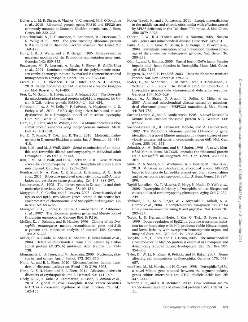
Figure 4 Disruption of mRpS33 causes cardiomyopathy. (A) Representa-tive OCT images of control w1118, transposon insertion PBac{WH}mRpS33f01766, tinC-Gal4 control, and UAS-RNAi against mRpS33 drivenby tinC-Gal4. (B) PBac{WH}mRpS33f01766, and heart-specific mRpS33knockdown have significant decrease in heart function compared to con-trols. n ¼ 8–16 per group shown in parentheses. *P , 0.05, **P , 0.01vs. w1118, one-way Anova with Dunnett Multiple Comparisons test.
Cardiomyopathy in Minute Drosophila 865
Dow
nloaded from https://academ
ic.oup.com/genetics/article/189/3/861/6063873 by guest on 21 D
ecember 2021
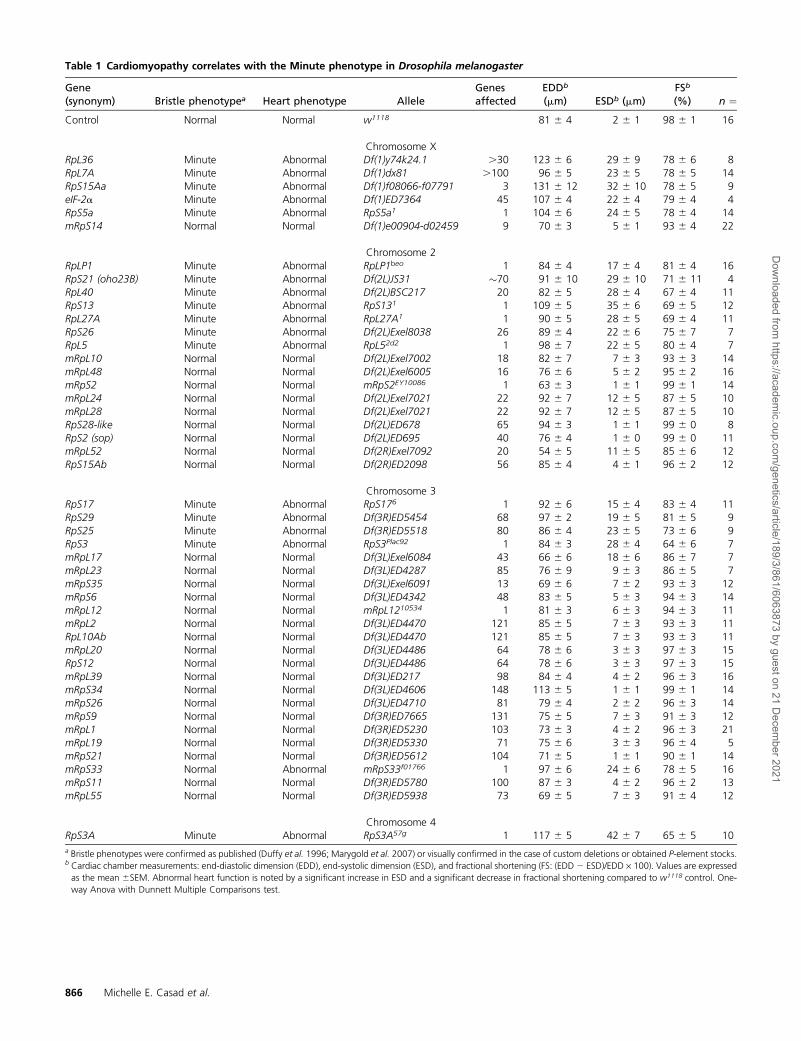
Table 1 Cardiomyopathy correlates with the Minute phenotype in Drosophila melanogaster
Gene(synonym) Bristle phenotypea Heart phenotype Allele
Genesaffected
EDDb
(mm) ESDb (mm)FSb
(%) n ¼Control Normal Normal w1118 81 6 4 2 6 1 98 6 1 16
Chromosome XRpL36 Minute Abnormal Df(1)y74k24.1 .30 123 6 6 29 6 9 78 6 6 8RpL7A Minute Abnormal Df(1)dx81 .100 96 6 5 23 6 5 78 6 5 14RpS15Aa Minute Abnormal Df(1)f08066-f07791 3 131 6 12 32 6 10 78 6 5 9eIF-2a Minute Abnormal Df(1)ED7364 45 107 6 4 22 6 4 79 6 4 4RpS5a Minute Abnormal RpS5a1 1 104 6 6 24 6 5 78 6 4 14mRpS14 Normal Normal Df(1)e00904-d02459 9 70 6 3 5 6 1 93 6 4 22
Chromosome 2RpLP1 Minute Abnormal RpLP1beo 1 84 6 4 17 6 4 81 6 4 16RpS21 (oho23B) Minute Abnormal Df(2L)JS31 �70 91 6 10 29 6 10 71 6 11 4RpL40 Minute Abnormal Df(2L)BSC217 20 82 6 5 28 6 4 67 6 4 11RpS13 Minute Abnormal RpS131 1 109 6 5 35 6 6 69 6 5 12RpL27A Minute Abnormal RpL27A1 1 90 6 5 28 6 5 69 6 4 11RpS26 Minute Abnormal Df(2L)Exel8038 26 89 6 4 22 6 6 75 6 7 7RpL5 Minute Abnormal RpL52d2 1 98 6 7 22 6 5 80 6 4 7mRpL10 Normal Normal Df(2L)Exel7002 18 82 6 7 7 6 3 93 6 3 14mRpL48 Normal Normal Df(2L)Exel6005 16 76 6 6 5 6 2 95 6 2 16mRpS2 Normal Normal mRpS2EY10086 1 63 6 3 1 6 1 99 6 1 14mRpL24 Normal Normal Df(2L)Exel7021 22 92 6 7 12 6 5 87 6 5 10mRpL28 Normal Normal Df(2L)Exel7021 22 92 6 7 12 6 5 87 6 5 10RpS28-like Normal Normal Df(2L)ED678 65 94 6 3 1 6 1 99 6 0 8RpS2 (sop) Normal Normal Df(2L)ED695 40 76 6 4 1 6 0 99 6 0 11mRpL52 Normal Normal Df(2R)Exel7092 20 54 6 5 11 6 5 85 6 6 12RpS15Ab Normal Normal Df(2R)ED2098 56 85 6 4 4 6 1 96 6 2 12
Chromosome 3RpS17 Minute Abnormal RpS176 1 92 6 6 15 6 4 83 6 4 11RpS29 Minute Abnormal Df(3R)ED5454 68 97 6 2 19 6 5 81 6 5 9RpS25 Minute Abnormal Df(3R)ED5518 80 86 6 4 23 6 5 73 6 6 9RpS3 Minute Abnormal RpS3Plac92 1 84 6 3 28 6 4 64 6 6 7mRpL17 Normal Normal Df(3L)Exel6084 43 66 6 6 18 6 6 86 6 7 7mRpL23 Normal Normal Df(3L)ED4287 85 76 6 9 9 6 3 86 6 5 7mRpS35 Normal Normal Df(3L)Exel6091 13 69 6 6 7 6 2 93 6 3 12mRpS6 Normal Normal Df(3L)ED4342 48 83 6 5 5 6 3 94 6 3 14mRpL12 Normal Normal mRpL1210534 1 81 6 3 6 6 3 94 6 3 11mRpL2 Normal Normal Df(3L)ED4470 121 85 6 5 7 6 3 93 6 3 11RpL10Ab Normal Normal Df(3L)ED4470 121 85 6 5 7 6 3 93 6 3 11mRpL20 Normal Normal Df(3L)ED4486 64 78 6 6 3 6 3 97 6 3 15RpS12 Normal Normal Df(3L)ED4486 64 78 6 6 3 6 3 97 6 3 15mRpL39 Normal Normal Df(3L)ED217 98 84 6 4 4 6 2 96 6 3 16mRpS34 Normal Normal Df(3L)ED4606 148 113 6 5 1 6 1 99 6 1 14mRpS26 Normal Normal Df(3L)ED4710 81 79 6 4 2 6 2 96 6 3 14mRpS9 Normal Normal Df(3R)ED7665 131 75 6 5 7 6 3 91 6 3 12mRpL1 Normal Normal Df(3R)ED5230 103 73 6 3 4 6 2 96 6 3 21mRpL19 Normal Normal Df(3R)ED5330 71 75 6 6 3 6 3 96 6 4 5mRpS21 Normal Normal Df(3R)ED5612 104 71 6 5 1 6 1 90 6 1 14mRpS33 Normal Abnormal mRpS33f01766 1 97 6 6 24 6 6 78 6 5 16mRpS11 Normal Normal Df(3R)ED5780 100 87 6 3 4 6 2 96 6 2 13mRpL55 Normal Normal Df(3R)ED5938 73 69 6 5 7 6 3 91 6 4 12
Chromosome 4RpS3A Minute Abnormal RpS3A57g 1 117 6 5 42 6 7 65 6 5 10a Bristle phenotypes were confirmed as published (Duffy et al. 1996; Marygold et al. 2007) or visually confirmed in the case of custom deletions or obtained P-element stocks.b Cardiac chamber measurements: end-diastolic dimension (EDD), end-systolic dimension (ESD), and fractional shortening (FS: (EDD 2 ESD)/EDD · 100). Values are expressedas the mean 6SEM. Abnormal heart function is noted by a significant increase in ESD and a significant decrease in fractional shortening compared to w1118 control. One-way Anova with Dunnett Multiple Comparisons test.
866 Michelle E. Casad et al.
Dow
nloaded from https://academ
ic.oup.com/genetics/article/189/3/861/6063873 by guest on 21 D
ecember 2021

the basis of these observations, we show that many Minutestocks exhibit cardiomyopathy in addition to the previouslydescribed characteristics that define the Minute phenotype.The Minute stocks screened included deletions of CRP genesas well as the one non-CRP gene eIF-2a (Marygold et al.2007). ManyMinute stocks have poor viability, and deletionsacross these Minute loci are often not available in many de-letion collections. Our results suggest that cardiac abnormal-ities may contribute to the poor viability of Minute flies.While we have not screened all the Minute loci (17 of 61loci were screened), we postulate that cardiomyopathy islikely a common phenotype of the Minute syndrome.
The mechanism for decreased cardiac function inMinutestocks is not known. Previous work has suggested thata specific balance of all subunits is needed in the ribosome
such that ribosome assembly and translation capacity isvery sensitive to the least abundant component available(Marygold et al. 2007). Recent work indicates that muta-tions in genes that specifically affect ribosomal assemblyresult in marked decrease of functional ribosomes (Freedet al. 2010). Prior work has also investigated possibleextra-ribosomal functions of some ribosomal proteins(Warner and Mcintosh 2009), as well as tissue specificribosomal activity (Kondrashov et al. 2011; Noben-Trauthand Latoche 2011). Alternatively, it has been proposed thatdifferences in basal expression of various ribosomal proteingenes could correspond with the differential sensitivity of thegenes to haplo-insufficiency (Marygold et al. 2007). There-fore, heart size and function may be sensitive to decreases inCRP-mediated translation, or subsets of CRPs could have
Figure 5 Cardiac-specific knockdown of ribo-somal proteins in the adult results in postdeve-lopmental dilated cardiac phenotype. (A)Representative OCT images of Drosophila con-taining the following transgenes: tub-Gal80ts,tinC-Gal4 drivers control; tub-Gal80ts, tinC-Gal4, and UAS-RNAi for RpS15Aa; tinC-Gal4and UAS-RNAi for RpS3; tub-Gal80ts, tinC-Gal4, and UAS-RNAi for RpS5a; tub-Gal80ts;tinC-Gal4 and UAS-RNAi for mRpS33. Flies de-veloped at 18� keeping the RNAi transgenefrom being expressed. Adult flies were eitherkept at 18� (no knockdown) or moved to 27�to turn on heart-specific knockdown in theadult. (B) Summary data showing that with driv-ers only, heart function remains normal whenadults are moved to 27�; however, heart func-tion is significantly impaired when RNAi expres-sion is induced in the adult heart. n ¼ 9–17 fliesper group shown in parentheses. *P , 0.05 vs.18� control, unpaired t-test. No error bars areshown on mRpS33 at 18� since all measure-ments of FS were 100%.
Cardiomyopathy in Minute Drosophila 867
Dow
nloaded from https://academ
ic.oup.com/genetics/article/189/3/861/6063873 by guest on 21 D
ecember 2021

heart-specific function as well. It is interesting that knock-down of CRPs in the adult fly can cause a postdevelopmentalphenotype, indicating that decreasing ribosomal subunit ex-pression in the adult fly can adversely affect heart function.The mechanism for the heart phenotype may be due to a de-crease in the translational capacity of cardiac cells, or as pre-viously mentioned, CRPs may have extra-ribosomal functionsimportant for the integrity of the heart.
MRPs are essential for cell growth and proliferation, andmutant alleles of several MRP genes have been character-ized in the fly, with defects in cell growth and development(Frei et al. 2005; Tselykh et al. 2005; Zhan et al. 2010). Inour screen, we observed that 28 of 29 disruptions of non-Minute CRP or MRP genes retained a normal adult cardiacfunction, representing 5 of 22 non-Minute CRPs and 24 of75 MRPs. While all of these genes are required to produceproteins for proper ribosome formation and protein trans-lation, only a subset of ribosomal genes cause a Minute phe-notype, including cardiomyopathy. mRpS33 is the onlyexception we identified in which a heterozygous deficiencyof a MRP gene causes significantly dilated cardiomyopathy.
Human CRP and MRP genes have been established ascandidates for causing human syndromes and diseases(Draptchinskaia et al. 1999; Ruggero and Pandolfi 2003;O’brien et al. 2005; Da Costa et al. 2010; Freed et al.2010; Ito et al. 2010; Narla and Ebert 2010). DiamondBlackfan anemia is associated with mutations in severalhaplo-insufficient CRP genes (Draptchinskaia et al. 1999;Da Costa et al. 2010; Ito et al. 2010) and is characterizedby congenital defects, including cardiac abnormalities (Itoet al. 2010; Doherty et al. 2010). Mutations in the geneencoding ribosomal protein RPS19 have been identified inapproximately 25% of Diamond Blackfan anemia familiesand haplo-insufficiency of several other CRP genes have sub-sequently been found in affected patients (Da Costa et al.2010). Studies in transgenic mice expressing a mutatedRPS19 gene suggest that one mechanism by which muta-tions in RPS19 can cause Diamond Blackfan anemia is by itseffect as a dominant negative protein (Devlin et al. 2010).
Other diseases that have decreased ribosomal biogenesisand function are commonly associated with increased sus-ceptibilities to cancer (Ruggero and Pandolfi 2003; Bilangesand Stokoe 2007; Montanaro et al. 2008; Narla et al. 2011).From human studies, disease-causing mutations often appearto disrupt the biogenesis of the ribosome, and thus mutationin one gene is able to greatly influence the translational ca-pacity of the cell. In humans, hematopoietic tissues seem tobe especially vulnerable to these ribosomopathies, whichcould be explained by the high proliferation rate in thesecells; however, the tissue specificity of these diseases of ubiq-uitous proteins is intriguing (Freed et al. 2010). In addition,several human MRP genes map to loci associated with disor-ders consistent with impaired oxidative phosphorylation(O’Brien et al. 2005). Mutations of human MRPS22 andMRP16 have been shown to cause severe disease in the ho-mozygous state (Miller et al. 2004; Saada et al. 2007; Smits
et al. 2010), showing that complete lack of a MRP is ex-tremely detrimental. Future investigation of human ribo-somal protein gene mutations, both in the heterozygousand homozygous states, may reveal insights into ribosomalbiology and cardiovascular disease.
Drosophila is a valuable model system for investigationinto human disease, including cardiomyopathy. Previouswork has found that alterations in structural and contractileproteins can alter contractility in the Drosophila heart (Wolfet al. 2006; Allikian et al. 2007; Taghli-Lamallem et al.2008). Additionally, signaling in such pathways as Notch,Rhomboid 3, SMAD, and insulin can change heart functionin flies (Wessells et al. 2004; Kim et al. 2010; Yu et al. 2010;Goldstein et al. 2011). In addition to the CRP genes that weidentify in this study, large-scale RNAi screening has identi-fied protein complexes important for Drosophila heart func-tion (Neely et al. 2010).
Our data support the concept that alterations in ribo-somal function can cause cardiac dysfunction as shown bythe marked cardiomyopathy in Drosophila with a Minutephenotype. Additional investigation is needed to addressthe mechanism underlying the sensitivity of cardiac tissuesin Drosophila to mutation in haplo-insufficient ribosomalprotein genes.
Acknowledgments
This work was supported by grants from the NationalInstitutes of Health HL-083965 to H.A.R., HL085072 toM.J.W, American Heart Association predoctoral fellowships0715314U and 09PRE2110019 to M.E.C., and AmericanHeart Association postdoctoral fellowship 0825499E toI.M.K.
Literature Cited
Alexander, S., N. Woodling, and B. Yedvobnick, 2006 Insertionalinactivation of the L13a ribosomal protein gene of Drosophilamelanogaster identifies a new Minute locus. Gene 368: 46–52.
Allikian, M. J., G. Bhabha, P. Dospoy, A. Heydemann, P. Ryder et al.,2007 Reduced life span with heart and muscle dysfunction inDrosophila sarcoglycan mutants. Hum. Mol. Genet. 16: 2933–2943.
Andersson, S., S. Saeboe-Larssen, A. Lambertsson, J. Merriam, andM. Jacobs-Lorena, 1994 A Drosophila third chromosome Min-ute locus encodes a ribosomal protein. Genetics 137: 513–520.
Bilanges, B., and D. Stokoe, 2007 Mechanisms of translationalderegulation in human tumors and therapeutic interventionstrategies. Oncogene 26: 5973–5990.
Da Costa, L., H. Moniz, M. Simansour, G. Tchernia, N. Mohandaset al., 2010 Diamond-Blackfan anemia, ribosome and erythro-poiesis. Transfus. Clin. Biol. 17: 112–119.
Devlin, E. E., L. Dacosta, N. Mohandas, G. Elliott, and D. M. Bodine,2010 A transgenic mouse model demonstrates a dominantnegative effect of a point mutation in the RPS19 gene associatedwith Diamond-Blackfan anemia. Blood 116: 2826–2835.
Dietzl, G., D. Chen, F. Schnorrer, K. C. Su, Y. Barinova et al.,2007 A genome-wide transgenic RNAi library for conditionalgene inactivation in Drosophila. Nature 448: 151–156.
868 Michelle E. Casad et al.
Dow
nloaded from https://academ
ic.oup.com/genetics/article/189/3/861/6063873 by guest on 21 D
ecember 2021
Doherty, L., M. R. Sheen, A. Vlachos, V. Choesmel, M. F. O’Donohueet al., 2010 Ribosomal protein genes RPS10 and RPS26 arecommonly mutated in Diamond-Blackfan anemia. Am. J. Hum.Genet. 86: 222–228.
Draptchinskaia, N., P. Gustavsson, B. Andersson, M. Pettersson, T.N. Willig et al., 1999 The gene encoding ribosomal proteinS19 is mutated in Diamond-Blackfan anaemia. Nat. Genet. 21:169–175.
Duffy, J. B., J. Wells, and J. P. Gergen, 1996 Dosage-sensitivematernal modifiers of the Drosophila segmentation gene runt.Genetics 142: 839–852.
Fauvarque, M., P. Laurenti, A. Boivin, S. Bloyer, R. Griffin-Sheaet al., 2001 Dominant modifiers of the polyhomeotic extra-sex-combs phenotype induced by marked P element insertionalmutagenesis in Drosophila. Genet. Res. 78: 137–148.
Freed, E. F., F. Bleichert, L. M. Dutca, and S. J. Baserga,2010 When ribosomes go bad: diseases of ribosome biogene-sis. Mol. Biosyst. 6: 481–493.
Frei, C., M. Galloni, E. Hafen, and B. A. Edgar, 2005 The Drosoph-ila mitochondrial ribosomal protein mRpL12 is required for Cy-clin D/Cdk4-driven growth. EMBO J. 24: 623–634.
Goldstein, J. A., S. M. Kelly, P. P. LoPresti, A. Heydemann, J. U.Earley et al., 2011 SMAD signaling drives heart and muscledysfunction in a Drosophila model of muscular dystrophy.Hum. Mol. Genet. 20: 894–904.
Hart, K., T. Klein, and M. Wilcox, 1993 A Minute encoding a ribo-somal protein enhances wing morphogenesis mutants. Mech.Dev. 43: 101–110.
Ito, E., Y. Konno, T. Toki, and K. Terui, 2010 Molecular patho-genesis in Diamond-Blackfan anemia. Int. J. Hematol. 92: 413–418.
Kim, I. M., and M. J. Wolf, 2009 Serial examination of an induc-ible and reversible dilated cardiomyopathy in individual adultDrosophila. PLoS One 4: e7132.
Kim, I. M., M. J. Wolf, and H. A. Rockman, 2010 Gene deletionscreen for cardiomyopathy in adult Drosophila identifies a newnotch ligand. Circ. Res. 106: 1233–1243.
Kondrashov, N., A. Pusic, C. R. Stumpf, K. Shimizu, A. C. Hsiehet al., 2011 Ribosome-mediated specificity in hox mRNA trans-lation and vertebrate tissue patterning. Cell 145: 383–397.
Lambertsson, A., 1998 The minute genes in Drosophila and theirmolecular functions. Adv. Genet. 38: 69–134.
Marygold, S., C. Coelho, and S. Leevers, 2005 Genetic analysis ofRpL38 and RpL5, two Minute genes located in the centric het-erochromatin of chromosome 2 of Drosophila melanogaster. Ge-netics 169: 683–695.
Marygold, S. J., J. Roote, G. Reuter, A. Lambertsson, M. Ashburneret al., 2007 The ribosomal protein genes and Minute loci ofDrosophila melanogaster. Genome Biol. 8: R216.
McKim, K., J. Dahmus, and R. Hawley, 1996 Cloning of the Dro-sophila melanogaster meiotic recombination gene mei-218:a genetic and molecular analysis of interval 15E. Genetics144: 215–228.
Miller, C., A. Saada, N. Shaul, N. Shabtai, E. Ben-Shalom et al.,2004 Defective mitochondrial translation caused by a ribo-somal protein (MRPS16) mutation. Ann. Neurol. 56: 734–738.
Montanaro, L., D. Trere, and M. Derenzini, 2008 Nucleolus, ribo-somes, and cancer. Am. J. Pathol. 173: 301–310.
Narla, A., and B. L. Ebert, 2010 Ribosomopathies: human disor-ders of ribosome dysfunction. Blood 115: 3196–3205.
Narla, A., S. N. Hurst, and B. L. Ebert, 2011 Ribosome defects indisorders of erythropoiesis. Int. J. Hematol. 93: 144–149.
Neely, G. G., K. Kuba, A. Cammarato, K. Isobe, S. Amann et al.,2010 A global in vivo Drosophila RNAi screen identifiesNOT3 as a conserved regulator of heart function. Cell 141:142–153.
Noben-Trauth, K., and J. R. Latoche, 2011 Ectopic mineralizationin the middle ear and chronic otitis media with effusion causedby RPL38 deficiency in the Tail-short (Ts) mouse. J. Biol. Chem.286: 3079–3093.
O’Brien, T. W., B. J. O’Brien, and R. A. Norman, 2005 NuclearMRP genes and mitochondrial disease. Gene 354: 147–151.
Parks, A. L., K. R. Cook, M. Belvin, N. A. Dompe, R. Fawcett et al.,2004 Systematic generation of high-resolution deletion cover-age of the Drosophila melanogaster genome. Nat. Genet. 36:288–292.
Qian, L., and R. Bodmer, 2009 Partial loss of GATA factor Pannierimpairs adult heart function in Drosophila. Hum. Mol. Genet.18: 3153–3163.
Ruggero, D., and P. P. Pandolfi, 2003 Does the ribosome translatecancer? Nat. Rev. Cancer 3: 179–192.
Ryder, E., M. Ashburner, R. Bautista-Llacer, J. Drummond, J.Webster et al., 2007 The DrosDel Deletion Collection: aDrosophila genomewide chromosomal deficiency resource.Genetics 177: 615–629.
Saada, A., A. Shaag, S. Arnon, T. Dolfin, C. Miller et al.,2007 Antenatal mitochondrial disease caused by mitochon-drial ribosomal protein (MRPS22) mutation. J. Med. Genet.44: 784–786.
Saeboe-Larssen, S., and A. Lambertsson, 1996 A novel DrosophilaMinute locus encodes ribosomal protein S13. Genetics 143:877–885.
Saeboe-Larssen, S., B. Urbanczyk Mohebi, and A. Lambertsson,1997 The Drosophila ribosomal protein L14-encoding gene,identified by a novel Minute mutation in a dense cluster of pre-viously undescribed genes in cytogenetic region 66D. Mol. Gen.Genet. 255: 141–151.
Schmidt, A., M. Hollmann, and U. Schafer, 1996 A newly iden-tified Minute locus, M(2)32D, encodes the ribosomal proteinL9 in Drosophila melanogaster. Mol. Gen. Genet. 251: 381–387.
Smits, P., A. Saada, S. B. Wortmann, A. J. Heister, M. Brink et al.,2010 Mutation in mitochondrial ribosomal protein MRPS22leads to Cornelia de Lange-like phenotype, brain abnormalitiesand hypertrophic cardiomyopathy. Eur. J. Hum. Genet. 19: 394–399.
Taghli-Lamallem, O., T. Akasaka, G. Hogg, U. Nudel, D. Yaffe et al.,2008 Dystrophin deficiency in Drosophila reduces lifespan andcauses a dilated cardiomyopathy phenotype. Aging Cell 7: 237–249.
Thibault, S. T., M. A. Singer, W. Y. Miyazaki, B. Milash, N. A.Dompe et al., 2004 A complementary transposon tool kit forDrosophila melanogaster using P and piggyBac. Nat. Genet. 36:283–287.
Torok, I., D. Herrmann-Horle, I. Kiss, G. Tick, G. Speer et al.,1999 Down-regulation of RpS21, a putative translation initia-tion factor interacting with P40, produces viable Minute imagosand larval lethality with overgrown hematopoietic organs andimaginal discs. Mol. Cell. Biol. 19: 2308–2321.
Tselykh, T. V., C. Roos, and T. I. Heino, 2005 The mitochondrialribosome-specific MrpL55 protein is essential in Drosophila anddynamically required during development. Exp. Cell Res. 307:354–366.
Tyler, D., W. Li, N. Zhuo, B. Pellock, and N. Baker, 2007 Genesaffecting cell competition in Drosophila. Genetics 175: 643–657.
van Beest, M., M. Mortin, and H. Clevers, 1998 Drosophila RpS3a,a novel Minute gene situated between the segment polaritygenes cubitus interruptus and dTCF. Nucleic Acids Res. 26:4471–4475.
Warner, J. R., and K. B. McIntosh, 2009 How common are ex-traribosomal functions of ribosomal proteins? Mol. Cell 34: 3–11.
Cardiomyopathy in Minute Drosophila 869
Dow
nloaded from https://academ
ic.oup.com/genetics/article/189/3/861/6063873 by guest on 21 D
ecember 2021

Wessells, R. J., E. Fitzgerald, J. R. Cypser, M. Tatar, and R. Bodmer,2004 Insulin regulation of heart function in aging fruit flies.Nat. Genet. 36: 1275–1281.
Wolf, M. J., H. Amrein, J. A. Izatt, M. A. Choma, M. C. Reedy et al.,2006 Drosophila as a model for the identification of genescausing adult human heart disease. Proc. Natl. Acad. Sci. USA103: 1394–1399.
Yin, Z., and M. Frasch, 1998 Regulation and function of tinmanduring dorsal mesoderm induction and heart specification inDrosophila. Dev. Genet. 22: 187–200.
Yu, L., T. Lee, N. Lin, and M. J. Wolf, 2010 Affecting Rhomboid-3function causes a dilated heart in adult Drosophila. PLoS Genet.6: e1000969.
Zhan, Y., N. Y. Melian, M. Pantoja, N. Haines, H. Ruohola-Bakeret al., 2010 Dystroglycan and mitochondrial ribosomal proteinl34 regulate differentiation in the Drosophila eye. PLoS One 5:e10488.
Communicating editor: T. C. Kaufman
870 Michelle E. Casad et al.
Dow
nloaded from https://academ
ic.oup.com/genetics/article/189/3/861/6063873 by guest on 21 D
ecember 2021