cardiac output monitoring - si.mahidol.ac.th · benefit of cardiac output monitoring diagnostic...
TRANSCRIPT

Cardiac output monitoringWhat the Fact !!
R2 Nachawan Gosiyaphant
R2 Chayaporn Subanphanichkul
Advisor : Aj.Chaowanan Khamtuikrua
Introduction
• Hemodynamic instability in patients who admitted in ICU causing a mismatch between oxygen delivery and demand that leading to multiple organ failure
• If basic vital parameters were failed there is an increased need for hemodynamic monitoring
ICU2016 Learning: Package Pulse Contour Cardiac Output Learning Package
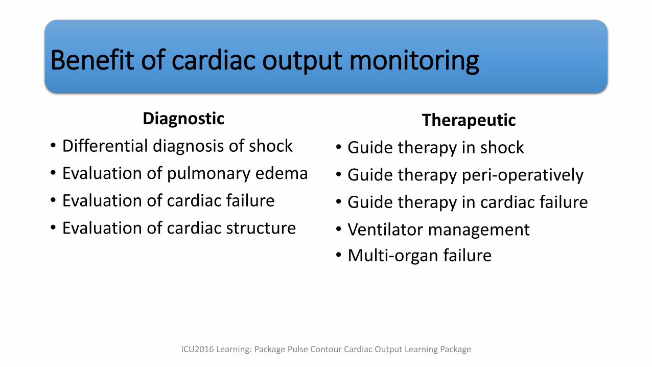
Benefit of cardiac output monitoring
Diagnostic
• Differential diagnosis of shock
• Evaluation of pulmonary edema
• Evaluation of cardiac failure
• Evaluation of cardiac structure
Therapeutic
• Guide therapy in shock
• Guide therapy peri-operatively
• Guide therapy in cardiac failure
• Ventilator management
• Multi-organ failure
ICU2016 Learning: Package Pulse Contour Cardiac Output Learning Package

History
• The first technique for measuring CO was described by Adolph Fick in 1870• Fick’s technique calculation of CO from measurements of the oxygen content
of arterial and mixed venous blood as well as oxygen consumption
• The Fick technique became the accepted standard for the measurement of CO
Cardiac Output Monitoring; Seminars in Cardiothoracic and Vascular Anesthesia 2010

• Fick principle
• CO = Cardiac Output• VO2 = Oxygen consumption (ml/min)• Ca = Oxygen concentration of arterial blood
• Cv = Oxygen concentration of mixed venous blood
Hemodynamic monitoring in the critically ill : an overview of current cardiac output monitoring methods ; F1000Reseach2016
History
• The dye dilution technique for CO determination is based on the work of Stewart in the late 19th century and later modified by Hamilton in the 1930s • Calculate CO by dividing the quantity of an injected indicator dye by the area
under the dilution curve measured downstream
• Dye dilution became an “accepted method of reference” in the measurement of CO
Cardiac Output Monitoring; Seminars in Cardiothoracic and Vascular Anesthesia 2010

History
• In the 1970’s by Swan, Ganz, and Forrester introduced pulmonary artery catheterization into clinical practice• Pulmonary artery catheters generate the thermodilution curves and analyze
them to determine CO
• Reliable, clinically relevant, and clinically useful
• “Gold” standard to which all new CO monitors are compared
• Several invasive and less-invasive methods have been developed during the last few decades to measure CO
Hemodynamic monitoring in the critically ill : an overview of current cardiac output monitoring methods ; F1000Reseach2016
Cardiac output monitoring
• Invasive method
• Less-invasive method
• Non-invasive method

Monitor Method Real time Calibrated
Invasive
Pulmonary artery catheter Thermodilution - +
Less-invasive
PiCCO® LiDCO® VolumeView®/EV1000®
TranspulmonaryThermodilution
+/- +
FloTrac®/Vigileo® Pulse contour and pulse pressure variation
+ -
NiCO® Partial CO2rebreathing + -
Transesophagealechocardiography
+ -
Esophageal Doppler + -

Monitor Method Real time Calibrated
Non-invasive
Transthoracic echocardiography
+ -
T-line® /Nexfin®/ CNAP® Non-invasive pulse contour systems
+ -
esCCO® Estimated continuous cardiac output
+ -
USCOM® Ultrasonic cardiac output monitoring
+ -

Cardiac output monitoring
• Invasive method
• Pulmonary artery catheter
• Less-invasive method
• Non-invasive method

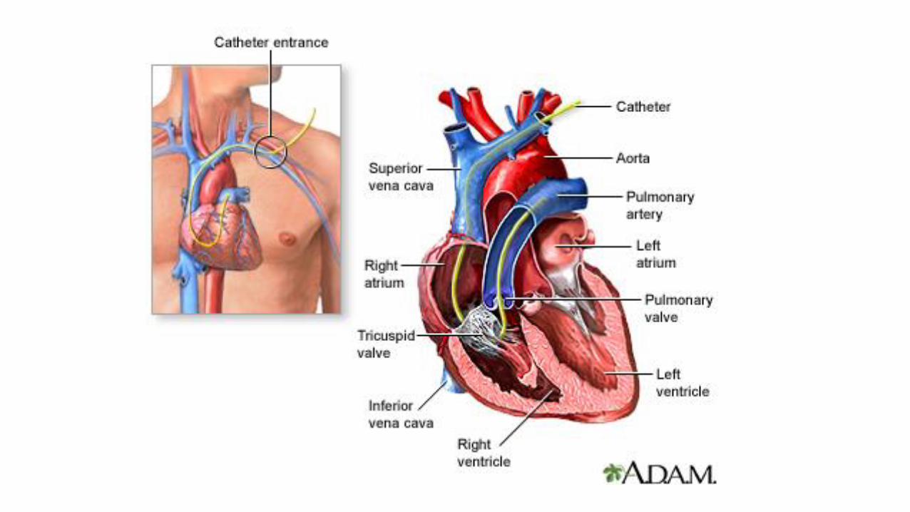
Pulmonary artery catheter
• Gold standard
• CO is measured with thermodilution
Hemodynamic monitoring in the critically ill : an overview of current cardiac output monitoring methods ; F1000Reseach2016
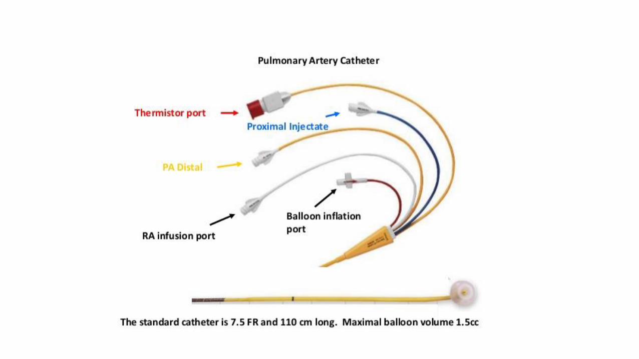


Factor Influencing Accuracy of Thermodilutioncardiac output measurement
• Intracardiac shunts
• Tricuspid or pulmonic valve regurgitation
• Inadequate delivery of thermal indicator
• Thermistor malfunction from fibrin or clot
• Pulmonary artery blood temperature fluctuations
• Respiratory cycle influences
Miller 8ed,2015, page 1,388

Cardiac output monitoring
• Invasive method
• Less-invasive method
• Transpulmonary thermodilution
• Pulse contour and pulse pressure variation
• Partial CO2 rebreathing
• Transesophageal echocardiography
• Esophageal doppler
• Non-invasive method
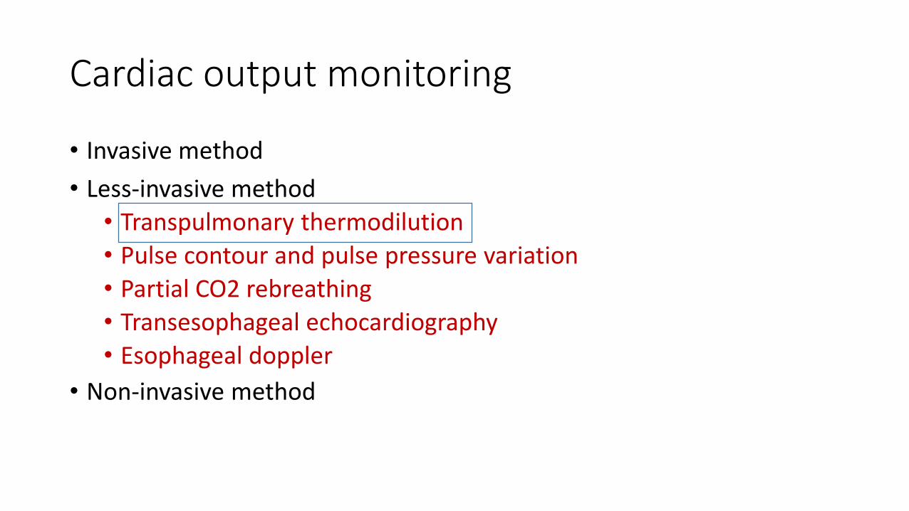
Cardiac output monitoring
• Invasive method
• Less-invasive method
• Transpulmonary thermodilution
• Pulse contour and pulse pressure variation
• Partial CO2 rebreathing
• Transesophageal echocardiography
• Esophageal doppler
• Non-invasive method

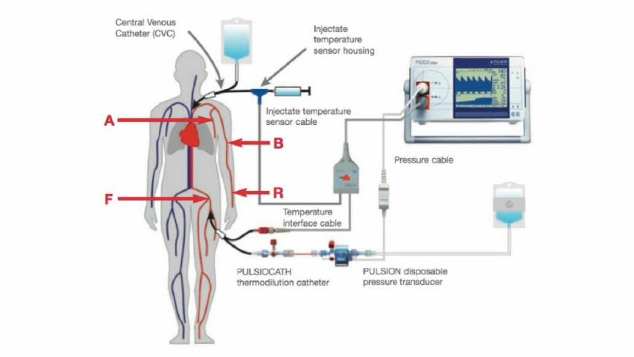
Transpulmonary thermodilutionPiCCO® system
• Use central venous catheter and arterial line with thermistor
• Injection of cold saline through a central line
• The arterial line measures the drop in blood temperature and from this obtain a Thermodilution Curve
• Use the area under thermodilution curve to calculate the CO
Hemodynamic monitoring in the critically ill : an overview of current cardiac output monitoring methods ; F1000Reseach2016
Transpulmonary thermodilutionVolume view® /EV1000 ®
• EV 1000 consist of PiCCO and FloTrac
• Volume view is an extra program that can measure Global end diastolic volume (GEDV)
Hemodynamic monitoring in the critically ill : an overview of current cardiac output monitoring methods ; F1000Reseach2016


Transpulmonary dye dilutionLiDCO® system
• LiDCO® system uses lithium as an intravascular indicator
• LiDCOrapid ® / PulseCO® The data are rapidly available and provide real-time beat-to-beat variations in CO by using pulse contour analysis system
Hemodynamic monitoring in the critically ill : an overview of current cardiac output monitoring methods ; F1000Reseach2016

Cardiac output monitoring
• Invasive method
• Less-invasive method
• Transpulmonary thermodilution
• Pulse contour and pulse pressure variation
• Partial CO2 rebreathing
• Transesophageal echocardiography
• Esophageal doppler
• Non-invasive method

Pulse contour and pulse pressure analysis
• FloTrac, Vigileo, ProAQT, Pulsioflex, LiDCOrapid, PulseCO
• Estimate CO from pulse pressure analysis, impedance, arterial compliance, SVR
• Lack accuracy in unstable patients
Hemodynamic monitoring in the critically ill : an overview of current cardiac output monitoring methods ; F1000Reseach2016

FloTrac®/Vigileo®
• Use PPV and vascular tone to calculate stroke volume and CO
Hemodynamic monitoring in the critically ill : an overview of current cardiac output monitoring methods ; F1000Reseach2016

Bersten and Soni’s” Oh's Intensive Care Manual”, 6th Edition
Cardiac output monitoring
• Invasive method
• Less-invasive method
• Transpulmonary thermodilution
• Pulse contour and pulse pressure variation
• Partial CO2 rebreathing
• Transesophageal echocardiography
• Esophageal doppler
• Non-invasive method

Partial CO2-rebreathing NiCO®
• Using CO2 instead of O2 as an indicator
• Measure the CO2 production by exhaled CO2 content by the respiratory minute volume
• Every three minutes, a partial rebreathing cycle should be started using a rebreathing loop, resulting in reduced CO2 elimination
• Difference between normal and rebreathing ratios are used to calculate CO
• Significant pulmonary disease can interfere with the measurements
Hemodynamic monitoring in the critically ill : an overview of current cardiac output monitoring methods ; F1000Reseach2016

Cardiac output monitoring
• Invasive method
• Less-invasive method
• Transpulmonary thermodilution
• Pulse contour and pulse pressure variation
• Partial CO2 rebreathing
• Transesophageal echocardiography
• Esophageal doppler
• Non-invasive method
Cardiac output monitoring
• Invasive method
• Less-invasive method
• Transpulmonary thermodilution
• Pulse contour and pulse pressure variation
• Partial CO2 rebreathing
• Transesophageal echocardiography
• Esophageal doppler
• Non-invasive method

Esophageal Doppler
• Flexible ultrasound probe
• The blood flow in the descending aorta is measured to determine stroke volume and CO
• Can provide real-time CO
Hemodynamic monitoring in the critically ill : an overview of current cardiac output monitoring methods ; F1000Reseach2016


Cardiac output monitoring
• Invasive method
• Less-invasive method
• Non-invasive method
• Transthoracic echocardiography
• Non-invasive pulse contour systems
• Estimated continuous cardiac output• Ultrasonic cardiac output monitoring

Cardiac output monitoring
• Invasive method
• Less-invasive method
• Non-invasive method
• Transthoracic echocardiography
• Non-invasive pulse contour systems
• Estimated continuous cardiac output• Ultrasonic cardiac output monitoring

Cardiac output monitoring
• Invasive method
• Less-invasive method
• Non-invasive method
• Transthoracic echocardiography
• Non-invasive pulse contour systems
• Estimated continuous cardiac output• Ultrasonic cardiac output monitoring
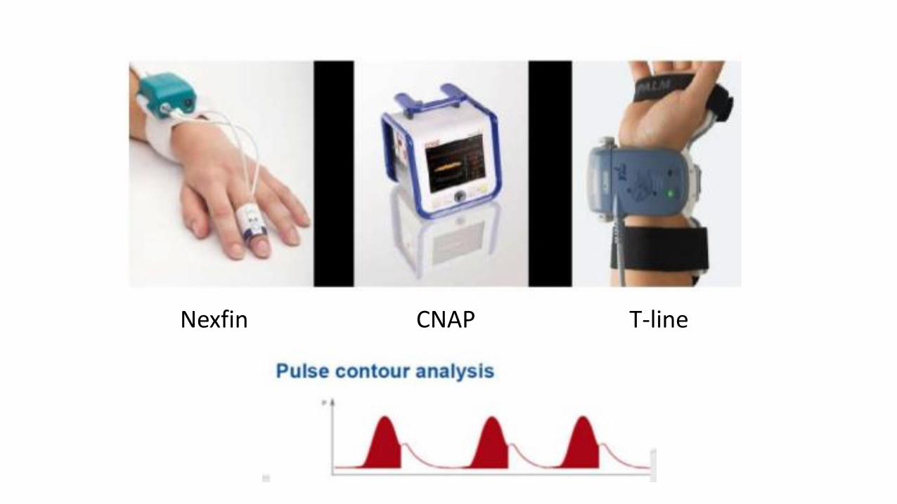
Nexfin CNAP T-line
Cardiac output monitoring
• Invasive method
• Less-invasive method
• Non-invasive method
• Transthoracic echocardiography
• Non-invasive pulse contour systems
• Estimated continuous cardiac output• Ultrasonic cardiac output monitoring

Estimated continuous cardiac output esCCO®
• Estimate the CO with an algorithm based on patient characteristics and measurement of HR, SpO2, NIBP
Hemodynamic monitoring in the critically ill : an overview of current cardiac output monitoring methods ; F1000Reseach2016

Pulse wave transit time
https://www.esahq.org


Cardiac output monitoring
• Invasive method
• Less-invasive method
• Non-invasive method
• Transthoracic echocardiography
• Non-invasive pulse contour systems
• Estimated continuous cardiac output• Ultrasonic cardiac output monitoring

Ultrasonic cardiac output monitoring USCOM®
• Measuring the flow velocity in the aortic and pulmonary outflow tracts
• Combines this with pre-calculated valve areas to estimate a CO
Hemodynamic monitoring in the critically ill : an overview of current cardiac output monitoring methods ; F1000Reseach2016

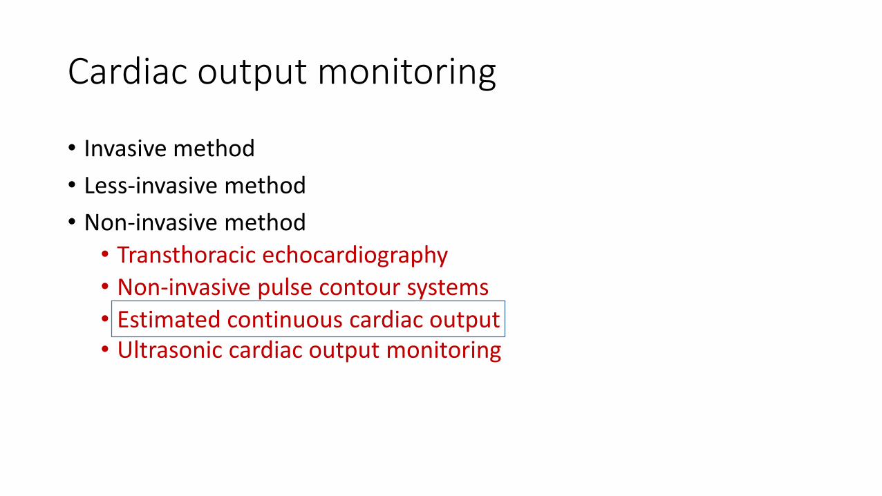
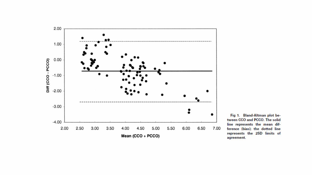
Design : Prospective, observational studyParticipants : Patients scheduled for elective CABGResult : Agreement between the CCO method and both less-invasive measurements was clinically acceptable. There were no adverse events associated with the use of either device.


Design : Prospective, observational single-center study.Participants : Patients with severe sepsis or septic shock requiring hemodynamic monitoring by VolumeView/EV1000 and receiving mechanical ventilationResult : Calibrated arterial pressure waveform analysis was more accurate and less dependent on vascular tone than uncalibrated method.

Take Home Message
• The thermodilution method is the clinical gold-standard for the measurement of CO
• Nowadays there are several less invasive method that had been developed for clinical use.
• Calibrated method is more accuracy than uncalibrated method.