canine hemangiosarcoma originates from hematopoietic precursors with potential for endothelial...
TRANSCRIPT

0
d
Experimental Hematology 34 (2006) 870–878
Canine hemangiosarcoma originates fromhematopoietic precursors with potential for endothelial differentiation
Angela R. Lamerato-Kozickia,*, Karen M. Helmb,Cristan M. Jubalaa, Gary C. Cutterc, and Jaime F. Modianoa,b
aDepartment of Immunology and AMC Cancer Research Center,
University of Colorado at Denver and Health Sciences Center; Denver, Colo., USA;bUniversity of Colorado Cancer Center, Denver, Colo., USA; cDepartment of Biostatistics, University of Alabama at Birmingham,
Birmingham, Ala., USA
(Received 16 September 2005; revised 27 March 2006; accepted 11 April 2006)
Objective. Two competing hypotheses can be formulated regarding the origin of caninehemangiosarcoma (HSA). One states HSA originates from differentiated vascular endothelialcells that undergo mutations which endow them with malignant potential. The other statesHSA originates from transformed hemangioblastic stem cells. This study was designed tobegin to distinguish between these possibilities, as well as to test if flow cytometry wassufficiently sensitive to detect malignant cells in blood samples from dogs with HSA.
Methods. We used multiparameter flow cytometry to examine expression of cell-surfacedeterminants associated with hematopoietic precursors (c-kit, CD34, CD133, CD45) or withlineage-committed cells (CD3, CD11b, CD14, CD21, CD105, CD146, avb3-integrin) in HSAcell lines and in blood samples from healthy dogs or dogs with HSA.
Results. The data show that HSA cells coexpress surface markers associated with hematopoi-etic precursors and with commitment to endothelial lineage, providing a means to identifytheir presence in circulation and distinguish them from normal or malignant white blood cells.The percentage of cells that coexpressed these markers ranged from 0.5 to 1.25% for HSAdogs, and was less than 0.3% for unaffected dogs or dogs with HSA that had the tumorsremoved within 48 hours prior to obtaining samples.
Conclusions. The results place the ontogeny of HSA with multipotential bone marrow–derived stem cells whose progeny arrest differentiation at the hemangioblast or angioblaststage. In addition, these expression patterns may assist to confirm an HSA diagnosis, monitorminimal residual disease, and detect the disease in early stages. � 2006 International Societyfor Experimental Hematology. Published by Elsevier Inc.
Hemangiosarcoma (HSA) is a common, aggressive malig-nant neoplasm of dogs that arises from vascular endothelialcells; however, its precise origins remain incompletelyunderstood. Two competing hypotheses can be formulatedregarding the ontogeny of HSA. One states that HSAoriginates from differentiated cells in the endothelial liningof blood vessels (i.e., cells that have matured to or past theangioblast stage) that undergo mutations which endowthem with malignant potential [1]. This assumes that anycell that can proliferate is equally susceptible to transforma-tion and retains the capacity for self-renewal. In this case,
Offprint requests to: Jaime F. Modiano, V.M.D., Ph.D., UCDHSC, 1600
Pierce Street, Denver, CO 80214; E-mail: [email protected]
*Current address: Department of Medical Sciences, School of Veteri-
nary Medicine, University of Wisconsin, Madison, WI.
301-472X/06 $–see front matter. Copyright � 2006 International Society fo
oi: 10.1016/j.exphem.2006.04.013
tumor development would be a stochastic process drivenby selection of mutations that favor proliferation andsurvival [2]. The competing hypothesis states that HSAoriginates from incompletely differentiated, bone marrow–derived, multipotential stem cells that are near or at thestage of endothelial commitment (i.e., hemangioblasts)[3]. Recent studies of leukemias, brain tumors, mesenchy-mal tumors, and mammary cancer support the concept of‘‘cancer stem cells’’ [2,4–7], and when considered in lightof the finding that early endothelial precursor cells migratefrom the bone marrow to peripheral vessels and may belargely responsible for physiologic and pathologic angio-genesis [3,8], it increases the plausibility that transforma-tion of such stem cells is responsible for HSA.
The mobilization of angiogenic progenitor cells in thebone marrow is mediated by matrix metalloproteinase-9,
r Experimental Hematology. Published by Elsevier Inc.

871A.R. Lamerato-Kozicki et al./ Experimental Hematology 34 (2006) 870–878
kit ligand, and vascular endothelial growth factor (VEGF)[9]. VEGF binding to VEGF receptor-2 (VEGFR-2)mediates a maturation cascade of endothelial commitmentprogressing from hemangioblast to angioblast to early andlate endothelial precursor cells (EPC) [3,9]. Hemangio-blasts are multipotent hematopoietic stem cells defined bythe expression of CD133, VEGFR-2, and possibly CD34and c-kit; they represent a small population with prolifera-tive potential capable to give rise to late endothelialoutgrowth [3,9]. EPC are virtually undetectable in thecirculation of normal individuals except by the most sensi-tive methods or by prolonged expansion in culture [9].
The course of hemangioblast differentiation remains in-completely understood: it has been proposed that these cellsgo through stages where they express the common leuko-cyte antigen CD45 and the myeloid antigen CD14, whichare downregulated in differentiated endothelial cells [9–12]; however, other investigators believe that monocytoidCD14þ cells that give rise to EPC represent a lineage thatis distinct from bone marrow hemangioblasts [13]. Thereis consensus, however, that mature endothelial cells com-monly express VEGFR, CD31, CD105, CD146, and FactorVIII–related antigen (vWF antigen). Both CD31 and vWFantigen are endothelial markers that are expressed in mostcases of canine HSA [14,15]. CD146 (Mel-CAM) is an ad-hesion molecule present on endothelial cells throughout thevascular tree [16]. CD105 (endoglin) is a receptor for thetransforming growth factors (TGF)-b1 and -b2, and isabundantly expressed on angiogenic endothelial cells[17]. In a previous study in our laboratory, stable expression
of CD146 and CD105 was confirmed in eight canine HSAcell lines after several passages [18]. Under conditions ofactivation that lead to proliferation, endothelial cells alsoupregulate avb3-integrin (vitronectin receptor) [19,20].
Our goal was to distinguish between the two alternativepossibilities of HSA ontogeny. Specifically, we surmisedthat the expression of markers associated with bone marrowprogenitor cells could distinguish EPC or EPC-like cells(hemangioblasts) from mature endothelial cells. Previouslywe demonstrated that HSA cells represented a ‘‘primitive’’lineage based on expression of c-kit [18]. Here, we confirmthat HSA originates from bone marrow progenitors at var-ious stages of hemangioblastic differentiation. In additionto their implications for a naturally occurring and accessi-ble model to study the biology of cancer stem cells, thesefindings offer added insights into the pathogenesis of canineHSA, they can be used for diagnostic purposes, and theymay be prognostically significant.
Materials and methods
Cell lines and tumor samplesHSA cell lines DD-1, Dal-4, CHAD-G4.1, CHAD-B7.4, Mocha-HSA, and Frog-HSA were derived and maintained as described[1,18]; Jurkat cells were also maintained as described previously[21]. Blood samples from 35 dogs, including 17 with biopsy-confirmed HSA, 16 that did not have HSA (9 healthy, 3 withleukemia, 3 with splenic hematoma, 1 with splenic nodularhyperplasia), and 2 with undiagnosed hemorrhagic effusions (peri-cardial, abdominal) were obtained with owner consent through
Table 1. Antibodies, source, and antigen specificities
Target CD antigen Clone Source and isotype Antigen specificity Vendor
CD3 CA17.2A12 Mouse IgG1 Canine CD3 Serotec1
CD5 YKIX322.3 Rat IgG2a Canine CD5 Serotec
CD45 YKIX716.13 Rat IgG2b Canine CD45 Serotec
CD11b CA16.3E10 Mouse IgG1 Canine CD11b Serotec
B cells CA2.1D6 Mouse IgG1 Canine CD212 Serotec
CD14 TuK4 Mouse IgG2a Human CD143 Serotec
CD34 2E9 Mouse IgG1 Canine CD34 BD Bioscience4
c-kit (CD117) YB5.B8 Mouse IgG1 Human c-kit5 BD Bioscience
CD133 13A4 Rat IgG1 Murine CD133 eBioscience4
CD146 P1H12 Mouse IgG1 Human CD1466 Chemicon7
avb3-integrin (CD51/61) LM609 Mouse IgG1 Human CD51/618 Chemicon
CD105 8E11 Mouse IgM Human CD1059 Southern Biotechnology
Associates10
1Raleigh, NC.2Precise identity of target antigen as CD21 has not been verified. Reported cross-reactivity by vendor includes horse and cat CD21 homologues.3Reported cross-reactivity by vendor includes dog CD14.4San Diego, CA.5Reported cross-reactivity by vendor includes dog c-Kit.6Reported cross-reactivity by vendor includes dog CD146.7Temecula, CA.8Reported cross-reactivity by vendor includes dog avb3-integrin.9Reported to cross-react with dog CD105 [16].10Birmingham, AL.

872 A.R. Lamerato-Kozicki et al. / Experimental Hematology 34 (2006) 870–878
a protocol reviewed by the Institutional Animal Care and UseCommittee and the Institutional Review Board of AMC CancerCenter.
Flow cytometry and immunofluorescenceFlow cytometric staining and immunofluorescence were done asdescribed [18,22]. Adherent cells were detached using 1 mMEDTA or Accutase (Innovative Cell Technologies, San Diego,CA, USA). Preservation of surface antigens (and thus signalstrength) was better with EDTA, but overall cellular viabilitywas higher with Accutase. Cell lines were stained with primaryantibodies described in Table 1, conjugated to fluorescein isothio-cyanate (FITC) or phycoerythrin (PE), or labeled with Zenonprobes (Alexa Fluor 488, Alexa-Fluor 647, or Alexa-Fluor APC,Invitrogen-Molecular Probes, Carlsbad, CA, USA), or with irrele-vant, isotype-matched antibody controls and analyzed using Coul-ter Cytomics FC500 (Beckman Coulter, Fullerton, CA, USA) orFACSCalibur (BD Immunocytometry Systems, San Jose, CA,USA) flow cytometers. We used a series of iterative steps to iden-tify circulating EPC or HSA cells in peripheral blood. First, weused single-color staining to define background levels for eachantibody and to verify that the relative numbers of leukocytes(CD21þ B cells, CD3þ and CD5þ T cells, CD14þ monocytes,and CD11bþ granulocytes) in our samples were within previouslyreported reference intervals [23–25]. Next, we used antibody com-binations for two-color staining. Color compensation was adjustedusing BD Biosciences CompBeads. Populations that stained posi-tive for one or more of three markers associated with bone marrowprogenitor cells (c-kit, CD34, CD133) and for a marker associatedwith endothelial cells (CD146) and/or proliferating endothelialcells (avb3-integrin) were ‘‘back-gated’’ to two-dimensional lightscatter dot plots to define the flow cytometric light scatter param-eters of HSA cells vs normal leukocytes. This procedure was usedfor all the samples included in the analyses; in addition, we further
modified the protocol for the last seven samples to exclude leuko-cytes by using antibodies against CD5, CD11b, and CD21 labeledwith FITC and/or Alexa Fluor 488 to establish a ‘‘dump gate,’’ andto detect EPC in the remaining population by dual-staining withantibodies against c-kit, CD34, or CD133 along with antibodiesagainst avb3-integrin or CD146. At least 100,000 cells were ana-lyzed for each antibody pair to ensure statistical validity for rare-event determination. A patent is pending for the assays to diagnoseangiosarcomas and hemangiosarcomas in samples from humansand animals, respectively.
StatisticsDescriptive statistics were used to characterize each subgroup inthe population. Nonparametric analyses (Kruskal-Wallace one-way analyses of variance with pairwise comparisons, using Dunn’smethod adjusted for three pairwise comparisons using a bonferroniadjusted p value of 0.01667) were used to determine if any twogroups were significantly different from each other. All analyseswere conducted using SAS Version 9.1 (SAS Institute, Cary,NC, USA).
ResultsTo define the ontogeny of HSA and to test the alternativehypothesis that this tumor arises from hemangioblasts, weexamined the expression of markers that would distinguishthese cells from other lineages of hematopoietic differenti-ation and from mature, differentiated leukocytes and vas-cular endothelial cells. We used flow cytometry andimmunofluorescence microscopy to examine expressionof surface proteins restricted to bone marrow precursorcells (e.g., c-kit, CD34, CD133) and proteins that define lin-eage commitment (CD3, CD21, CD11b, CD105, CD146,
Figure 1. Light scatter profiles of HSA cell lines. (A) Two-dimensional light scatter dot plots (FS vs SS) for five HSA cell lines analyzed using the Coulter
Cytomics FC500. Each axis has 1024 channels (0–1023). Events excluded from the gated regions represent dead cells and debris, or cell aggregates. The cell
lines Frog-HSA and Mocha-HSA are very early passage lines that show the pleomorphic features of the cells when they are first established in culture. DD-1,
Dal-4, and CHAD-B7 cells represent established lines that tend to be less pleomorphic. (B) Dal-4 cells were labeled with CFSE and mixed with human Jurkat
T leukemia cells at a ratio of 1 Dal-4 cell for every 100 Jurkat cells. Cells were analyzed using the FACSCalibur; the left dot plot shows right angle side
scatter (SSC-H) in 256 channels as a function of CFSE staining. On the right dot plot, CFSEþ cells were ‘‘backgated’’ onto the light scatter (FSC-H vs SSC-
H) dot plots to show the relative distribution of HSA cells as compared to mononuclear lymphoblasts.
873A.R. Lamerato-Kozicki et al./ Experimental Hematology 34 (2006) 870–878
avb3-integrin) in six HSA cell lines. We also evaluated ex-pression of CD14 and CD45, both of which are expressedby hemangioblastic cells at various stages of lineage com-mitment and differentiation [3,26,27]. Cultured HSA cellswere moderately pleomorphic, generating diffuse light scat-ter signatures (Fig. 1). The forward angle light scatter (FS)of HSA cells was larger than large mononuclear leukocytesand granulocytes; the right angle (side) light scatter (SS)was larger than that of mononuclear leukocytes and similarto that of granulocytes. In fact, when we labeled Dal-4 HSAcells with CFSE and mixed with Jurkat lymphoma cells atratios ranging from 1:1 to 1:100, the cells were distinguish-able from each other (Fig. 1).
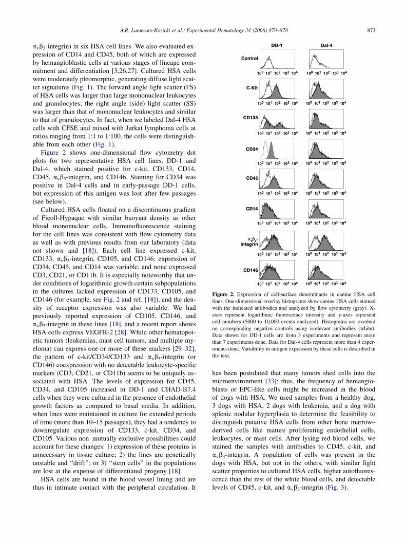
Figure 2 shows one-dimensional flow cytometry dotplots for two representative HSA cell lines, DD-1 andDal-4, which stained positive for c-kit, CD133, CD14,CD45, avb3-integrin, and CD146. Staining for CD34 waspositive in Dal-4 cells and in early-passage DD-1 cells,but expression of this antigen was lost after few passages(see below).
Cultured HSA cells floated on a discontinuous gradientof Ficoll-Hypaque with similar buoyant density as otherblood mononuclear cells. Immunofluorescence stainingfor the cell lines was consistent with flow cytometry dataas well as with previous results from our laboratory (datanot shown and [18]). Each cell line expressed c-kit,CD133, avb3-integrin, CD105, and CD146; expression ofCD34, CD45, and CD14 was variable, and none expressedCD3, CD21, or CD11b. It is especially noteworthy that un-der conditions of logarithmic growth certain subpopulationsin the cultures lacked expression of CD133, CD105, andCD146 (for example, see Fig. 2 and ref. [18]), and the den-sity of receptor expression was also variable. We hadpreviously reported expression of CD105, CD146, andavb3-integrin in these lines [18], and a recent report showsHSA cells express VEGFR-2 [28]. While other hematopoi-etic tumors (leukemias, mast cell tumors, and multiple my-eloma) can express one or more of these markers [29–32],the pattern of c-kit/CD34/CD133 and avb3-integrin (orCD146) coexpression with no detectable leukocyte-specificmarkers (CD3, CD21, or CD11b) seems to be uniquely as-sociated with HSA. The levels of expression for CD45,CD34, and CD105 increased in DD-1 and CHAD-B7.4cells when they were cultured in the presence of endothelialgrowth factors as compared to basal media. In addition,when lines were maintained in culture for extended periodsof time (more than 10–15 passages), they had a tendency todownregulate expression of CD133, c-kit, CD34, andCD105. Various non–mutually exclusive possibilities couldaccount for these changes: 1) expression of these proteins isunnecessary in tissue culture; 2) the lines are geneticallyunstable and ‘‘drift’’; or 3) ‘‘stem cells’’ in the populationsare lost at the expense of differentiated progeny [18].
HSA cells are found in the blood vessel lining and arethus in intimate contact with the peripheral circulation. It
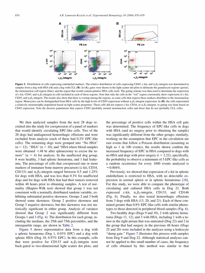
has been postulated that many tumors shed cells into themicroenvironment [33]; thus, the frequency of hemangio-blasts or EPC-like cells might be increased in the bloodof dogs with HSA. We used samples from a healthy dog,3 dogs with HSA, 2 dogs with leukemia, and a dog withsplenic nodular hyperplasia to determine the feasibility todistinguish putative HSA cells from other bone marrow–derived cells like mature proliferating endothelial cells,leukocytes, or mast cells. After lysing red blood cells, westained the samples with antibodies to CD45, c-kit, andavb3-integrin. A population of cells was present in thedogs with HSA, but not in the others, with similar lightscatter properties to cultured HSA cells, higher autofluores-cence than the rest of the white blood cells, and detectablelevels of CD45, c-kit, and avb3-integrin (Fig. 3).
Figure 2. Expression of cell-surface determinants in canine HSA cell
lines. One-dimensional overlay histograms show canine HSA cells stained
with the indicated antibodies and analyzed by flow cytometry (gray). X-
axes represent logarithmic fluorescence intensity and y-axes represent
cell numbers (5000 to 10,000 events analyzed). Histograms are overlaid
on corresponding negative controls using irrelevant antibodies (white).
Data shown for DD-1 cells are from 3 experiments and represent more
than 7 experiments done. Data for Dal-4 cells represent more than 4 exper-
iments done. Variability in antigen expression by these cells is described in
the text.
874 A.R. Lamerato-Kozicki et al. / Experimental Hematology 34 (2006) 870–878
Figure 3. Distribution of cells expressing endothelial markers. The relative distribution of cells expressing CD45, c-kit, and avb3-integrin was determined in
samples from a dog with HSA (A) and a dog with CLL (B). In (A), gates were drawn in the light scatter dot plots to delineate the granulocyte regions (green),
the mononuclear cell region (blue), and the region that would contain putative HSA cells (red). The gating scheme was then used to determine the expression
of c-kit, CD45, and avb3-integrin in cells included in each of those regions. Note that only the cells in the ‘‘red’’ region consistently show expression of c-kit,
CD45, and avb3-integrin. The results also show that there is overlap among the regions, as some cells that express these markers distribute to the mononuclear
region. Monocytes can be distinguished from HSA cells by the high levels of CD45 expression without avb3-integrin expression. In (B), the cells represented
a relatively monomorphic population based on light scatter properties. These cells did not express c-kit, CD34, or avb3-integrin, so gating was done based on
CD45 expression. Note the discrete populations that express CD45 (probably normal mononuclear cells) and those that do not (probably CLL cells).
We then analyzed samples from the next 28 dogs re-cruited into the study for coexpression of a panel of markersthat would identify circulating EPC-like cells. Two of the28 dogs had undiagnosed hemorrhagic effusions and wereexcluded from analysis (each of these had 0.2% EPC-likecells). The remaining dogs were grouped into ‘‘No HSA’’(n 5 12), ‘‘HSA’’ (n 5 10), and ‘‘HSA where blood sampleswere obtained !48 hr after splenectomy to excise the tu-mors’’ (n 5 4) for analysis. In the unaffected dog group,8 were healthy, 3 had splenic hematoma, and 1 had leuke-mia. The percentage of cells that coexpressed one or moremarkers of immature bone marrow precursors (c-kit, CD34,CD133) and avb3-integrin ranged between 0.5 and 1.25%for dogs with HSA, and was less than 0.3% for unaffecteddogs and for dogs with HSA that had their tumors removedwithin 48 hours prior to obtaining samples. A test of nor-mality (Shapiro-Wilk test) showed that group 1 was notconsistent with a normally distributed random variable, ex-hibiting substantial positive skewness. Groups 2 and 3 bothshowed some skewness: Group 2 positive skewness andGroup 3 negative skewness, but this skewness was not sta-tistically significant in either group. Analysis of varianceshowed that Group 2 was significantly different fromGroups 1 and 3 (Fig. 4). The distribution for each group, in-cluding the medians, the 25th and 75th percentiles, and theinterquartile range, are shown in Table 2.
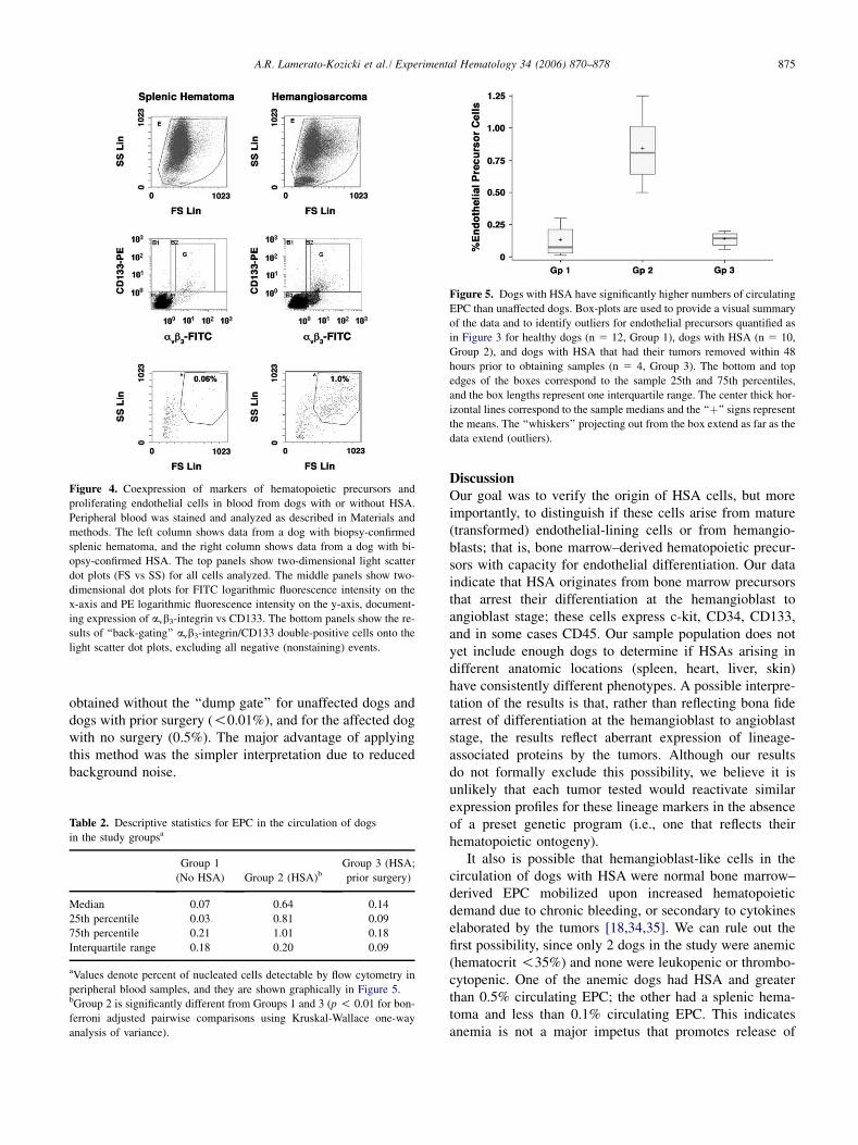
Figure 5 shows representative data from a dog witha splenic hematoma (Dog 1, 0.03% EPC) and a dog withsplenic HSA (Dog 18, 0.97% EPC). In this example, cellsthat were positive for CD133 and avb3-integrin wereback-gated to two-dimensional light scatter dot plots, and
the percentage of positive cells within the HSA cell gatewas determined. The frequency of EPC-like cells in dogswith HSA (and no surgery prior to obtaining the sample)was significantly different from the other groups; similarly,working on the assumption that EPC in the circulation arerare events that follow a Poisson distribution (assuming ashigh as 1 in 100 events), the results shown confirm theincreased frequency of EPC in HSA compared to dogs withno HSA and dogs with prior surgery (t 5 2.22); specifically,the probability to observe a minimum of 5 EPC-like cells asa random occurrence for every 1000 events analyzed isw0.004%.
Previously, we showed that expression of c-kit in splenicendothelium is restricted to HSA, with no detectable ex-pression in normal spleen or in splenic hematomas [18].For this study, we were able to compare the phenotype ofcirculating and cultured HSA cells in Dog 21. Bothexpressed c-kit, avb3-integrin, CD133, and CD146(Fig. 6). Finally, we also tested hemorrhagic effusionsfrom 3 dogs with HSA (13, 20, and 21). Each of these con-tained greater than 0.8% EPC-like cells with similar pheno-types to those detected in peripheral blood samples (Fig. 6).
Two healthy dogs (Dogs 9 and 10), 2 with splenic hema-toma (Dogs 11, 12), and 3 with HSA, including 1 with a tu-mor in the right atrium that was untreated (Dog 22) and 2 inthe group that had surgery in the previous 48 hours (Dogs25 and 26) were included in the analyses using a leukocyte‘‘dump gate.’’ Figure 7 illustrates this process with samplesfrom Dog 9 and Dog 22. Although statistical analyses can-not be applied to this small number of cases, the frequencyof cells obtained by this method was similar to that
875A.R. Lamerato-Kozicki et al./ Experimental Hematology 34 (2006) 870–878
obtained without the ‘‘dump gate’’ for unaffected dogs anddogs with prior surgery (!0.01%), and for the affected dogwith no surgery (0.5%). The major advantage of applyingthis method was the simpler interpretation due to reducedbackground noise.
Figure 4. Coexpression of markers of hematopoietic precursors and
proliferating endothelial cells in blood from dogs with or without HSA.
Peripheral blood was stained and analyzed as described in Materials and
methods. The left column shows data from a dog with biopsy-confirmed
splenic hematoma, and the right column shows data from a dog with bi-
opsy-confirmed HSA. The top panels show two-dimensional light scatter
dot plots (FS vs SS) for all cells analyzed. The middle panels show two-
dimensional dot plots for FITC logarithmic fluorescence intensity on the
x-axis and PE logarithmic fluorescence intensity on the y-axis, document-
ing expression of avb3-integrin vs CD133. The bottom panels show the re-
sults of ‘‘back-gating’’ avb3-integrin/CD133 double-positive cells onto the
light scatter dot plots, excluding all negative (nonstaining) events.
Table 2. Descriptive statistics for EPC in the circulation of dogs
in the study groupsa
Group 1
(No HSA) Group 2 (HSA)bGroup 3 (HSA;
prior surgery)
Median 0.07 0.64 0.14
25th percentile 0.03 0.81 0.09
75th percentile 0.21 1.01 0.18
Interquartile range 0.18 0.20 0.09
aValues denote percent of nucleated cells detectable by flow cytometry in
peripheral blood samples, and they are shown graphically in Figure 5.bGroup 2 is significantly different from Groups 1 and 3 (p ! 0.01 for bon-
ferroni adjusted pairwise comparisons using Kruskal-Wallace one-way
analysis of variance).
DiscussionOur goal was to verify the origin of HSA cells, but moreimportantly, to distinguish if these cells arise from mature(transformed) endothelial-lining cells or from hemangio-blasts; that is, bone marrow–derived hematopoietic precur-sors with capacity for endothelial differentiation. Our dataindicate that HSA originates from bone marrow precursorsthat arrest their differentiation at the hemangioblast toangioblast stage; these cells express c-kit, CD34, CD133,and in some cases CD45. Our sample population does notyet include enough dogs to determine if HSAs arising indifferent anatomic locations (spleen, heart, liver, skin)have consistently different phenotypes. A possible interpre-tation of the results is that, rather than reflecting bona fidearrest of differentiation at the hemangioblast to angioblaststage, the results reflect aberrant expression of lineage-associated proteins by the tumors. Although our resultsdo not formally exclude this possibility, we believe it isunlikely that each tumor tested would reactivate similarexpression profiles for these lineage markers in the absenceof a preset genetic program (i.e., one that reflects theirhematopoietic ontogeny).
It also is possible that hemangioblast-like cells in thecirculation of dogs with HSA were normal bone marrow–derived EPC mobilized upon increased hematopoieticdemand due to chronic bleeding, or secondary to cytokineselaborated by the tumors [18,34,35]. We can rule out thefirst possibility, since only 2 dogs in the study were anemic(hematocrit !35%) and none were leukopenic or thrombo-cytopenic. One of the anemic dogs had HSA and greaterthan 0.5% circulating EPC; the other had a splenic hema-toma and less than 0.1% circulating EPC. This indicatesanemia is not a major impetus that promotes release of
Figure 5. Dogs with HSA have significantly higher numbers of circulating
EPC than unaffected dogs. Box-plots are used to provide a visual summary
of the data and to identify outliers for endothelial precursors quantified as
in Figure 3 for healthy dogs (n 5 12, Group 1), dogs with HSA (n 5 10,
Group 2), and dogs with HSA that had their tumors removed within 48
hours prior to obtaining samples (n 5 4, Group 3). The bottom and top
edges of the boxes correspond to the sample 25th and 75th percentiles,
and the box lengths represent one interquartile range. The center thick hor-
izontal lines correspond to the sample medians and the ‘‘þ’’ signs represent
the means. The ‘‘whiskers’’ projecting out from the box extend as far as the
data extend (outliers).

876 A.R. Lamerato-Kozicki et al. / Experimental Hematology 34 (2006) 870–878
Figure 6. HSA cells in circulation and malignant effusions have similar properties to cultured HSA cells derived from the same tumor. Peripheral blood and
abdominal effusion from Dog 21 were analyzed as described in Figure 4. A cell line (FROG-HSA) established from a surgical biopsy of this dog’s tumor was
subsequently analyzed using the same parameters. The differences in quadrant gating are due to autofluorescence, especially by the cultured cells in the FITC
spectrum.
EPC into the circulation of dogs with HSA. We cannotcompletely exclude the possibility that VEGF and other cy-tokines released by the tumors mobilize EPC; however,there are at least two observations that make this unlikely.First, VEGF expression appears to be comparable in HSAand in splenic hematomas [1]. Nevertheless, HSA might re-lease other cytokines that contribute to release of EPC fromthe bone marrow. Second, the EPC had distinct cell-surfacephenotypes, but the patterns were not the same for each dogwith HSA. That is, cells might coexpress CD34 and avb3-integrin, c-kit and avb3-integrin, CD133 and avb3-integrin,or combinations thereof, but these patterns differed amongthe dogs. Although we were only able to compare the phe-notype of circulating EPC to that of the primary tumor inone case (we did not have fresh tumor specimens fromthe other dogs tested), these showed predictable similari-ties. In contrast, we believe that normal bone marrowEPC mobilized by tumor cytokines should share similarphenotypes among all dogs, rather than the different pheno-types we observed in these cases. Finally, CD133þ cellsrepresent the earliest identifiable multipotent cells in thehemangioblastic lineage, which is more consistent withrelease of tumor ‘‘stem cells’’ than with mobilization ofbone marrow EPC. Experiments are in progress to enrichthese populations by flow-sorting to assess their lineage dif-ferentiation potential in culture and to determine if theyharbor typical mutations of the PTEN C-terminal domainsor other gene expression profiles that may be peculiar tocanine HSA [1].
Ultimately, whether the cells originate from bone mar-row–derived stem cells that reside in the vascular endothe-lial lining or from bone marrow–derived stem cells thathome to blood vessels after transformation still remainsto be determined. Whichever the case, the nature of the tu-mor and the properties of the cells might offer an environ-ment that favors entry of cells into the circulation. Whilethis would help to explain the metastatic behavior ofHSA, it also might provide the ability to develop a diagnos-tically useful assay that could be similarly adapted to detect
circulating malignant hemangioblasts in human patients atrisk for developing angiosarcoma [18].
EPC in the circulation can increase under various physio-logic or pathologic conditions in humans and mice. Physio-logic conditions are mainly related to intense physicaltraining. Pathologic conditions include stem cell mobiliza-tion after cytokine administration and vascular injury(including myocardial infarcts and early congestive heartfailure, oxidative stress, and pulmonary hypertension) [9].
Figure 7. Coexpression of markers of hematopoietic precursors and
proliferating endothelial cells in blood from dogs with or without HSA.
Peripheral blood was analyzed as described in Figure 4, except that cells
labeled with CD5, CD21, and CD11b-FITC (red population) were
excluded from the analysis, and the middle panels show two-dimensional
dot plots for PE logarithmic fluorescence intensity on the x-axis and
Alexa-647 logarithmic fluorescence intensity on the y-axis, documenting
expression of c-kit vs avb3-integrin.

877A.R. Lamerato-Kozicki et al./ Experimental Hematology 34 (2006) 870–878
Nevertheless, even under pathologic conditions EPC in thecirculation generally remain well below 0.5%. Unlike hu-mans, risk factors that might elevate the number of circulat-ing EPC would be rare in dogs, with intense conditioning orearly congestive heart failure as the only common risk factorsthat might lead to increased numbers of EPC. Conversely, webelieve that HSA lesions would shed EPC-like cells into thecirculation continuously, meaning that a diagnostic proce-dure to detect EPC-like cells in excess of a normal referenceinterval would be a sensitive means to diagnose canine HSA(and possibly human angiosarcoma), even in early stages.
Our results suggest the assay might also be useful to1) identify malignant cells in effusions, which may be pre-dictive for the development of metastatic disease, 2) moni-tor remission and minimal residual disease (cells wereundetectable in the 4 dogs that had their tumors removedwithin 24 hours), and 3) allow detection of the disease inearly stages in at-risk patients in the prevention setting.The variable expression of CD45 and CD14 suggests thatHSA cells might attain different stages of differentiationthat affect response to therapy, although CD14 expressioncan be confounded by the overlapping morphology andlight scatter of monocyte-derived cells, EPC-like cells,and possibly mesenchymal stem cells. Given the smallnumber of dogs with HSA that achieve objective responsesand the suitability of this disease to model responses of hu-man patients with angiosarcoma [18], prospective trials toexplore these questions are warranted.
AcknowledgmentsWe wish to thank owners and veterinarians who contributed casesfor this project; Susan Fosmire, Lori Gardner, Stacie Bianco,Daniel Davila, and Evan Pushchak for technical help; MichaelAshton and Christine Childs for assistance with flow cytometry;and Drs. Susan Majka, Jonni Moore, Anne Avery, and Nao Ter-ada for helpful discussions. This work was supported by a reten-tion fund from the AMC Cancer Center, the Department ofImmunology, and the University of Colorado Cancer Center; bygrants from the Portuguese Water Dog Foundation, the Portu-guese Water Dog Club of America, the Starlight Fund, and Idexx,Inc.; and by a postdoctoral fellowship from the University of Col-orado Cancer Center to ARL.
References1. Dickerson EB, Thomas R, Fosmire SP, et al. Mutations of phosphatase
and tensin homolog deleted from chromosome 10 in canine hemangio-
sarcoma. Vet Pathol. 2005;42:618–632.
2. Frank SA, Nowak MA. Cell biology: Developmental predisposition to
cancer. Nature. 2003;422:494–494.
3. Schatteman GC, Awad O. Hemangioblasts, angioblasts, and adult en-
dothelial cell progenitors. Anat Rec A Discov Mol Cell Evol Biol.
2004;276:13–21.
4. Huntly BJ, Gilliland DG. Leukaemia stem cells and the evolution of
cancer-stem-cell research. Nat Rev Cancer. 2005;5:311–321.
5. Serakinci N, Guldberg P, Burns JS, et al. Adult human mesenchymal
stem cell as a target for neoplastic transformation. Oncogene. 2004;23:
5095–5098.
6. Singh SK, Hawkins C, Clarke ID, et al. Identification of human brain
tumour initiating cells. Nature. 2004;432:396–401.
7. Smith GH. Mammary cancer and epithelial stem cells: a problem or
a solution? Breast Cancer Res. 2002;4:47–50.
8. Ingram DA, Mead LE, Moore DB, Woodard W, Fenoglio A, Yoder
MC. Vessel wall derived endothelial cells rapidly proliferate because
they contain a complete hierarchy of endothelial progenitor cells.
Blood. 2005;105:2783–2786.
9. Hristov M, Weber C. Endothelial progenitor cells: characterization,
pathophysiology, and possible clinical relevance. J Cell Mol Med.
2004;8:498–508.
10. Kim SY, Park SY, Kim JM, et al. Differentiation of endothelial cells
from human umbilical cord blood AC133�CD14þ cells. Ann Hematol.
2005;84:417–422.
11. Quirici N, Soligo D, Caneva L, Servida F, Bossolasco P, Deliliers GL.
Differentiation and expansion of endothelial cells from human bone
marrow CD133þ cells. Br J Haematol. 2001;115:186–194.
12. Zhao Y, Glesne D, Huberman E. A human peripheral blood mono-
cyte–derived subset acts as pluripotent stem cells. Proc Natl Acad
Sci U S A. 2003;100:2426–2431.
13. Khan SS, Solomon MA, McCoy JP Jr. Detection of circulating endo-
thelial cells and endothelial progenitor cells by flow cytometry. Cy-
tometry B Clin Cytom. 2005;64:1–8.
14. Ferrer L, Fondevila D, Rabanal RM, Vilafranca M. Immunohisto-
chemical detection of CD31 antigen in normal and neoplastic canine
endothelial cells. J Comp Pathol. 1995;112:319–326.
15. von Beust BR, Suter MM, Summers BA. Factor VIII–related antigen
in canine endothelial neoplasms: an immunohistochemical study. Vet
Pathol. 1988;25:251–255.
16. Delorme B, Basire A, Gentile C, et al. Presence of endothelial progen-
itor cells, distinct from mature endothelial cells, within human
CD146þ blood cells. Thromb Haemost. 2005;94:1270–1279.
17. Warrington K, Hillarby MC, Li C, Letarte M, Kumar S. Functional
role of CD105 in TGF-b1 signalling in murine and human endothelial
cells. Anticancer Res. 2005;25:1851–1864.
18. Fosmire SP, Dickerson EB, Scott AM, et al. Canine malignant heman-
giosarcoma as a model of primitive angiogenic endothelium. Lab In-
vest. 2004;84:562–572.
19. Friedlander M, Brooks PC, Shaffer RW, Kincaid CM, Varner JA,
Cheresh DA. Definition of two angiogenic pathways by distinct av in-
tegrins. Science. 1995;270:1500–1502.
20. Soldi R, Mitola S, Strasly M, Defilippi P, Tarone G, Bussolino F. Role
of a-v/b-3 integrin in the activation of vascular endothelial growth fac-
tor receptor-2. EMBO J. 1999;18:882–892.
21. Frazer-Abel AA, Baksh S, Fosmire SP, et al. Nicotine activates
NFATc2 and prevents cell cycle entry in T cells. J Pharmacol Exp
Ther. 2004;311:758–769.
22. Jubala CM, Wojcieszyn JW, Valli VEO, et al. CD20 expression in nor-
mal canine B cells and in canine non-Hodgkin’s lymphoma. Vet Path-
ol. 2005;42:468–476.
23. Byrne KM, Kim HW, Chew BP, Reinhart GA, Hayek MG. A standard-
ized gating technique for the generation of flow cytometry data for
normal canine and normal feline blood lymphocytes. Vet Immunol Im-
munopathol. 2000;73:167–182.
24. Gauthier MJ, Aubert I, Abrams-Ogg A, Woods JP, Bienzle D. The im-
munophenotype of peripheral blood lymphocytes in clinically healthy
dogs and dogs with lymphoma in remission. J Vet Intern Med. 2005;
19:193–199.
25. Reis AB, Carneiro CM, Carvalho MG, et al. Establishment of a micro-
plate assay for flow cytometric assessment and it is use for the evalu-
ation of age-related phenotypic changes in canine whole blood
leukocytes. Vet Immunol Immunopathol. 2005;103:173–185.

878 A.R. Lamerato-Kozicki et al. / Experimental Hematology 34 (2006) 870–878
26. Dahlke MH, Lauth OS, Jager MD, et al. In vivo depletion of hemato-
poietic stem cells in the rat by an anti-CD45 (RT7) antibody. Blood.
2002;99:3566–3572.
27. Shaw JP, Basch R, Shamamian P. Hematopoietic stem cells and endo-
thelial cell precursors express Tie-2, CD31 and CD45. Blood Cells
Mol Dis. 2004;32:168–175.
28. MacDonald VS, Dickerson EB, Akhtar N, Helfand SC. Tyrosine
kinase blockade in canine hemangiosarcoma (Abstract). Vet Comp
Oncol. 2005;3:153.
29. Ria R, Vacca A, Ribatti D, Di Raimondo F, Merchionne F, Dammacco
F. a(v)b(3) integrin engagement enhances cell invasiveness in human
multiple myeloma. Haematologica. 2002;87:836–845.
30. Vacca A, Ria R, Presta M, et al. a(v)b(3) integrin engagement modu-
lates cell adhesion, proliferation, and protease secretion in human lym-
phoid tumor cells. Exp Hematol. 2001;29:993–1003.
31. London CA, Kisseberth WC, Galli SJ, Geissler EN, Helfand SC. Ex-
pression of stem cell factor receptor (c-kit) by the malignant mast cells
from spontaneous canine mast cell tumours. J Comp Pathol. 1996;115:
399–414.
32. Vernau W, Moore PF. An immunophenotypic study of canine leuke-
mias and preliminary assessment of clonality by polymerase chain re-
action. Vet Immunol Immunopathol. 1999;69:145–164.
33. Wong CW, Lee A, Shientag L, et al. Apoptosis: an early event in met-
astatic inefficiency. Cancer Res. 2001;61:333–338.
34. Akhtar N, Padilla M, Dickerson E, et al. Interleukin-12 inhibits tumor
growth in a novel angiogenesis canine hemangiosarcoma xenograft
model. Neoplasia. 2004;6:106–116.
35. Dickerson EB, Akhtar N, Steinberg H, et al. Enhancement of the anti-
angiogenic activity of interleukin-12 by peptide targeted delivery of
the cytokine to av/b3 integrin. Mol Cancer Res. 2004;2:663–673.