campylobacter-induced interleukin-8 responses in human intestinal epithelial cells and primary...
TRANSCRIPT

www.elsevier.com/locate/vetmic
Veterinary Microbiology 124 (2007) 115–124
Campylobacter-induced interleukin-8 responses in human
intestinal epithelial cells and primary intestinal chick cells
Erika Borrmann *, Angela Berndt, Ingrid Hanel, Heike Kohler
Friedrich-Loeffler-Institute, Institute of Molecular Pathogenesis, Naumburger Str. 96 a, D-07743 Jena, Germany
Received 15 December 2006; received in revised form 30 March 2007; accepted 4 April 2007
Abstract
Campylobacter (C.) jejuni and C. coli can cause gastrointestinal disorders in humans characterized by acute inflammation.
Inflammatory signals are initiated during interaction between these pathogens and human intestinal cells, but nothing is known
about the stimulation of avian intestinal cells by Campylobacter. Interleukin-8 (IL-8) as a proinflammatory chemokine plays an
important role in mobilizing cellular defence mechanism. IL-8 mRNA expression in both human intestinal cells (INT 407) and
primary intestinal chick cells (PIC) was determined by quantitative real-time RT-PCR. The secretion of IL-8 protein by INT407
was measured using ELISA. Although C. jejuni and C. coli are considered to be harmless commensals in the gut of birds, the
avian Campylobacter isolates investigated were able to induce the proinflammatory IL-8 in PIC as well as in INT407. In an in
vitro system, C. jejuni as well as C. coli were able to induce IL-8 mRNA in PIC. Relation between the virulence properties like
toxin production, the ability to invade and to survive in Caco-2 cells and the level of IL-8 mRNA produced by INT 407 and PIC
after infection with Campylobacter strains was also investigated.
# 2007 Elsevier B.V. All rights reserved.
Keywords: Campylobacter jejuni; Campylobacter coli; Interleukin-8; Human intestinal epithelial cells; Primary intestinal chick cells; Real-
time quantitative PCR
1. Introduction
Campylobacter (C.) jejuni and its close relative C.
coli are important human pathogens. They can cause
diseases such as gastroenteritis characterized by
severe inflammation of the intestinal mucosa with
an influx of professional phagocytes (Ketley, 1997;
* Corresponding author. Tel.: +49 3641 804 427;
fax: +49 3641 804 228.
E-mail address: [email protected] (E. Borrmann).
0378-1135/$ – see front matter # 2007 Elsevier B.V. All rights reserved
doi:10.1016/j.vetmic.2007.04.041
Altekruse et al., 1999; Jones et al., 2003). Campy-
lobacteriosis is described as a multifactorial process
involving the intake of the Campylobacter strains in
the gastrointestinal tract, followed by adherence to
intestinal epithelial cells, secretion of virulence
proteins and cell invasion (Raphael et al., 2005).
Epithelial cells are able to secrete chemotactic
mediators after contact with pathogenic bacteria as
described for Salmonella typhimurium, Helicobacter
pylori and others (Thorpe et al., 1999; Aubert et al.,
2000; Gewirtz et al., 2000; Backhed et al., 2003). They
.
E. Borrmann et al. / Veterinary Microbiology 124 (2007) 115–124116
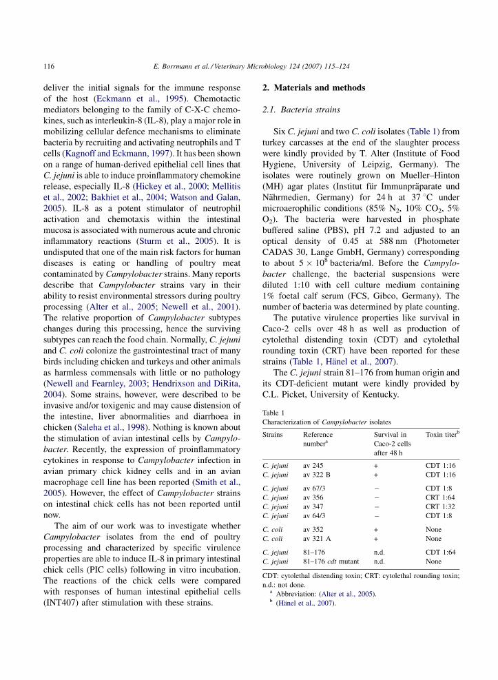
Table 1
Characterization of Campylobacter isolates
Strains Reference
numbera
Survival in
Caco-2 cells
after 48 h
Toxin titerb
C. jejuni av 245 + CDT 1:16
C. jejuni av 322 B + CDT 1:16
C. jejuni av 67/3 � CDT 1:8
C. jejuni av 356 � CRT 1:64
C. jejuni av 347 � CRT 1:32
C. jejuni av 64/3 � CDT 1:8
C. coli av 352 + None
C. coli av 321 A + None
C. jejuni 81–176 n.d. CDT 1:64
C. jejuni 81–176 cdt mutant n.d. None
CDT: cytolethal distending toxin; CRT: cytolethal rounding toxin;
n.d.: not done.a Abbreviation: (Alter et al., 2005).b (Hanel et al., 2007).
deliver the initial signals for the immune response
of the host (Eckmann et al., 1995). Chemotactic
mediators belonging to the family of C-X-C chemo-
kines, such as interleukin-8 (IL-8), play a major role in
mobilizing cellular defence mechanisms to eliminate
bacteria by recruiting and activating neutrophils and T
cells (Kagnoff and Eckmann, 1997). It has been shown
on a range of human-derived epithelial cell lines that
C. jejuni is able to induce proinflammatory chemokine
release, especially IL-8 (Hickey et al., 2000; Mellitis
et al., 2002; Bakhiet et al., 2004; Watson and Galan,
2005). IL-8 as a potent stimulator of neutrophil
activation and chemotaxis within the intestinal
mucosa is associated with numerous acute and chronic
inflammatory reactions (Sturm et al., 2005). It is
undisputed that one of the main risk factors for human
diseases is eating or handling of poultry meat
contaminated by Campylobacter strains. Many reports
describe that Campylobacter strains vary in their
ability to resist environmental stressors during poultry
processing (Alter et al., 2005; Newell et al., 2001).
The relative proportion of Campylobacter subtypes
changes during this processing, hence the surviving
subtypes can reach the food chain. Normally, C. jejuni
and C. coli colonize the gastrointestinal tract of many
birds including chicken and turkeys and other animals
as harmless commensals with little or no pathology
(Newell and Fearnley, 2003; Hendrixson and DiRita,
2004). Some strains, however, were described to be
invasive and/or toxigenic and may cause distension of
the intestine, liver abnormalities and diarrhoea in
chicken (Saleha et al., 1998). Nothing is known about
the stimulation of avian intestinal cells by Campylo-
bacter. Recently, the expression of proinflammatory
cytokines in response to Campylobacter infection in
avian primary chick kidney cells and in an avian
macrophage cell line has been reported (Smith et al.,
2005). However, the effect of Campylobacter strains
on intestinal chick cells has not been reported until
now.
The aim of our work was to investigate whether
Campylobacter isolates from the end of poultry
processing and characterized by specific virulence
properties are able to induce IL-8 in primary intestinal
chick cells (PIC cells) following in vitro incubation.
The reactions of the chick cells were compared
with responses of human intestinal epithelial cells
(INT407) after stimulation with these strains.
2. Materials and methods
2.1. Bacteria strains
Six C. jejuni and two C. coli isolates (Table 1) from
turkey carcasses at the end of the slaughter process
were kindly provided by T. Alter (Institute of Food
Hygiene, University of Leipzig, Germany). The
isolates were routinely grown on Mueller–Hinton
(MH) agar plates (Institut fur Immunpraparate und
Nahrmedien, Germany) for 24 h at 37 8C under
microaerophilic conditions (85% N2, 10% CO2, 5%
O2). The bacteria were harvested in phosphate
buffered saline (PBS), pH 7.2 and adjusted to an
optical density of 0.45 at 588 nm (Photometer
CADAS 30, Lange GmbH, Germany) corresponding
to about 5 � 108 bacteria/ml. Before the Campylo-
bacter challenge, the bacterial suspensions were
diluted 1:10 with cell culture medium containing
1% foetal calf serum (FCS, Gibco, Germany). The
number of bacteria was determined by plate counting.
The putative virulence properties like survival in
Caco-2 cells over 48 h as well as production of
cytolethal distending toxin (CDT) and cytolethal
rounding toxin (CRT) have been reported for these
strains (Table 1, Hanel et al., 2007).
The C. jejuni strain 81–176 from human origin and
its CDT-deficient mutant were kindly provided by
C.L. Picket, University of Kentucky.

E. Borrmann et al. / Veterinary Microbiology 124 (2007) 115–124 117
2.2. Cells
2.2.1. Human intestinal cell line INT407
Human intestinal epithelial cells (INT407; ECACC
No.: 85051004) were maintained in minimum essen-
tial medium (MEM, Sigma–Aldrich, Germany) with
non-essential amino acids (NEAA, Sigma–Aldrich)
supplemented with 10% FCS and 2 mM L-glutamine.
2.2.2. Primary intestinal chick cells (PIC)
Specific pathogen free (SPF) chickens (White Leg-
horn) were hatched at the facilities of the institute
from eggs received from Charles River Deutschland
GmbH (Extertal, Germany).
The isolation of the intestinal cells from these
chickens was performed as described by Athmann
et al. (2002). Briefly, the intestines from 1-day-old
chicks were removed and put in PBS with gentamicin
(150 mg/ml) and amphotericin B (25 mg/ml). The
intestines were slit open and cut into small fragments
which were intensively washed with Hanks balanced
salt solution, pH 7.4 (HBSS, Sigma–Aldrich) and then
sliced into smaller pieces. These pieces were digested
for 4 h at 37 8C using 300 units/ml collagenase
(Sigma–Aldrich) in HBSS. Afterwards, the cell
suspension was pipetted vigorously and left to
sediment under gravity for several times. Finally,
the supernatant was transferred in Dulbecco’s modi-
fied Eagle’s medium (DMEM, Sigma–Aldrich)
supplemented with 2.5% FCS and 2% sorbitol
(Sigma–Aldrich). After centrifugation at 100 � g
for 3 min the cell pellet was resuspended in MEM
with NEAA containing 2 mM L-glutamine, 5% FCS,
5% chick serum (Sigma–Aldrich), 0.5% chick embryo
extract (MP Biomedicals, Germany) and 120 mg/ml
sodium pyruvate. Cells were counted with a haemo-
cytometer and viability was assessed by trypan blue
exclusion. Cells (6 � 104 cells/cm2) were seeded in
culture flasks coated with collagen type II (Sigma–
Aldrich). The cell morphology was assessed by light
microscopy.
2.2.3. Immunohistochemistry
For immunohistochemical investigations, cells
(INT407, PIC) grown in chamber slides (Becton
Dickinson, United States) were prepared as described
previously (Berndt and Methner, 2001). Briefly,
monolayers were fixed with acetone and subsequently
incubated with a chick cross-reactive monoclonal
mouse anti human cytokeratin type II antibody (clone:
MCA888H; Serotec, Germany), secondary goat-anti
mouse immunoglobulin (Sigma–Aldrich) and peroxi-
dase-anti peroxidase complex (Sigma–Aldrich). The
enzyme-linked antibody was visualized by reaction
with 3,30-diaminobenzidine (Merck, Germany) and
hydrogen peroxide. As a negative control, slides were
incubated with normal mouse serum instead of the
primary monoclonal antibody. The cells were counter-
stained with haematoxylin and mounted with Canada
balsam (Riedel de Haen, Germany). The analysis of
cell staining was performed by light microscopy. The
percentage of cytokeratin positively stained cells was
calculated by counting positive and negative stained
cells of at least 100 cells per well.
2.3. Campylobacter challenge
Both cell types (INT407 and PIC) were grown to a
confluent monolayer in cell culture flasks, washed and
released using trypsin-EDTA (Sigma–Aldrich). The
cells were resuspended in the respective cell culture
medium and seeded at 0.5 ml per well in 24-well tissue
culture plates. For PIC cells plates were coated with
collagen type II. The cell numbers were 2 � 105 cells
per well for INT407 and 3 � 105 cells per well for
PIC. The cell monolayer was allowed to reform during
24 h incubation in a humidified atmosphere at 37 8Cand 5% CO2. After washing with PBS, the bacterial
suspensions were added to the cells at a MOI of about
1:100. Cell culture media and phorbol myristate
acetate (PMA, 10 ng/ml, Sigma–Aldrich) were used as
negative and positive controls, respectively. The 24-
well plates were incubated for 3 h at 37 8C and 5%
CO2, and afterwards the supernatants were removed.
Cells were washed two times with PBS and incubated
in fresh cell culture medium with 1% FCS. Finally, the
supernatants were harvested after total incubation
times of 4 h (3 h incubation of the cells with bacteria
and 1 h incubation with cell culture medium), 8 h
(3 h + 5 h) and 24 h (3 h + 21 h), filtered through 0.22-
mm pore-size syringe filters and stored at�80 8C until
analyzed for cytokine. The cell pellets were harvested
by addition of trypsin-EDTA. After resuspension in
cell culture medium, centrifugation at 300 � g for
5 min and washing two times with PBS, the cells were
stored at �80 8C until RNA isolation.
E. Borrmann et al. / Veterinary Microbiology 124 (2007) 115–124118
In preliminary tests the ability of PIC to produce
IL-8 mRNA was checked using lipopolysaccharide
(LPS) from E. coli 026:B6 (Sigma–Aldrich) in the
concentration range from 0.1 mg/ml to 100 mg/ml.
The isolated RNA from cell pellets was used as
positive control in further investigations. Since
INT407 did not react to LPS up to a concentration
of 400 mg/ml we used PMA as positive control for the
production of IL-8 mRNA.
2.4. RNA extraction and cDNA production
Total cell RNA was extracted using the RNeasy
mini kit (Qiagen, Germany) according to the
manufacturer’s instructions. The method was slightly
modified by inclusion of a DNA digestion step with
RNase-Free DNase (Qiagen, Germany). The quantity
and purity of the RNA samples were determined
spectrophotometrically by measuring the absorbance
at 260 nm and the 260/280 nm absorbance ratio
(Biophotometer, Eppendorf, Germany). The isolated
RNA was stored immediately at �80 8C. Before
starting the reverse transcription (RT), the isolated
RNA was adjusted to 0.05 mg/ml and finally denatured
for 5 min at 65 8C. RT was performed in 20 ml final
volume with OmniscriptR reverse transcriptase (Qia-
gen, Germany) in the presence of RNase inhibitor
(Promega, Germany) using random hexamer primer
pd(N)6 (Roche Diagnostics, Germany). The cDNA
was analyzed immediately or stored at �20 8C until
use.
2.5. Qualitative PCR
The qualitative PCR was performed in a 50 ml final
volume containing 25 ml Hot Star Taq master mix
(Qiagen, Germany) with 2.5 U Hot Star Taq Poly-
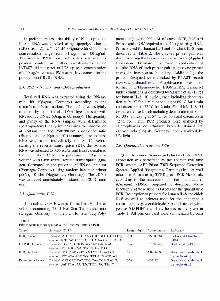
Table 2
Primer sequences for qualitative PCR and real-time RT-PCR
Target Sequence (50–30)
IL-8, human Forward: ATG ACT TCC AAG CTG GCC GTG
reverse: TCT CAG CCC TCT TCA AAA ACT TC
GAPDH, human Forward: TGG GTG TGA ACC ATG AGA AG;
reverse: GCT AAG CAG TTG GTG GTG C
IL-8, chicken Forward: ATG AAC GGC AAG CTT GGA GCT;
reverse: GCC ATA AGT GCC TTT ACG ATC AG
Beta-actin, chicken Forward: CAT CAC CAT TGG CAA TGA GAG G
reverse: GAT TCA TCG TAC TCC TGC TTG C
merase (Qiagen), 200 mM of each dNTP, 0.45 mM
Primer and cDNA equivalent to 15 ng starting RNA.
Primers used for human IL-8 and for chick IL-8 were
described in Table 2. The chicken primer pair was
designed using the Primers express software (Applied
Biosystems, Germany). To avoid amplification of
cellular DNA of each primer pair, at least one primer
spans an intron-exon boundary. Additionally, the
primers designed were checked by BLAST search
(www.ncbi.nlm.nih.gov) Amplification was per-
formed in a Thermocycler (BIOMETRA, Germany)
under conditions as described by Sharma et al. (1995)
for human IL-8: 30 cycles, each including denatura-
tion at 94 8C for 1 min, annealing at 60 8C for 1 min
and extension at 72 8C for 2 min. For chick IL-8, 35
cycles were used, each including denaturation at 94 8Cfor 30 s, annealing at 57 8C for 30 s and extension at
72 8C for 1 min. PCR products were analyzed by
electrophoresis on ethidium bromide stained 2%
agarose gels (Peqlab, Germany) and visualized by
UV-light.
2.6. Quantitative real-time PCR
Quantification of human and chicken IL-8 mRNA
expression was performed by the Taqman real time
PCR system (ABI Prism 7000 Sequence Detection
System, Applied Biosystems, Germany) in a 96-well
microtiter format using SYBR green PCR Mastermix
according to the instructions of the manufacturer
(Quiagen). cDNA’s prepared as described above
(Section 2.4) were used as targets for the quantitative
PCR. Description of primers for human IL-8 and chick
IL-8 as well as primers used for the endogenous
control genes glyceraldehyde-3-phosphate-dehydro-
genase (GAPDH) and chick beta-actin are given in
Table 2. All primers used were synthesized by Jena
Length (bp) Accession no. Reference
GCT;
T C
289 NM000584 Sarkar and Chaudhuri
(2004)
76 BC029340 Brink et al. (2000)
281 AJ009800 Berndt et al. (submitted
for publication)
; 354 X00182 Berndt et al. (submitted
for publication)
E. Borrmann et al. / Veterinary Microbiology 124 (2007) 115–124 119
Bioscience (Jena, Germany). IL-8 and the respective
endogenous control genes GAPDH or beta-actin were
amplified in separate wells of 96-well plates. The PCR
reactions contained either 300 nM of each primer for
human IL-8 and human GAPDH or 450 nM for chick
IL-8 and chick beta-actin and commercially available
PCR Mastermix (QuantiTectTM SYBR green PCR,
Qiagen, Germany) which includes SYBR Green I as a
fluorescent reporter and Rox as internal reference for
normalization of the fluorescence signal. A 7.5 ng
cDNA template was analyzed per reaction. In every
run, three no-template controls (DEPC-water) were
included. Amplification conditions were 2 min at
50 8C, 15 min at 95 8C, followed by 35 cycles of 15 s
at 94 8C, 30 s at 60 8C and 30 s at 72 8C. For the chick
reactions 40 cycles were carried out and the annealing
temperature was 61 8C. Subsequently, melting curve
analysis was performed to measure the specificity of
amplification. Final quantification was done using the
comparative Ct-method and is reported as relative
transcription or the n-fold difference relative to a
calibrator cDNA, here non-infected cells (Leuteneg-
ger et al., 2000; Sarkar and Chaudhuri, 2004). The
threshold cycle number (Ct) of triplicate reactions was
calculated using the ABI Prism software 1.23 (relative
quantification). The levels of IL-8 expression were
normalized to GAPDH and beta-actin, respectively,
using the formula 2�DDCt in which �DDCt = DCt
(sample) � DCt (calibrator) with DCt as difference
between Ct of target gene (IL-8) and Ct of house-
keeping gene (GAPDH or beta-actin). For the
comparative Ct-method the amplification efficiencies
of the target and the endogenous control were
estimated. For this, dilution steps of cDNA prepara-
tions in triplicate were amplified to obtain standard
curves and the slopes of curves for IL-8 and the
housekeeping gene were compared. An amount of
0.0075 ng cDNA was detectable for both human and
chick IL-8.
2.7. Determination of human IL-8 protein by
ELISA
The determination of IL-8 protein in cell super-
natants was performed using a commercially available
ELISA following the manufacturer’s instructions
(Biosource, CA, USA). Briefly, the supernatants and
the standard in serial dilution were pipetted in wells of
anti-human-IL-8 monoclonal antibody coated plates
and a biotinylated antibody was immediately added
to all wells. After incubation of 2 h and intensive
washing steps streptavidin-horseradish peroxidase
conjugate was added. Tetramethyl benzidine served
as substrate reagent. The absorbance was measured at
450 nm using an ELISA reader (Tecan, Germany)
and the amount of IL-8 protein was calculated in
comparison to the standard by the reader software.
The results from at least three independent tests are
presented as means � standard deviation (S.D.).
Statistical analyses of the comparison between control
and samples as well as between C. coli and C. jejuni
were performed by the Kruskal–Wallis- and Mann–
Whitney-test. A P value of <0.05 was considered
statistically significant.
3. Results
3.1. Comparison of the growth of PIC and INT407
The cultured PIC grew in a fibroblast-like manner
characterized by their elongated and spindle shape.
Compared to the INT407, the PIC showed a lower
tendency to confluent growth. The PIC could be
cultured without loss of viability or morphological
organization up to 10 passages.
The structure of the cells was determined using a
chick cross-reactive monoclonal mouse anti human
cytokeratin type II antibody and compared with the
human epithelial cells of INT407 (Fig. 1). About 90%
of PIC cells and about 70% of INT407 cells were
positively stained for cytokeratin. Light microscopy of
PIC revealed a moderate staining intensity and a
characteristic cytoplasmic network of cytokeratin
fibres indicating epithelial properties of the primary
cells. The reason for the moderate staining intensity
could be the use of a chick cross-reactive anti human
antibody. In comparison, the cytokeratin staining of
INT407 cells was more intensive and showed a more
diffuse character. The primary cells can be considered
as a culture with epithelial properties.
3.2. TaqMan amplification efficiencies
The DDCt method for relative quantification
requires that efficiencies of target and endogenous

E. Borrmann et al. / Veterinary Microbiology 124 (2007) 115–124120
Fig. 2. A representative agarose gel visualized by ethidium bro-
mide. The gel shows the expression of human-IL-8 (upper band) and
the control (GAPDH) genes (lower band) in INT407 and the
expression of chick IL-8 (lower band) and the control (beta-actin)
genes (upper band) in PIC cells after incubation with C. coli av 352
at 8 h total incubation time as determined by gene-specific co-
amplification of the genes by RT-PCR.
Fig. 1. Immunohistochemical staining of INT407 (A) and PIC (B) for cytokeratin II (brown). A typical pattern of keratin fiber staining can be
seen in the cytoplasm of PIC. INT407 cells show a more diffuse staining pattern.
control amplified in different wells are approximately
equal. Therefore, 10-fold dilution steps of human and
chick IL-8 cDNA were amplified in triplicate. The
standard curves calculated run approximately parallel
and the differences of the slopes between curves
obtained from GAPDH and human IL-8 was 0.04 and
for beta-actin and chick IL-8 was 0.05.
3.3. IL-8 mRNA expression in PIC and INT407
after infection with Campylobacter isolates and
PMA
The ability of PIC cells to produce IL-8 mRNA was
checked using lipopolysaccharide (LPS) from E. coli
026:B6 in different concentrations. Since the expres-
sion of IL-8 mRNA could be induced by LPS, it was
possible to compare the effects of avian Campylo-
bacter isolates on human and chick intestinal cells
with regard to IL-8 mRNA expression.
All Campylobacter isolates tested were able to
induce IL-8 mRNA in PIC as well as in INT 407.
Representative results of IL-8 mRNA expression
after 8 h total incubation time are shown in Fig. 2.
Campylobacter strains adhered and invaded both
human INT 407 and PIC to the same extent after 3-h-
postinfection (data not shown). Both cell types
produced significantly higher IL-8 mRNA levels in
comparison to non-infected cells at a total incubation
time of 4 h (Figs. 3 and 4). The C. jejuni av 322 B
induced the highest level of mRNA from all C. jejuni
isolates investigated in INT407 at 8 h and in PIC at
4 h. In PIC a time-independent production for all
isolates apart from av 322 B was determined. Six from
eight isolates produced the highest amount of IL-8
mRNA after 4 h. Marked differences between C.
jejuni and C. coli in the time-dependent course and the
amount of IL-8 mRNA were not determined. In
INT407, however, a time-dependent production of IL-
8 mRNA was detected for both C. coli strains (av 352
and av 321 A). The infection of INT407 with these
strains resulted in an increase in the amount of IL-8
mRNA from 4 h to 24 h, while the C. jejuni strains
E. Borrmann et al. / Veterinary Microbiology 124 (2007) 115–124 121
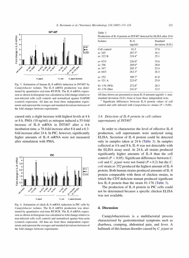
Fig. 3. Estimation of human IL-8 mRNA induction in INT407 by
Campylobacter isolates. The IL-8 mRNA production was deter-
mined by quantitative real-time RT-PCR. The IL-8 mRNA expres-
sion as shown in histogram was calculated as fold change relative to
non-infected cells (cell control) and normalized against GAPDH
(control) expression. All data are from three independent experi-
ments and represent the averages and standard deviations between of
the fold changes between experiments.
Table 3
Production of IL-8 protein in INT407 detected by ELISA after 24 h
Isolates IL-8
(pg/ml)
Standard
deviation (S.D.)
Cell control 91.5 37.8
av 245 287.5a 16.1
av 322 B 219.4a 25.1
av 67/3 226.0a 35.6
av 356 269.0a 30.0
av 347 205.3a 52.4
av 64/3 263.3a 26.3
av 352 309.7a 41.8
av 321 A 223.0a 25.0
81–176 (WS) 272.8a 20.3
81–176 (Mu) 241.6a 12.5
All data shown are presented as mean IL-8 amounts (pg/ml) � stan-
standard deviation (S.D.) from at least three independent tests.a Significant differences between IL-8 protein values of cell
control and cells infected with Campylobacter strains (P < 0.05).
caused only a slight increase with highest levels at 4 h
or 8 h. PMA (10 ng/ml) as mitogen induced a 55-fold
increase of IL-8 mRNA in INT407 after a 4-h
incubation time, a 78-fold increase after 8 h and a 6.3-
fold increase after 24 h. In PIC, however, significantly
higher amounts of IL-8 mRNA were not measured
after stimulation with PMA.
Fig. 4. Estimation of chick IL-8 mRNA induction in PIC cells by
Campylobacter isolates. The IL-8 mRNA production was deter-
mined by quantitative real-time RT-PCR. The IL-8 mRNA expres-
sion as shown in histogram was calculated as fold change relative to
non-infected cells (cell control) and normalized against beta-actin
(control) expression. All data are from three independent experi-
ments and represent the averages and standard deviations between of
the fold changes between experiments.
3.4. Detection of IL-8 protein in cell culture
supernatants of INT407
In order to characterize the level of effective IL-8
production, cell supernatants were analyzed using
ELISA. Secretion of IL-8 protein could be detected
only in samples taken at 24 h (Table 3). In samples
collected at 4 h and 8 h, IL-8 was not detectable with
the ELISA assay used. At 24 h, all strains produced
significantly higher amounts of IL-8 than the cell
control (P < 0.05). Significant differences between C.
coli and C. jejuni were not found (P = 0.2) but the C.
coli strain av 352 produced the highest amount of IL-8
protein. Both human strains produced amounts of IL-8
protein comparable with them of chicken strains, in
which the CDT-deficient mutant produced significant
less IL-8 protein than the strain 81-176 (Table 3).
The production of IL-8 protein in PIC cells could
not be determined because a specific chicken ELISA
was not available.
4. Discussion
Campylobacteriosis is a multifactorial process
characterized by gastrointestinal symptoms such as
diarrhoea, cramping, abdominal pain, and fever. A
hallmark of this human disorder caused by C. jejuni or

E. Borrmann et al. / Veterinary Microbiology 124 (2007) 115–124122
C. coli is the inflammation of the intestinal mucosa.
These pathogens are able to initiate inflammatory
signals by interaction (adhesion and/or invasion)
with host cells and activate signalling pathways that
induce the production of inflammatory cytokines and
recruitment of phagocytes, especially neutrophils, into
infected tissue (Jones et al., 2003; Everest, 2005). The
function of chemotactic cytokines (chemokines) such
as IL-8 produced by human cells after contact with
pathogens is described as crucial for acute inflam-
matory diseases. IL-8 and other proinflammatory
mediators are important in intiating the host mucosal
inflammatory response, which is critical for both the
induction of diarrhoea and the clearance of infection
(Watson and Galan, 2005). However, in the vast
majority of animals colonised with C. jejuni or C. coli
intestinal inflammation or diseases are absent. Little is
known about the interaction between avian intestinal
cells and Campylobacter strains. The biological role
of avian IL-8 has not been defined clearly yet (Smith
et al., 2005). Any differences in innate responses to
this pathogen between the human and avian hosts
should lead to a greater understanding of the disease
process in humans. Therefore, we used the human
intestinal cell line INT407 and cells isolated from the
avian intestine (PIC) to study the IL-8 mRNA
production after infection of the cells with different
Campylobacter strains. The primary intestinal cell
type (PIC) from 1-day-old chicks prepared for our
investigations reacted to LPS already at a concentra-
tion of 0.1 mg/ml with the production of IL-8 mRNA
and showed epithelial properties. On this reason, these
cells were assumed to be a suitable model to check the
interaction between avian isolates and avian intestinal
cells.
The results of the study clearly show that in vitro C.
jejuni as well as C. coli were able to induce IL-8
mRNA in avian intestinal cells to a level comparable
to human intestinal cells. The strains investigated were
characterized by the putative virulence properties
toxin production and survival in Caco-2 cells (Table 1,
Hanel et al., 2007). PIC cell type reacted in a similar
manner like the human intestinal cell type although
some differences between the reactions of both cell
types following infection with Campylobacter strains,
PMA and LPS were detected. PMA as mitogen
induced a high level of IL-8 mRNA in INT407,
whereas PIC did not react to PMA. The reason for this
is unclear, because the interaction between PMA and
PIC has not yet been studied in detail. Otherwise,
INT407 did not react to LPS up to a concentration of
400 mg/ml. Investigation by flow cytometry using the
monoclonal antibody anti-human CD 14 showed that
CD 14 was not detectable on INT407. The stimulation
with C. jejuni strains of both PIC and INT407 resulted
in an increase in the IL-8 mRNA level at 4 h, which, in
general, began to decrease after this time. The two C.
coli strains, however, showed another time course of
IL-8 induction in human intestinal cells than the C.
jejuni strains. For these strains mRNA level increased
up to 24 h, but this effect could not be confirmed in
PIC.
In both cell types, PIC and INT407, the strain av
322 B induced the highest level of mRNA from all C.
jejuni isolates investigated. This strain is characterized
by several putative virulence properties like produc-
tion of CDT, the ability to invade and to survive in
Caco-2 cells. Bakhiet et al. (2004) showed that the
induction of chemokines by intestinal cells is most
probably mediated by the action of CDT. However,
Hickey et al. (1999, 2000) suggested that two
independent mechanisms, one of which requires
adherence and/or invasion and the second of which
requires CDT production, are necessary for the IL-8
release from INT407 cells. This thesis is supported by
the high IL-8 mRNA response to strain av 322 B
producing CDT and surviving in Caco-2 cells.
Otherwise, the C. jejuni strains which invade Caco-
2 cells but do not have the ability to survive in cells
induced amounts of mRNA in INT407 as well as in
PIC cells which were only slightly higher than the
values of non-infected cells (Table 1, Figs. 3 and 4).
The ability of both C. coli strains to survive in Caco-2
cells and possibly in INT 407 could explain the strong
IL-8 mRNA induction in human intestinal cells up to
24 h. PIC cell types could react more sensitive to
CDT/CRT. C. jejuni strains, which produced toxin,
induced higher mRNA amounts in PIC than in
INT407, but both C. coli strains without toxin
production induced lower IL-8 mRNA levels in
PIC than in INT 407. After challenge of INT 407 as
well as PIC with the human strains, the levels of IL-
8 mRNA were comparable with those obtained with
the chicken strains. Any differences between the
level of IL-8 mRNA of strain 81–176 and its mutant
was not determined on both cells at 4 h. The amount

E. Borrmann et al. / Veterinary Microbiology 124 (2007) 115–124 123
of IL-8 mRNA induced by strain 81-176, however,
was significantly higher than the amount of the
CDT-deficient mutant in PIC at 8 h. Additionally,
the CDT-deficient mutant produced significant less
IL-8 protein than the strain 81-176 (Table 3). This
result agreed with the results of Hickey et al. (1999,
2000).
In addition to the quantitative determination of
mRNA induction, the secretion of IL-8 protein by
INT407 was measured in order to characterize the
level of effective IL-8 production after 24 h. Our
results of human IL-8 protein measured by ELISA
are comparable to published values (Hickey et al.,
2000). Since the expression of mRNA does not
necessarily result in translation to protein, it would
be of interest to determine the amounts of IL-8
protein secreted by PIC. Differences in the ability to
produce biologically active IL-8 may dictate the
outcome of the balance between colonization and
disease. Unfortunately methods for the detection of
chicken IL-8 protein are not available at present. To
sum up, this work presents the first results of the
comparison between the effects of avian and human
Campylobacter strains on intestinal primary chick
cells and a human intestinal cell line. It could be
shown that the primary intestinal cell type isolated
from 1-day-old chicks were able to express IL-8
mRNA after stimulation with avian Campylobacter
strains. Our data confirm the assumption that
Campylobacter strains can stimulate the avian host
in a proinflammatory manner. The lack of inflam-
matory symptoms in chicks could be possibly
attributed either to the protective role of anti-
Campylobacter maternal antibodies (Sahin et al.,
2003) or to the induction of an exclusively local
response (Smith et al., 2005). The putative virulence
properties of the strains could influence the strength
of the interaction between Campylobacter and the
chick immune system.
Acknowledgements
We thank C. Muselmann, S. Drexler and A.
Hinsching for the excellent technical assistance.
Furthermore, we would like to thank T. Alter for
providing the Campylobacter strains and F. Schulze
for helpful discussions.
References
Altekruse, S.F., Stern, N.J., Fields, P.I., Swerdlow, D.L., 1999.
Campylobacter jejuni—an emerging foodborne pathogen.
Emerg. Infect. Dis. 5, 28–35.
Alter, T., Gaull, F., Froeb, A., Fehlhaber, K., 2005. Distribution of
Campylobacter jejuni strains at different stages of a turkey
slaughter line. Food Microbiol. 22, 345–351.
Athmann, R., Niewohner, J., Louvard, D., Robine, S., 2002. Epithe-
lial cells: establishment of primary cultures and immortalization.
In: Methods in Microbiology, vol. 31. Academic Press Ltd.,
pp.94–113.
Aubert, V., Schneeberger, D., Sauty, A., Winter, J., Sperisen, P.,
Aubert, J.-D., Spertini, F., 2000. Induction of tumor necrosis
factor alpha and interleukin-8 gene expression in bronchial
epithelial cells by toxic shock syndrome toxin 1. Infect. Immun.
68, 120–124.
Backhed, F., Torstensson, E., Seguin, D., Richter-Dahlfors, A.,
Rokbi, B., 2003. Helicobacter pylori infection induces inter-
leukin-8 receptor expression in the human gastric epithelium.
Infect. Immun. 71, 3357–3360.
Bakhiet, M., Al-Salloom, F.S., Qareiballa, A., Bindayana, K., Farid,
I., Botta, G.A., 2004. Induction of a and b chemokines by
intestinal epithelial cells stimulated with Campylobacter jejuni.
J. Infect. 48, 236–244.
Berndt, A., Methner, U., 2001. Gamma/delta T cell response of
chicks after oral administration of attenuated and non-attenuated
Salmonella typhimurium strains. Vet. Immunol. Immunopathol.
78, 143–161.
Berndt,A., Jugert, C., Wilhelm, A., Pieper, J. Methner, U., submitted
for publication. Chicken caecal immune response to differently
invasive Salmonella serovars.
Eckmann, L., Kagnoff, M.K., Fierer, J., 1995. Intestinal epithelial
cells as watchdogs for the natural immune system. Trends
Microbiol. 3, 118–120.
Brink, N., Szamel, M., Young, A.R., Wittern, K.P., Bergemann, J.,
2000. Comparative quantificationof IL-1b, IL-10, IL-10r, TNFa
and IL-7 mRNA levels in UV-irradiated human skin in vivo.
Inflamm. Res. 49, 290–296.
Everest, P., 2005. Intestinal inflammatory responses. In: Ketley,
J.M., Konkel, M.E. (Eds.), Campylobacter; Molecular and
Cellular Biology. Horizon Bioscience, Wymondham, pp.
421–434.
Gewirtz, A.T., Rao, A.S., Simon Jr., P.O., Merlin, D., Carnes, D.,
Madara, J.L., Neish, A.S., 2000. Salmonella typhimurium
induces epithelial IL-8 expression via Ca2+-mediated activation
of the NF-kB pathway. J. Clin. Invest. 105, 79–92.
Hanel, I., Borrmann, E., Muller, J., Alter, T., 2007. Relationships
between bacterial genotypes and in vitro virulence properties of
Campylobacter jejuni and Campylobacter coli isolated from
turkeys. J. Appl. Microbiol. 102, 433–441.
Hendrixson, D.R., DiRita, V.J., 2004. Identification of Campylo-
bacter jejuni genes involved in commensal colonization of the
chick gastrointestinal tract. Mol. Microbiol. 52, 471–484.
Hickey, T.E., Baqar, S., Bourgeois, L., Ewing, C.P., Guerry, P., 1999.
Campylobacter jejuni-stimulated secretion of interleukin-8 by
INT407 cells. Infect. Immun. 67, 88–93.

E. Borrmann et al. / Veterinary Microbiology 124 (2007) 115–124124
Hickey, T.E., McVeigh, A.L., Scott, D.A., Michielutti, R.E., Bixby,
A., Carrol, S.A., Bourgeois, L., Guerry, P., 2000. Campylobacter
jejuni cytolethal distending toxin mediates release of interleu-
kin-8 from intestinal epithelial cells. Infect. Immun. 68, 6535–
6541.
Jones, M.A., Totemeyer, S., Maskell, D.J., Bryant, C.E., Barrow,
P.A., 2003. Induction of proinflammatory responses in the
human monocytic cell line THP-1 by Campylobacter jejuni.
Infect. Immun. 71, 2626–2633.
Kagnoff, M.K., Eckmann, L., 1997. Epithelial cells as sensor for
microbial infections. J. Clin. Invest. 100, 6–10.
Ketley, J.M., 1997. Pathogenesis of enteric infection by Campylo-
bacter. Microbiology 143, 5–21.
Leutenegger, C.M., Alluwaimi, A.M., Smith, W.L., Perani, L.,
Cullor, J.S., 2000. Quantitation of bovine cytokine mRNA in
milk cells of healthy cattle by real-time TAqManR polymerase
chain reaction. Vet. Immunol. Immunopathol. 77, 275–287.
Mellitis, K., Mullen, J., Wand, M., Armbruster, G., Patel, A.,
Connerton, P., Skelly, M., Connerton, I., 2002. Activation of
the transcription factor NF-kB by Campylobacter jejuni. Micro-
biology 148, 2753–2763.
Newell, D.G., Fearnley, C., 2003. Sources of Campylobacter colo-
nization in broiler chicks. Appl. Environ. Microbiol. 69, 4343–
4351.
Newell, D.G., Shreve, J.E., Toszeghy, M., Domingue, G., Bull, S.,
Humphrey, T., Mead, G., 2001. Changes in the carriage of
Campylobacter strains by poultry carcasses during processing
in abattoirs. Appl. Environ. Microbiol. 67, 2636–2640.
Raphael, B.H., Monteville, M.R., Klena, J.D., Joens, L.A., Konkel,
M.E., 2005. In: Ketley, J.M., Konkel, M.E. (Eds.), Campylo-
bacter; Molecular and Cellular Biology. Horizon Bioscience,
Wymondham, pp. 397–414.
Sahin, O., Lua, N., Huang, S., Zhang, Q., 2003. Effect of Campy-
lobacter-specific maternal antibodies on Campylobacter jejuni
colonization in young chicks. Appl. Environ. Microbiol. 69,
5372–5379.
Saleha, A.A., Mead, G.C., Ibrahim, A.L., 1998. Campylobacter
jejuni in poultry production and processing in relation to public
health. World’s Poultry Sci. J. 54, 49–58.
Sarkar, M., Chaudhuri, K., 2004. Association of adherence and
motility in interleukin 8 induction in human intestinal epithelial
cells by Vibrio cholerae. Microbes Infect. 6, 676–685.
Sharma, S.A., Tummuru, M.K.R., Miller, G.G., Blaser, M.J., 1995.
Interleukin-8 response of gastric epithelial cell lines to Helico-
bacter pylori stimulation in vitro. Infect. Immun. 63, 1681–
1687.
Smith, C.K., Kaiser, P., Rothwell, L., Humphrey, T., Barrow, P.A.,
Jones, M.J., 2005. Campylobacter-jejuni-induced cytokine
responses in avian cells. Infect. Immun. 73, 2094–2100.
Sturm, A., Baumgart, D.C., Harder d’Heureuse, J., Hotz, A., Wie-
denmann, B., Dignass, A.U., 2005. CXCL8 modulates human
intestinal epithelial cells through a CXCR 1 dependent pathway.
Cytokine 29, 42–48.
Thorpe, C.M., Hurley, B.P., Lincicome, L.L., Jacewicz, M.J.,
Keusch, G.T., Acheson, D.W.K., 1999. Shiga toxins stimulate
secretion of interleukin-8 from intestinal epithelial cells. Infect.
Immun. 67, 5985–5993.
Watson, R.O., Galan, J.E., 2005. Signal transduction in Campylo-
bacter jejuni-induced cytokine production. Cell. Microbiol. 7
(2005), 655–665.