burkholderia cenocepacia et12 strain activates tnfr1 signalling in
TRANSCRIPT

Burkholderia cenocepacia ET12 strain activates TNFR1signalling in cystic fibrosis airway epithelial cells
Umadevi S. Sajjan,1* Marc B. Hershenson,1
Janet F. Forstner2 and John J. LiPuma1
1Department of Pediatrics and Communicable Diseases,University of Michigan Medical School, 1150 W. MedicalCenter Drive, Ann Arbor, MI 48109, USA.2Department of Biochemistry and Structural Biology,Hospital for Sick Children, Toronto, Ontario, CanadaM5G 1X8.
Summary
Burkholderia cenocepacia is an important pulmonarypathogen in individuals with cystic fibrosis (CF). Infec-tion is often associated with severe pulmonary inflam-mation, and some patients develop a fatal necrotizingpneumonia and sepsis (‘cepacia syndrome’). Themechanisms by which this species causes severepulmonary inflammation are poorly understood. Here,we demonstrate that B. cenocepacia BC7, a potentiallyvirulent representative of the epidemic ET12 lineage,binds to tumour necrosis factor receptor 1 (TNFR1)and activates TNFR1-related signalling pathwaysimilar to TNF-a, a natural ligand for TNFR1. Thisinteraction participates in stimulating a robust IL-8production from CF airway epithelial cells. In contrast,BC45, a less virulent ET12 representative, and ATCC25416, an environmental B. cepacia strain, do not bindto TNFR1 and stimulate only minimal IL-8 productionfrom CF cells. Further, TNFR1 expression is increasedin CF airway epithelial cells compared with non-CFcells. We also show that B. cenocepacia ET12 straincolocaizes with TNFR1 in vitro and in the lungs of CFpatients who died due to infection with B. cenocepa-cia, ET12 strain. Together, these results suggest thatinteraction of B. cenocepacia, ET12 strain with TNFR1may contribute to robust inflammatory responses elic-ited by this organism.
Introduction
Cystic fibrosis (CF) is caused by mutations in the geneencoding the CF transmembrane conductance regulator
(Riordan et al., 1989; Rommens et al., 1989). Chroniclung disease accounts for most of the morbidity and mor-tality in CF, and is characterized by airway obstruction,chronic bacterial infection and vigorous inflammation thatresults in bronchiectasis and eventual pulmonary failure.Although Pseudomonas aeruginosa is the most commonrespiratory pathogen in CF, Burkholderia cenocepacia, amember of B. cepacia complex (Bcc), is as an importantopportunistic pathogen associated with increased rates ofmorbidity and mortality. B. cenocepacia-infected patientsshow an unpredictable and variable clinical course,ranging from asymptomatic carriage to fatal necrotizingpneumonia and sepsis (‘cepacia syndrome’) (Isles et al.,1984). Among the epidemic lineages of B. cenocepacia,the ET12 lineage appears to be disproportionately asso-ciated with cepacia syndrome-related deaths (Ledsonet al., 2002), and patients infected with this strain are atespecially high risk of poor outcome after lung transplan-tation (De Soyza et al., 2001; 2004a).
Although pulmonary inflammation is a critical elementof host defence, it also contributes significantly to the lungdamage that is a hallmark of CF. Pathogens that arecapable of inducing an intense and sustained chemokineresponse from the airway epithelium likely add to theseverity of lung disease in CF. Bacterial products fromboth P. aeruginosa and members of the Bcc stimulate anexcessive and prolonged pro-inflammatory response inepithelial cells and/or monocytes; however, products fromBcc species are more potent inducers of this responsethan P. aeruginosa (Shaw et al., 1995; Zughaier et al.,1999; Kube et al., 2001).
Epithelial cells lining the airway mucosa expressseveral cell surface receptors, including evolutionarilyconserved toll-like receptors (TLRs). These receptors rec-ognize particular molecular patterns of invading patho-gens and signal cells to respond appropriately byexpressing neutrophil-attracting C-X-C chemokines,including IL-8, which is required for effective clearance ofpathogens (Bals and Hiemstra, 2004). P. aeruginosa-induced stimulation of IL-8 from CF airway epithelial cellsinvolves interaction of whole bacteria or bacterial compo-nents (pili, flagella or exoenzyme) with asialo-GM1 andTLRs 2, 4 and 5 (Bryan et al., 1998; Adamo et al., 2004;Epelman et al., 2004; Soong et al., 2004). Similarly,members of the Bcc interact with TLRs 4 and 5 andactivate downstream signalling pathways, leading to IL-8
Received 27 April, 2007; revised 13 July, 2007; accepted 17 July,2007. *For correspondence. E-mail [email protected]; Tel.(+1) 734 615 3862; Fax (+1) 734 936 7083.
Cellular Microbiology (2008) 10(1), 188–201 doi:10.1111/j.1462-5822.2007.01029.xFirst published online 14 August 2007
© 2007 The AuthorsJournal compilation © 2007 Blackwell Publishing Ltd
production (Reddi et al., 2003; Urban et al., 2004). Bac-terial ligands responsible for this interaction were identi-fied as lipopolysaccharide (LPS) and flagella, which arecommon to all members of the Bcc. We and others haveshown that B. cenocepacia stimulates a greater pro-inflammatory response than the other species of Bcc inairway epithelial cells (Palfreyman et al., 1997; Fink et al.,2003; Sajjan et al., 2004). More recently, the lipid A moietyfrom LPS, a key determinant of pro-inflammatory poten-tial, was shown to vary among B. cenocepacia isolates,with the lipid A from B. cenocepacia strain BC7 being arelatively weak cytokine inducer in monocytic cell lines(De Soyza et al., 2004b). BC7 was originally isolated froma patient who died of ‘cepacia syndrome’, and is capableof stimulating relatively high IL-8 production from CF bron-chial epithelial cells and causing intense inflammation ininfected CF mice (Sajjan et al., 2001a; 2004). Theseobservations suggest that, although the interaction ofB. cenocepacia components (LPS, lipid A or flagella) withTLRs 4 and 5 plays a significant role in stimulatingchemokine responses in monocytes and pneumocytes, itmay not explain the robust inflammatory response elicitedby B. cenocepacia BC7 in CF bronchial epithelial cells.Therefore, we hypothesized that B. cenocepacia BC7binds to yet other undefined receptors on CF bronchialepithelial cells to activate downstream signalling eventsthat leads to the production of IL-8, a potent neutrophilchemoattractant. To test this hypothesis, a cell surfacereceptor that interacts particularly with B. cenocepaciaBC7 was identified and the contribution of this interactionto the pro-inflammatory response in CF airway epithelialcells was investigated.
Results
Burkholderia cenoceapcia BC7 binds to tumour necrosisfactor (TNF) receptor 1 (TNFR1)
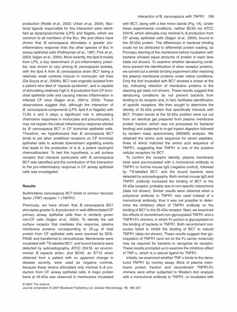
Previously, we have shown that B. cenocepacia BC7stimulates greater IL-8 production in well-differentiated CFprimary airway epithelial cells than in similarly grownnon-CF cells (Sajjan et al., 2004). To identify the cellsurface receptor that mediates this response, plasmamembrane proteins corresponding to 25 mg of totalprotein from CF epithelial cells were resolved by SDS-PAGE and transferred to nitrocellulose. Membranes wereincubated with 35S-labelled BC7, and bound bacteria weredetected by autoradiography. ATCC 25416, an environ-mental B. cepacia strain, and BC45, an ET12 strainobtained from a patient with no apparent change indisease severity, were used as negative controls,because these strains stimulated only minimal IL-8 pro-duction from CF airway epithelial cells. A major proteinband at 55 kDa was observed in membranes incubated
with BC7, along with a few minor bands (Fig. 1A). Underthese experimental conditions, neither BC45 nor ATCC25416, which stimulate only minimal IL-8 production fromCF airway epithelial cells (Sajjan et al., 2004), bound tothe 55 kDa protein. The differences in bacterial bindingcould not be attributed to differential protein loading, asPonceau staining of the membrane before incubation withbacteria showed equal amounts of protein in each lane(data not shown). To examine whether denaturing condi-tions prevent the identification of other receptor proteins,we carried out a similar binding experiment after resolvingthe plasma membrane proteins under native conditions.Only the blot incubated with BC7 showed a smear at thetop, indicating retention of membrane proteins in thestacking gel (data not shown). These results suggest thatdenaturing conditions do not prevent bacteria frombinding to its receptor and, in fact, facilitates identificationof specific receptors. We then sought to determine theidentity of 55 kDa protein that specifically interacts withBC7. Protein bands at the 55 kDa position were cut outfrom an identical gel prepared from plasma membraneprotein fraction (which was not processed for bacterialbinding) and subjected to in-gel trypsin digestion followedby tandem mass spectrometry (MS/MS) analysis. Weobtained the amino acid sequence for seven peptides,three of which matched the amino acid sequence ofTNFR1, suggesting that TNFR1 is one of the putativecellular receptors for BC7.
To confirm the receptor identity, plasma membraneblots were pre-incubated with a monoclonal antibody toTNFR1 or normal mouse IgG (negative control), followedby 35S-labelled BC7, and the bound bacteria weredetected by autoradiography. Both normal mouse IgG andTNFR1 antibody increased the binding of BC7 to the55 kDa receptor, probably due to non-specific interactions(data not shown). Similar results were obtained when apolyclonal antibody to TNFR1 was used instead of amonoclonal antibody; thus it was not possible to deter-mine the inhibitory effect of TNFR1 antibody on thebinding of BC7 to the 55 kDa receptor. Next, we examinedthe effects of recombinant non-glycosylated TNFR1 and aTNFR1/Fc chimera, in which Fc portion is glycosylated onthe binding of bacteria to TNFR1. Both recombinant mol-ecules failed to inhibit the binding of BC7 to naturalTNFR1 (data not shown). These results suggest that gly-cosylation of TNFR1 (and not on the Fc carrier molecule)may be required for bacteria to recognize its receptor.These results prompted us to examine the inhibitory effectof TNF-a, which is a natural ligand for TNFR1.
Initially, we examined whether TNF-a binds to the dena-tured TNFR1 by overlay assay. Blots of plasma mem-brane protein fraction and recombinant TNFR1/Fcchimera were either subjected to Western blot analysiswith a monoclonal antibody to TNFR1, or incubated with
Interaction of B. cenocepacia with TNFR1 189
© 2007 The AuthorsJournal compilation © 2007 Blackwell Publishing Ltd, Cellular Microbiology, 10, 188–201

TNF-a, and the bound TNF-a was detected by using apolyclonal antibody to TNF-a. Antibody to TNFR1detected a band at 55 kDa in the plasma membraneprotein fraction. Recombinant TNFR1/Fc chimera wasdetected at a slightly higher position (Fig. 1B). Althoughthe molecular weight of TNFR1/Fc chimera is 48 kDa, therecombinant protein migrates as a 60 kDa protein as aresult of glycosylation in the Fc portion, according to themanufacturer’s information sheet (R&D Systems). Nearlyidentical bands were observed in blots incubated withTNF-a, confirming that TNF-a binds to denatured TNFR1and could be used to inhibit the binding of BC7 to TNFR1(Fig. 1C). TNF-a also binds to TNFR2, which has amolecular weight of 75 kDa. Absence of a positive band atthis position in plasma membrane protein fraction was notsurprising, because airway epithelial cells do not expressTNFR2 (Gomez et al., 2004).
To assess the effect of TNF-a on the binding of BC7 toTNFR1, blots of epithelial cell plasma membrane fractionwere pre-incubated with TNF-a or IL-1b, incubated with35S-labelled BC7, and the bound bacteria detected byautoradiography. TNF-a inhibited the binding of BC7 toTNFR1 in a dose-dependent manner and almost com-pletely abrogated the binding at a concentration of
5 mg ml-1 (Fig. 1D). In contrast, IL-1b had no effect on thebinding of BC7 to TNFR1 even at the highest concentra-tion used. These results suggest that TNF-a receptor(TNFR1) may function as a receptor for BC7.
Tumour necrosis factor receptor 1 (TNFR1) expressionis increased in primary cultures of CF airwayepithelial cells
Airway epithelial cells obtained from two CF and twonon-CF individuals were cultured at an air/liquid interface.Total protein extracts from these cells were subjected toimmunoblot analysis using TNFR1 antibody (Fig. 2A).Compared with non-CF cells, CF airway epithelial cellsshowed more intense 55 kDa immunoreactive bands,although intensity of b-actin bands was similar in eachlane, an indication of increased expression of TNFR1 inCF cells. To determine the distribution of TNFR1, paraffinsections of CF and non-CF cultures were immunostainedwith antibody to TNFR1. Both CF and non-CF cellsexpressed TNFR1, but there was a difference in distribu-tion of this receptor in these cell types. CF cells appear tohave more TNFR1 on the cell membrane (Fig. 2B and C).Sections incubated with normal mouse IgG (negative
Fig. 1. Identification of cellular receptor for B. cenocepacia.A. Binding of bacteria to 55 kDa protein. A fraction enriched in membrane proteins prepared from CF airway epithelial cells differentiated into amucociliary phenotype was subjected to SDS-PAGE. Proteins were transferred to a nitrocellulose membrane, blocked with gelatin, incubatedwith 35S-labelled bacteria, and bound bacteria were detected by autoradiography.B and C. Binding of TNF-a to denatured TNFR1. Denatured plasma membrane fraction or TNFR1/Fc chimera was subjected to SDS-PAGE,and proteins were transferred to nitrocellulose. Blots were either subjected to Western blot analysis with monoclonal antibody to TNFR1 (B), orincubated with TNF-a, and the bound protein was detected by using polyclonal antibody to TNF-a (C).D. Inhibition of BC7 binding to 55 kDa protein by TNF-a. Blots of plasma membrane proteins were pre-incubated with TNF-a or IL-1b, followedby incubation with 35S-labelled bacteria. Bound bacteria were detected by autoradiography and semiquantified by densitometry.
190 U. S. Sajjan, M. B. Hershenson, J. F. Forstner and J. J. LiPuma
© 2007 The AuthorsJournal compilation © 2007 Blackwell Publishing Ltd, Cellular Microbiology, 10, 188–201

control) instead of TNFR1 antibody were negative, indi-cating the specificity of the antibody (Fig. 2D and E).
Tumour necrosis factor receptor 1 functions as areceptor for BC7 in airway epithelial cells
To determine the role of TNFR1 as a receptor forB. cenocepacia in intact cells, we used immortalized CF(IB3) cells, because these cells retain the characteristicsof CF cells and, at the same time, do not produce mucus,
which might hinder the accessibility of cell surface mol-ecules to potential inhibitors. TNFR1 expression on thesurface of IB3 cells was confirmed by fluorescence-assisted cell sorting (FACS) analysis. Non-permeabilizedIB3 cells incubated with a TNFR1 antibody showedincreased fluorescence as indicated by shift in the histo-gram to the right (Fig. 3A) compared with normal IgG-treated cells (control), suggesting that IB3 cells expressabundant amounts of TNFR1 on their surface. Bacterialbinding to cells was then determined by incubating IB3
Fig. 2. Expression of TNFR1 in CF andnon-CF airway epithelial cells.A. Western blot analysis. Cell lysates from CFand non-CF primary airway epithelial cellsdifferentiated into a mucociliary phenotypewere probed with monoclonal antibody toTNFR1. N1 and N2 are normal airwayepithelial cells from two different donors; CF1and CF2 are airway epithelial cells from twodifferent CF patients.B–E. Immunofluorescence detection ofTNFR1. Paraffin-embedded sections preparedfrom CF (C) and non-CF (B) mucociliarycultures were stained with monoclonalantibody to TNFR1, and the bound antibodywas detected by anti-mouse IgG conjugatedwith Alexa Fluor-598. Arrowheads representTNFR1 expression on the apical surface.Sections stained with normal IgG are shownin D and E.
Fig. 3. Binding of bacteria to TNFR1 in intactcells.A. Expression of TNFR1: non-permeabilizedIB3 cells were incubated with monoclonalantibody to TNFR1, and bound antibody wasdetected with anti-mouse IgG conjugated withAlexa Fluor-488 and analysed by FACS. Datapresented are representative of threeindependent experiments.B. Binding of bacteria to cells: IB3 cells werelightly fixed with 0.5% paraformaldehyde,washed and incubated with FITC-labelledBC7 or ATCC 25416. Cells were harvestedafter removal of unbound bacteria andanalysed by FACS. Data presented arerepresentative of three independentexperiments.C and D. Effect of TNF-a or TNFR1 antibodyon binding of bacteria to IB3 cells: lightly fixedIB3 cells were incubated with TNF-a orTNFR1 antibody for 30 min, followed byincubation with FITC-labelled BC7. Bacterialbinding was then assessed by FACS. Datarepresent mean � SEM of four independentexperiments carried out in triplicates;*P < 0.05, ANOVA.
Interaction of B. cenocepacia with TNFR1 191
© 2007 The AuthorsJournal compilation © 2007 Blackwell Publishing Ltd, Cellular Microbiology, 10, 188–201

cells with fluorescein isothiocyanate (FITC)-labelled BC7or ATCC 25416, followed by FACS analysis. Only cellsincubated with BC7 showed a shift in the histogram, indi-cating that BC7, but not ATCC 25416, binds to IB3 cells(Fig. 3B). Pre-incubation of IB3 cells with TNF-a, a naturalligand for TNFR1 (Fig. 3C), and antibody to TNFR1(Fig. 3D), each inhibited the binding of BC7 to IB3 cells ina dose-dependent manner, with maximum reductions of43% and 45% respectively, indicating that TNFR1 mayserve as a cellular receptor for BC7. IL-1b or normal IgG,which were used as negative controls, had no effect onBC7 binding to IB3 cells (data not shown). The partialinhibition of BC7 binding to epithelial cells by TNF-a con-trasts with previous results, in which TNF-a completelyinhibited BC7 binding to TNFR1 (Fig. 1D), because in thisexperiment, the binding of BC7 to isolated TNFR1 wasbeing determined rather than the intact cells. Takentogether, these data suggest that in intact cells, BC7 mayalso bind to other cellular receptors in addition to TNFR1.
BC7 colocalizes with TNFR1 in cell culturesdifferentiated into mucociliary phenotype
Well-differentiated CF and normal cell cultures were incu-bated with BC7 and incubated for 2 h. Cell cultures werewashed to remove unbound bacteria, fixed and embed-ded in paraffin. Thin sections from these cultureswere immunostained with antibodies to TNFR1 andB. cenocepacia and visualized by confocal fluorescencemicroscopy, taking optical sections at 0.2 m intervals. Ingeneral, CF cell cultures showed more bound bacteriathan the similarly treated normal cells, as observed pre-
viously (Sajjan et al., 2004). Further, at least 40–50% ofthe bound bacteria colocalized with TNFR1 in both CFand normal cells (Fig. 4C and D).
Colocalization of B. cenocepacia with TNFR1 in CF andnormal lung sections
Paraffin sections prepared from lungs of a CF patientwho died due to infection with B. cenocepacia ET12,were immunostained with antibodies to TNFR1 andB. cenocepacia (Figs 5C–F). Lung sections obtained froma normal donor served as a negative control (Fig. 5A andB). CF lung sections showed increased expressionof TNFR1 compared with normal lung sections.As expected, normal lung sections did not showB. cenocepacia (Fig. 5B). On the other hand, CF sectionsshowed B. cenocepacia organisms in both the airwaysand parenchyma, which frequently colocalized withTNFR1 in bronchial and alveolar epithelial cells (Fig. 5Dand F). The B. cenocepacia isolate (BCM132) obtainedfrom this patient belongs to ET12 lineage as determinedby PFGE analysis. It showed binding to 55 kDa TNFR1and also stimulated IL-8 from CF cells similar to BC7(Fig. 6A and B). These observations suggest thatB. cenocepacia ET12 may interact with TNFR1 in thelungs of infected CF patients.
Burkholderia cenocepacia BC7 activates TNFR1-relatedsignalling pathway
We sought to examine the pro-inflammatory signallingpathways activated by BC7 in CF airway epithelial cells.
Fig. 4. Colocalization of B. cenocepacia BC7 with TNFR1 in non-CF and CF cell cultures: Paraffin sections of non-CF (A and C) and CF (Band D) primary cultures infected with BC7 were incubated with antibodies to TNFR1 and B. cenocepacia, and the bound antibodies weredetected by Alexa Fluor-488 (for detection of TNFR1) and anti-rabbit Alexa Fluor-598 (for detection of bacteria). Arrowheads in C and Dindicate colocalization of bacteria with TNFR1. A and B are phase-contrast micrographs and correspond to C and D respectively. The inset inD is a digital magnification of an area marked in rectangle to show colocalization of bacteria with TNFR1 (yellow).
192 U. S. Sajjan, M. B. Hershenson, J. F. Forstner and J. J. LiPuma
© 2007 The AuthorsJournal compilation © 2007 Blackwell Publishing Ltd, Cellular Microbiology, 10, 188–201

Fig. 5. Colocalization of B. cenocepacia withTNFR1 in lungs of a CF patient. Lungsections from normal donor (A and B) or a CFpatient who died due to B. cenocepaciainfection (C–F) were incubated with antibodiesto TNFR1 and B. cenocepacia, and the boundantibody was detected as described in Fig. 4.A, C and E are phase-contrast micrographsand correspond to B, D and F respectively.Arrowheads indicate colocalization of bacteriawith TNFR1. The inset in D and F is a digitalmagnification of an area marked in rectangleto demonstrate colocalization of bacteria withTNFR1 (yellow) in the lung sections.
Fig. 6. A. Binding of BCM132 to 55 kDaTNFR1. Western blots of plasma membraneproteins were either incubated with35S-labelled BCM132 or BC7. Bound bacteriawere detected by autoradiography.B. Stimulation of IL-8 response in IB3 cells byBCM132. IB3 cells were incubated withBCM132 or BC7 at a moi of 0.1 for 24 h, andthe IL-8 in cell culture media was measuredby ELISA. *P < 0.05, ANOVA.
Interaction of B. cenocepacia with TNFR1 193
© 2007 The AuthorsJournal compilation © 2007 Blackwell Publishing Ltd, Cellular Microbiology, 10, 188–201
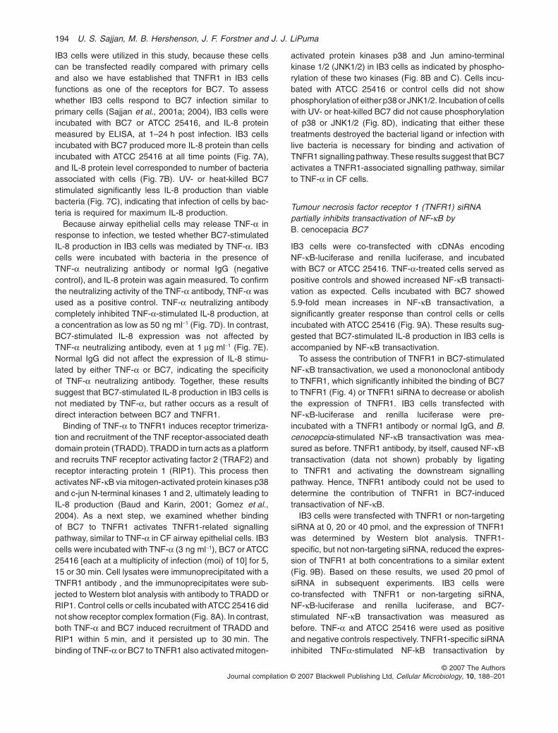
IB3 cells were utilized in this study, because these cellscan be transfected readily compared with primary cellsand also we have established that TNFR1 in IB3 cellsfunctions as one of the receptors for BC7. To assesswhether IB3 cells respond to BC7 infection similar toprimary cells (Sajjan et al., 2001a; 2004), IB3 cells wereincubated with BC7 or ATCC 25416, and IL-8 proteinmeasured by ELISA, at 1–24 h post infection. IB3 cellsincubated with BC7 produced more IL-8 protein than cellsincubated with ATCC 25416 at all time points (Fig. 7A),and IL-8 protein level corresponded to number of bacteriaassociated with cells (Fig. 7B). UV- or heat-killed BC7stimulated significantly less IL-8 production than viablebacteria (Fig. 7C), indicating that infection of cells by bac-teria is required for maximum IL-8 production.
Because airway epithelial cells may release TNF-a inresponse to infection, we tested whether BC7-stimulatedIL-8 production in IB3 cells was mediated by TNF-a. IB3cells were incubated with bacteria in the presence ofTNF-a neutralizing antibody or normal IgG (negativecontrol), and IL-8 protein was again measured. To confirmthe neutralizing activity of the TNF-a antibody, TNF-a wasused as a positive control. TNF-a neutralizing antibodycompletely inhibited TNF-a-stimulated IL-8 production, ata concentration as low as 50 ng ml-1 (Fig. 7D). In contrast,BC7-stimulated IL-8 expression was not affected byTNF-a neutralizing antibody, even at 1 mg ml-1 (Fig. 7E).Normal IgG did not affect the expression of IL-8 stimu-lated by either TNF-a or BC7, indicating the specificityof TNF-a neutralizing antibody. Together, these resultssuggest that BC7-stimulated IL-8 production in IB3 cells isnot mediated by TNF-a, but rather occurs as a result ofdirect interaction between BC7 and TNFR1.
Binding of TNF-a to TNFR1 induces receptor trimeriza-tion and recruitment of the TNF receptor-associated deathdomain protein (TRADD). TRADD in turn acts as a platformand recruits TNF receptor activating factor 2 (TRAF2) andreceptor interacting protein 1 (RIP1). This process thenactivates NF-kB via mitogen-activated protein kinases p38and c-jun N-terminal kinases 1 and 2, ultimately leading toIL-8 production (Baud and Karin, 2001; Gomez et al.,2004). As a next step, we examined whether bindingof BC7 to TNFR1 activates TNFR1-related signallingpathway, similar to TNF-a in CF airway epithelial cells. IB3cells were incubated with TNF-a (3 ng ml-1), BC7 or ATCC25416 [each at a multiplicity of infection (moi) of 10] for 5,15 or 30 min. Cell lysates were immunoprecipitated with aTNFR1 antibody , and the immunoprecipitates were sub-jected to Western blot analysis with antibody to TRADD orRIP1. Control cells or cells incubated with ATCC 25416 didnot show receptor complex formation (Fig. 8A). In contrast,both TNF-a and BC7 induced recruitment of TRADD andRIP1 within 5 min, and it persisted up to 30 min. Thebinding of TNF-a or BC7 to TNFR1 also activated mitogen-
activated protein kinases p38 and Jun amino-terminalkinase 1/2 (JNK1/2) in IB3 cells as indicated by phospho-rylation of these two kinases (Fig. 8B and C). Cells incu-bated with ATCC 25416 or control cells did not showphosphorylation of either p38 or JNK1/2. Incubation of cellswith UV- or heat-killed BC7 did not cause phosphorylationof p38 or JNK1/2 (Fig. 8D), indicating that either thesetreatments destroyed the bacterial ligand or infection withlive bacteria is necessary for binding and activation ofTNFR1 signalling pathway. These results suggest that BC7activates a TNFR1-associated signalling pathway, similarto TNF-a in CF cells.
Tumour necrosis factor receptor 1 (TNFR1) siRNApartially inhibits transactivation of NF-kB byB. cenocepacia BC7
IB3 cells were co-transfected with cDNAs encodingNF-kB-luciferase and renilla luciferase, and incubatedwith BC7 or ATCC 25416. TNF-a-treated cells served aspositive controls and showed increased NF-kB transacti-vation as expected. Cells incubated with BC7 showed5.9-fold mean increases in NF-kB transactivation, asignificantly greater response than control cells or cellsincubated with ATCC 25416 (Fig. 9A). These results sug-gested that BC7-stimulated IL-8 production in IB3 cells isaccompanied by NF-kB transactivation.
To assess the contribution of TNFR1 in BC7-stimulatedNF-kB transactivation, we used a mononoclonal antibodyto TNFR1, which significantly inhibited the binding of BC7to TNFR1 (Fig. 4) or TNFR1 siRNA to decrease or abolishthe expression of TNFR1. IB3 cells transfected withNF-kB-luciferase and renilla luciferase were pre-incubated with a TNFR1 antibody or normal IgG, and B.cenocepcia-stimulated NF-kB transactivation was mea-sured as before. TNFR1 antibody, by itself, caused NF-kBtransactivation (data not shown) probably by ligatingto TNFR1 and activating the downstream signallingpathway. Hence, TNFR1 antibody could not be used todetermine the contribution of TNFR1 in BC7-inducedtransactivation of NF-kB.
IB3 cells were transfected with TNFR1 or non-targetingsiRNA at 0, 20 or 40 pmol, and the expression of TNFR1was determined by Western blot analysis. TNFR1-specific, but not non-targeting siRNA, reduced the expres-sion of TNFR1 at both concentrations to a similar extent(Fig. 9B). Based on these results, we used 20 pmol ofsiRNA in subsequent experiments. IB3 cells wereco-transfected with TNFR1 or non-targeting siRNA,NF-kB-luciferase and renilla luciferase, and BC7-stimulated NF-kB transactivation was measured asbefore. TNF-a and ATCC 25416 were used as positiveand negative controls respectively. TNFR1-specific siRNAinhibited TNFa-stimulated NF-kB transactivation by
194 U. S. Sajjan, M. B. Hershenson, J. F. Forstner and J. J. LiPuma
© 2007 The AuthorsJournal compilation © 2007 Blackwell Publishing Ltd, Cellular Microbiology, 10, 188–201

Fig. 7. Effect of TNF-a neutralizing antibody on B. cenocepacia-stimulated IL-8 response in IB3 cells.A. IL-8 response: IB3 cells were incubated with B. cenocepacia BC7 or ATCC 25416 for 1, 3, 6 or 24 h, and IL-8 in the cell culture media wasquantified by ELISA.B. Binding of bacteria to cells. Cells incubated with bacteria for 3, 6 or 24 h were fixed, and bacteria associated were immunostained with Bccantibody and quantified by immunofluorescence microscopy.C. IL-8 response to UV- or heat-killed BC7: IB3 cells were incubated with live, UV-killed or heat-killed BC7 for 24 h, and IL-8 in the cell culturemedia was quantified by ELISA.D and E. Effect of TNF-a neutralizing antibody: IB3 cells were incubated with 3 ng of TNF-a (D) or B. cenocepacia BC7 at a moi of 0.1 (E) inthe presence or absence of varying concentrations of TNF-a neutralizing antibody or normal IgG for 24 h. IL-8 in cell culture supernatants wasquantified by ELISA. Bars represent mean � SEM of four independent experiments carried out in triplicate; *P < 0.05, ANOVA.
Interaction of B. cenocepacia with TNFR1 195
© 2007 The AuthorsJournal compilation © 2007 Blackwell Publishing Ltd, Cellular Microbiology, 10, 188–201

almost 90%, indicating that TNF-a-driven NF-kB transac-tivation is dependent on TNF-a ligation to TNFR1 underthe present experimental conditions. BC7-stimulatedNF-kB transactivation was partially inhibited by TNFR1-specific siRNA (Fig. 9C), suggesting that binding of BC7to TNFR1 is required for maximal BC7-stimulated cellactivation. Non-targeting siRNA did not affect NF-kBtransactivation stimulated by either TNF-a or BC7, indi-
cating the specificity of TNFR1-siRNA. ATCC 25416-induced transactivation of NF-kB was not affected byeither TNFR1-siRNA or non-targeting siRNA. Takentogether, these results suggest that TNFR1 may functionas one of the cellular receptors for potentially virulentET12 strains of B. cenocepacia, and this binding interac-tion contributes to the robust IL-8 response from CFairway epithelial cells.
Fig. 8. Activation of TNFR1-related signallingpathway by B. cenocepacia.A. Western blot analysis ofimmunoprecipitates: IB3 cells were incubatedwith either TNF-a (3 ng ml-1) orB. cenocepacia, BC7 or ATCC 25416 at a moiof 10 for 5, 15 or 30 min and lysed. Celllysates were immunoprecipitated with aTNFR1 antibody and subjected to Westernblot analysis with either TRADD or RIP1antibody.B and C. Phosphorylation of p38 and JNK:lysates from the same experiment weresubjected to immunoblot analysis withantibodies to phosho-p38, or total p38 (B)phospho-JNK1/2, or total JNK1/2 (C).D. Phosphorylation of p38 and JNK in IB3cells incubated with heat-killed or UV-killedBC7: cell lysates from IB3 cells incubated withlive, UV- or heat-killed bacteria for 30 minwere subjected to immunoblot analysis withantibodies to phosho-p38, or total p38phospho-JNK1/2, or total JNK1/2 (D). Datapresented are representative of threeindependent experiments. Std, molecularmass standards; cell lysate: total-cell lysatewas used to confirm the band positions ofTRADD or RIP1 observed inimmunoprecipitates; Con, untreated IB3 cells.
196 U. S. Sajjan, M. B. Hershenson, J. F. Forstner and J. J. LiPuma
© 2007 The AuthorsJournal compilation © 2007 Blackwell Publishing Ltd, Cellular Microbiology, 10, 188–201

Discussion
Previously, we demonstrated that the interaction ofBC7 with cytokeratin 13 (CK13) influenced the pro-inflammatory response from squamous epithelial cells(Sajjan et al., 2000a; 2002). While CK13 is normally
expressed in nasal mucosa and trachea, expression isincreased in the lower airway epithelium after repeatedinjury, which is common in CF lungs (Moll et al., 1982;Nagle et al., 1985; Sajjan et al., 2000b). However, wealso showed that B. cenocepacia BC7 can stimulateintense and sustained IL-8 production in CF airway epi-thelial cells differentiated into a mucociliary phenotype,which do not express CK13 on their apical surface(Sajjan et al., 2004). We further observed that B. ceno-cepacia interacts with alveolar epithelial cells in thelungs of CF patients, where CK13 expression was notevident (Sajjan et al., 2000b; 2001b). TLR4 and TLR5have been shown to be potential receptors for B. ceno-cepacia LPS and flagella respectively (Reddi et al.,2003; Urban et al., 2004). However, in preliminarystudies, we observed that HEK293 cells, which lackTLRs, responded to infection with BC7 by producing IL-8(data not shown). Together, these observations sug-gested that BC7 interacts with a receptor other thanCK13 or TLRs in CF airway epithelial cells.
In this study, we demonstrated that BC7, one of thepotentially virulent ET12 strains of B. cenocepacia, bindsto TNFR1 and this binding interaction induces associationof TNFR1 with TRADD and RIP, as well as activation ofp38 and JNK, similar to the receptor’s natural ligandTNF-a. Further, we found that TNFR1 siRNA partiallyinhibited NF-kB transactivation stimulated by BC7. Con-focal microscopy revealed that B. cenocepacia, BC7 andisolate BCM132 colocalize with TNFR1 in primary humanmucociliary-differentiated airway epithelial cells, and inthe lungs of a CF patient who died due to B. cenocepaciainfection respectively. Taken together, these data suggestthat interaction of potentially virulent ET12 strains withTNFR1 may contribute to the intense inflammationobserved in infected CF patients.
Tumour necrosis factor receptor 1 (TNFR1) belongs to asuperfamily of TNF receptors and plays an important role ininnate host defence (Pfeffer et al., 1993; Castanos-Velezet al., 1998; Gomez et al., 2004; Hehlgans and Pfeffer,2005). Binding of TNF-a to TNFR1 activates the NF-kB andJNK pathways, leading to transcriptional activation ofgenes encoding pro-inflammatory proteins, including IL-8(Stanger et al., 1995; Hsu et al., 1996a; Wright et al.,2004). Binding of TNF-a to TNFR1 may also activate thecaspase cascade, ultimately inducing apoptosis (Hsuet al., 1996b). The inflammatory pathway deviates from theapoptotic pathway very early in the signalling process.Binding of TNF-a to TNFR1 induces trimerization andrecruitment of TRADD, which then recruits either RIP1 andTRAF2 or FAS-associated death domain, activatinginflammatory or apoptotic pathways respectively (Baudand Karin, 2001; Hehlgans and Pfeffer, 2005). Weobserved that binding of B. cenocepacia to CF airwayepithelial cells did not induce apoptosis (data not shown);
Fig. 9. Effect of TNFR1 siRNA on transactivation of NF-kB byB. cenocepacia BC7.A. Transactivation of NF-kB by bacteria: IB3 cells wereco-transfected with NF-kB luciferase and renilla luciferase, and thenincubated with BC7, ATCC 25146 at a moi of 0.1 or TNF-a(3 ng ml-1) for 24 h. Cells were lysed, and luciferase activity wasmeasured and expressed as fold increase over control.B. Western blot analysis: IB3 cells were transfected with TNFR1-specific or non-targeting siRNA, and incubated for 72 h. Cell lysateswere subjected to Western blot analysis with a TNFR1 antibody .C. Inhibition of transactivation of NF-kB by TNFR1 siRNA: IB3 cellswere co-transfected with TNFR1-specific or non-targeting siRNA,NF-kB luciferase and renilla luciferase, and incubated for 24 h. Cellswere serum starved overnight and then incubated with bacteria at amoi of 0.1 or TNF-a (3 ng ml-1) for 24 h, and luciferase activity wasmeasured as before. Bars represent mean � SEM of six indepen-dent experiments carried out in duplicates; *P < 0.05, ANOVA.
Interaction of B. cenocepacia with TNFR1 197
© 2007 The AuthorsJournal compilation © 2007 Blackwell Publishing Ltd, Cellular Microbiology, 10, 188–201

rather, this interaction stimulated increased production ofIL-8. This effect was preceded by the recruitment ofTRADD and RIP1 to TNFR1 and phosphorylation of p38and JNK, similar to cells treated with TNF-a, a naturalligand for TNFR1. Neutralizing antibody to TNF-a did notinhibit the production of IL-8 from IB3 cells incubated withBC7, indicating that the observed effect was not mediatedby TNF-a. On the other hand, knockdown of TNFR1expression by specific siRNA significantly attenuatedBC7-induced NF-kB transactivation. These observationssuggest that direct binding of BC7 to TNFR1 in CF cells isrequired to activate a TNFR1-stimulated inflammatory sig-nalling pathway.
The IL-8 response stimulated by B. cenocepacia maynot depend solely on the interaction of bacteria withTNFR1. We and others have demonstrated thatB. cenocepacia also binds to other cellular receptors(Sajjan et al., 2002; Reddi et al., 2003; Urban et al.,2004). In the present study, our cell culture studies dem-onstrated that approximately half of the bound bacteriacolocalized with TNFR1 receptor in both CF and non-CFcells. Although TNF-a completely abrogated the bindingof BC7 to isolated TNFR1 in vitro, it only partially inhibitedthe binding of BC7 to CF cells even at the highest con-centration used. Similarly, blocking of TNFR1 expressionby TNFR1-specific siRNA partially repressed the NF-kBtransactivation associated with IL-8 production. Thus, thecapacity of BC7 to interact with TNFR1 may increase itspotential to stimulate pro-inflammatory response from CFcells, but is not sufficient for the maximal response.
Although activation of TNFR1 plays a critical role in hostdefence, pathogenic bacteria have evolved to utilize andsubvert this pathway to their advantage in susceptiblehosts. As we have shown for B. cenocepacia, Staphylo-coccus aureus also has been shown to bind to TNFR1 inairway epithelial cells and induce a TNFR1-related signal-ling pathway, leading to NF-kB activation and IL-8 produc-tion (Gomez et al., 2004). Protein A, a major surfaceprotein present in almost all strains of S. aureus, wasidentified as a bacterial ligand. In a murine model of lunginfection, the interaction of protein A with TNFR1 resultedin the mobilization of PMNs to airways, leading to severeinflammation. Although recruitment of PMNs to airways isa critical component of host defence, excessive andrepeated infiltration of PMNs can cause lung damage.
Previously we have shown that cable pili and the asso-ciated 22 kDa adhesin are required to stimulate maximumIL-8 production from squamous-differentiated bronchialepithelial cells. Preliminary studies indicated that isogenicmutants of BC7 lacking either cable pili or adhesinretained their capacity to bind to TNFR1 (data not shown),suggesting a role for yet another undefined bacterialligand. Identification and characterization of this bacterialligand is in progress.
Gene array analysis of CF airway epithelial cellsinfected with P. aeruginosa points to increased expressionof TNF receptor and other related genes (Eidelman et al.,2001). In the present study, we showed that expression ofTNFR1 is increased in cultured CF airway epithelial cells,as well as in the lungs of CF patients. These observa-tions implicate that chronic respiratory infection withP. aeruginosa may, in fact, increase the expression ofTNFR1 in the lungs of CF patients. Thus, B. cenocepaciaET12 strain, capable of binding to TNFR1 and activatingdownstream signalling pathway may cause severe inflam-mation in infected CF patients.
Experimental procedures
Bacterial strains and growth conditions
Burkholderia cenocepacia isolates BC7 and BC45 were obtainedfrom CF patients, and have been described previously (Sajjanet al., 1992; 2002). Both BC7 and BC45 belong to ET12 lineageand are indistinguishable by PFGE analysis and ribotyping, butdiffer phenotypically. BC7, which was obtained from a patientwho died of ‘cepacia syndrome’, binds to mucin glycopeptidesand CK13, induces cell damage and a robust IL-8 response fromprimary CF airway epithelial cells, and causes severe lung inflam-mation and necrotizing pneumonia in CF mice. In contrast, BC45,which was obtained from a patient with no apparent change indisease severity, does not bind to mucin or cytokeratin 13, stimu-lates low amounts of IL-8 from CF cells, and causes relativelymild cell damage in vitro (Sajjan et al., 1992; 2000a; 2001a;2002; 2004). BCM132 was isolated from post-mortem lungs of aCF patient who died of cepacia syndrome, and also belongs toET12 lineage. ATCC 25416 is an environmental strain purchasedfrom the American Tissue Culture Collection (Manassas, VA).Frozen stocks of BC7, BC45 and ATCC 25416 were subculturedon BHI agar and incubated at 37°C for 24–36 h. A single colonywas inoculated into 10 ml tryptic soy broth and grown at 37°C for12–16 h in the presence or absence of 35S-Translabel (GEHealthcare, Mississauga, ON, Canada). Bacteria were harvestedby centrifugation, washed with PBS, and suspended in serum-free cell culture media or PBS containing 1% bovine serumalbumin (BSA) to a desired concentration by measuring theoptical density at 600 nm (1 unit at OD600 corresponds to~1 ¥ 109 cfu ml-1). In some experiments, bacteria were labelledwith FITC (Pierce Biotechnology, Rockford, IL) as described pre-viously (Sajjan et al., 2000b).
Cell cultures
Cystic fibrosis and normal primary airway epithelial cells atpassage one were cultured at air/liquid interface as previouslydescribed (Sajjan et al., 2004), and were kindly provided by DrKeshavjee (University Health Network, Toronto, Ontario,Canada). Immortalized CF bronchial epithelial cells (IB3 cells)(Zeitlin et al., 1991) were kindly provided by P. Zeitlin (JohnsHopkins Medical Institute, Baltimore, Maryland). IB3 cells wereroutinely grown in LHC-8 cell culture medium (Invitrogen, Carls-bad, CA) supplemented with 2 mM glutamine, 5% fetal bovine
198 U. S. Sajjan, M. B. Hershenson, J. F. Forstner and J. J. LiPuma
© 2007 The AuthorsJournal compilation © 2007 Blackwell Publishing Ltd, Cellular Microbiology, 10, 188–201

serum, and 50 U ml-1 penicillin and 50 mg ml-1 streptomycin at37°C in 5% CO2.
Binding assays
Bacterial overlay assay. Binding of bacteria to isolated plasmamembrane proteins was determined by a bacterial overlay assayas described previously (Sajjan, 1993). Briefly, blots of CF airwayepithelial cell plasma membrane proteins were blocked with 1%gelatin, washed and incubated with PBS-BSA containing35S-labelled BC7 or BC45, or ATCC 25416. Blots were washedwith PBS-BSA to remove unbound bacteria, and bound bacteriawere detected by autoradiography. In some experiments, blots ofplasma membrane proteins were pre-incubated with TNF-a (R&DSystems, Minneapolis, MN) or IL-1b. Autoradiographs were semi-quantified using the ChemiDoc XRS system combined withQuantity One 1-D analysis software (BIO-RAD Laboratories,Hercules, CA).
Binding of bacteria to epithelial cells. IB3 cell monolayers grownin six-well plates were lightly fixed with 0.5% paraformaldehydefor 30 min and washed with PBS containing 0.5% BSA. FITC-labelled bacteria were added to cells at a moi of 1, centrifuged at200 g and incubated for 1 h at 37°C. Cells were washed withPBS-BSA to remove unbound bacteria, and cells along withbound bacteria were detached from the plate and analysedwith a Becton Dickinson FACSCalibur using CellQuest software.The autofluorescence contributed by cells was subtracted fromall experimental samples. In inhibition assays, cells were pre-incubated with potential inhibitors (TNF-a, or antibody to TNFR1)for 1 h, washed with PBS and then incubated with FITC-labelledbacteria. Cells incubated with normal IgG or IL-1b instead ofinhibitors served as negative controls.
Bacterial binding to cells by microscopy. Binding of bacteria toIB3 cells was determined by quantitative immunofluorescencemicroscopy as previously described (Sajjan et al., 2000a;2002).
Identification of receptor protein
The plasma membrane-enriched fraction from primary CF airwayepithelial cells was subjected to SDS-PAGE and stained withCoomassie blue. Protein bands at 55 kDa were cut out andpartially digested with trypsin. Peptides isolated from the gel weresubjected to peptide mapping and MS/MS sequencing withApplied Biosystems/MDS Sciex API QSTAR XL MALDI QTOF(Urban et al., 2004). Peptide fingerprinting of the in-gel digestswas analysed by database searching with ProFound (129.85.19.192/profound_bin/WebProFound.exe), and the peptidesequence obtained by MS/MS spectra was interpreted manuallyand used to search for protein homologies (http://www.ncbi.nlm.nih.gov/blast).
Infection of cell cultures
IB3 cells were shifted to serum- and antibiotic-free media for atleast 16 h prior to infection with bacteria. Bacteria were added tocells at a moi of 0.1, 1 or 10 and centrifuged at 200 g, and
incubated as indicated in the Results section. Medium was col-lected and centrifuged, and IL-8 present in the supernatants wasquantified by ELISA (R&D Systems). In some experiments, cellswere incubated with bacteria in the presence of neutralizingantibody to TNF-a (R&D Systems, MN) or normal IgG. Well-differentiated airway epithelial cell cultures were shifted toantibiotic-free media at least 24 h prior to infection. Cultures wereincubated with ATCC 25416 or BC7 (1 ¥ 105 cfu equivalent to moiof 0.1) apically for 24 h. Apical surface of cell cultures waswashed, embedded in agarose and fixed in 10% buffered forma-lin, and finally embedded in paraffin as described previously(Sajjan et al., 2002).
Detection of cell surface TNFR1 by flow cytometry
IB3 cells were incubated with monoclonal antibody specific toTNFR1 or normal mouse IgG (matched isotype control) andanalysed by flow cytometry as previously described (Gomezet al., 2004; Sajjan et al., 2006).
Immunoprecipitation and Western blot analysis
After relevant treatment, epithelial cells were washed and lysed inice-cold RIPA buffer containing complete protease inhibitors,1 mM sodium orthovandate and 1 mM sodium fluoride. Cellularproteins were resolved by SDS-PAGE and transferred to anImmobilonTM-P membrane (Millipore, Billerica, MA). Membraneswere probed with primary antibodies to phosphorylated or totalp38, JNK1, JNK2 (Cell Signaling Technology, Danvers, MA),TNFR1, TRADD or RIP1 (Santa Cruz Biotechnology, Santa Cruz,CA). Signals were amplified and visualized with horseradishperoxidase-conjugated secondary antibodies (Bio-Rad Laborato-ries) and chemiluminescent substrate (Pierce). Immunoprecipita-tion of cell lysates with TNFR1 antibody was performed usingExactaCruzTM B immunoprecipitation kit (Santa Cruz Biotechnol-ogy) following the manufacturer’s instructions. Immunoprecipi-tates were solubilized in reducing buffer and subjected toWestern blotting with either TRADD or RIP1 antibodies. Whole-cell lysates were used to confirm the band position of TRADD andRIP1 observed in the immunoprecipitates.
Transfection of cells and measurement ofNF-kB activation
IB3 cells grown in six-well plates were transiently transfected withhNF-kB-luciferase (0.2 mg; Stratagene, La Jolla, CA) and renillaluciferase (10 ng) using Lipofectamine 2000 (Invitrogen). Thefollowing day, the cells were shifted to serum-free media andstimulated with either B. cenocepacia BC7 at a moi of 0.1 or TNF-a(3 ng ml-1). After incubation for 24 h, the cells were harvested andluciferase activity was measured using a luminometer. Changes inpromoter activity were normalized for transfection efficiency bydividing luciferase light units by renilla luciferase light units.Results were then reported as fold increase over the emptyvector/untreated control. When siRNA was used to inhibit theexpression of TNFR1 protein, cells were transfected with 20 or40 pmol of TNFR1 siRNA (Santa Cruz Biotechnology) along withhNF-kB and renilla luciferase. Cells transfected with non-targetingsiRNA instead of TNFR1 siRNA was used as control.
Interaction of B. cenocepacia with TNFR1 199
© 2007 The AuthorsJournal compilation © 2007 Blackwell Publishing Ltd, Cellular Microbiology, 10, 188–201

Immunofluorescence
Paraffin lung sections from a CF patient who succumbed tocepacia syndrome was provided by Dr E. Tullis (University HealthNetwork, Toronto, Canada). Collection and utilization of lungtissue was approved by the hospital ethics committee. Paraffinsection of non-CF lung was obtained from the pathology tissuelibrary at the Hospital for Sick Children, Toronto, Canada. Depar-affinized and hydrated sections of lungs or primary airway epi-thelial cell cultures were heated in 10 mM citric acid buffer underpressure for 90 s to unmask the antigens and blocked withnormal donkey serum. Sections were incubated with a TNFR1antibody or a mixture of TNFR1 antibody and antibody toB. cepacia complex (R418) (Sajjan et al., 2001b), and the boundantibodies were detected with anti-goat conjugated with AlexaFluor-488 (for detection of TNFR1) and anti-rabbit Alexa Flour-598 (for detection of bacteria) (Molecular Probes, Portland, OR).Sections were visualized by confocal fluorescent microscopy witha Zeiss LSM 510 confocal microscope mounted on a Zeiss Axio-vert 100 M inverted microscope. Optical sections at 0.2 m intervalwere taken to assess the colocalization of bacteria with TNFR1as described previously (Adamo et al., 2004; Gomez et al., 2004;2005; 2007). Sections treated with normal IgG instead of primaryantibody served as negative controls.
Data analysis
Statistical significance was assessed by analysis of variance(ANOVA). Differences identified by ANOVA were pinpointed by theTukey-Kramer test.
Acknowledgements
This work was supported by the grants from the Cystic FibrosisFoundation (SAJJAN06I0 to U.S.S. and LIPUMA97P0 to J.J.L.)and the National Institutes of Health (HL82550 to M.B.H.). U.S.S.was partially supported by the Carroll Hass Research Fund inCystic Fibrosis.
References
Adamo, R., Sokol, S., Soong, G., Gomez, M.I., and Prince, A.(2004) Pseudomonas aeruginosa flagella activate airwayepithelial cells through asialoGM1 and toll-like receptor 2as well as toll-like receptor 5. Am J Respir Cell Mol Biol 30:627–634.
Bals, R., and Hiemstra, P.S. (2004) Innate immunity inthe lung: how epithelial cells fight against respiratorypathogens. Eur Respir J 23: 327–333.
Baud, V., and Karin, M. (2001) Signal transduction by tumornecrosis factor and its relatives. Trends Cell Biol 11: 372–377.
Bryan, R., Kube, D., Perez, A., Davis, P., and Prince, A.(1998) Overproduction of the CFTR R domain leads toincreased levels of AsialoGM1 and increased Pseudomo-nas aeruginosa binding by epithelial cells. Am J Respir CellMol Biol 19: 269–277.
Castanos-Velez, E., Maerlan, S., Osorio, L.M., Aberg, F.,Biberfeld, P., Orn, A., and Rottenberg, M.E. (1998) Trypa-
nosoma cruzi infection in tumor necrosis factor receptorp55-deficient mice. Infect Immun 66: 2960–2968.
De Soyza, A., McDowell, A., Archer, L., Dark, J.H., Elborn,S.J., Mahenthiralingam, E., et al. (2001) Burkholderiacepacia complex genomovars and pulmonary transplanta-tion outcomes in patients with cystic fibrosis. Lancet 358:1780–1781.
De Soyza, A., Morris, K., McDowell, A., Doherty, C., Archer,L., Perry, J., et al. (2004a) Prevalence and clonality ofBurkholderia cepacia complex genomovars in UK patientswith cystic fibrosis referred for lung transplantation. Thorax59: 526–528.
De Soyza, A., Ellis, C.D., Khan, C.M., Corris, P.A., andDemarco de Hormaeche, R. (2004b) Burkholderia cenoce-pacia lipopolysaccharide, lipid A, and proinflammatoryactivity. Am J Respir Crit Care Med 170: 70–77.
Eidelman, O., Srivastava, M., Zhang, J., Leighton, X., Murtie,J., Jozwik, C., et al. (2001) Control of the proinflammatorystate in cystic fibrosis lung epithelial cells by genes fromthe TNF-alphaR/NFkappaB pathway. Mol Med 7: 523–534.
Epelman, S., Stack, D., Bell, C., Wong, E., Neely, G.G.,Krutzik, S., et al. (2004) Different domains of Pseudomo-nas aeruginosa exoenzyme S activate distinct TLRs.J Immunol 173: 2031–2040.
Fink, J., Steer, J.H., Joyce, D.A., McWilliam, A.S., andStewart, G.A. (2003) Pro-inflammatory effects of Burkhold-eria cepacia on cystic fibrosis respiratory epithelium. FEMSImmunol Med Microbiol 38: 273–282.
Gomez, M.I., Lee, A., Reddy, B., Muir, A., Soong, G., Pitt, A.,et al. (2004) Staphylococcus aureus protein A inducesairway epithelial inflammatory responses by activatingTNFR1. Nat Med 10: 842–848.
Gomez, M.I., Sokol, S.H., Muir, A.B., Soong, G., Bastien, J.,and Prince, A.S. (2005) Bacterial induction of TNF-alphaconverting enzyme expression and IL-6 receptor alphashedding regulates airway inflammatory signaling.J Immunol 175: 1930–1936.
Gomez, M.I., Seaghdha, M.O., and Prince, A.S. (2007) Sta-phylococcus aureus protein A activates TACE throughEGFR-dependent signaling. EMBO J 26: 701–709.
Hehlgans, T., and Pfeffer, K. (2005) The intriguing biology ofthe tumor necrosis factor/tumor necrosis factor receptorsuperfamily: players, rules and the games. Immunology115: 1–20.
Hsu, H., Huang, J., Shu, H.B., Baichwal, V., and Goeddel,D.V. (1996a) TNF-dependent recruitment of the proteinkinase RIP to the TNF receptor-1 signaling complex. Immu-nity 4: 387–396.
Hsu, H., Shu, H.B., Pan, M.G., and Goeddel, D.V. (1996b)TRADD-TRAF2 and TRADD–FADD interactions define twodistinct TNF receptor 1 signal transduction pathways. Cell84: 299–308.
Isles, A., Maclusky, I., Corey, M., Gold, R., Prober, C.,Fleming, P., and Levison, H. (1984) Pseudomonas cepaciainfection in cystic fibrosis: an emerging problem. J Pediatr104: 206–210.
Kube, D., Sontich, U., Fletcher, D., and Davis, P.B. (2001)Proinflammatory cytokine responses to P. aeruginosainfection in human airway epithelial cell lines. Am J PhysiolLung Cell Mol Physiol 280: L493–L502.
Ledson, M.J., Gallagher, M.J., Jackson, M., Hart, C.A., and
200 U. S. Sajjan, M. B. Hershenson, J. F. Forstner and J. J. LiPuma
© 2007 The AuthorsJournal compilation © 2007 Blackwell Publishing Ltd, Cellular Microbiology, 10, 188–201

Walshaw, M.J. (2002) Outcome of Burkholderia cepaciacolonisation in an adult cystic fibrosis centre. Thorax 57:142–145.
Moll, R., Franke, W.W., Schiller, D.L., Geiger, B., andKrepler, R. (1982) The catalog of human cytokeratins: pat-terns of expression in normal epithelia, tumors and culturedcells. Cell 31: 11–24.
Nagle, R.B., Moll, R., Weidauer, H., Nemetschek, H., andFranke, W.W. (1985) Different patterns of cytokeratinexpression in the normal epithelia of the upper respiratorytract. Differentiation 30: 130–140.
Palfreyman, R.W., Watson, M.L., Eden, C., and Smith, A.W.(1997) Induction of biologically active interleukin-8 fromlung epithelial cells by Burkholderia (Pseudomonas)cepacia products. Infect Immun 65: 617–622.
Pfeffer, K., Matsuyama, T., Kundig, T.M., Wakeham, A.,Kishihara, K., Shahinian, A., et al. (1993) Mice deficient forthe 55 kd tumor necrosis factor receptor are resistant toendotoxic shock, yet succumb to L. monocytogenesinfection. Cell 73: 457–467.
Reddi, K., Phagoo, S.B., Anderson, K.D., and Warburton, D.(2003) Burkholderia cepacia-induced IL-8 gene expressionin an alveolar epithelial cell line: signalling through CD14and mitogen-activated protein kinase. Pediatr Res 54:297–305.
Riordan, J.R., Rommens, J.M., Kerem, B., Alon, N., Rozma-hel, R., Grzelczak, Z., et al. (1989) Identification of thecystic fibrosis gene: cloning and characterization ofcomplementary DNA. Science 245: 1066–1073.
Rommens, J.M., Iannuzzi, M.C., Kerem, B., Drumm, M.L.,Melmer, G., Dean, M., et al. (1989) Identification of thecystic fibrosis gene: chromosome walking and jumping.Science 245: 1059–1065.
Sajjan, U.S., and Forstner, J.F. (1993) Role of a 22-kilodaltonpilin protein in binding of Pseudomonas cepacia to buccalepithelial cells. Infect Immun 61: 3157–3163.
Sajjan, U.S., Corey, M., Karmali, M.A., and Forstner, J.F.(1992) Binding of Pseudomonas cepacia to normal humanintestinal mucin and respiratory mucin from patients withcystic fibrosis. J Clin Invest 89: 648–656.
Sajjan, U.S., Sylvester, F.A., and Forstner, J. (2000a) Cable-piliated Burkholderia cepacia bind to cytokeratin 13 of epi-thelial cells. Infect Immun 68: 1787–1795.
Sajjan, U., Wu, Y., Kent, G., and Forstner, J. (2000b) Pref-erential adherence of cable-piliated Burkholderia cepaciato respiratory epithelia of CF knockout mice and humancystic fibrosis lung explants. J Med Microbiol 49: 875–885.
Sajjan, U., Thanassoulis, G., Cherapanov, V., Lu, A., Sjolin,C., Steer, B., et al. (2001a) Susceptibility of Cftr (–/–) mice
to pulmonary infection with Burkholderia cepacia. InfectImmun 69: 5138–5150.
Sajjan, U., Corey, M., Humar, A., Tullis, E., Cutz, E., Acker-ley, C., and Forstner, J. (2001b) Immunolocalization ofBurkholderia cepacia in the lungs of cystic fibrosis patients.J Med Microbiol 50: 535–546.
Sajjan, U., Ackerley, C., and Forstner, J. (2002) Interactionof cblA/Adhesin-positive Burkholderia cepacia with squa-mous epithelium. Cell Microbiol 4: 73–86.
Sajjan, S., Keshavjee, S., and Forstner, J. (2004)Responses of well-differentiated airway epithelial cell cul-tures from healthy donors and patients with cystic fibrosisto Burkholderia cenocepacia infection. Infect Immun 72:4188–4199.
Sajjan, U.S., Jia, Y., Newcomb, D.C., Bentley, J.K., Lukacs,N.W., LiPuma, J.J., and Hershenson, M.B. (2006) H. influ-enzae potentiates airway epithelial cell responses to rhi-novirus by increasing ICAM-1 and TLR3 expression.FASEB J 20: 2121–2123.
Shaw, D., Poxton, I.R., and Govan, J.R. (1995) Bio-logical activity of Burkholderia (Pseudomonas) cepacialipopolysaccharide. FEMS Immunol Med Microbiol 11:99–106.
Soong, G., Reddy, B., Sokol, S., Adamo, R., and Prince, A.(2004) TLR2 is mobilized into an apical lipid raft receptorcomplex to signal infection in airway epithelial cells. J ClinInvest 113: 1482–1489.
Stanger, B.Z., Leder, P., Lee, T.H., Kim, E., and Seed, B.(1995) RIP: a novel protein containing a death domain thatinteracts with Fas/APO-1 (CD95) in yeast and causes celldeath. Cell 81: 513–523.
Urban, T.A., Griffith, A., Torok, A.M., Smolkin, M.E., Burns,J.L., and Goldberg, J.B. (2004) Contribution of Burkhold-eria cenocepacia flagella to infectivity and inflammation.Infect Immun 72: 5126–5134.
Wright, T.W., Pryhuber, G.S., Chess, P.R., Wang, Z., Notter,R.H., and Gigliotti, F. (2004) TNF receptor signaling con-tributes to chemokine secretion, inflammation, and respi-ratory deficits during Pneumocystis pneumonia. J Immunol172: 2511–2521.
Zeitlin, P.L., Lu, L., Cutting, G., Stetten, G., Kieffer, K.A.,Craig, R., and Guggino, W.B. (1991) A cystic fibrosis bron-chial epithelial cell line: immortalization by adeno-12-SV40infection. Am J Respir Cell Mol Biol 4: 313–319.
Zughaier, S.M., Ryley, H.C., and Jackson, S.K. (1999)Lipopolysaccharide (LPS) from Burkholderia cepacia ismore active than LPS from Pseudomonas aeruginosa andStenostrophomonas maltophilia in stimulating tumor necro-sis factor alpha from human monocytes. Infect Immun 67:1505–1507.
Interaction of B. cenocepacia with TNFR1 201
© 2007 The AuthorsJournal compilation © 2007 Blackwell Publishing Ltd, Cellular Microbiology, 10, 188–201