bulgarian chemical communications, volume 53, special
TRANSCRIPT
35
Bulgarian Chemical Communications, Volume 53, Special Issue A (pp. 035 – 041) 2021 DOI: 10.34049/bcc.53.А.0005
Natural medicine: an evaluation of the in vitro cytotoxic effect of several Bulgarian fungal species on two panels of cancer cell lines
A. Dushkov, M. Petrova, J. Todorova, A. Gospodinov, I. Ugrinova* Institute of Molecular Biology, Bulgarian Academy of Sciences, 1113, Sofia, Bulgaria
Received January 14, 2021; Revised March 01, 2021
The medicinal potential of Bulgaria’s wild-growing fungi has, until recently, largely gone unexplored by the native scientific community. While many of the region’s mushrooms have long been prized by residents as a valuable food source, they have generally not been thought of as having the medicinal value of certain herbs, for example. However, the growing body of scientific literature confirming the diverse beneficial effects of many Asian mushrooms (long held in high regard by ancient medicinal traditions of their respective regions), as well as the somewhat surprising discovery that some of these same mushrooms can be found in Bulgaria, has sparked our interest in exploring the effects they may have on different types of cancer, with the goal of either complementing existing treatments or, perhaps, uncovering new treatments based on compounds isolated from fungal extracts. We have prepared different ethanol and water extracts from the mushrooms Trametes versicolor, Lenzites betulina, Fomes fomentarius, Fomitopsis betulina and Amanita muscaria and have examined the primary cytotoxic effect of these crude extracts on a panel of human skin and lung cancer cells in vitro, with the goal of establishing their IC50 values via MTT (3-(4,5-Dimethylthiazol-2-yl)-2,5-diphenyltetrazolium bromide) assay, comparing them, and gaining perspective for future research. Our results show that all extracts exhibit varying degrees of cytotoxicity even at low concentrations, warranting further inquiries into their anti-tumor potential.
Key words: fungi; cancer; cytotoxicity; replication; natural medicine
INTRODUCTION
Historically, different civilizations’ attitudes toward mushrooms in general have been very dissimilar. As Gordon and Valentina Pavlovna Wasson point out [1], there is a clear dichotomy between civilizations that readily accept mushrooms as a food source and even medicine (termed ‘mycophilic’) and those that disregard them or outright reject them as such (‘mycophobic’). A good example of a mycophilic civilization is ancient China, where mentions of mushrooms can be found in some of the oldest known medicinal texts, such as the Huangdi Neijing. China is also the home of some of the first known mushroom cultivation attempts – the fungus Auricularia auricula-judae is known to have first been grown intentionally in China around the year 600 A.D ‘[2]. In comparison, after the Roman Age, many Western European civilizations have either not paid much attention to mushrooms in general, or regarded them as unnatural and dangerous due to their proclivity for growing on dead and rotting matter, their sudden appearance after rainfall as compared to the slow-growing plants (even appearing in circles, whose common name ‘fairy rings’ reflects their supposed supernatural origin in
the Western European mind), and their curious shapes, colors and odors. Bulgaria is, in some ways, no exception to this - while some of the local mushrooms have been well regarded as delicious edibles, evidence for their medicinal use is scarce. However, the times are changing, and with the growing availability of information accumulated on the medicinal properties of staple Asian mushrooms, the scientific community is gaining an interest in exploring the beneficial effects of their consumption and the mechanisms of action of their active compounds. The realization that some of the best medicinal fungi from Asia – the well-known Trametes versicolor (kawaratake, 雲芝 yun zhi), Ganoderma lucidum (reishi), Grifola frondosa (maitake), and Inonotus obliquus (chaga), for example – can be found in Bulgaria’s forests, has prompted us to take active interest in examining the effects of several local species on cancerous cells in vitro.
The species of mushroom we studied in this work – all wood-growing fungi, except for Amanita muscaria - have each been indicated to contain compounds that exhibit some kind of anti-cancer activity via various mechanisms. Trametes versicolor, or ‘turkey’s tail’, contains a compound known as polysaccharide K, or Krestin (PSK), a protein-bound polysaccharide used as an adjuvant
© 2021 Bulgarian Academy of Sciences, Union of Chemists in Bulgaria * To whom all correspondence should be sent: E-mail: [email protected]
A. Dushkov et al.: Natural medicine: an evaluation of the in vitro cytotoxic effect of several Bulgarian fungal species on two…
36
immunotherapeutic in Japan for more than three decades [3] that exhibits both immunopotentiating antitumor effects [4] and an ability to directly induce apoptosis in cell cutures in vitro [5]. A water extract derived from Lenzites betulina has been shown to have mild anti-tumor activity against the highly malignant murine tumor cell line Sarcoma 180 [6], while petroleum ether and ethyl acetate extracts from the fungus have exhibited a high degree of toxicity towards HeLa (human cervix epitheloid) and SMMC-7721 (human hepatoma) tumor cell lines compared to the positive control – quercetin [7]. An ethanol extract derived from Fomes fomentarius, a fungus curiously found (alongside Fomitopsis betulina) in the possession of ‘the Iceman’ Ötzi, a 5000 year old mummy [8], has been discovered to induce apoptosis in MDA-MB-231 breast cancer cells via AKT targeting [9], while the ‘water-extractable’ polysaccharide MFKF-AP1β isolated from F. fomentarius fruiting bodies has shown a growth-inhibiting and apoptosis-inducing (via single strand DNA breakage) effect in human lung carcinoma A549 cells [10]. A pentacyclic triterpene known as betulinic acid, a compound found in the bark of white birch trees (Betula pendula) and the fruiting bodies of Fomitopsis betulina [11] (also known in literature as Piptoporus betulinus), a fungus which grows almost exclusively on them, has been demonstrated to be a ‘melanoma-specific cytotoxic agent’, effectively inhibiting tumor growth in athymic (nude) mice injected with human malignant melanoma MEL-2 and MEL-1 cells by inducing apoptosis via cell cycle arrest in the G0/G1 phase and generation of 50-kbp DNA fragments [12]. Finally, the iconic mushroom Amanita muscaria became a subject of interest after Vladimir Vazharov, author of the book ‘Medicinal Fungi in Bulgaria’ (2016, second ed., Biana, Sofia), personally brought to our attention the use of ethanol extracts of its fruiting bodies as an adjuvant therapy in the treatment of different cancers by many people in Russia and Japan, as well as recovered patients whose development he has observed himself; a study by Yoshida et. al. reports the presence of the β-(1-6)-branched (1-3)-β-D-glucans lentinan and schizophyllan in the fruiting bodies, which, despite not showing direct cytotoxicity against tumor cells, are thought to ‘prime’ the immune response to tumors and have been shown to have the effect of increasing the ratio of macrophages in murine peritoneal exudate cells by more than 50% when administered to mice [13].
In this study, we performed an evaluation of the primary cytotoxicity of crude extracts – ethanol, water, and a 1:1 mixture of the two – from the fruiting bodies of the fungi on human skin and lung cancer cell panels, with a non-cancerous lung fibroblast cell line used as a point of comparison. The IC50
values for the different extracts, taking into account the species of mushroom and the type of extraction method used, were established and compared. The wood-growing fungi (all the species in the study except A. muscaria) were tested on the skin cancer cell lines, while A. muscaria was tested on the lung cancer cell lines.
EXPERIMENTAL
Fungi
All the fungal fruiting bodies used in this study, with the exception of A. muscaria, were collected by our team in the Vitosha mountain nature reserve near Sofia, Bulgaria. The fruiting bodies were dried out completely at 65 oC before being crushed thoroughly for extraction.
Extraction methods
All the extracts used were prepared in our laboratory, with the exception of the A. muscaria ethanol and ethanol/DMSO extracts, which were kindly provided to us by Vladimir Vazharov. Two types of extract were prepared for each wood fungus species using water and ethanol.
The water extraction was carried out by placing 5 grams of dried, crushed fruiting bodies in 50mL tubes before pouring boiling water inside (30mL for T. versicolor, 45mL for all other mushrooms except A. muscaria) and leaving them to soak at 80 oC for 24 hours. After that, the tubes were centrifuged at 6000 rpm for 10 minutes and the supernatant of each tube was removed and filtered separately through four layers of gauze in order to remove all residual pieces of the fruiting bodies. The leftover biomaterial from each mushroom was thereafter placed in the same respective gauze used for that species and the remaining liquid was pressed out thoroughly and collected with the rest of the respective extract. The extracts were again centrifuged under the above mentioned conditions, after which the supernatant was collected and ran through filters with a pore diameter of 0.45 μm to ensure sterility.
The ethanol extraction was performed by placing 5 grams of dried, crushed fruiting bodies in 50mL tubes, then adding 96% ethanol (20mL for T.
A. Dushkov et al.: Natural medicine: an evaluation of the in vitro cytotoxic effect of several Bulgarian fungal species on two…
37
versicolor, 45mL for all other mushrooms except A. muscaria). After soaking for 24h with occasional stirring, the extracts were subjected to the same centrifugation and filtering steps (using gauze and 0.45 μm filters) as the water extracts.
In the experiment, both the activities of the pure water and ethanol extracts, as well as the activity of a 1:1 mixture of the two (with the exception of A. muscaria), were evaluated.
The A.muscaria extracts were prepared by Vladimir Vazharov. The ethanol extract was prepared by filling a 500 mL glass bottle with dried mushroom caps, torn into small pieces, and pouring a 50% water-ethanol mixture inside until all the caps were covered completely, then leaving them to soak at room temperature and away from light for 18 days. When the time was up, the liquid was poured out (the caps were once again pressed in gauze to collect the entire quantity). The extract was then ran through a funnel with a cotton filled tube in order to filter out residual biomaterials, and finally filtered through a 0.45 μm filter. The ethanol/DMSO extract was prepared in an identical way with added DMSO for a final concentration of 5%.
Cell lines
The cell lines used in this study include a panel of lung cells - the healthy lung fibroblasts MRC-5, the human non-small cell lung carcinoma cell line H1299 and the adenocarcinomic human alveolar basal epithelial cell line A549 – a panel of skin cancer cells, consisting of the human epithelial malignant melanoma cell lines A375 and A375 KRAS and the human melanoma cell lines Hs 895 and Hs 895.T. – and the epithelial human breast cancer cell line МDA-MB231. H1299 cells were grown in RPMI 1640 medium, A549 – in F-12K medium; A375, A375 KRAS, Hs 895 and Hs 895.T were grown in DMEM medium; MRC-5 were grown in MEM medium, while MDA-MB231 cells were grown in Leibovitz's L-15 medium. All media was purchased from Thermo Fischer Scientific. We added fetal bovine serum (FBS) (final concentration in the media - 10%) and penicillin/streptomycin (Thermo Fischer Scientific) to all media. The cells were grown in an incubator under conditions of 37 oC and 5% CO2.
Cytotoxicity test (MTT assay)
In order to evaluate the extracts’ cytotoxicity, we performed an MTT cytotoxicity assay. This
method is based on the addition of the yellow tetrazoleum salt MTT (3-(4,5-Dimethylthiazol-2-yl)-2,5-diphenyltetrazolium bromide) to the growth medium and its biotransformation by the cells to a violet formazan. Viable cells contain NAD(P)H-dependent oxidoreductase enzymes, which reduce MTT to formazan. This changes the color of the cell media with MTT to varying shades of violet; the darker the solution, the greater the number of viable, metabolically active cells.
The cells from all cell lines were seeded in 96-well plates (100 μL/well) at a density of 1 × 10⁵ cells per milliliter and, after 24h of incubation at 37 oC, were treated with different concentrations of extract added to the medium. As a positive control, in the plates used to test the ethanol extracts, additional wells were treated only with EtOH or a 1:1 EtOH-water mixture with the same concentrations as those that would be present in the wells with the extract. After letting the cells grow in the presence of the extract (the time of treatment being 72h for all fungi), we removed the medium with extract from the wells and replaced it with a phenol-free medium with added MTT (Invitrogen) (final concentration of 0.5 mg/mL). After keeping the cells in our incubator for 2.5 more hours, the medium was removed from the wells and DMSO aliquots of 100 μL were added into each well as a solvent for the formazan crystals that had formed. The cellular viability was measured on a multifunctional microplate reader Varioscan Lux. GraphPad Prism 6 software was used to establish the IC50 values of each extract on each cell line for each of the amounts of time of treatment and to generate graphs.
In the plates, the amount of medium used per well for incubation and treatment was 100 μL. For all fungi except A. muscaria, the amounts of extract used, both water and ethanol, were 10, 5, 2.5, 1.25 and 0.625 μL per 100 μL of medium (the same values can also be thought of as percentage of extract per well), with three-well repeats for each amount. In the case of the A.muscaria extract, the final concentration of EtOH in the wells after extract addition was used to define the amount of extract in that well, i.e. we treated the extract as a 50% ethanol solution and represented the extract concentrations in the wells as the final diluted ethanol content of each well after extract addition (a 1% A.muscaria extract concentration shown on the graphs represents a 1% EtOH content in that well). For A. muscaria, we used concentrations of 1%, 0.9%, 0.8%, 0.7%, 0.6%, 0.5%, 0.4% and 0.3%, with four-well repeats for each of them.
A. Dushkov et al.: Natural medicine: an evaluation of the in vitro cytotoxic effect of several Bulgarian fungal species on two…
38
RESULTS AND DISCUSSION
Our primary experiments were focused on setting up a framework within which to compare the different extraction methods’ efficacy, taking into account the species of fungus which they were applied to, in producing an extract capable of inducing a cytotoxic response in our cell cultures. To that end, we carried out an MTT assay, which allowed us to establish the half-effective lethal dose (IC50) for many of the extracts against the two cell line panels. The cells from all tested cell lines were treated with water and ethanol extracts from wood-growing fungi for 72h. The results, shown in Table 1 as IC50 values given in amount of μL of extract per 100 μL of medium (or % of extract per well), indicate a varied response to the different extracts’ presence, depending on the solvent used for extraction (and thus the compounds present in the extract) and the type of cell line involved.
All the extracts displayed varying degrees of primary cytotoxic activity when applied to the wells, albeit with varying degrees of success among the different cell lines. A comparison of the IC50 values of the wood-growing fungi (excluding A. muscaria) yields the conclusion that out of these species, the extracts derived from F. betulina have the overall highest cytotoxic effect when applied directly to the cancer cells, likely due to the presence of betulinic acid, which is soluble in ethanol. This is consistent with the IC50 values of all the F. betulina extracts – the ethanol extract’s value is noticeably lower as compared to the water and water/ethanol extracts from the same species. Betulinic acid dissolves even more readily in DMSO, which could be considered for addition to the solvent during the extraction process in order to increase the quantity of the compound in the extract, although its toxicity to cells must be taken into account if it is to be used. The F. fomentarius ethanol extract displayed by far the least activity
against Hs 895.T cells among all the cell lines treated with it, and a noticeably higher activity against another two of the cell lines, one of which was the MDA-MB-231 breast cancer cells, confirming previous studies describing the ethanol extract’s apoptosis-inducing effects [9], and the other - the A375 malignant melanoma cells; the extract’s similar IC50 values for the two cell lines (the value for A375 being slightly lower) raise the question of whether or not the cytotoxic effect against A375 cells is due to AKT signaling, as in the case of the MDA-MB-231 cells [9]. The water F. fomentarius extract had significantly higher IC50 values than the ethanol one against in all the cell lines, suggesting either that perhaps more time is needed for extraction of a sufficient quantity of the polysaccharide MFKF-AP1β needed to observe an effect similar to that described for the A549 lung cancer cell line [10], or that the compound’s amount is sufficient but does not have the same apoptosis-inducing effect on these skin and breast cancer cell lines as on the A549 cells. We note that the F. fomentarius water extract’s effect on the A375 cells was significantly more noticeable than on the MDA-MB-231 and Hs 895.T cells. The T. versicolor extracts, both water and ethanol, were toxic to the A375 melanoma cells to a greater extent than to the other cell lines treated with them, showing only slightly less cytotoxicity than F. betulina against this particular cell line. Although PSK is known to be soluble in hot water and insoluble in a variety of organic solvents [14], the ethanol extract had a lower IC50 value, which might indicate that optimization of the extraction process is needed. The data for Lenzites betulina shows that overall it was the least toxic of all the species examined, which might indicate the absence of ethanol soluble directly cytotoxic compounds in its fruiting bodies, despite evidence for cytotoxic activity when using petroleum ether and ethyl
Table 1. IC50 values of extracts from wood-growing fungi, represented in μL of extract per 100 μL of growth medium (same as percentage of extract per well).NT – Not Tested.
Cells/ Species
Trametes versicolor
Lenzites betulina Fomes fomentarius Fomitopsis betulina
Positive control
Extract type
H2O EtOH H2O+ EtOH
H2O EtOH H2O+ EtOH
H2O EtOH H2O+ EtOH
H2O EtOH H2O+ EtOH
EtOH H2O+ EtOH
A375 2.4 1.22 1.7 3.8 1.7 2.6 4.9 1.9 3.2 1.8 0.84 1.4 1.5 2 A375 KRAS
NT NT 3.3 NT NT 5 NT NT 5 NT NT NT NT 4.1
Hs 895 NT NT 1.6 NT NT 2.6 NT NT 3.8 NT NT NT NT 2.3 Hs 895.T 9.9 7.2 8.4 15.8 8 9.2 12.7 8.5 9.4 4.8 6.6 7.8 7.9 8.8 MDA-MB231
5.2 2.7 4.8 6.2 3.8 5.6 9.6 3.2 5.4 NT NT NT 3 5.2
A. Dushkov et al.: Natural medicine: an evaluation of the in vitro cytotoxic effect of several Bulgarian fungal species on two…
39
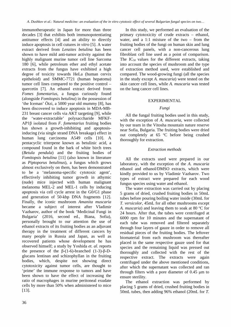
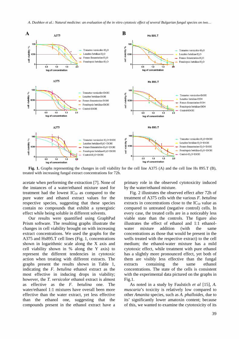
Fig. 1. Graphs representing the changes in cell viability for the cell line A375 (A) and the cell line Hs 895.T (B),
treated with increasing fungal extract concentrations for 72h.
acetate when performing the extraction [7]. None of the instances of a water/ethanol mixture used for treatment had the lowest IC50 as compared to the pure water and ethanol extract values for the respective species, suggesting that these species contain no compounds that exhibit a synergistic effect while being soluble in different solvents.
Our results were quantified using GraphPad Prism software. The resulting graphs illustrate the changes in cell viability brought on with increasing extract concentrations. We used the graphs for the A375 and Hs895.T cell lines (Fig. 1, concentrations shown in logarithmic scale along the X axis and cell viability shown in % along the Y axis) to represent the different tendencies in cytotoxic action when treating with different extracts. The graphs present the results shown in Table 1, indicating the F. betulina ethanol extract as the most effective in inducing drops in viability; however, the T. versicolor ethanol extract is almost as effective as the F. betulina one. The water/ethanol 1:1 mixtures have overall been more effective than the water extract, yet less effective than the ethanol one, suggesting that the compounds present in the ethanol extract have a
primary role in the observed cytotoxicity induced by the water/ethanol mixture.
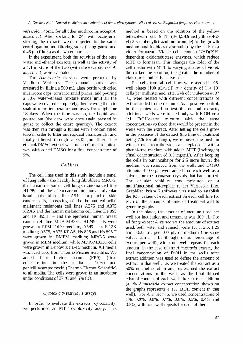
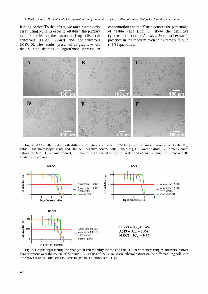
Fig. 2 illustrates the observed effect after 72h of treatment of A375 cells with the various F. betulina extracts in concentrations close to the IC50 value as compared to untreated (negative control) cells. In every case, the treated cells are in a noticeably less viable state than the controls. The figure also illustrates the effect of ethanol and 1:1 ethanol-water mixture addition (with the same concentrations as those that would be present in the wells treated with the respective extract) to the cell medium; the ethanol-water mixture has a mild cytotoxic effect, while treatment with pure ethanol has a slightly more pronounced effect, yet both of them are visibly less effective than the fungal extracts containing the same ethanol concentrations. The state of the cells is consistent with the experimental data pictured on the graphs in Fig.1.
As noted in a study by Faulstich et al [15], A. muscaria’s toxicity is relatively low compared to other Amanita species, such as A. phalloides, due to its’ significantly lower amatoxin content; because of this, we wanted to examine the cytotoxicity of its
A. Dushkov et al.: Natural medicine: an evaluation of the in vitro cytotoxic effect of several Bulgarian fungal species on two…
40
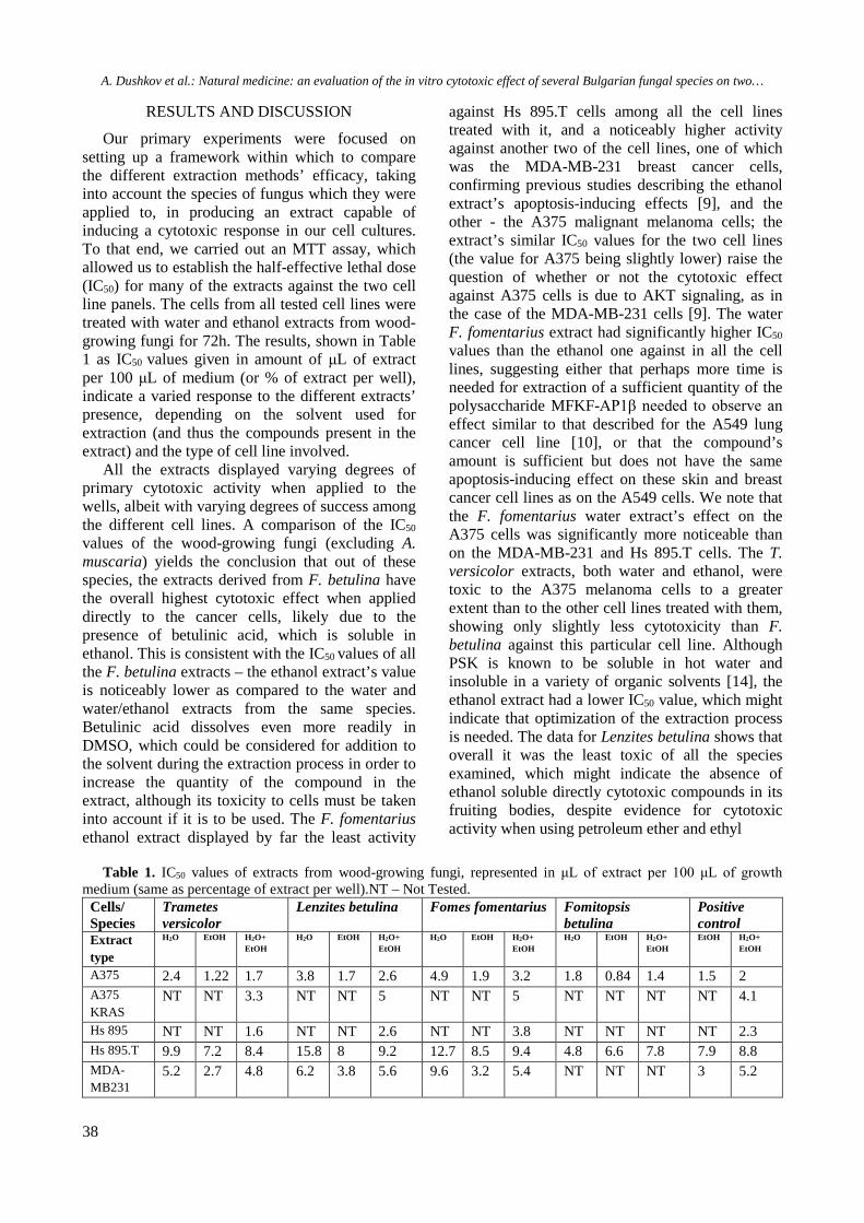
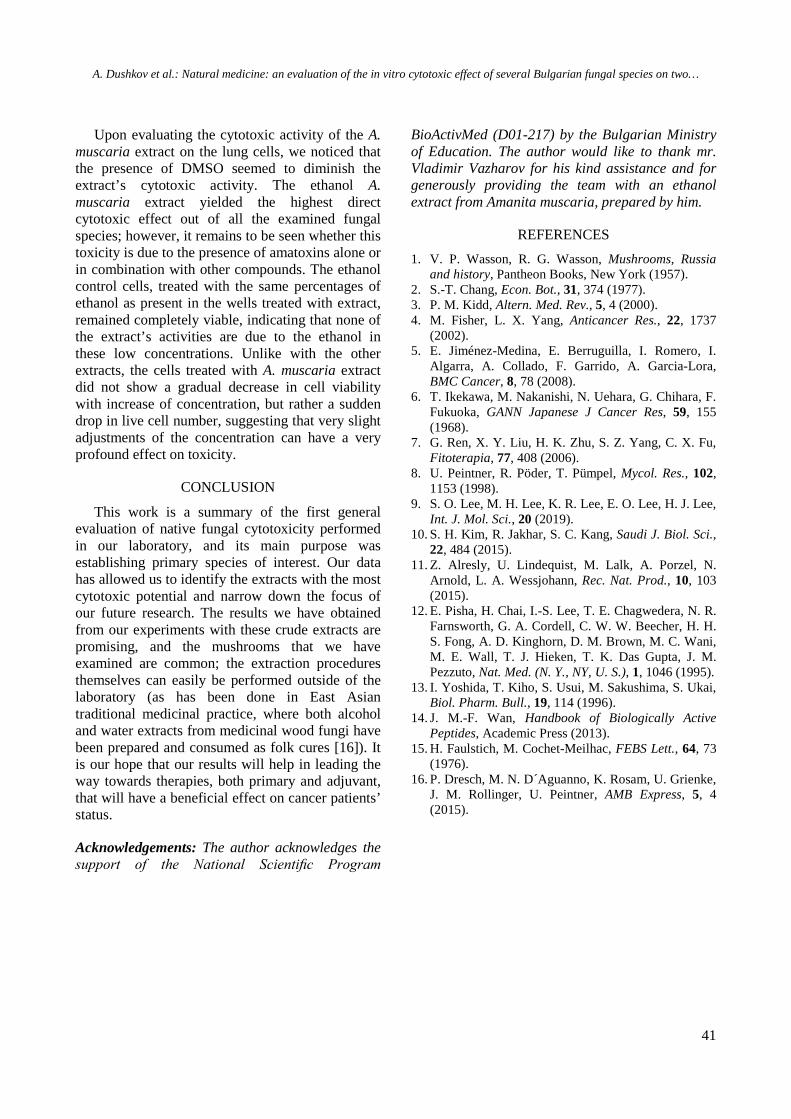
fruiting bodies. To that effect, we ran a cytotoxicity assay using MTT in order to establish the primary cytotoxic effect of the extract on lung cells, both cancerous (H1299, A549) and non-cancerous (MRC-5). The results, presented as graphs where the X axis denotes a logarithmic increase in
concentration and the Y axis denotes the percentage of viable cells (Fig. 3), show the definitive cytotoxic effect of the A. muscaria ethanol extract’s presence in the medium even in extremely minute (<1%) quantities.
Fig. 2. A375 cells treated with different F. betulina extracts for 72 hours with a concentration equal to the IC50 value; light microscopy, magnified 10x. A – negative control cells (untreated), B – water extract, C – water-ethanol extract mixture, D – ethanol extract, E – control cells treated with a 1:1 water and ethanol mixture, F – control cells treated with ethanol.
Fig. 3. Graphs representing the changes in cell viability for the cell line H1299 with increasing A. muscaria extract
concentrations over the course of 72 hours. IC50 values of the A. muscaria ethanol extract on the different lung cell lines are shown here as a final ethanol percentage concentration per 100 μL.
A. Dushkov et al.: Natural medicine: an evaluation of the in vitro cytotoxic effect of several Bulgarian fungal species on two…
41
Upon evaluating the cytotoxic activity of the A. muscaria extract on the lung cells, we noticed that the presence of DMSO seemed to diminish the extract’s cytotoxic activity. The ethanol A. muscaria extract yielded the highest direct cytotoxic effect out of all the examined fungal species; however, it remains to be seen whether this toxicity is due to the presence of amatoxins alone or in combination with other compounds. The ethanol control cells, treated with the same percentages of ethanol as present in the wells treated with extract, remained completely viable, indicating that none of the extract’s activities are due to the ethanol in these low concentrations. Unlike with the other extracts, the cells treated with A. muscaria extract did not show a gradual decrease in cell viability with increase of concentration, but rather a sudden drop in live cell number, suggesting that very slight adjustments of the concentration can have a very profound effect on toxicity.
CONCLUSION
This work is a summary of the first general evaluation of native fungal cytotoxicity performed in our laboratory, and its main purpose was establishing primary species of interest. Our data has allowed us to identify the extracts with the most cytotoxic potential and narrow down the focus of our future research. The results we have obtained from our experiments with these crude extracts are promising, and the mushrooms that we have examined are common; the extraction procedures themselves can easily be performed outside of the laboratory (as has been done in East Asian traditional medicinal practice, where both alcohol and water extracts from medicinal wood fungi have been prepared and consumed as folk cures [16]). It is our hope that our results will help in leading the way towards therapies, both primary and adjuvant, that will have a beneficial effect on cancer patients’ status.
Acknowledgements: The author acknowledges the support of the National Scientific Program
BioActivMed (D01-217) by the Bulgarian Ministry of Education. The author would like to thank mr. Vladimir Vazharov for his kind assistance and for generously providing the team with an ethanol extract from Amanita muscaria, prepared by him.
REFERENCES 1. V. P. Wasson, R. G. Wasson, Mushrooms, Russia
and history, Pantheon Books, New York (1957). 2. S.-T. Chang, Econ. Bot., 31, 374 (1977). 3. P. M. Kidd, Altern. Med. Rev., 5, 4 (2000). 4. M. Fisher, L. X. Yang, Anticancer Res., 22, 1737
(2002). 5. E. Jiménez-Medina, E. Berruguilla, I. Romero, I.
Algarra, A. Collado, F. Garrido, A. Garcia-Lora, BMC Cancer, 8, 78 (2008).
6. T. Ikekawa, M. Nakanishi, N. Uehara, G. Chihara, F. Fukuoka, GANN Japanese J Cancer Res, 59, 155 (1968).
7. G. Ren, X. Y. Liu, H. K. Zhu, S. Z. Yang, C. X. Fu, Fitoterapia, 77, 408 (2006).
8. U. Peintner, R. Pöder, T. Pümpel, Mycol. Res., 102, 1153 (1998).
9. S. O. Lee, M. H. Lee, K. R. Lee, E. O. Lee, H. J. Lee, Int. J. Mol. Sci., 20 (2019).
10. S. H. Kim, R. Jakhar, S. C. Kang, Saudi J. Biol. Sci., 22, 484 (2015).
11. Z. Alresly, U. Lindequist, M. Lalk, A. Porzel, N. Arnold, L. A. Wessjohann, Rec. Nat. Prod., 10, 103 (2015).
12. E. Pisha, H. Chai, I.-S. Lee, T. E. Chagwedera, N. R. Farnsworth, G. A. Cordell, C. W. W. Beecher, H. H. S. Fong, A. D. Kinghorn, D. M. Brown, M. C. Wani, M. E. Wall, T. J. Hieken, T. K. Das Gupta, J. M. Pezzuto, Nat. Med. (N. Y., NY, U. S.), 1, 1046 (1995).
13. I. Yoshida, T. Kiho, S. Usui, M. Sakushima, S. Ukai, Biol. Pharm. Bull., 19, 114 (1996).
14. J. M.-F. Wan, Handbook of Biologically Active Peptides, Academic Press (2013).
15. H. Faulstich, M. Cochet-Meilhac, FEBS Lett., 64, 73 (1976).
16. P. Dresch, M. N. D´Aguanno, K. Rosam, U. Grienke, J. M. Rollinger, U. Peintner, AMB Express, 5, 4 (2015).