budesonide ameliorates lung injury induced by large volume
TRANSCRIPT
RESEARCH ARTICLE Open Access
Budesonide ameliorates lung injuryinduced by large volume ventilationYing-Nan Ju1, Kai-Jiang Yu1 and Guo-Nian Wang2*
Abstract
Background: Ventilation-induced lung injury (VILI) is a health problem for patients with acute respiratory dysfunctionsyndrome. The aim of this study was to investigate the effectiveness of budesonide in treating VILI.
Methods: Twenty-four rats were randomized to three groups: a ventilation group, ventilation/budesonide group, andsham group were ventilated with 30 ml/kg tidal volume or only anesthesia for 4 hor saline or budesonide airwayinstillation immediately after ventilation. The PaO2/FiO2and wet-to-dry weight ratios, protein concentration, neutrophilcount, and neutrophil elastase levels in bronchoalveolar lavage fluid (BALF) and the levels of inflammation-relatedfactors were examined. Histological evaluation of and apoptosis measurement inthe lung were conducted.
Results: Compared with that in the ventilation group, the PaO2/FiO2 ratio was significantly increased by treatment withbudesonide. The lung wet-to-dry weight ratio, total protein, neutrophil elastase level, and neutrophilcount in BALF weredecreased in the budesonide group. The BALF and plasma tumor necrosis factor (TNF)-α, interleukin (IL)-1β, IL-6,intercellular adhesion molecule (ICAM)-1, and macrophage inflammatory protein (MIP)-2 levels were decreased,whereas the IL-10 level was increased in the budesonide group. The phosphorylated nuclear factor (NF)-kBlevelsin lung tissue were inhibited by budesonide. The histological changes in the lung and apoptosis were reducedby budesonide treatment. Bax, caspase-3, and cleaved caspase-3 were down-regulated, and Bcl-2 was up-regulated bybudesonide.
Conclusions: Budesonide ameliorated lung injury induced by large volume ventilation, likely by improving epithelialpermeability, decreasing edema, inhibiting local and systemic inflammation, and reducing apoptosis in VILI.
Keywords: Budesonide, Lung injury, Mechanical ventilation
BackgroundMechanical ventilation (MV) is indispensable for pa-tients with acute respiratory distress syndrome (ARDS),and it is required for about 39 % patients in intensivecare units [1]. However, MV can damage injured lungsin patients with ARDS [2]. Studies have shown thatabout 24 % of ARDS patients treated with MV devel-oped ventilator-induced lung injury (VILI) [3], whichresulted in a 40–50 % mortality rate [4]. MV with a largevolume may lead to alveolar overstretching, increasealveolar-capillary permeability, and cause pulmonaryedema [5] and lung focal inflammation [6]. Small tidalvolume MV can reduce the lung injury and lower the
mortality of ARDS [4]; however, ARDS remains a majorproblem still associated witha mortality of 25–45 % inintensive care units [7]. Therefore, it is imperative to de-velop alternative therapies to attenuate VILI.Studies have shown that the imbalance of pro- and
anti-inflammatory cytokines plays a critical role in thepathogenesis of VILI [8, 9]. During VILI, cytokines arereleased, leucocytes are recruited to the lung, and lungpermeability is increased, resulting in lung edema anddeterioration of pulmonary gas exchange [10]. Moreover,the cytokines released from injured endothelial and epi-thelial can enter the blood and cause systemic inflamma-tion and injury to other organs.Glucocorticoids can ameliorate the VILI [11, 12]. How-
ever, the systemic use of glucocorticoids may cause im-munosuppression and steroid resistance [13]. In addition,systemic use of glucocorticoids was not found toimprove
* Correspondence: [email protected] of Anesthesiology, Cancer Hospital of Harbin MedicalUniversity, Pain Research Institute of Heilongjiang Academy of MedicalSciences, No. 150 Haping Rd., Nangang District, Harbin 150081, ChinaFull list of author information is available at the end of the article
© 2016 The Author(s). Open Access This article is distributed under the terms of the Creative Commons Attribution 4.0International License (http://creativecommons.org/licenses/by/4.0/), which permits unrestricted use, distribution, andreproduction in any medium, provided you give appropriate credit to the original author(s) and the source, provide a link tothe Creative Commons license, and indicate if changes were made. The Creative Commons Public Domain Dedication waiver(http://creativecommons.org/publicdomain/zero/1.0/) applies to the data made available in this article, unless otherwise stated.
Ju et al. BMC Pulmonary Medicine (2016) 16:90 DOI 10.1186/s12890-016-0251-z
the outcome of ARDS, butinstead led to neuromuscularweakness and increased mortality risk for patients withARDS [14]. In contrast, administration of glucocorticoidsthrough inhalation relieves symptoms with less clinicalside effects. We also found that budesonide can amelioratethe lung injury induced by one-lung ventilation or endo-toxin in our clinical work and experiments [15, 16]. Otherstudies also have shown that budesonide can attenuatelung injury induced by chlorine gas, surfactant-depletion,or aspiration [17–20]. Therefore, we hypothesized thatbudesonide can reduce the incidence of VILI. In thisstudy, we investigated the effect of budesonide on VILIusing a rat model. Our data indicated that budesonidemay reduce VILI, providing an alternative approach toattenuating VILI.
MethodsAnimal experimentAll Wistar male rats were fasted and provided with waterad libitum for 24 h before the study. Twenty-four rats wererandomized to three groups: a sham group (S), a ventila-tion group (V), and a ventilation/budesonide group (VB)(n = 8 per group). Rats in the V and VB groups were venti-lated for 4 h with tidal volume 30 ml/kg [21, 22] (respira-tory rate: 50/min, inspiratory expiratory ratio: 1:1). All ratswere anesthetized using 3 % pentobarbital sodium (30 mg/kg intraperitoneally). The S group only received anesthesia.A tracheotomy was performed for rats in the V and VBgroups. The caudal vein and artery were cannulated to col-lect blood, analyze blood gas, and perform injection. Afterinjection of rocuronium (0.6 mg/kg), the rats in the V andVB groups received saline or budesonide 1 mg/kg by air-way instillation immediately after ventilation. All therats were maintained under anesthesia with 3 % pento-barbital sodium (10 mg/kg) and rocuronium (0.6 mg/kg) for a 1-h interval. The arterial blood analyses wereperformed, and the peripheral blood samples were col-lected at baseline (immediately after ventilation), 1, 2,
and 4 h after ventilation (T0-T3). After ventilation for4 h, all the rats were sacrificed after anesthesia, and thelungs were collected for further analysis.
Arterial blood gas analysisThe arterial blood gases from T0 to T3 were analyzedusing a Bayer Rapidlab 348 (Bayer Diognostics, Germany).PaO2/FiO2 ratios were calculated.
Pulmonary alveolocapillary permeabilityAfter ventilation for 4 h, the right upper lungs wereweighed and then dried at 60 °C for 48 h. The ratio ofwet/dry weight (W/D) was calculated.
Preparation of bronchoalveolar lavage fluid (BALF)BALF was collected from the left lung by infusing chilledsaline (4 °C, 15 ml/kg) containing (EDTA)-2Na and with-drawal five times. Cell differentiation was determined bystaining using a cytocentrifuged spin preparation (CF-RD,Sakura, Tokyo, Japan) of the BALF. The BALF was centri-fuged at 1000 g at 4 °C for 15 min. After centrifugation,the BALF were immediately stored at -80 °C. The neutro-phil levels in the BALF were counted with a cell counter.
Histopathologic analysis of lung tissueThe right lower lung was fixed with 10 % formalin, em-bedded in paraffin, and cut into 4-μm sections. Thesections were stained with hematoxylin and eosin. Twoindependent pathologists analyzed and scored the lunginjury under light microscopy from 0 to 4 (0, minimumdamage; 1, mild damage; 2, moderate damage; 3, severedamage; and 4, maximum damage), according to the as-sessment of alveolar congestion, edema, neutrophil in-filtration in the airspace or vessel wall, hemorrhage, thethickness of the alveolar wall, and hyaline membraneformation.
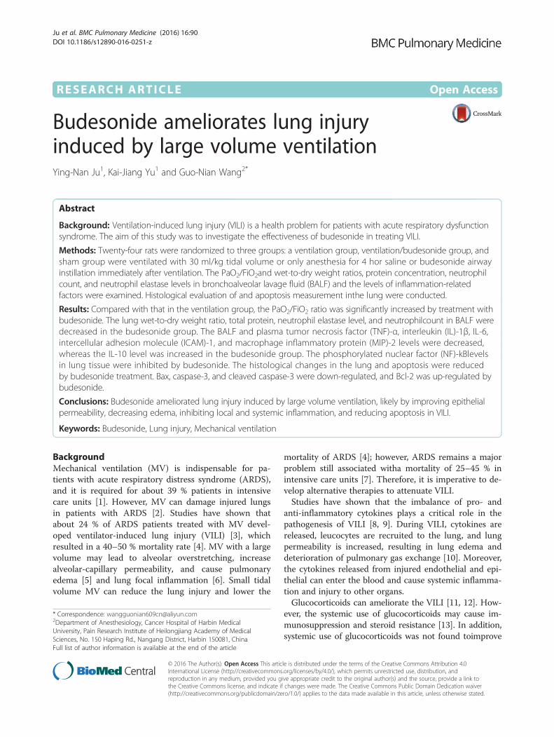
Fig. 1 The effect of budesonide on wet/dry weight ratio, protein concentration, and PaO2/FiO2 in VILI. *P < 0.05, compared with the S group;#P < 0.05, compared with the V group. ( , S group; , V group; , VB group)
Ju et al. BMC Pulmonary Medicine (2016) 16:90 Page 2 of 10
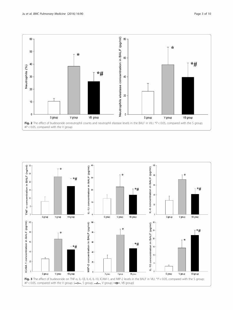
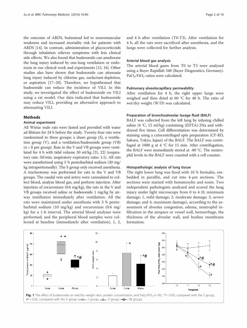
Fig. 2 The effect of budesonide onneutrophil counts and neutrophil elastase levels in the BALF in VILI. *P < 0.05, compared with the S group;#P < 0.05, compared with the V group
Fig. 3 The effect of budesonide on TNF-α, IL-1β, IL-6, IL-10, ICAM-1, and MIP-2 levels in the BALF in VILI. *P < 0.05, compared with the S group;#P < 0.05, compared with the V group. ( , S group; , V group; , VB group)
Ju et al. BMC Pulmonary Medicine (2016) 16:90 Page 3 of 10
TUNEL staining of lung sectionsA lobe of each right lung was examined for apoptosis usingTUNEL staining with an Apoptosis Assay kit (Roche Diag-nostics GmbH, Science, Mannheim, Germany). The slideswere incubated with proteinase Kfor 30 min and rinsedtwice with phosphate-buffered saline (PBS). Then theywere immersed in TUNEL reaction mixture at 37 °C for60 min. After washing with PBS three times, the endogen-ous peroxidase activity was quenched with 0.3 % H2O2
and covered with extra-avidin peroxidase, followed byimmersion in adiaminobenzidine solution. The slideswere counterstained with Mayer-hematoxylin, dehy-drated, and mounted. The cells showing brownishstaining in the nuclei were judged as apoptotic. Ten im-ages were randomly selected from each section, and atleast 1,000 cells were counted to calculate the apoptosisindex by independent pathologists.
Western blottingThe soluble protein was extracted from right lung tissueusing lysis buffer containing protein inhibitors (BeyotimeBiotechnology, Jiangsu, China). The concentration of thesample protein was determined using the Bradford assay.Aliquots of homogenate protein were resolved in polyacryl-amide gels and transferred onto polyvinylidene fluoride
membranes. The membranes were blocked with 5 % drymilk and then probed with antibodies for Bax, Bcl-2,caspase-3, phosphorylated NF-kB (Santa Cruz Biotechnol-ogy, Santa Cruz, CA, USA), and cleaved caspase-3 (Sigma-Aldrich, St. Louis, Missouri, USA), followed by incubationwith horseradish peroxidase-linked secondary antibodies(Santa Cruz Biotechnology). The bands were visualized viaenhanced chemiluminescence.
Statistical analysisAll normally distributed data are presented as mean andstandard deviation (SD) and were analyzed using SPSS11.0 (SPSS, Chicago, IL, USA). The normally distributeddata were analyzedusing the unpaired t test for a singletime-point or repeated measures analysis of variance.The non-normally distributed data were analyzed usingMann-Whitney rank sum test, and histologic data wereanalyzed using the Wilcoxon U-test.
ResultsBudesonide improves alveolocapillary permeability andthe W/D weight ratio and total protein in BALF in VILIWe evaluated the effect of budesonide on alveolocapillarypermeability in VILI. The results showed that the oxygenindex was significantly decreased after large volume
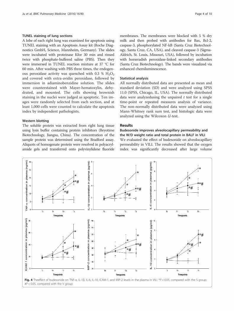
Fig. 4 Theeffect of budesonide on TNF-α, IL-1β, IL-6, IL-10, ICAM-1, and MIP-2 levels in the plasma in VILI. *P < 0.05, compared with the S group;#P < 0.05, compared with the V group
Ju et al. BMC Pulmonary Medicine (2016) 16:90 Page 4 of 10
ventilation, compared with that in the S group. Budeso-nide dramatically increased the oxygen index in the VBgroup (Fig. 1). The W/D weight ratio and total protein inBALF were significantly greater in the V and VB groups,compared to the S group, but were significantly less in theVB group compared to the V group (Fig. 1). These resultssuggested that budesonide improved alveolocapillary per-meability and the W/D weight ratio and total protein inBALF in VILI.
Budesonide inhibits inflammation in VILIWe evaluated the effect of budesonide on inflammatio-nin VILI. The results showed that the levels of neutro-phils in BALF were higher in the V and VB groups thaninthe S group, but were significantly lower in the VBgroup compared to theV group (Fig. 2). In addition, theconcentration of neutrophil elastase was significantlygreater in the V and VB groups compared to the S groupand lower in the VB group than in the V group (Fig. 2).The BALF and plasma TNF-α, IL-1β, IL-6, ICAM-1, andMIP-2 levels were significantly higher in the V and VBgroup than in theS group. Compared to the V group, the
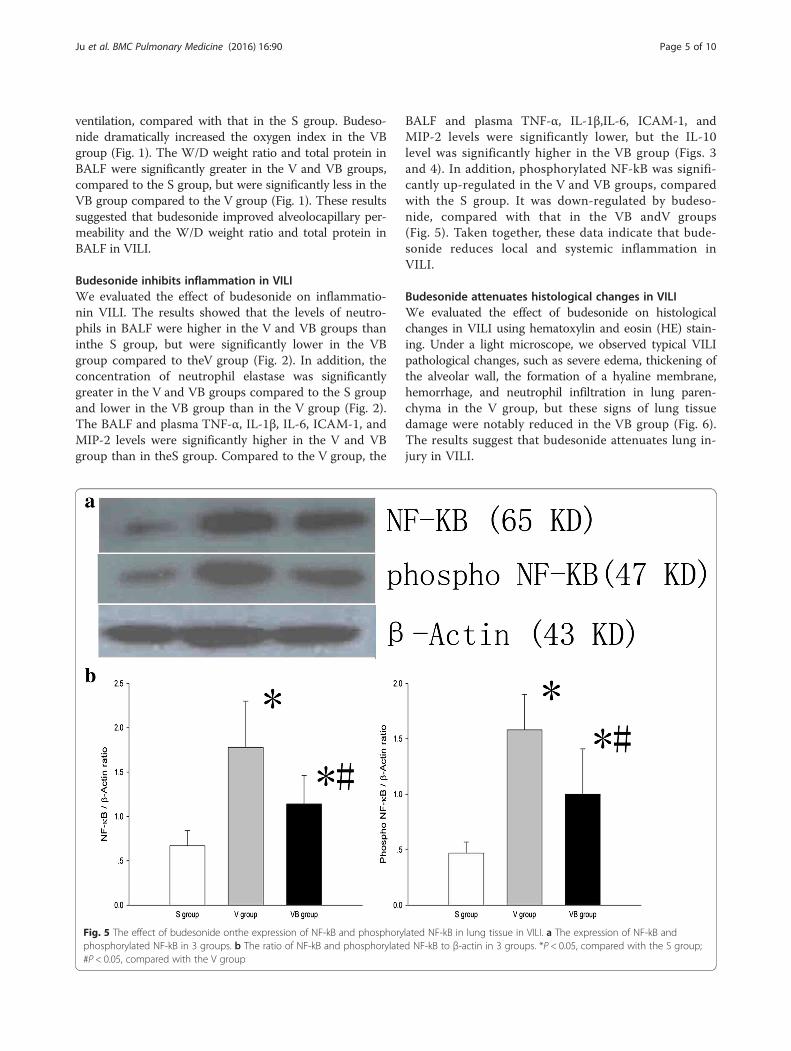
BALF and plasma TNF-α, IL-1β,IL-6, ICAM-1, andMIP-2 levels were significantly lower, but the IL-10level was significantly higher in the VB group (Figs. 3and 4). In addition, phosphorylated NF-kB was signifi-cantly up-regulated in the V and VB groups, comparedwith the S group. It was down-regulated by budeso-nide, compared with that in the VB andV groups(Fig. 5). Taken together, these data indicate that bude-sonide reduces local and systemic inflammation inVILI.
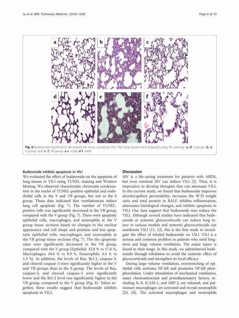
Budesonide attenuates histological changes in VILIWe evaluated the effect of budesonide on histologicalchanges in VILI using hematoxylin and eosin (HE) stain-ing. Under a light microscope, we observed typical VILIpathological changes, such as severe edema, thickening ofthe alveolar wall, the formation of a hyaline membrane,hemorrhage, and neutrophil infiltration in lung paren-chyma in the V group, but these signs of lung tissuedamage were notably reduced in the VB group (Fig. 6).The results suggest that budesonide attenuates lung in-jury in VILI.
Fig. 5 The effect of budesonide onthe expression of NF-kB and phosphorylated NF-kB in lung tissue in VILI. a The expression of NF-kB andphosphorylated NF-kB in 3 groups. b The ratio of NF-kB and phosphorylated NF-kB to β-actin in 3 groups. *P < 0.05, compared with the S group;#P < 0.05, compared with the V group
Ju et al. BMC Pulmonary Medicine (2016) 16:90 Page 5 of 10
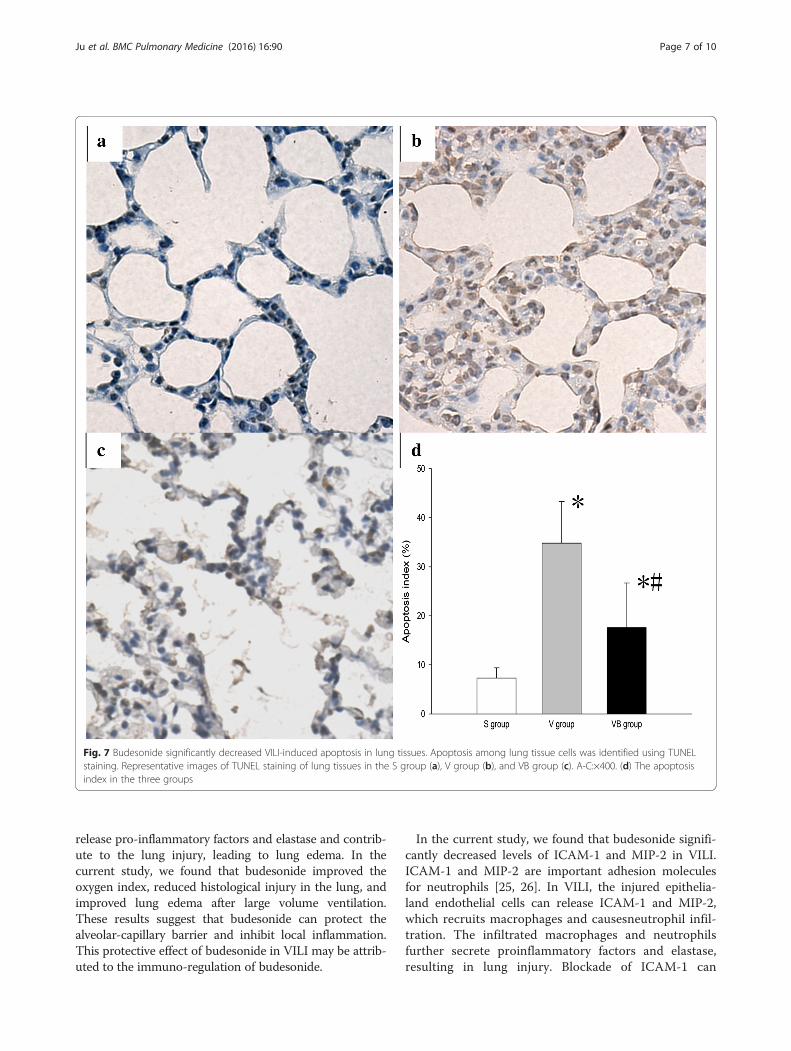
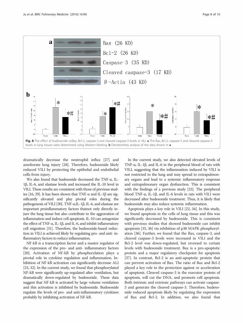
Budesonide inhibits apoptosis in VILIWe evaluated the effect of budesonide on the apoptosis oflung tissues in VILI using TUNEL staining and Westernblotting. We observed characteristic chromatin condensa-tion in the nuclei of TUNEL-positive epithelial and endo-thelial cells in the V and VB groups, but not in the Sgroup. These data indicated that ventilationcan inducelung cell apoptosis (Fig. 7). The number of TUNEL-positive cells was significantly decreased in the VB group,compared with the V group (Fig. 7). There were apoptoticepithelial cells, macrophages, and neutrophils in the Vgroup tissue sections based on changes in the nuclearappearance and cell shape and position and less apop-totic epithelial cells, macrophages, and neutrophils inthe VB group tissue sections (Fig. 7). The the apoptoticrates were significantly decreased in the VB group,compared with the V group (Epthelial: 32.8 % vs 17.6 %,Macrophages: 18.6 % vs 8.9 %, Neutrophils: 4.4 % vs1.5 %). In addition, the levels of Bax, Bcl-2, caspase-3,and cleaved caspase-3 were significantly higher in the Vand VB groups than in the S group. The levels of Bax,caspase-3, and cleaved caspase-3 were significantlylower and the Bcl-2 level was significantly higher in theVB group, compared to the V group (Fig. 8). Taken to-gether, these results suggest that budesonide inhibitsapoptosis in VILI.
DiscussionMV is a life-saving treatment for patients with ARDS,but even minimal MV can induce VILI [2]. Thus, it isimperative to develop therapies that can attenuate VILI.In the current study, we found that budesonide improvesalveolocapillary permeability, increases the W/D weightratio and total protein in BALF, inhibits inflammation,attenuates histological changes, and inhibits apoptosis inVILI. Our data support that budesonide may reduce theVILI. Although several studies have indicated that bude-sonide or systemic glucocorticoids can reduce lung in-jury in various models and systemic glucocorticoids canameliorate VILI [11, 12], this is the first study to investi-gate the effect of inhaled budesonide on VILI. VILI is aserious and common problem in patients who need long-term and large volume ventilation. The major injury isfound in their lungs. In this study, we administered bude-sonide through inhalation to avoid the systemic effect ofglucocorticoids and strengthen its local efficacy.During large volume ventilation, overstretching of epi-
thelial cells activates NF-kB and promotes NF-kB phos-phorylation. Under stimulation of mechanical ventilation,many chemoattractant and proinflammatory factors, in-cluding IL-8, ICAM-1, and MIP-2, are released, and pul-monary macrophages are activated and recruit neutrophils[23, 24]. The activated macrophages and neutrophils
Fig. 6 Budesonide significantly decreased the injury caused by VILI. The lung tissues were analyzed using HE staining: (a, d) S group; (b, e)V group; and (c, f) VB group. a-c, ×200; d-f, ×400
Ju et al. BMC Pulmonary Medicine (2016) 16:90 Page 6 of 10
release pro-inflammatory factors and elastase and contrib-ute to the lung injury, leading to lung edema. In thecurrent study, we found that budesonide improved theoxygen index, reduced histological injury in the lung, andimproved lung edema after large volume ventilation.These results suggest that budesonide can protect thealveolar-capillary barrier and inhibit local inflammation.This protective effect of budesonide in VILI may be attrib-uted to the immuno-regulation of budesonide.
In the current study, we found that budesonide signifi-cantly decreased levels of ICAM-1 and MIP-2 in VILI.ICAM-1 and MIP-2 are important adhesion moleculesfor neutrophils [25, 26]. In VILI, the injured epithelia-land endothelial cells can release ICAM-1 and MIP-2,which recruits macrophages and causesneutrophil infil-tration. The infiltrated macrophages and neutrophilsfurther secrete proinflammatory factors and elastase,resulting in lung injury. Blockade of ICAM-1 can
Fig. 7 Budesonide significantly decreased VILI-induced apoptosis in lung tissues. Apoptosis among lung tissue cells was identified using TUNELstaining. Representative images of TUNEL staining of lung tissues in the S group (a), V group (b), and VB group (c). A-C:×400. (d) The apoptosisindex in the three groups
Ju et al. BMC Pulmonary Medicine (2016) 16:90 Page 7 of 10
dramatically decrease the neutrophil influx [27] andameliorate lung injury [28]. Therefore, budesonide likelyreduced VILI by protecting the epithelial and endothelialcells from injury.We also found that budesonide decreased the TNF-α, IL-
1β, IL-6, and elastase levels and increased the IL-10 level inVILI. These results are consistent with those of previous stud-ies [16, 29]. It has been shown that TNF-α and IL-1β are sig-nificantly elevated and play pivotal roles during thepathogenesis of VILI [30]. TNF-α,IL-1β, IL-6, and elastase areimportant proinflammatory factors thatnot only directly in-jure the lung tissue but also contribute to the aggravation ofinflammation and induce cell apoptosis. IL-10 can antagonizethe effect of TNF-α, IL-1β, and IL-6, and inhibit inflammatorycell migration [31]. Therefore, the budesonide-based reduc-tion in VILI is achieved likely by regulating pro- and anti- in-flammatory factors to reduce inflammation.NF-kB is a transcription factor and a master regulator of
the expression of the pro- and anti- inflammatory factors[20]. Activation of NF-kB by phosphorylation plays apivotal role in cytokine regulation and inflammation. In-hibition of NF-kB activation can significantly decrease ALI[23, 32]. In the current study, we found that phosphorylatedNF-kB were significantly up-regulated after ventilation, butdramatically down-regulated by budesonide. These datasuggest that NF-kB is activated by large volume ventilationand this activation is inhibited by budesonide. Budesonideregulate the levels of pro- and anti-inflammatory cytokinesprobably by inhibiting activation of NF-kB.
In the current study, we also detected elevated levels ofTNF-α, IL-1β, and IL-6 in the peripheral blood of rats withVILI, suggesting that the inflammation induced by VILI isnot restricted to the lung and may spread to extrapulmon-ary organs and lead to a systemic inflammatory responseand extrapulmonary organ dysfunction. This is consistentwith the findings of a previous study [33]. The peripheralblood TNF-α, IL-1β, and IL-6 levels in rats with VILI weredecreased after budesonide treatment. Thus, it is likely thatbudesonide may also reduce systemic inflammation.Apoptosis plays a key role in VILI [22, 34]. In this study,
we found apoptosis in the cells of lung tissue and this wassignificantly decreased by budesonide. This is consistentwith previous studies that showed budesonide can inhibitapoptosis [35, 36] via inhibition of p38 MAPK phosphoryl-ation [36]. Further, we found that the Bax, caspase-3, andcleaved caspase-3 levels were increased in VILI and theBcl-2 level was down-regulated, but reversed to certainlevels with budesonide treatment. Bax is a pro-apoptoticprotein and a major regulatory checkpoint for apoptosis[37]. In contrast, Bcl-2 is an anti-apoptotic protein thatcan prevent activation of Bax. The ratio of Bax and Bcl-2played a key role in the protection against or accelerationof apoptosis. Cleaved caspase-3 is the executor protein ofapoptosis, will cut the DNA, and promote cell apoptosis.Both intrinsic and extrinsic pathways can activate caspase-3 and generate the cleaved caspase-3. Therefore, budeso-nide reduced apoptosis likely by regulating the expressionof Bax and Bcl-2. In addition, we also found that
Fig. 8 The effect of budesonide onBax, Bcl-2, caspase-3 and cleaved caspase-3 levels in VILI. a The Bax, Bcl-2, caspase-3 and cleaved caspase-3levels in lung tissues were determined using Western blotting. b Densitometry analysis of the data shown in a
Ju et al. BMC Pulmonary Medicine (2016) 16:90 Page 8 of 10
macropahges and neutrophils underwent apoptosis. Dur-ing VILI, the macrophages and neutrophils were activatedand phagocytized the necrotic cells and then underwentapoptosis. However, in this study, we only compared theapoptosis of epithelial cells to evaluate the effect of bude-sonide on VILI. We can differentiate the macrophages andneutrophils from epithelial cells based on the position,shape, and nuclear characteristics of these cells.This study has several limitations. First, rats were venti-
lated with a tidal volume of 30 ml/kg, which is substan-tially higher than volumes used in clinical application. Ourpreliminary study showed that a lower tidal volume (10–15 ml/kg) did not cause a decline in the PaO2/FiO2 ratioand VILI. Therefore, we increased the tidal volume to30 ml/kg, and we successfully established the signifi-cantly decreased PaO2/FiO2and mild acute respiratorydistress syndrome. Therefore, we used the tidal volumeof 30 ml/kg to establish VILI. This is consistent withthe study by Li et al whoalso used the 30 ml/kg tidalvolume to induce ALI [21, 22]. Second, in this study,budesonide was administered at the onset of VILI, sup-porting the use of budesonide as a preventative treatment.Clinically, however, patients need mechanical ventilationsupport before dysfunction of or injury to the lung occurs.Third, we did not evaluate the purity of neutrophils inBALF, which may influence the judgment of the effects ofbudesonide on neutrophils in VILI. We will address theselimitations in our future studies.
ConclusionIn conclusion, in the current study, we found that budeso-nide ameliorated lung injury probably by improving epi-thelial permeability, decreasing edema, inhibiting localand systemic inflammation, and reducing apoptosis inVILI. We speculate that inhalation of budesonide reduceslung injury and edema via inhibition of NF-kB phosphor-ylation and decreased secretion of adhesion moleculesand pro-inflammatory factors, which reduceslocal and sys-temic inflammation. Budesonide inhalation may be a po-tential approach for ARDS therapy in clinical practice.
AbbreviationsARDS, acute respiratory distress syndrome; BALF, bronchoalveolar lavagefluid; FiO2, fraction of inspired of oxygen; HE, hematoxylin and eosin; ICAM,intercellular adhesion molecule; IL, interleukin; MAPK, mitogen-activatedprotein kinase; MIP, macrophage inflammatory protein; MV, mechanicalventilation; NF, nuclear factor; PaO2, partial pressure of arterial oxygen; PBS,phosphate-buffered saline; TNF, tumor necrosis factor; TUNEL, terminaldeoxynucleotidyl transferase-mediated biotinylated UTP nick end labeling;VILI, ventilation-induced lung injury; W/D, Wet/dry weight;
AcknowledgementWe appreciated doctor Fang-fang Niu for measuring the designing this studyand preparing the manuscript.
FundingThis research was supported by funds from the Translational MedicineSpecial Foundation of China Russia Medical Research Center (no. 201519 and
CR1418), the T echnological and Innovative Talent Foundation of Harbin(2012RFXXS041), and the Hai Yan Foundation of the Cancer Hospital ofHarbin Medical University (JJQN2016-02).
Availability of data and materialsAll the data and material can be available.
Authors’ contributionsY-NJ carried out the molecular genetic studies and drafted the manuscript.K-JY carried out the immunoassays. K-JY and G-NW participated in the designof the study and performed the statistical analysis. Y-NJ, K-JY and G-NWconceived of the study, and participated in its design and coordinationand helped to draft the manuscript. All authors read and approved thefinal manuscript.
Competing interestsThe authors declare that they have no competing interests.
Consent for publicationNot applicable.
Ethics approval and consent to participateThis research project was approved by the Second Affiliated Hospital ofHarbin Medical University, Harbin, China. All treatments were carried out inaccordance with the Institutional Animal Care and Use Committee of SecondAffiliated Hospital of Harbin Medical University and followed nationalguidelines for the treatment of animals. This study adheres to the AnimalResearch: Reporting of In Vivo Experiments guidelines.
Author details1Department of ICU, Cancer Hospital of Harbin Medical University, Harbin150081, China. 2Department of Anesthesiology, Cancer Hospital of HarbinMedical University, Pain Research Institute of Heilongjiang Academy ofMedical Sciences, No. 150 Haping Rd., Nangang District, Harbin 150081,China.
Received: 28 January 2016 Accepted: 26 May 2016
References1. Esteban A, Anzueto A, Alia I, Gordo F, Apezteguia C, Palizas F, Cide D,
Goldwaser R, Soto L, Bugedo G, et al. How is mechanical ventilation employedin the intensive care unit? An international utilization review. Am J Respir CritCare Med. 2000;161(5):1450–8.
2. Wolthuis EK, Vlaar AP, Choi G, Roelofs JJ, Juffermans NP, Schultz MJ.Mechanical ventilation using non-injurious ventilation settings causes lunginjury in the absence of pre-existing lung injury in healthy mice. Crit Care.2009;13(1):R1.
3. Gajic O, Dara SI, Mendez JL, Adesanya AO, Festic E, Caples SM, Rana R, StSauver JL, Lymp JF, Afessa B, et al. Ventilator-associated lung injury inpatients without acute lung injury at the onset of mechanical ventilation.Crit Care Med. 2004;32(9):1817–24.
4. Tobin MJ. Culmination of an era in research on the acute respiratorydistress syndrome. N Engl J Med. 2000;342(18):1360–1.
5. Kneyber MC, Zhang H, Slutsky AS. Ventilator-induced lung injury. Similarityand differences between children and adults. Am J Respir Crit Care Med.2014;190(3):258–65.
6. Caruso P. Ventilator-induced lung injury distribution: the key tounderstanding injury mechanisms. Am J Respir Crit Care Med. 2007;175(1):95–6. author reply 96.
7. Force ADT, Ranieri VM, Rubenfeld GD, Thompson BT, Ferguson ND, CaldwellE, Fan E, Camporota L, Slutsky AS. Acute respiratory distress syndrome: theBerlin Definition. JAMA. 2012;307(23):2526–33.
8. Wilson MR, O’Dea KP, Zhang D, Shearman AD, van Rooijen N, Takata M.Role of lung-marginated monocytes in an in vivo mouse model ofventilator-induced lung injury. Am J Respir Crit Care Med. 2009;179(10):914–22.
9. Wilson MR, Choudhury S, Goddard ME, O’Dea KP, Nicholson AG, Takata M.High tidal volume upregulates intrapulmonary cytokines in an in vivomouse model of ventilator-induced lung injury. J Appl Physiol. 2003;95(4):1385–93.
Ju et al. BMC Pulmonary Medicine (2016) 16:90 Page 9 of 10
10. Verbrugge SJ, Lachmann B, Kesecioglu J. Lung protective ventilatorystrategies in acute lung injury and acute respiratory distress syndrome: fromexperimental findings to clinical application. Clin Physiol Funct Imaging.2007;27(2):67–90.
11. Hegeman MA, Hennus MP, Cobelens PM, Kavelaars A, Jansen NJ, Schultz MJ,van Vught AJ, Heijnen CJ. Dexamethasone attenuates VEGF expression andinflammation but not barrier dysfunction in a murine model of ventilator-induced lung injury. PLoS One. 2013;8(2):e57374.
12. Ohta N, Shimaoka M, Imanaka H, Nishimura M, Taenaka N, Kiyono H,Yoshiya I. Glucocorticoid suppresses neutrophil activation in ventilator-induced lung injury. Crit Care Med. 2001;29(5):1012–6.
13. Peter JV, John P, Graham PL, Moran JL, George IA, Bersten A. Corticosteroidsin the prevention and treatment of acute respiratory distress syndrome(ARDS) in adults: meta-analysis. BMJ. 2008;336(7651):1006–9.
14. Steinberg KP, Hudson LD, Goodman RB, Hough CL, Lanken PN, Hyzy R,Thompson BT, Ancukiewicz M, National Heart L, Blood Institute AcuteRespiratory Distress Syndrome Clinical Trials N. Efficacy and safety ofcorticosteroids for persistent acute respiratory distress syndrome. N Engl JMed. 2006;354(16):1671–84.
15. Gao W, Ju N. Budesonide inhalation ameliorates endotoxin-induced lunginjury in rabbits. Exp Biol Med. 2015;240(12):1708–16.
16. Ju NY, Gao H, Huang W, Niu FF, Lan WX, Li F, Gao W. Therapeutic effect ofinhaled budesonide (Pulmicort(R) Turbuhaler) on the inflammatory responseto one-lung ventilation. Anaesthesia. 2014;69(1):14–23.
17. Wang J, Zhang L, Walther SM. Inhaled budesonide in experimental chlorinegas lung injury: influence of time interval between injury and treatment.Intensive Care Med. 2002;28(3):352–7.
18. Mokra D, Mokry J, Drgova A, Petraskova M, Bulikova J, Calkovska A.Intratracheally administered corticosteroids improve lung function inmeconium-instilled rabbits. J Physiol Pharmacol. 2007;58(Suppl 5(Pt 1)):389–98.
19. Kohno M, Haramoto M, Nakajima O, Yang L, Hinotsu S, Yokohira M, ImaidaK, Kawakami K. Antedrug budesonide by intrapulmonary treatmentattenuates bleomycin-induced lung injury in rats with minimal systemicadverse effects. Biol Pharm Bull. 2010;33(7):1206–11.
20. Jansson AH, Eriksson C, Wang X. Effects of budesonide and N-acetylcysteineon acute lung hyperinflation, inflammation and injury in rats. VasculPharmacol. 2005;43(2):101–11.
21. Li LF, Liao SK, Huang CC, Hung MJ, Quinn DA. Serine/threonine kinase-protein kinase B and extracellular signal-regulated kinase regulate ventilator-induced pulmonary fibrosis after bleomycin-induced acute lung injury: aprospective, controlled animal experiment. Crit Care. 2008;12(4):R103.
22. Huang CS, Kawamura T, Lee S, Tochigi N, Shigemura N, Buchholz BM, KlokeJD, Billiar TR, Toyoda Y, Nakao A. Hydrogen inhalation amelioratesventilator-induced lung injury. Crit Care. 2010;14(6):R234.
23. Held HD, Boettcher S, Hamann L, Uhlig S. Ventilation-induced chemokineand cytokine release is associated with activation of nuclear factor-kappaB andis blocked by steroids. Am J Respir Crit Care Med. 2001;163(3 Pt 1):711–6.
24. Halbertsma FJ, Vaneker M, Scheffer GJ, van der Hoeven JG. Cytokines andbiotrauma in ventilator-induced lung injury: a critical review of theliterature. Neth J Med. 2005;63(10):382–92.
25. Miyao N, Suzuki Y, Takeshita K, Kudo H, Ishii M, Hiraoka R, Nishio K, TamataniT, Sakamoto S, Suematsu M, et al. Various adhesion molecules impairmicrovascular leukocyte kinetics in ventilator-induced lung injury. Am J PhysiolLung Cell Mol Physiol. 2006;290(6):L1059–68.
26. Li LF, Liao SK, Lee CH, Huang CC, Quinn DA. Involvement of Akt andendothelial nitric oxide synthase in ventilation-induced neutrophilinfiltration: a prospective, controlled animal experiment. Crit Care. 2007;11(4):R89.
27. Beck-Schimmer B, Madjdpour C, Kneller S, Ziegler U, Pasch T, Wuthrich RP,Ward PA, Schimmer RC. Role of alveolar epithelial ICAM-1 inlipopolysaccharide-induced lung inflammation. Eur Respir J. 2002;19(6):1142–50.
28. Gao X, Xu N, Sekosan M, Mehta D, Ma SY, Rahman A, Malik AB. Differentialrole of CD18 integrins in mediating lung neutrophil sequestration andincreased microvascular permeability induced by Escherichia coli in mice. JImmunol. 2001;167(5):2895–901.
29. Rudiger JJ, Gencay M, Yang JQ, Bihl M, Tamm M, Roth M. Fast beneficialsystemic anti-inflammatory effects of inhaled budesonide and formoterolon circulating lymphocytes in asthma. Respirology. 2013;18(5):840–7.
30. Wilson MR, Choudhury S, Takata M. Pulmonary inflammation induced byhigh-stretch ventilation is mediated by tumor necrosis factor signaling inmice. Am J Physiol Lung Cell Mol Physiol. 2005;288(4):L599–607.
31. Opal SM, DePalo VA. Anti-inflammatory cytokines. Chest. 2000;117(4):1162–72.32. Yu PJ, Li JR, Zhu ZG, Kong HY, Jin H, Zhang JY, Tian YX, Li ZH, Wu XY,
Zhang JJ, et al. Praeruptorin D and E attenuate lipopolysaccharide/hydrochloric acid induced acute lung injury in mice. Eur J Pharmacol. 2013;710(1–3):39–48.
33. Belperio JA, Keane MP, Lynch 3rd JP, Strieter RM. The role of cytokinesduring the pathogenesis of ventilator-associated and ventilator-inducedlung injury. Semin Respir Crit Care Med. 2006;27(4):350–64.
34. Li LF, Liao SK, Ko YS, Lee CH, Quinn DA. Hyperoxia increases ventilator-induced lung injury via mitogen-activated protein kinases: a prospective,controlled animal experiment. Crit Care. 2007;11(1):R25.
35. Straumann A, Conus S, Degen L, Felder S, Kummer M, Engel H,Bussmann C, Beglinger C, Schoepfer A, Simon HU. Budesonide iseffective in adolescent and adult patients with active eosinophilicesophagitis. Gastroenterology. 2010;139(5):1526–37. 1537 e1521.
36. Gallelli L, Pelaia G, Fratto D, Muto V, Falcone D, Vatrella A, Curto LS, Renda T,Busceti MT, Liberto MC, et al. Effects of budesonide on P38 MAPK activation,apoptosis and IL-8 secretion, induced by TNF-alpha and Haemophilusinfluenzae in human bronchial epithelial cells. Int J ImmunopatholPharmacol. 2010;23(2):471–9.
37. Cartron PF, Juin P, Oliver L, Meflah K, Vallette FM. Impact of proapoptoticproteins Bax and Bak in tumor progression and response to treatment.Expert Rev Anticancer Ther. 2003;3(4):563–70.
• We accept pre-submission inquiries
• Our selector tool helps you to find the most relevant journal
• We provide round the clock customer support
• Convenient online submission
• Thorough peer review
• Inclusion in PubMed and all major indexing services
• Maximum visibility for your research
Submit your manuscript atwww.biomedcentral.com/submit
Submit your next manuscript to BioMed Central and we will help you at every step:
Ju et al. BMC Pulmonary Medicine (2016) 16:90 Page 10 of 10