brief definitive report sars-cov-2 triggered neutrophil
TRANSCRIPT

BRIEF DEFINITIVE REPORT
SARS-CoV-2–triggered neutrophil extracellular trapsmediate COVID-19 pathologyFlavio Protasio Veras1,2, Marjorie Cornejo Pontelli3,4, Camila Meirelles Silva1,2, Juliana E. Toller-Kawahisa1,2, Mikhael de Lima1,2,Daniele Carvalho Nascimento1,2, Ayda Henriques Schneider1,2, Diego Caetite1,2, Lucas Alves Tavares3,4, Isadora M. Paiva1,2,Roberta Rosales4, David Colón1,2, Ronaldo Martins3,4, Italo Araujo Castro3,4, Glaucia M. Almeida1,2, Maria Isabel Fernandes Lopes5,Maıra Nilson Benatti5, Letıcia Pastorelli Bonjorno5, Marcela Cavichioli Giannini5, Rodrigo Luppino-Assad5, Sergio Luna Almeida5,Fernando Vilar5, Rodrigo Santana5, Valdes R. Bollela5, Maria Auxiliadora-Martins5, Marcos Borges5, Carlos Henrique Miranda5,Antonio Pazin-Filho5, Luis Lamberti P. da Silva3,4, Larissa Dias Cunha4, Dario S. Zamboni1,4, Felipe Dal-Pizzol8, Luiz O. Leiria1,2, Li Siyuan6,Sabrina Batah6, Alexandre Fabro6, Thais Mauad7, Marisa Dolhnikoff7, Amaro Duarte-Neto7, Paulo Saldiva7, Thiago Mattar Cunha1,2,Jose Carlos Alves-Filho1,2, Eurico Arruda3,4, Paulo Louzada-Junior1,5, Rene Donizeti Oliveira5, and Fernando Queiroz Cunha1,2
Severe COVID-19 patients develop acute respiratory distress syndrome that may progress to cytokine storm syndrome, organdysfunction, and death. Considering that neutrophil extracellular traps (NETs) have been described as important mediators oftissue damage in inflammatory diseases, we investigated whether NETs would be involved in COVID-19 pathophysiology. Acohort of 32 hospitalized patients with a confirmed diagnosis of COVID-19 and healthy controls were enrolled. Theconcentration of NETs was augmented in plasma, tracheal aspirate, and lung autopsies tissues from COVID-19 patients, andtheir neutrophils released higher levels of NETs. Notably, we found that viable SARS-CoV-2 can directly induce the release ofNETs by healthy neutrophils. Mechanistically, NETs triggered by SARS-CoV-2 depend on angiotensin-converting enzyme 2,serine protease, virus replication, and PAD-4. Finally, NETs released by SARS-CoV-2–activated neutrophils promote lungepithelial cell death in vitro. These results unravel a possible detrimental role of NETs in the pathophysiology of COVID-19.Therefore, the inhibition of NETs represents a potential therapeutic target for COVID-19.
IntroductionThe coronavirus disease 2019 (COVID-19) became pandemic,affecting more than 4 million people worldwide, with more than300,000 deaths by May 2020. Caused by the severe acute res-piratory syndrome coronavirus 2 (SARS-CoV-2), COVID-19 re-sembles influenza, with a clinical picture ranging from mildupper airway symptoms in the majority of cases to severe lowerairway symptoms in a subgroup of patients, in which acuterespiratory distress syndrome develops and may rapidly pro-gress to respiratory failure due to intense acute lung injury, itsmajor cause of death (Lai et al., 2020). It is also known that thissubgroup of patients has cytokine storm syndrome, which seemsto be responsible for multi-organ failure (Chen et al., 2020). Inaddition, COVID-19 patients develop signs and symptoms similar
to those observed in sepsis, many of which result in micro-thrombosis, organ dysfunction, and eventually shock (Wu andMcGoogan, 2020; Magro et al., 2020; Guan et al., 2020). The firststep in SARS-CoV-2 infection is the molecular interactionbetween virus membrane glycoprotein spike (S) and theangiotensin-converting enzyme 2 (ACE2), which is expressedin the several host cells, including lung pneumocytes, epithelialcells, and endothelial cells (Qi et al., 2020; Lovren et al., 2008).To complete the fusion process, S protein needs to be cleaved byserine proteases such as TMPRSS2 (Shulla et al., 2011; Hoffmannet al., 2020).
The increased number of circulating neutrophils has beendescribed as an indicator of the severity of respiratory
.............................................................................................................................................................................1Center of Research in Inflammatory Diseases, Ribeirão Preto Medical School, University of São Paulo, Ribeirão Preto, São Paulo, Brazil; 2Department of Pharmacology,Ribeirão Preto Medical School, University of São Paulo, Ribeirão Preto, São Paulo, Brazil; 3Virology Research Center, Ribeirão Preto Medical School, University of São Paulo,Ribeirão Preto, São Paulo, Brazil; 4Department of Cell and Molecular Biology, Ribeirão Preto Medical School, University of São Paulo, Ribeirão Preto, São Paulo, Brazil;5Divisions of Clinical Immunology, Emergency, Infectious Diseases and Intensive Care Unit, Ribeirão Preto Medical School, University of São Paulo, Ribeirão Preto, São Paulo,Brazil; 6Pathology and Legal Medicine, Ribeirão Preto Medical School, University of São Paulo, Ribeirão Preto, São Paulo, Brazil; 7Department Pathology, School ofMedicine, University of São Paulo, São Paulo, Brazil; 8Laboratory of Experimental Pathophysiology, Graduate Program in Health Sciences, Health Sciences Unit, Universityof Southern Santa Catarina, Criciuma, Santa Catarina, Brazil.
Correspondence to Fernando Q. Cunha: [email protected]; Flavio P. Veras: [email protected]; Thiago Mattar Cunha: [email protected].
© 2020 Veras et al. This article is distributed under the terms of an Attribution–Noncommercial–Share Alike–No Mirror Sites license for the first six months after thepublication date (see http://www.rupress.org/terms/). After six months it is available under a Creative Commons License (Attribution–Noncommercial–Share Alike 4.0International license, as described at https://creativecommons.org/licenses/by-nc-sa/4.0/).
Rockefeller University Press https://doi.org/10.1084/jem.20201129 1 of 12
J. Exp. Med. 2020 Vol. 217 No. 12 e20201129
Dow
nloaded from http://rupress.org/jem
/article-pdf/217/12/e20201129/1414690/jem_20201129.pdf by guest on 12 M
arch 2022

symptoms and a poor clinical outcome in COVID-19 (Guan et al.,2020). Among effector mechanisms of neutrophils in inflam-matory diseases, neutrophil-derived extracellular traps (NETs)are some of the most important (Brinkmann et al., 2004;Papayannopoulos and Zychlinsky, 2009; Kaplan and Radic,2012; Jorch and Kubes, 2017). NETs are networks of extracel-lular fibers composed of DNA containing histones and granule-derived enzymes, such as myeloperoxidase (MPO) and elastase(Brinkmann et al., 2004). The process of NET formation byneutrophils, called NETosis, has been widely studied. In gen-eral, the process starts with neutrophil activation by patternrecognition receptors or chemokines, followed by ROS pro-duction and calcium mobilization, which leads to the activationof protein arginine deiminase 4 (PAD-4), an intracellular en-zyme involved in the deimination of arginine residues on his-tones (Li et al., 2010). In 2004, Brinkmann et al. (2004) initiallydescribed NETs as microbicidal mechanisms released by neu-trophils (Brinkmann et al., 2004). However, accumulating ev-idence demonstrated that NETs have double-edged–swordactivities. Besides their microbicidal activity, NETs have alsobeen implicated in tissue injury and, consequently, in thepathogenesis of several diseases, including rheumatoid arthri-tis (Khandpur et al., 2013; Sur Chowdhury et al., 2014), diabetes(Wong et al., 2015), and sepsis. Regarding sepsis, our group andothers have described that during experimental and clinicalsepsis, NETs are found in high concentrations in the blood andare positively correlated with biomarkers of vital organ injuriesand sepsis severity. Furthermore, disruption or inhibition ofNET release by pharmacological treatment with recombinanthuman DNase (rhDNase) or PAD-4 inhibitors, respectively,markedly reduced organ damage, especially in the lungs, andincreased the survival rate of severe septic mice (Colón et al.,2019; Czaikoski et al., 2016; Kambas et al., 2012; Martinod et al.,2015; Altrichter et al., 2010; Clark et al., 2007). The well-knownsimilarities between sepsis and key events involved in theCOVID-19 pathophysiology, such as cytokine overproduction(Mehta et al., 2020), microthrombosis (Magro et al., 2020;Dolhnikoff et al., 2020), and acute respiratory distress syn-drome (Lai et al., 2020), led us to hypothesize that NETs aretriggered during SARS-CoV-2 infection and might contribute totissue injury in COVID-19 patients. In this context, recent evi-dence indicates an increase of NETs in the plasma and lungs ofCOVID-19 patients (Middleton et al., 2020; Skendros et al.,2020; Zuo et al., 2020). However, the cellular and molecularmechanisms underlying NET production and their immuno-pathological role in COVID-19 are not fully understood. Here,we demonstrated that the concentration of NETs increases inthe plasma, tracheal aspirate, and lung tissue specimens ofautopsies from COVID-19 patients. Furthermore, we found thatcirculating neutrophils are infected with SARS-CoV-2 and re-lease high levels of NETs. Importantly, SARS-CoV-2 can di-rectly induce the release of NETs by healthy neutrophils, whichis dependent on ACE2–serine protease axis, virus replication,and PAD-4 signaling. Finally, NETs released by SARS-CoV-2–activated neutrophils promote lung epithelial apoptosis. Theseresults describe novel cellular and molecular mechanisms in-volved in the production of NETs by SARS-CoV-2 infection and
their possible detrimental role in the pathophysiology ofCOVID-19. Therefore, the inhibition of NET release or actionscould represent a potential therapeutic target for COVID-19.
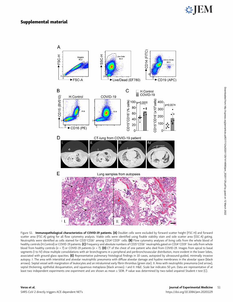
Results and discussionCOVID-19 patients’ blood neutrophils produce higher levels ofNETsAlthough the number of patients with COVID-19 is growingexponentially worldwide, there is no effective treatment for thisdisease. The knowledge of the mechanisms by which the hostdeals with the SARS-CoV-2 infection will certainly allow thedevelopment of new therapeutic strategies aiming to treatCOVID-19. Here, we aimed to investigate the possible role ofNETs in the pathophysiology of COVID-19. First, we confirmedthe SARS-CoV-2 infection of all patients (32 patients) by RT-PCRof nasopharyngeal samples using a standard protocol (Nallaet al., 2020; Table S1) and/or detection of specific antibodiesIgM and IgG against SARS-CoV-2 in plasma samples. The de-mographic and clinical characteristics of COVID-19 patients areshown in Table S1. The increased number of circulating neu-trophils is an indicator of a worse outcome in COVID-19 (Huanget al., 2020; Chen et al., 2020). We evaluated the number ofcirculating neutrophils (CD15+CD16+ cells) in COVID-19 patientsby flow cytometry. As recently reported (Magro et al., 2020), theCOVID-19 patients of the present cohort also showed significantneutrophilia (Fig. S1, A–C).
The levels of NETs in circulation and organ tissues are in-creased during sepsis (Colón et al., 2019; Czaikoski et al., 2016;Kambas et al., 2012; Martinod et al., 2015; Altrichter et al., 2010;Clark et al., 2007). Considering that several sepsis events such ascytokine storm and vital organs lesions are also observed inpatients with severe COVID-19 (Chen et al., 2020; Guan et al.,2020), we then determined the concentration of soluble NETs inthe plasma by measuring MPO–DNA complexes (Colón et al.,2019). Confirming previous findings (Middleton et al., 2020;Skendros et al., 2020; Zuo et al., 2020), higher levels of NETswere found in the plasma of COVID-19 patients compared withhealthy controls (Fig. 1 A). To verify whether the higher con-centration of NETs in the plasma of COVID-19 patients was aconsequence of the increased number of circulating neutrophilsor their enhanced ability to release these mediators, we purifiedblood neutrophils from controls and COVID-19 patients to ana-lyze the release of NETs in vitro. Using a normalized number ofcells, we found that blood neutrophils from COVID-19 patientsreleased higher levels of NETs compared with controls (Fig. 1 B).NETs can be microscopically visualized as extracellular fibers ofDNA colocalizing with MPO and citrullinated histone H3 (H3Cit;Brinkmann et al., 2004). Confirming these data, confocal mi-croscopy analysis also showed the increased ability of neu-trophils from COVID-19 patients to release NETs (Fig. 1 C). Thesedata were confirmed by Pearson’s correlation coefficientanalysis of colocalization between DAPI and MPO (Fig. 1 D).NETs from COVID-19 patients are typical rigid rods. Notably,more than 80% of neutrophils from COVID-19 patients werepositive for the NETs structures (Fig. 1 E). In addition, thebranch lengths of the released NETs by neutrophils from
Veras et al. Journal of Experimental Medicine 2 of 12
SARS-CoV-2 directly triggers ACE-dependent NETs https://doi.org/10.1084/jem.20201129
Dow
nloaded from http://rupress.org/jem
/article-pdf/217/12/e20201129/1414690/jem_20201129.pdf by guest on 12 M
arch 2022
COVID-19 patients were also larger than those from healthycontrols (Fig. 1 F).
One of the most important intracellular mechanisms thatmediate the release of NETs by neutrophils is the activation ofPAD-4. This enzyme catalyzes the citrullination of arginineresidues on histones to promote chromatin decondensation andextrusion of NETs (Li et al., 2010). Notably, the release of NETsby blood neutrophils from COVID-19 patients was reduced bythe incubation of cells with Cl-Amidine, an inhibitor of PAD-4(Fig. 1, G and H). These results indicate that in COVID-19 pa-tients, circulating neutrophils are more susceptible to the re-lease of PAD-4–dependent NETs, which might cause a systemicincrease of soluble NETs.
NETs are highly detected in the tracheal aspirate and lungtissue from COVID-19 patientsLung inflammation is the primary cause of life-threateningrespiratory disorder in critical and severe forms of COVID-19(Guan et al., 2020). NETs have been identified in the lung tissueof viral and nonviral infected patients and experimental animals(Sivanandham et al., 2018; Dicker et al., 2018). We next inves-tigated the content of NETs in the tracheal aspirate obtainedfrom patients with severe COVID-19 under mechanical ventila-tion admitted to the intensive care unit or in the lung sectionsfrom postmortem COVID-19. First, we found a higher concen-tration of NETs in the tracheal aspirate from COVID-19 patientscompared with airway fluid from healthy controls (Fig. 2 A). Theconcentration of NETs in the tracheal aspirate was 10 timeshigher than observed in the plasma of COVID-19 patients (Fig. 1A and Fig. 2 A). The confocal microscopy analysis of pellet cellsfrom the tracheal aspirate also revealed typical NETs structureswith extracellular DNA costaining with MPO and H3Cit(Fig. 2 B).
We next investigated whether lung tissue damage observedin COVID-19 patients was associated with the local presence ofNETs. First, we confirmed extensive injury in the lungs ofCOVID-19 patients by computed tomography (CT) analysis. Weobserved in CT imaging multiple consolidations with air bron-chograms in all pulmonary fields, with peripheral and peri-bronchovascular distribution, more evident in the lower lobes,associated with ground-glass opacities, and compatible withdiffuse alveolar damage (Fig. S1 D). Next, we performed aminimally invasive autopsy to harvest lung tissue and analyzedhistopathologic features. The histopathological analysis of thelung tissue from COVID-19 autopsies exhibited exudative andproliferative diffuse alveolar damage (Fig. S1, E [I]); intense al-veolar and small airway epithelial changes with cytopathic ef-fects and squamous metaplasia (Fig. S1 E [II]); mild lymphocyticinfiltration; and changes in pulmonary arterioles (endothelialswelling and small fibrinous thrombi). Neutrophilic pneumoniawas observed in samples from 6 out of 10 patients, in variabledegrees (Fig. S1 E). Confocal microscopy analysis of lung tissuesfrom COVID-19 postmortem individuals with the presence ofneutrophils also revealed the presence of characteristic NETstructure, with extracellular DNA staining colocalizing withMPO and H3Cit (Fig. 2 C). NETs were not found in the healthytissues obtained from lobectomy due to heart failure (Fig. 2 C).
These results indicate that NETs are released in the lung tissueand are associated with lung damage in COVID-19 patients.
SARS-CoV-2 replication induces the release of NETsSeveral stimuli trigger NETosis, including pathogen-associatedmolecular patterns, damage-associated molecular patterns, andinflammatory mediators, including cytokines and chemokines(Brinkmann et al., 2004; Keshari et al., 2012). There is also ev-idence that infection of neutrophils with several viruses trig-gered the formation of NETs (Saitoh et al., 2012; Funchal et al.,2015; Hiroki et al., 2020), which led us to investigate whetherSARS-CoV-2 is able to directly promote NET release by humanneutrophils. For that, neutrophils isolated from the blood ofhealthy controls were cultured in the presence of differentmultiplicities of infection (MOIs) of viable and inactivatedSARS-CoV-2. We found that viable, but not inactivated, SARS-CoV-2 increased the release of NETs in a MOI-dependent man-ner (Fig. 3, A and B). Importantly, SARS-CoV-2–induced releaseof NETs was abrogated by Cl-Amidine, a PAD-4 inhibitor (Fig. 3,A and B).
Similar to what we observed in neutrophils from COVID-19patients, NETs released by in vitro cultured healthy neutrophilsin the presence of SARS-CoV-2 have longer branch lengths(Fig. 3 C). Notably, we detected neutrophils with SARS-CoV-2antigens (Fig. 3 D). Neutrophils positive for SARS-CoV-2 an-tigens undergo higher NETosis than antigen-negative neu-trophils (Fig. 3, D and E). SARS-CoV-2 antigens were alsodetected in the blood neutrophils isolated in the samples from5 out of 11 COVID-19 patients, and these SARS-CoV-2+ neu-trophils are also more efficient at producing NETs (Fig. S2, Aand B). These results indicate that SARS-CoV-2 might directlyactivate neutrophils to release NETs. The lack of NET releasein the culture with inactivated SARS-CoV-2 indicates thatactive viral infection and/or replication might be necessary totrigger NET release.
To investigate this hypothesis, we treated SARS-CoV-2–exposed neutrophils with tenofovir disoproxil fumarate (TDF),an inhibitor of RNA polymerase that has been shown to reduceSARS-CoV-2 replication in Vero cells (Elfiky, 2020; Clososkiet al., 2020). TDF is a drug of the same class of remdesivir,which is effective in vitro against virus replication and accel-erates the recovery of COVID-19 patients (Yin et al., 2020; Beigelet al., 2020). Notably, TDF abrogated the release of NETs byneutrophils from healthy donors induced by SARS-CoV-2(Fig. 3 F), which was also associated with a reduction in theintracellular viral load (Fig. 3 G). Of note, TDF did not reducePMA-induced NET release, ruling out off-target effects of TDFon NET production (Fig. 3 H). Finally, to further support thereplicative process of SARS-CoV-2 in neutrophils, we assessedthe presence of double-stranded RNA (dsRNA) in situ by im-munofluorescence. dsRNA is generated by viral RNA polymer-ase as an intermediate in the genome replication of RNA viruses,including SARS-CoV-2 (Weber et al., 2006). Importantly, dsRNAwas detected in SARS-CoV-2–infected neutrophils 2 and 4 h afterinfection (Fig. 3 I). We did not detect dsRNA staining in non-infected cells or in cells incubated with an inactivated virus(Fig. 3 I). Taken together, these data suggest that the viral
Veras et al. Journal of Experimental Medicine 3 of 12
SARS-CoV-2 directly triggers ACE-dependent NETs https://doi.org/10.1084/jem.20201129
Dow
nloaded from http://rupress.org/jem
/article-pdf/217/12/e20201129/1414690/jem_20201129.pdf by guest on 12 M
arch 2022

replication cycle is crucial for the release of NETs by SARS-CoV-2–activated neutrophils. It is worth noting that our datadid not confirm that SARS-CoV-2 completes its replication cyclewith release of infectious progeny before the initiation of NE-Tosis process. Another important question that emerges fromthese data is regarding the molecular mechanism by whichactive SARS-CoV-2 replication promotes NET formation. Onepossible candidate would be TLR7, since it is known that thisintracellular sensor is involved in the recognition of single-stranded RNA virus (Lund et al., 2004). Indeed, TLR7 has
been implicated in the production of NETs by neutrophils in-fected with chikungunya virus and HIV (Hiroki et al., 2020;Saitoh et al., 2012). Although our data imply that SARS-CoV-2 isable to directly stimulate NETs, considering that the levels ofcytokines and chemokines are increased during severe COVID-19 (Gong et al., 2020 Preprint), we cannot ignore that cytokinesand chemokines are also activating NETosis during the ongoingCOVID-19. For instance, the serum of COVID-19 patients is ableto increase the release of NETs by healthy neutrophils(Middleton et al., 2020; Zuo et al., 2020).
Figure 1. COVID-19 patients produces high concen-trations of NETs. Plasma and neutrophils were isolatedfrom healthy controls and COVID-19 patients. (A) NETquantification by MPO-DNA PicoGreen assay in plasmafrom healthy controls (H.Control; n = 21) or COVID-19patients (n = 32). (B) Supernatants from cultures ofblood isolated neutrophils from healthy controls (n = 10)or COVID-19 patients (n = 11). NET quantification wasperformed using MPO-DNA PicoGreen assay. (C) Rep-resentative confocal analysis of NETs release by neu-trophils isolated from healthy controls (n = 10) orCOVID-19 patients (n = 11), cultured for 4 h at 37°C. Cellswere stained for nuclei (DAPI, blue), MPO (green), andH3Cit (red). Scale bar indicates 50 µm. (D) Colocaliza-tion of DAPI and MPO between healthy controls (n = 10)and COVID-19 (n = 10). The data depicts Pearson’scorrelation coefficient assessed by Fiji/ImageJ software.(E) Percentage of NETosis in neutrophil from COVID-19patients (n = 7). (F) NET length quantification. (G) NETquantification by MPO-DNA PicoGreen assay in the su-pernatants of blood-isolated neutrophils from COVID-19patients (n = 3) preincubated, or not, with PAD-4 in-hibitor (Cl-Amidine; 200 µM) for 4 h at 37°C. (H) Rep-resentative confocal images showing the presence ofNETs in isolated neutrophils from COVID-19 patients,treated or not, with Cl-Amidine (200 µM). Cells werestained for nuclei (DAPI, blue), MPO (green), and H3Cit(red). Scale bar indicates 50 µm. Data are representativeof at least two independent experiments and are shownas mean ± SEM. P value were determined by two-tailedunpaired (A, B, and D–F) or paired (G) Student t test.
Veras et al. Journal of Experimental Medicine 4 of 12
SARS-CoV-2 directly triggers ACE-dependent NETs https://doi.org/10.1084/jem.20201129
Dow
nloaded from http://rupress.org/jem
/article-pdf/217/12/e20201129/1414690/jem_20201129.pdf by guest on 12 M
arch 2022

SARS-CoV-2–induced NETs requires ACE2 and serine proteaseactivityCell entry of coronaviruses depends on the binding of viral spike(S) proteins to cellular receptors and on S protein priming byhost cell proteases. Evidence demonstrates that SARS-CoV-2uses ACE2 and the serine protease TMPRSS2 for entry in dif-ferent cell types (Li et al., 2003; Hoffmann et al., 2020; Lan et al.,2020; Yan et al., 2020). Thus, we investigated whether the re-lease of NETs in SARS-CoV-2–infected neutrophils depends onthe ACE2–TMPRSS2 axis. First, we determined ACE2 expressionby conventional PCR and Western blot in neutrophils from fourhealthy donors. As positive controls, we assessed ACE expres-sion in Caco-2 cell line (Yamashita et al., 2005) and HeLa cellstransduced with lentivirus expressing human ACE2 (Hela-ACE2). As a negative control, we used nontransduced HeLa cells.We detected mRNA and protein expression of ACE2 in neu-trophils from all donors (Fig. S2 C and Fig. 4 A). In accordancewith this, large-scale transcriptome data from six public data-bases also showed ACE2 expression in immune system cells,including neutrophils (Li et al., 2020). We then investigated therole of ACE2 and SARS-CoV-2 S protein interaction in the releaseof NETs by SARS-CoV-2. Thus, isolated neutrophils were treatedwith a neutralizing anti-hACE2 antibody (αACE2) or camostat,an inhibitor of the serine protease TMPRSS2 that blocks earlyinteractions of S protein with the ACE2 receptor (Hoffmannet al., 2020). Notably, both drugs abrogated SARS-CoV-2–induced NETs released by neutrophils (Fig. 4, B and C). The viralload of neutrophils exposed to SARS-CoV-2was also inhibited by
αACE2 or camostat (Fig. 4 D). These treatments did not modifyNET production by PMA-activated neutrophils (Fig. 4 E), sug-gesting that the ACE2/TMPRSS2 pathway is crucial for SARS-CoV-2 entry and release of NETs by neutrophils.
SARS-CoV-2–activated neutrophils induce lung epithelial celldeath through the release of NETsThe release of NETs has been associated with tissue damage inseveral immune-mediated diseases, including chronic obstruc-tive pulmonary disease, rheumatoid arthritis, and sepsis (Dickeret al., 2018; Khandpur et al., 2013; Colón et al., 2019). NETs causetissue damage by extracellular exposure of DNA, granular pro-teins such as MPO, and histone, inducing apoptosis and fibrosisprocesses (Thammavongsa et al., 2013; Yadav et al., 2019).COVID-19 is characterized by extensive damage in the alveolarepithelium (Dolhnikoff et al., 2020). To understand the possiblerole of released NETs in the pathophysiology of COVID-19, wenext explored the hypothesis that NETs would be involved in thedamage of lung epithelial cells. To this end, blood-isolated neu-trophils from healthy controls incubatedwith SARS-CoV-2 (MOI =1.0) were co-cultured with A549 cells, a human alveolar basalepithelial cell, and cell viability was determined (annexin V+ cells)by flow cytometry (Fig. 5 A).
We found that SARS-CoV-2–activated neutrophils increasedthe apoptosis of A549 cells compared with nonactivated neu-trophils. Apoptosis was prevented by treatment of the co-culturewith Cl-Amidine (Fig. 5 B and C). Besides releasing higher levelsof NETs (Fig. 1 B), neutrophils isolated from COVID-19 patients
Figure 2. NET release in the lungs of COVID-19 patients.(A) NET quantification by MPO-DNA PicoGreen assay in thetracheal aspirate from COVID-19 patients (n = 12) and insaline-induced airway fluids from healthy control (H.Con-trol; n = 9). (B) Representative confocal analysis of NETs inneutrophil from the tracheal aspirate of COVID-19 patients(n = 5). Cells were stained for nuclei (DAPI, blue), MPO(green), and H3Cit (red). Scale bar indicates 50 µm.(C) Representative confocal images of the presence of NETsin the lung tissue from autopsies of negative controls (n = 3)or COVID-19 (n = 6) patients. Cells were stained for nuclei(DAPI, blue), MPO (green), and H3Cit (red). Scale bar in-dicates 50 µm. Data are representative of at least two in-dependent experiments and are shown as mean ± SEM. Pvalue was determined by two-tailed unpaired Student t test(A).
Veras et al. Journal of Experimental Medicine 5 of 12
SARS-CoV-2 directly triggers ACE-dependent NETs https://doi.org/10.1084/jem.20201129
Dow
nloaded from http://rupress.org/jem
/article-pdf/217/12/e20201129/1414690/jem_20201129.pdf by guest on 12 M
arch 2022

also showed a higher cytotoxic effect on A549 cells than neu-trophils isolated from healthy controls (Fig. S2, D and E).
To confirm the deleterious effect of NETs on lung epithelialcells, we evaluate whether NETs purified from in vitro PMA-
activated neutrophils (from healthy controls) are also able tocause lung epithelial cells death. We found that the exposure ofA549 cells to purified NETs significantly increased the per-centage of apoptotic annexin V+ cells compared with untreated
Figure 3. SARS-CoV-2 induces the release ofNETs by healthy neutrophils. Neutrophils wereisolated from healthy controls and incubatedwith Mock, inactivated SARS-CoV-2, or SARS-CoV-2 (MOI = 0.5 or 1.0). One group of cells in-cubated with SARS-CoV-2 MOI = 1.0 was pre-treated with a PAD-4 inhibitor (Cl-Amidine, 200µM). (A) Representative images of NET release.Cells were stained for nuclei (DAPI, blue), MPO(green), and H3Cit (red). Scale bar indicates 50µm. (B and C) NET quantification by MPO-DNAPicoGreen assay (B) and quantification of NETslength in these neutrophils supernatants (C; n =6). (D) Representative images showing im-munostaining for nuclei (DAPI, white), MPO(green), H3Cit (red), and SARS-CoV-2 (cyan) inneutrophils incubated with Mock or SARS-CoV-2 (MOI = 1.0). Scale bar indicates 50 µm.(E) Percentage of NETs positive cells stained, ornot, for SARS-CoV-2 antigens (10 fields wereanalyzed). SARS-CoV-2–infected neutrophils(MOI = 1.0, n = 3) were pretreated with 10 µMTDF, an RNA polymerase inhibitor. (F and G)NETs quantification by MPO-DNA PicoGreenassay (F) and SARS-CoV-2 viral load detection inneutrophil cell pellet by RT-PCR, 4 h after in-fection (G). Fold change relative to SARS-CoV-2 group was used. (H) Neutrophils from healthycontrols (n = 3) were stimulated with PMA pre-treated or not with 10 µM TDF. NET quantifi-cation was assessed by MPO-DNA PicoGreenassay in neutrophil supernatants after 4 h incu-bation. (I) Detection of replication by im-munostaining for dsRNA (red) 2 and 4 h afterinfection. Nuclei (DAPI, blue) and MPO (green)were used as control of neutrophil staining. Scalebar indicates 50 µm. Data are representative ofat least two independent experiments and areshown as mean ± SEM. P values were deter-mined by one-way ANOVA followed by Bonfer-roni’s post hoc test (B, C, and E–H).
Veras et al. Journal of Experimental Medicine 6 of 12
SARS-CoV-2 directly triggers ACE-dependent NETs https://doi.org/10.1084/jem.20201129
Dow
nloaded from http://rupress.org/jem
/article-pdf/217/12/e20201129/1414690/jem_20201129.pdf by guest on 12 M
arch 2022

cells (Fig. 5, D and E). Importantly, the addition of rhDNase,which degrades NETs, prevented NET-induced A549 cell apo-ptosis to the similar levels observed in untreated cells (Fig. 5, Dand E). Down-regulation of cytokeratin-17 expression has beenassociated with reduced viability of epithelial cells (Mikamiet al., 2017). The expression of cytokeratin-17 was substantiallyreduced in A549 cells by exposure to purified NETs and pre-vented by the treatment with rhDNase (Fig. 5, F and G). To-gether, these results indicate that NETs might be a potentialharmful mediator from neutrophils that cause lung epithelialcell damage during SARS-CoV-2 activation. Moreover, NETscould also activate different pattern recognition receptors, in-cluding TLR4 and 7, that mediate the release of inflammatorymediators, which in turn can amplify the direct effects of NETsin COVID-19 patients (Saitoh et al., 2012; Funchal et al., 2015;Hiroki et al., 2020).
In this context, during severe COVID-19, it was observed thatapoptosis of lung epithelial and endothelial cells, events thatcompromise the lung function, worsening the severity of thedisease (Dolhnikoff et al., 2020; Zhang et al., 2020a). In thiscontext, severe COVID-19 is characterized by apoptosis of lungepithelial and endothelial cells, events that compromise the lungfunction. In the same way, we showed that the presence ofneutrophil-releasing NETs, as well as a high concentration ofNETs of COVID-19 patients’ tracheal aspirate, are related to theCOVID-19 severity. Finally, the analysis of lung tissues sectionsobtained by autopsies of COVID-19 patients presented neu-trophils releasing NETs, located in the alveolar space.
Thrombogenic events might contribute to damage to themicrocirculation of lungs, hearts, and kidneys in severe
COVID-19 patients (Magro et al., 2020; Dolhnikoff et al., 2020;Su et al., 2020; Zhang et al., 2020b). In this context, NETs havebeen linked with initiation and enhancement of thrombogenicevents in different diseases, directly or through activation ofplatelets (Perdomo et al., 2019). Although we did not addressthe possible involvement of NETs in thrombogenesis duringCOVID-19, the fact that a systemic increase of NET productionwas observed makes it plausible to infer that NETs also triggerthese events. Accordingly, recent studies showed the presenceof NETs colocalized with platelet-derived thrombolytic factorin cultured neutrophils and in lung autopsies from COVID-19patients (Middleton et al., 2020; Skendros et al., 2020).
In summary, in the present study, we demonstrated that inCOVID-19 patients, circulating and lung-infiltrating neutrophilsare releasing higher levels of NETs. We also showed that SARS-CoV-2 directly stimulates neutrophils to release NETs in mech-anisms dependent on ACE2 and serine protease activity axis andeffective viral replication. Finally, our findings demonstrate thepotentially deleterious role of NETs for lung epithelial cells,which might explain part of the pathophysiology of severeCOVID-19. In conclusion, our study supports the use of in-hibitors of NETs synthesis or promoters of NETs fragmentation,as a strategy to ameliorate multi-organ damage during theclinical course.
Materials and methodsPatientsWe enrolled 17 critical and 15 severe patients with COVID-19,according to the Chinese Center for Disease Control and
Figure 4. SARS-CoV-2 infection in neu-trophils depends on ACE2 and serine proteaseTMPRSS2 pathway for the NETs formation.(A) Expression of ACE2 was assessed byWesternblot (A) in Caco-2, HeLa cells transduced withhACE2 (Hela-ACE2), HeLa cells, and isolatedneutrophils from healthy controls. β-Actin ex-pression was used as load control for proteinexpression. SARS-CoV-2–infected neutrophils(MOI = 1.0) were pretreated with neutralizinganti-ACE2 antibody (αACE2, 0.5 µg/ml) and ca-mostat (10 µM), a serine protease TMPRSS2 in-hibitor. (B) NETs quantification by MPO-DNAPicoGreen assay in these neutrophils super-natants (n = 6). (C) Immunostaining for nuclei(DAPI, blue), MPO (green), and H3Cit (red). Scalebar indicates 50 µm. (D) SARS-CoV-2 viral loaddetection in neutrophil cell pellet (n = 3) by RT-PCR 4 h after infection. Fold change relative toSARS-CoV-2 group. (E) PMA-stimulated neu-trophils from healthy controls (n = 3) were pre-treated or not with 0.5 µg/ml αACE2 and 10 µMcamostat. NET quantification was assessed byMPO-DNA PicoGreen assay in neutrophils su-pernatants after 4 h of PMA stimulation. Data arerepresentative of at least two independent ex-periments and are shown as mean ± SEM. Pvalues were determined by one-way ANOVAfollowed by Bonferroni’s post hoc test (B, D, andE).
Veras et al. Journal of Experimental Medicine 7 of 12
SARS-CoV-2 directly triggers ACE-dependent NETs https://doi.org/10.1084/jem.20201129
Dow
nloaded from http://rupress.org/jem
/article-pdf/217/12/e20201129/1414690/jem_20201129.pdf by guest on 12 M
arch 2022

Prevention (Wu and McGoogan, 2020), confirmed by RT-PCR asdescribed previously (Nalla et al., 2020), or by the specific an-tibodies IgM and IgG for SARS-CoV-2 (Asan Easy Test COVID-19IgM/IgG kits; Asan Pharmaceutical Co.). Table S1 summarizesclinical, laboratory, and treatment records. We performed CT ofthe chest for all patients. For comparisons of NET assays in bloodand tracheal aspirate, we collected samples from 21 age- andgender-matched healthy controls (age, 40.57 ± 15.29; 24% female).
Plasma and neutrophils isolationPeripheral blood samples were collected from patients andhealthy controls by venipuncture and centrifuged at 450 g forplasma separation. The blood cells were then resuspended inHank’s balanced salt solution (Corning; cat. 21-022-CV), and theneutrophil population was isolated by Percoll (GE Healthcare;cat. 17-5445-01) density gradient. Isolated neutrophils were re-suspended in RPMI 1640 (Corning; cat. 15-040-CVR) supplemented
with 0.1% BSA. The neutrophil purity was >95% as determined byRosenfeld-colored Cytospin (Laborclin; cat. 620529).
Tracheal aspirateThe tracheal fluid was obtained by aspiration of the orotrachealtube using an aseptic technique. The collected fluids were mixed 1:1with 0.1 M dithiothreitol (Thermo Fisher Scientific; cat. R0862) andincubated for 15 min at 37°C. The solution obtained was centrifuged750 g at 4°C for 10 min. The cells were pelletized in coverslips withpoly-L-lysine solution 0.1% for immunostaining, and the super-natants were used for measurement of NETs. To stimulate airwayfluid production and collection from healthy controls, inhalation of5 ml of hypertonic saline (3%) was performed.
Virus stock productionThe SARS-CoV-2 Brazil/SPBR-02/2020 strain was used for in-vitro experiments and was initially isolated from the first
Figure 5. NETs induce the apoptosis of lungepithelial cells. (A) Blood isolated neutrophils(106 cells) from healthy donors, pretreated, ornot, with Cl-Amidine (200 µM) were incubated,or not, with viable SARS-CoV-2 (n = 6). Createdwith BioRender.com. After 1 h, these neutrophilswere co-cultured with A549 lung epithelial cells(5 × 10⁴ cells) for 24 h at 37°C. (B) Represen-tative dot plots of FACS analysis for Annexin V+
cells. (C) Frequency of Annexin V+ A549 cells.(D) NETs were purified from healthy neutrophilsstimulated with PMA (50 nM) for 4 h at 37°C.Representative dot plots of FACS analysis ofAnnexin V+ A549 cells incubated with purifiedNETs (10 ng/ml) pretreated, or not, withrhDNase (0.5 mg/ml) for 24 h at 37°C. (E) Fre-quency of Annexin V+ A549 cells. (F) Immunoflu-orescence analysis of cytokeratin-17 expression inA549 cells incubated for 24 h with purified NETs(10 ng/ml). Cells were stained for nuclei (DAPI,blue) and cytokeratin-17 (red), an epithelialmarker. Scale bar indicates 50 µm. (G) Quantifi-cation of mean fluorescence intensity (MFI) ofcytokeratin-17 in A549 cells (15 fields/group). Dataare representative of at least two independentexperiments and are shown as mean ± SEM. Pvalues were determined by one-way ANOVA fol-lowed by Bonferroni’s post hoc test (C, E, and G).
Veras et al. Journal of Experimental Medicine 8 of 12
SARS-CoV-2 directly triggers ACE-dependent NETs https://doi.org/10.1084/jem.20201129
Dow
nloaded from http://rupress.org/jem
/article-pdf/217/12/e20201129/1414690/jem_20201129.pdf by guest on 12 M
arch 2022

Brazilian cases of COVID-19. Stocks were amplified in Vero E6cell line monolayers maintained in DMEM (Corning; cat. 15-013-CVR). Titers were expressed as the 50% tissue culture infectiousdose (TCID50), determined by the Reed-Muench method, andplotted in TCID50 per volume inoculated. For experiments usingan inactivated virus, stocks were incubated with formaldehydeat a final concentration of 0.2% overnight at 37°C.
Analysis of SARS-CoV-2 viral loadSARS-CoV-2 detection was performed with primer-probe setsfor 2019-nCoV_N1 and N2 (Integrated DNA Technologies), ac-cording to the US Centers for Disease Control protocol (Nallaet al., 2020). N1 and N2 genes, additionally to RNase P house-keeping gene, were tested by RT-PCR using total nucleic acidsextracted with Trizol (Invitrogen; cat. 15596026) from 250 µl ofhomogenized cells pellet in order to determine the genome viralload in vitro assays. All RT-PCR assays were done using the Viia7 Real-time PCR System (Applied Biosystems). Briefly, after re-verse transcription reaction was done with 200 ng of extractedRNA primed with random hexamers using High-Capacity cDNAReverse transcription kit (Applied Biosystems, cat. 10400745),RT-PCR for SARS-CoV-2 was done in a final volume of 10 µlusing 2 µl of complementary DNA (cDNA), 20 µM forward andreverse primers, 5 µM probe, and 4 µl of Taqman UniversalMaster Mix II, no uracil-N-glycosylase (Applied Biosystems; cat.4440038), with the following parameters: 50°C for 2 min, 95°Cfor 10 min, and then 40 cycles of 95°C for 15 s, 60°C for 1 min.The data were represented by fold change relative expression ofSARS-CoV-2 group.
Production of NETs by isolated neutrophilsNeutrophils (106 cells) obtained from COVID-19 patients orhealthy controls were incubated with RPMI 1640 supplementedwith 0.1% BSA treated or not with Cl-Amidine (200 µM; Sigma-Aldrich; cat. 506282), tenofovir disoproxil fumarate (TDF;10 µM; as described in Clososki et al., 2020), neutralizing anti-ACE2 antibody (αACE2; 0.5 µg/ml; Rhea Biotech; cat. IM-0060), camostat mesylate (Camostat; 10 µM; Sigma-Aldrich;cat. SML0057). All compounds were used 1 h before infectionwith SARS-CoV-2 (MOI = 1.0) or stimulation with 50 nM PMA.The viral load in cell pellet and concentration of NETs in su-pernatants was determined 4 h after infection. In anothercontext, neutrophils from healthy controls were incubated withSARS-CoV-2 (MOI = 0.5 and 1.0) for 4 h at 37°C or inactivatedSARS-CoV-2. The concentration of NETs in supernatants wasdetermined. A total of 5 × 10⁴ isolated neutrophils were at-tached to coverslips coated with poly-L-lysine solution 0.1%(Sigma-Aldrich; cat. P8920) incubated for 4 h at 37°C for NETimmunostaining.
Quantification of NETsBriefly, plasma or supernatant from neutrophils culture wasincubated overnight in a plate precoated with anti-MPO anti-body (Thermo Fisher Scientific; cat. PA5-16672). The DNA boundto MPO was quantified using the Quant-iT PicoGreen kit (In-vitrogen; cat. P11496), as described (Colón et al., 2019; Czaikoskiet al., 2016).
Immunostaining and confocal microscopySamples were fixed and stained with the following antibodies:rabbit anti-histoneH3 (H3Cit; Abcam; cat. ab5103; 1:500), mouseanti-MPO (2C7; Abcam; cat. ab25989, 1:500), rabbit anti-Cytokeratin 17 (Abcam; ab53707; 1:400). The samples werewashed in PBS and incubated with secondary antibodies donkeyanti-mouse IgG AlexaFluor 647 (Thermo Fisher Scientific; cat.A32787; 1:800) or AlexaFluor 488 (Abcam; cat. ab150061; 1:800)and donkey anti-rabbit IgG AlexaFluor 488 (Abcam; cat.ab150065; 1:800) or AlexaFluor 594 (Abcam; cat. ab150076;1:800). The nuclei were stained with DAPI (Life Technologies; cat.D1306; 1:1,000). To detect SARS-CoV-2 for immunostaining, weused a human serum kindly provided by Dr. Edison Durigon(University of São Paulo, São Paulo, Brazil) from a recoveredCOVID-19 patient (1:400). We used anti-human IgG biotin-conjugated (Sigma-Aldrich; cat. B-1140; 1:1,000) followed byamplification kit TSA Cyanine 3 System (PerkinElmer; cat. NE-L704A001KT), according to the manufacturer’s protocol. For thedetection of virus replication, we used mouse anti-dsRNA J2 (J2-1909; SCICONS English & Scientific Consulting Kft.; cat. 10010200;1:1,000), as described (Weber et al., 2006). Secondary antibodiesand preimmune fluorescence controls were performed (Fig. S3, Aand B). Images were acquired by Axio Observer combined withLSM 780 confocal device with 630× magnification (Carl Zeiss).The NETs were identified as the colocalization area of both anti-bodies (DAPI, MPO, and H3Cit) and quantified using the Fiji/Im-ageJ software. To determine the colocalization we used a ratio ofDAPI:MPO in each sample by performed the Pearson’s correlationcoefficient.
Quantification of NET lengthThe immunostained images were analyzed to examine thelength of NETs. DAPI, MPO, and H3Cit staining were selected toidentify pixels present in the NETs. We used the adapted pluginSimple Neurite Tracer from Fiji (ImageJ).
Purification of NETsIsolated neutrophils (1.5 × 10⁷ cells) from healthy controls werestimulated with 50 nM of PMA (Sigma-Aldrich; cat. P8139) for4 h at 37°C. The medium containing the NETs was centrifuged at450 g to remove cellular debris, and NET-containing super-natants were collected and centrifuged at 18,000 g. Supernatantswere removed, and pellets were resuspended in PBS. NETs werethen quantified through GeneQuant (Amersham BiosciencesCorporation).
hACE2 expression analysis by conventional PCRIsolated neutrophils from healthy controls, HeLa cells, HeLa cellstransduced with hAce2-HA, and Caco-2 cells were used for ACE2gene expression evaluation. For each cell sample (106 cells), 150ng of total RNA extracted using Trizol (Invitrogen; cat.15596026) were converted into cDNA by High-Capacity cDNAReverse transcription kit (Applied Biosystems; cat. 10400745),according to the manufacturer’s instructions. Conventional PCRwas performed with Phusion High Fidelity polymerase (ThermoFisher Scientific; cat. F531S) using primers for ACE2 (forward,59-TCCTAACCAGCCCCCTGTT-39 and reverse, 59-TGACAATGC
Veras et al. Journal of Experimental Medicine 9 of 12
SARS-CoV-2 directly triggers ACE-dependent NETs https://doi.org/10.1084/jem.20201129
Dow
nloaded from http://rupress.org/jem
/article-pdf/217/12/e20201129/1414690/jem_20201129.pdf by guest on 12 M
arch 2022

CAACCACTATCACT-39), and GAPDH (forward, 59-GTCTCCTCTGACTTCAACAGCG-39 and reverse, 59-ACCACCCTGTTGCTGTAGCCAA-39), as the internal control. After cDNA syn-thesis, samples were amplified using the following protocol: 95°Cfor 2 min; followed by 40 cycles of amplification at 95°C for 30 s,60°C for 30 s, and 72°C for 30 s; and a final extension at 72°C for7minusing aViriti 96-well Thermal Cycler (AppliedBiosystem). ThePCR products were observed by electrophoresis in 2% agarose gels.
Western blottingHeLa cells and HeLa cells transduced with Ace2-HA were cul-tivated in DMEM (Life Technologies; cat. 12800017) supple-mented with 0.1 mg of streptomycin/ml, 100 units of penicillin/ml, and 10% (vol/vol) fetal bovine serum (Thermo Fisher Sci-entific; cat. 12657029). Caco-2 cells were cultivated as describedabove, but DMEM was supplemented with 20% FBS and MEMnon-essential amino acid solution (GIBCO; cat. 11140–050). HeLacells, Caco-2 cells, and HeLa cells transducedwith Ace2-HAwerelysed with lysis buffer (50 mM Tris-HCl [pH 7.5], 150 mMNaCl,10% [vol/vol] glycerol, 5 mM EDTA, 1% [vol/vol] Triton X-100)supplemented with a protease inhibitor cocktail (Sigma-Aldrich;cat. P8340) on ice for 20 min and centrifuged for 20 min,14,000 g at 4°C. The supernatants were collected, and the proteinlevels from each sample were measured using the Bio-RadProtein Assay (cat. 5000006) to equalize total protein levels.Equal amounts of sample buffer (4% SDS; 160 mM Tris-HCl [pH6.8], 20% [vol/vol] glycerol, 100 mM dithiothreitol, and 0.1%bromophenol blue) were added to the samples and boiled for3 min. Primary human neutrophils from healthy controls werelysed with Laemmli sample buffer (Bio-Rad; cat. 1610737EDU)supplemented with 2-mercaptoethanol (Sigma-Aldrich; cat.M6250) at 95°C. The samples were resolved by SDS-PAGE 10%gel under reducing condition and transferred onto a nitrocel-lulose membrane (GE Healthcare Biosciences; cat. 10600002).The membranes were washed three times with PBS-T (PBS,0.5% Tween 20) and blocked with 5% nonfat dry milk and 1%BSA (Invitrogen; cat. 15561020) in PBS-T for 1 h and then in-cubated overnight at 4°C with rabbit anti-ACE2 (Rhea Biotech;cat. IM-0060; 1:1,000) or mouse anti–β-actin (Santa Cruz Bio-technology; cat. sc-47778; 1:1,000) in PBS with 1% BSA. Then,themembraneswerewashed five timeswith PBS-T (5min for eachwash) and incubated with HRP conjugated with anti-mouse IgG(GE Healthcare Biosciences; cat. NA931, 1:10,000) or HRP conju-gated with anti-rabbit IgG (Sigma-Aldrich; cat. GENA934; dilution1:10,000) in PBS-T with 5% nonfat dry milk and 1% BSA for 1 h.Afterward, the membranes were washed again five times withPBS-T (5 min for each wash), and the proteins were detected withenhanced chemiluminescence solutions (solution 1: 1 M Tris-HCl[pH 8.5], 250mM luminol, 90mMp-coumaric acid; and solution 2:30% H2O2, 1 M Tris-HCl [pH 8.5]) and visualized with ChemiDocImaging Systems (Bio-Rad).
Epithelial cell damage assayHuman alveolar basal epithelial A549 cell line (5 × 10⁴), main-tained in DMEM, was co-cultured with Cl-Amidine (200 µM)preincubated (30 min) neutrophils from healthy controls (106)and SARS-CoV-2 (MOI = 1.0) for 2 h. The A549 cells were also
co-cultured with neutrophils from COVID-19 patients or withpurified NETs (10 ng/ml) pretreated, or not, with rhDNase(0.5 mg/ml; Roche; 2 h at 37°C). The co-cultures of A549 cellswith neutrophils or NETs were incubated 24 h at 37°C, and theA549 viability was determined by flow cytometric analysis ofAnnexin V staining or immunostaining analysis for cytokeratin-17.
Flow cytometryBriefly, whole blood leukocytes were stained with Fixable Via-bility Dye eFluor 780 (eBioscience; cat. 65–0865-14; 1:3,000) andmonoclonal antibodies specific for CD14 (M5E2; BD; cat. 557153;1:50), CD19 (HIB19; BioLegend; cat. 302212; 1:200), CD15 (W6D3;BD; cat. 563141; 1:200) and CD16 (ebioCB16(CB16); eBioscience;cat. 12–0168-42; 1:200) for 30 min at 4°C. A549 cells (5 × 10⁴)were stained with FITC ApoScreen Annexin V Apoptosis Kit(SouthernBiotech; cat. 10010–02), according to the manu-facturer’s instructions. All data were collected on FACSVerseflow cytometers (BD Biosciences) for further analysis usingFlowJo (TreeStar) software.
Lung samples from autopsies10 COVID-19 patients were autopsied with the ultrasound-guided minimally invasive approach. The COVID19-HC-FMUSPautopsy study was approved by the HC-FMUSP Ethical Com-mittee (protocol #3951.904) and performed at the Image Plat-form in the Autopsy Room (https://pisa.hc.fm.usp.br/). Thesampling protocol was previously described (Duarte-Neto et al.,2019). Pulmonary tissue samples were stained with H&E andimmunostaining.
StatisticsStatistical significance was determined by either two-tailedpaired or unpaired Student t test and one-way ANOVA fol-lowed by Bonferroni’s post hoc test; P < 0.05 was consideredstatistically significant. Statistical analyses and graph plots wereperformed with GraphPad Prism 8.4.2 software.
Study approvalThe procedures followed in the study were approved bythe National Ethics Committee, Brazil (CONEP, CAAE:30248420.9.0000.5440). Written informed consent wasobtained from recruited patients.
Online supplemental materialFig. S1 shows immunopathological characteristics of COVID-19patients in this cohort. Fig. S2 shows the effect of neutrophilsinfected from COVID-19 patients in apoptosis of the lung epi-thelial cells. Fig. S3 shows specificity of immunostaining inSARS-CoV-2–infected neutrophils experiments. Table S1 showsclinical characteristics of COVID-19 patients.
AcknowledgmentsWe are grateful to Marcella Daruge Grando, Livia Maria C.S.Ambrósio, Muriel C.R.O. Berti, Basılica Botelho Muniz, andJuliana Trench Abumansur for technical assistance.
Veras et al. Journal of Experimental Medicine 10 of 12
SARS-CoV-2 directly triggers ACE-dependent NETs https://doi.org/10.1084/jem.20201129
Dow
nloaded from http://rupress.org/jem
/article-pdf/217/12/e20201129/1414690/jem_20201129.pdf by guest on 12 M
arch 2022

This research was supported by Fundação de Amparo aPesquisa do Estado de São Paulo grants (2013/08216-2 and 2020/05601-6), Conselho Nacional de Desenvolvimento Cientıfico eTecnológico and Coordenação de Aperfeiçoamento de Pessoal deNıvel Superior grants.
Author contributions: F.P. Veras, M.C. Pontelli, F.Q. Cunha,R.D. Oliveira, J.C. Alves-Filho, T.M. Cunha, P. Louzada-Junior, E.Arruda, L.O. Leiria, and L.D. Cunha contributed to the studydesign. J.E. Toller-Kawahisa, M. de Lima, D.C. Nascimento, andD. Colón performed neutrophil isolation. F.P. Veras, D. Caetite,Roberta Rosales, and G.M. Almeida performed immunostainingand confocal analysis. J.E. Toller-Kawahisa, A.H. Schneider, andD. Caetite performed NET quantification. C.M. Silva performedtracheal fluid experiments. L. Siyuan, S. Batah, A. Fabro, T.Mauad, M. Dolhnikoff, A. Duarte-Neto, and P. Saldiva contrib-uted to lung autopsy analysis. F.P. Veras and M.C. Pontelli per-formed A549 epithelial cell damage assay. A.H. Schneiderperformed the purification of NETs. F.P. Veras, M.C. Pontelli, R.Martins, and I.A. Castro performed SARS-CoV-2 experiments. R.Martins and I.M. Paiva performed RT-PCR and IgM/IgG assayfor SARS-CoV-2. F.P. Veras, M.C. Pontelli, and A.H. Schneiderperformed experiments with αACE2, camostat, and tenofovirdisoproxil fumarate in infected neutrophils. I.M. Paiva per-formed PCR for ACE2 gene expression. L.A. Tavares and L.L.P. daSilva contributed to the transduction of ACE2 in HeLa cells andperformed experiments of ACE2 protein expression. F.P. Veras,M. de Lima, and D.C. Nascimento performed FACS analysis.M.I.F. Lopez, M.N. Benatti, L.P. Bonjorno, M.C. Giannini, R.Luppino-Assad, S.L. Almeida, F. Vilar, R. Santana, V.R. Bollela,M. Auxiliadora-Martins, M. Borges, C.H. Miranda, A. Pazin-Filho, and F. Dal-Pizzol contributed to the collection of clinicalspecimens and demographic and clinical characteristics analysisfrom COVID-19 patients. F.Q. Cunha, R.D. Oliveira, T.M. Cunha,F.P. Veras, J.C. Alves-Filho, M.C. Pontelli, P. Louzada-Junior, E.Arruda, D.S. Zamboni, and L.O. Leiria wrote the manuscript. Allauthors approved the manuscript.
Disclosures: The authors declare no competing interests exist.
Submitted: 6 June 2020Revised: 11 August 2020Accepted: 31 August 2020
ReferencesAltrichter, J., S. Zedler, R. Kraft, E. Faist, S.R. Mitzner, M. Sauer, J. Windolf,
M. Scholz, and T. Logters. 2010. Neutrophil-derived circulating freeDNA (cf-DNA/NETs), a potential prognostic marker for mortality inpatients with severe burn injury. Eur. J. Trauma Emerg. Surg. 36:551–557. https://doi.org/10.1007/s00068-010-0013-1
Beigel, J.H., K.M. Tomashek, L.E. Dodd, A.K. Mehta, B.S. Zingman, A.C. Kalil,E. Hohmann, H.Y. Chu, A. Luetkemeyer, S. Kline, et al; ACTT-1 StudyGroup Members. 2020. Remdesivir for the Treatment of Covid-19 -Preliminary Report. N. Engl. J. Med. NEJMoa2007764. https://doi.org/10.1056/NEJMoa2007764
Brinkmann, V., U. Reichard, C. Goosmann, B. Fauler, Y. Uhlemann, D.S. Weiss,Y. Weinrauch, and A. Zychlinsky. 2004. Neutrophil extracellular traps killbacteria. Science. 303:1532–1535. https://doi.org/10.1126/science.1092385
Chen, T., D. Wu, H. Chen, W. Yan, D. Yang, G. Chen, K. Ma, D. Xu, H. Yu, H.Wang, et al. 2020. Clinical characteristics of 113 deceased patients with
coronavirus disease 2019: retrospective study. BMJ. 368:m1091. https://doi.org/10.1136/bmj.m1091
Clark, S.R., A.C. Ma, S.A. Tavener, B. McDonald, Z. Goodarzi, M.M. Kelly, K.D.Patel, S. Chakrabarti, E.McAvoy, G.D. Sinclair, et al. 2007. Platelet TLR4activates neutrophil extracellular traps to ensnare bacteria in septicblood. Nat. Med. 13:463–469. https://doi.org/10.1038/nm1565
Clososki, G.C., R.A. Soldi, R.M. Da Silva, T. Guaratini, J.N.C. Lopes, P.R.R.Pereira, J.L.C. Lopes, T. Dos Santos, R.B. Martins, C.S. Costa, et al. 2020.Tenofovir Disoproxil Fumarate: New Chemical Developments and En-couraging in vitro Biological Results for SARS-CoV-2. J. Braz. Chem. Soc.31:1552–1556. https://doi.org/10.21577/0103-5053.20200106
Colón, D.F., C.W. Wanderley, M. Franchin, C.M. Silva, C.H. Hiroki, F.V.S.Castanheira, P.B. Donate, A.H. Lopes, L.C. Volpon, S.K. Kavaguti, et al.2019. Neutrophil extracellular traps (NETs) exacerbate severity of in-fant sepsis. Crit. Care. 23:113. https://doi.org/10.1186/s13054-019-2407-8
Czaikoski, P.G., J.M.S.C. Mota, D.C. Nascimento, F. Sonego, F.V.E.S. Cas-tanheira, P.H. Melo, G.T. Scortegagna, R.L. Silva, R. Barroso-Sousa, F.O.Souto, et al. 2016. Neutrophil extracellular traps induce organ damageduring experimental and clinical sepsis. PLoS One. 11. e0148142. https://doi.org/10.1371/journal.pone.0148142
Dicker, A.J., M.L. Crichton, E.G. Pumphrey, A.J. Cassidy, G. Suarez-Cuartin, O.Sibila, E. Furrie, C.J. Fong, W. Ibrahim, G. Brady, et al. 2018. Neutrophilextracellular traps are associated with disease severity and microbiotadiversity in patients with chronic obstructive pulmonary disease.J. Allergy Clin. Immunol. 141:117–127. https://doi.org/10.1016/j.jaci.2017.04.022
Dolhnikoff, M., A.N. Duarte-Neto, R.A. de Almeida Monteiro, L.F.F. da Silva,E.P. de Oliveira, P.H.N. Saldiva, T. Mauad, and E.M. Negri. 2020.Pathological evidence of pulmonary thrombotic phenomena in severeCOVID-19. J. Thromb. Haemost. 18:1517–1519. https://doi.org/10.1111/jth.14844
Duarte-Neto, A.N., R.A.A. Monteiro, J. Johnsson, M.D.P. Cunha, S.Z. Pour,A.C. Saraiva, Y.L. Ho, L.F.F. da Silva, T. Mauad, P.M.A. Zanotto, et al.2019. Ultrasound-guided minimally invasive autopsy as a tool for rapidpost-mortem diagnosis in the 2018 Sao Paulo yellow fever epidemic:Correlation with conventional autopsy. PLoS Negl. Trop. Dis. 13.e0007625. https://doi.org/10.1371/journal.pntd.0007625
Elfiky, A.A. 2020. Ribavirin, Remdesivir, Sofosbuvir, Galidesivir, and Teno-fovir against SARS-CoV-2 RNA dependent RNA polymerase (RdRp): Amolecular docking study. Life Sci. 253. 117592. https://doi.org/10.1016/j.lfs.2020.117592
Funchal, G.A., N. Jaeger, R.S. Czepielewski, M.S. Machado, S.P. Muraro, R.T.Stein, C.B.C. Bonorino, and B.N. Porto. 2015. Respiratory syncytial virusfusion protein promotes TLR-4-dependent neutrophil extracellular trapformation by human neutrophils. PLoS One. 10. e0124082. https://doi.org/10.1371/journal.pone.0124082
Gong, J., H. Dong, S.Q. Xia, Y.Z. Huang, D. Wang, Y. Zhao, W. Liu, S. Tu, M.Zhang, Q. Wang, and F. Lu. 2020. Correlation Analysis Between DiseaseSeverity and Inflammation-related Parameters in Patients withCOVID-19 Pneumonia. medRxiv. https://doi.org/10.1101/2020.02.25.20025643 (Preprint posted February 27, 2020)
Guan, W.J., Z.Y. Ni, Y. Hu, W.H. Liang, C.Q. Ou, J.X. He, L. Liu, H. Shan, C.L.Lei, D.S.C. Hui, et al; China Medical Treatment Expert Group for Covid-19. 2020. Clinical Characteristics of Coronavirus Disease 2019 in China.N. Engl. J. Med. 382:1708–1720. https://doi.org/10.1056/NEJMoa2002032
Hiroki, C.H., J.E. Toller-Kawahisa, M.J. Fumagalli, D.F. Colon, L.T.M. Fig-ueiredo, B.A.L.D. Fonseca, R.F.O. Franca, and F.Q. Cunha. 2020. Neu-trophil Extracellular Traps Effectively Control Acute ChikungunyaVirus Infection. Front. Immunol. 10:3108. https://doi.org/10.3389/fimmu.2019.03108
Hoffmann, M., H. Kleine-Weber, S. Schroeder, N. Krüger, T. Herrler, S.Erichsen, T.S. Schiergens, G. Herrler, N.-H. Wu, A. Nitsche, et al. 2020.SARS-CoV-2 Cell Entry Depends on ACE2 and TMPRSS2 and Is Blockedby a Clinically Proven Protease Inhibitor. Cell. 181:271–280.e8. https://doi.org/10.1016/j.cell.2020.02.052
Huang, C., Y.Wang, X. Li, L. Ren, J. Zhao, Y. Hu, L. Zhang, G. Fan, J. Xu, X. Gu,et al. 2020. Clinical features of patients infected with 2019 novel co-ronavirus in Wuhan, China. Lancet. 395:497–506. https://doi.org/10.1016/S0140-6736(20)30183-5
Jorch, S.K., and P. Kubes. 2017. An emerging role for neutrophil extracellulartraps in noninfectious disease. Nat. Med. 23:279–287. https://doi.org/10.1038/nm.4294
Kambas, K., I. Mitroulis, E. Apostolidou, A. Girod, A. Chrysanthopoulou, I.Pneumatikos, P. Skendros, I. Kourtzelis, M. Koffa, I. Kotsianidis, et al.2012. Autophagy mediates the delivery of thrombogenic tissue factor to
Veras et al. Journal of Experimental Medicine 11 of 12
SARS-CoV-2 directly triggers ACE-dependent NETs https://doi.org/10.1084/jem.20201129
Dow
nloaded from http://rupress.org/jem
/article-pdf/217/12/e20201129/1414690/jem_20201129.pdf by guest on 12 M
arch 2022

neutrophil extracellular traps in human sepsis. PLoS One. 7. e45427.https://doi.org/10.1371/journal.pone.0045427
Kaplan, M.J., and M. Radic. 2012. Neutrophil extracellular traps: double-edged swords of innate immunity. J. Immunol. 189:2689–2695. https://doi.org/10.4049/jimmunol.1201719
Keshari, R.S., A. Jyoti, M. Dubey, N. Kothari, M. Kohli, J. Bogra, M.K. Barthwal,and M. Dikshit. 2012. Cytokines induced neutrophil extracellular trapsformation: implication for the inflammatory disease condition. PLoS One.7. e48111. https://doi.org/10.1371/journal.pone.0048111
Khandpur, R., C. Carmona-Rivera, A. Vivekanandan-Giri, A. Gizinski, S.Yalavarthi, J.S. Knight, S. Friday, S. Li, R.M. Patel, V. Subramanian, et al.2013. NETs are a source of citrullinated autoantigens and stimulateinflammatory responses in rheumatoid arthritis. Sci. Transl. Med. 5.178ra40. https://doi.org/10.1126/scitranslmed.3005580
Lai, C.C., T.P. Shih, W.C. Ko, H.J. Tang, and P.R. Hsueh. 2020. Severe acute res-piratory syndrome coronavirus 2 (SARS-CoV-2) and coronavirus disease-2019 (COVID-19): The epidemic and the challenges. Int. J. Antimicrob. Agents.55. 105924. https://doi.org/10.1016/j.ijantimicag.2020.105924
Lan, J., J. Ge, J. Yu, S. Shan, H. Zhou, S. Fan, Q. Zhang, X. Shi, Q. Wang, L.Zhang, et al. 2020. Structure of the SARS-CoV-2 spike receptor-bindingdomain bound to the ACE2 receptor. Nature. 581:215–220. https://doi.org/10.1038/s41586-020-2180-5
Li, W., M.J. Moore, N. Vasilieva, J. Sui, S.K. Wong, M.A. Berne, M. Soma-sundaran, J.L. Sullivan, K. Luzuriaga, T.C. Greenough, et al. 2003.Angiotensin-converting enzyme 2 is a functional receptor for the SARScoronavirus.Nature. 426:450–454. https://doi.org/10.1038/nature02145
Li, P., M. Li, M.R. Lindberg, M.J. Kennett, N. Xiong, and Y. Wang. 2010. PAD4 isessential for antibacterial innate immunity mediated by neutrophil extracel-lular traps. J. Exp. Med. 207:1853–1862. https://doi.org/10.1084/jem.20100239
Li, G., X. He, L. Zhang, Q. Ran, J.Wang, A. Xiong, D.Wu, F. Chen, J. Sun, and C.Chang. 2020. Assessing ACE2 expression patterns in lung tissues in thepathogenesis of COVID-19. J. Autoimmun. 112. 102463. https://doi.org/10.1016/j.jaut.2020.102463
Lovren, F., Y. Pan, A. Quan, H. Teoh, G. Wang, P.C. Shukla, K.S. Levitt, G.Y.Oudit, M. Al-Omran, D.J. Stewart, et al. 2008. Angiotensin convertingenzyme-2 confers endothelial protection and attenuates atherosclero-sis. Am. J. Physiol. Heart Circ. Physiol. 295:H1377–H1384. https://doi.org/10.1152/ajpheart.00331.2008
Lund, J.M., L. Alexopoulou, A. Sato, M. Karow, N.C. Adams, N.W. Gale, A.Iwasaki, and R.A. Flavell. 2004. Recognition of single-stranded RNAviruses by Toll-like receptor 7. Proc. Natl. Acad. Sci. USA. 101:5598–5603.https://doi.org/10.1073/pnas.0400937101
Magro, C., J.J. Mulvey, D. Berlin, G. Nuovo, S. Salvatore, J. Harp, A. Baxter-Stoltzfus, and J. Laurence. 2020. Complement associated microvascularinjury and thrombosis in the pathogenesis of severe COVID-19 infec-tion: A report of five cases. Transl. Res. 220:1–13. https://doi.org/10.1016/j.trsl.2020.04.007
Martinod, K., T.A. Fuchs, N.L. Zitomersky, S.L.Wong, M. Demers, M. Gallant,Y. Wang, and D.D. Wagner. 2015. PAD4-deficiency does not affectbacteremia in polymicrobial sepsis and ameliorates endotoxemic shock.Blood. 125:1948–1956. https://doi.org/10.1182/blood-2014-07-587709
Mehta, P., D.F. McAuley, M. Brown, E. Sanchez, R.S. Tattersall, and J.J.Manson; HLH Across Speciality Collaboration, UK. 2020. COVID-19:consider cytokine storm syndromes and immunosuppression. Lancet.395:1033–1034. https://doi.org/10.1016/S0140-6736(20)30628-0
Middleton, E.A., X.-Y. He, F. Denorme, R.A. Campbell, D. Ng, S.P. Salvatore,M. Mostyka, A. Baxter-Stoltzfus, A.C. Borczuk, M. Loda, et al. 2020.Neutrophil extracellular traps contribute to immunothrombosis inCOVID-19 acute respiratory distress syndrome. Blood. 136:1169–1179.https://doi.org/10.1182/blood.2020007008
Mikami, Y., S. Fujii, K. Nagata, H. Wada, K. Hasegawa, M. Abe, R.U. Yoshi-moto, S. Kawano, S. Nakamura, and T. Kiyoshima. 2017. GLI-mediatedKeratin 17 expression promotes tumor cell growth through the anti-apoptotic function in oral squamous cell carcinomas. J. Cancer Res.Clin. Oncol. 143:1381–1393. https://doi.org/10.1007/s00432-017-2398-2
Nalla, A.K., A.M. Casto, M.W. Huang, G.A. Perchetti, R. Sampoleo, L. Shres-tha, Y. Wei, H. Zhu, K.R. Jerome, and A.L. Greninger. 2020. Compara-tive Performance of SARS-CoV-2 Detection Assays Using SevenDifferent Primer-Probe Sets and One Assay Kit. J. Clin. Microbiol. 58.e00557-20. https://doi.org/10.1128/JCM.00557-20
Papayannopoulos, V., and A. Zychlinsky. 2009. NETs: a new strategy forusing old weapons. Trends Immunol. 30:513–521. https://doi.org/10.1016/j.it.2009.07.011
Perdomo, J., H.H.L. Leung, Z. Ahmadi, F. Yan, J.J.H. Chong, F.H. Passam, andB.H. Chong. 2019. Neutrophil activation and NETosis are the major
drivers of thrombosis in heparin-induced thrombocytopenia. Nat.Commun. 10:1322. https://doi.org/10.1038/s41467-019-09160-7
Qi, F., S. Qian, S. Zhang, and Z. Zhang. 2020. Single cell RNA sequencing of 13human tissues identify cell types and receptors of human coronavi-ruses. Biochem. Biophys. Res. Commun. 526:135–140. https://doi.org/10.1016/j.bbrc.2020.03.044
Saitoh, T., J. Komano, Y. Saitoh, T. Misawa, M. Takahama, T. Kozaki, T.Uehata, H. Iwasaki, H. Omori, S. Yamaoka, et al. 2012. Neutrophil ex-tracellular traps mediate a host defense response to human immuno-deficiency virus-1. Cell Host Microbe. 12:109–116. https://doi.org/10.1016/j.chom.2012.05.015
Shulla, A., T. Heald-Sargent, G. Subramanya, J. Zhao, S. Perlman, and T.Gallagher. 2011. A transmembrane serine protease is linked to the se-vere acute respiratory syndrome coronavirus receptor and activatesvirus entry. J. Virol. 85:873–882. https://doi.org/10.1128/JVI.02062-10
Sivanandham, R., E. Brocca-Cofano, N. Krampe, E. Falwell, S.M.K. Venkatraman,R.M. Ribeiro, C. Apetrei, and I. Pandrea. 2018. Neutrophil extracellulartrap production contributes to pathogenesis in SIV-infected nonhumanprimates. J. Clin. Invest. 128:5178–5183. https://doi.org/10.1172/JCI99420
Skendros, P., A. Mitsios, A. Chrysanthopoulou, D.C. Mastellos, S. Metallidis,P. Rafailidis, M. Ntinopoulou, E. Sertaridou, V. Tsironidou, C. Tsigalou,et al. 2020. Complement and tissue factor-enriched neutrophil extra-cellular traps are key drivers in COVID-19 immunothrombosis. J. Clin.Invest. 141374. https://doi.org/10.1172/JCI141374
Su, H., M. Yang, C. Wan, L.X. Yi, F. Tang, H.Y. Zhu, F. Yi, H.C. Yang, A.B.Fogo, X. Nie, et al. 2020. Renal histopathological analysis of 26 post-mortem findings of patients with COVID-19 in China. Kidney Int. 98:219–227. https://doi.org/10.1016/j.kint.2020.04.003
Sur Chowdhury, C., S. Giaglis, U.A. Walker, A. Buser, S. Hahn, and P. Hasler.2014. Enhanced neutrophil extracellular trap generation in rheumatoidarthritis: analysis of underlying signal transduction pathways and po-tential diagnostic utility. Arthritis Res. Ther. 16:R122. https://doi.org/10.1186/ar4579
Thammavongsa, V., D.M. Missiakas, and O. Schneewind. 2013. Staphylo-coccus aureus degrades neutrophil extracellular traps to promote im-mune cell death. Science. 342:863–866. https://doi.org/10.1126/science.1242255
Weber, F., V. Wagner, S.B. Rasmussen, R. Hartmann, and S.R. Paludan. 2006.Double-stranded RNA is produced by positive-strand RNA viruses and DNAviruses but not in detectable amounts by negative-strand RNA viruses.J. Virol. 80:5059–5064. https://doi.org/10.1128/JVI.80.10.5059-5064.2006
Wong, S.L., M. Demers, K. Martinod, M. Gallant, Y. Wang, A.B. Goldfine, C.R.Kahn, and D.D. Wagner. 2015. Diabetes primes neutrophils to undergoNETosis, which impairs wound healing. Nat. Med. 21:815–819. https://doi.org/10.1038/nm.3887
Wu, Z., and J.M. McGoogan. 2020. Characteristics of and Important LessonsFrom the Coronavirus Disease 2019 (COVID-19) Outbreak in China:Summary of a Report of 72 314 Cases From the Chinese Center forDisease Control and Prevention. JAMA. 323:1239. https://doi.org/10.1001/jama.2020.2648
Yadav, R., D.G. Yoo, J.M. Kahlenberg, S.L. Bridges, Jr., O. Oni, H. Huang, A.Stecenko, and B. Rada. 2019. Systemic levels of anti-PAD4 autoanti-bodies correlate with airway obstruction in cystic fibrosis. J. Cyst. Fibros.18:636–645. https://doi.org/10.1016/j.jcf.2018.12.010
Yamashita, M., M. Yamate, G.M. Li, and K. Ikuta. 2005. Susceptibility of humanand rat neural cell lines to infection by SARS-coronavirus. Biochem. Bio-phys. Res. Commun. 334:79–85. https://doi.org/10.1016/j.bbrc.2005.06.061
Yan, R., Y. Zhang, Y. Li, L. Xia, Y. Guo, and Q. Zhou. 2020. Structural basis forthe recognition of SARS-CoV-2 by full-length human ACE2. Science. 367:1444–1448. https://doi.org/10.1126/science.abb2762
Yin, W., C. Mao, X. Luan, D.D. Shen, Q. Shen, H. Su, X. Wang, F. Zhou, W.Zhao, M. Gao, et al. 2020. Structural basis for inhibition of the RNA-dependent RNA polymerase from SARS-CoV-2 by remdesivir. Science.368:1499–1504. https://doi.org/10.1126/science.abc1560
Zhang, H., P. Zhou, Y.Wei, H. Yue, Y.Wang, M. Hu, S. Zhang, T. Cao, C. Yang,M. Li, et al. 2020a. Histopathologic Changes and SARS-CoV-2 Im-munostaining in the Lung of a Patient With COVID-19. Ann. Intern. Med.172:629–632. https://doi.org/10.7326/M20-0533
Zhang, Y., M. Xiao, S. Zhang, P. Xia, W. Cao, W. Jiang, H. Chen, X. Ding, H.Zhao, H. Zhang, et al. 2020b. Coagulopathy and Antiphospholipid An-tibodies in Patients with Covid-19. N. Engl. J. Med. 382:e38. https://doi.org/10.1056/NEJMc2007575
Zuo, Y., S. Yalavarthi, H. Shi, K. Gockman, M. Zuo, J.A. Madison, C. Blair, A.Weber, B.J. Barnes, M. Egeblad, et al. 2020. Neutrophil extracellular trapsin COVID-19. JCI Insight. 5. https://doi.org/10.1172/jci.insight.138999
Veras et al. Journal of Experimental Medicine 12 of 12
SARS-CoV-2 directly triggers ACE-dependent NETs https://doi.org/10.1084/jem.20201129
Dow
nloaded from http://rupress.org/jem
/article-pdf/217/12/e20201129/1414690/jem_20201129.pdf by guest on 12 M
arch 2022
Supplemental material
Figure S1. Immunopathological characteristics of COVID-19 patients. (A) Doublet cells were excluded by forward scatter height (FSC-H) and forwardscatter area (FSC-A) gating for all flow cytometry analysis. Viable cells were identified using fixable viability stain and side scatter area (SSC-A) gating.Neutrophils were identified as cells stained for CD15+CD16+ among CD14−CD19− cells. (B) Flow cytometry analyses of living cells from the whole blood ofhealthy controls (H.Control) or COVID-19 patients. (C) Frequency and absolute numbers of CD15+CD16+ neutrophils gated on CD14−CD19− live cells fromwholeblood from healthy controls (n = 7) or COVID-19 patients (n = 7). (D) CT of the chest of one patient who died from COVID-19. Images from apical to basalsegments (I to IV) show multiple consolidations with air bronchograms in a peripheral and peribronchovascular distribution, more evident in the lower lobes,associated with ground-glass opacities. (E) Representative pulmonary histological findings in 10 cases, autopsied by ultrasound-guided, minimally invasiveautopsy. I: The area with interstitial and alveolar neutrophilic pneumonia with diffuse alveolar damage and hyaline membranes in the alveolar space (blackarrows). Septal vessel with margination of leukocytes and an intraluminal early fibrin thrombus (green star). II: Area with neutrophilic pneumonia (red arrow),septal thickening, epithelial desquamation, and squamous metaplasia (black arrows). I and II: H&E. Scale bar indicates 50 µm. Data are representative of atleast two independent experiments one experiment and are shown as mean ± SEM. P value was determined by two-tailed unpaired Student t test (C).
Veras et al. Journal of Experimental Medicine S1
SARS-CoV-2 directly triggers ACE-dependent NETs https://doi.org/10.1084/jem.20201129
Dow
nloaded from http://rupress.org/jem
/article-pdf/217/12/e20201129/1414690/jem_20201129.pdf by guest on 12 M
arch 2022

Figure S2. Infected neutrophils from COVID-19 patients induce apoptosis in lung epithelial cells. (A) Representative confocal images showing thedetection of SARS-CoV-2 antigens in blood neutrophils from COVID-19 patients (n = 5), but not in neutrophils from healthy controls (H.Control; n = 5). Cellswere stained for nuclei (DAPI, white), MPO (green), H3Cit (red), and SARS-CoV-2 (Cyan). Scale bar indicates 50 µm. (B) Percentage of NET-positive cellsstained, or not, for SARS-CoV-2 antigens. Blood-isolated neutrophils (106 cells) from healthy controls (n = 3) or COVID-19 patients (n = 3) were co-cultured withA549 lung epithelial cells (5 × 10⁴ cells) for 24 h at 37°C. (C) Expression of ACE2 was assessed by conventional PCR (C) in Caco-2, HeLa cells transduced withhACE2 (Hela-ACE2), HeLa cells, and isolated neutrophils from healthy controls. GAPDH expression was used as load control for gene expression. The sampleswere run on the same gel and the dotted line represents unrelated lanes. (D) Representative dot plots of FACS analysis for A549 Annexin V+ cells.(E) Frequency of Annexin V+ A549 cells. Data are representative of at least two independent experiments and are shown as mean ± SEM. P values weredetermined by two-tailed unpaired Student t test (two fields/patient; B) or one-way ANOVA followed by Bonferroni’s post hoc test (E).
Veras et al. Journal of Experimental Medicine S2
SARS-CoV-2 directly triggers ACE-dependent NETs https://doi.org/10.1084/jem.20201129
Dow
nloaded from http://rupress.org/jem
/article-pdf/217/12/e20201129/1414690/jem_20201129.pdf by guest on 12 M
arch 2022

Table S1 is provided online as a separate Word document and lists demographic and clinical characteristics of COVID-19 patients.
Figure S3. Immunostaining in SARS-CoV-2–infected neutrophils. Neutrophils from healthy donors (n = 3) were incubated with Mock or SARS-CoV-2 (MOI= 1.0) for 4 h. (A) Cells were stained for nuclei (DAPI, white), donkey anti-rabbit IgG AlexaFluor 488 (green), donkey anti-mouse IgG AlexaFluor 647 (red), andanti-human IgG biotin–conjugated incubated with tyramine Cy3 (cyan). Scale bar indicates 50 µm. (B) Representative confocal images showing the presence ofNETs in isolated neutrophils stained for nuclei (DAPI, white), MPO (green), and H3Cit (red). Pre-immune serum from healthy donor was used with control ofSARS-CoV-2 staining (cyan). Scale bar indicates 50 µm.
Veras et al. Journal of Experimental Medicine S3
SARS-CoV-2 directly triggers ACE-dependent NETs https://doi.org/10.1084/jem.20201129
Dow
nloaded from http://rupress.org/jem
/article-pdf/217/12/e20201129/1414690/jem_20201129.pdf by guest on 12 M
arch 2022