breast ultrasound: improving your skills & patient carencus.org/files/spring2014/breast.pdf ·...
TRANSCRIPT
4/4/2014
1
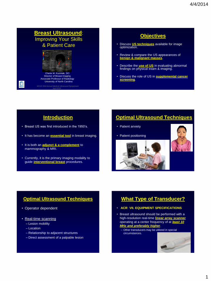
Breast Ultrasound:
Improving Your Skills
& Patient Care
Cherie M. Kuzmiak, DO
Director of Breast Imaging
Associate Professor of Radiology
University of North Carolina
NCUS 33rd Annual Medical Ultrasound Symposium
April 2014
Objectives
• Discuss US techniques available for image optimization.
• Review & compare the US appearances of benign & malignant masses.
• Describe the use of US in evaluating abnormal findings on physical exam & imaging.
• Discuss the role of US in supplemental cancer screening.
Introduction
• Breast US was first introduced in the 1950’s.
• It has become an essential tool in breast imaging.
• It is both an adjunct & a complement to
mammography & MRI.
• Currently, it is the primary imaging modality to
guide interventional breast procedures.
Optimal Ultrasound Techniques
• Patient anxiety
• Patient positioning
Optimal Ultrasound Techniques
• Operator dependent
• Real-time scanning
– Lesion mobility
– Location
– Relationship to adjacent structures
– Direct assessment of a palpable lesion
What Type of Transducer?
• ACR VII. EQUIPMENT SPECIFICATIONS
• Breast ultrasound should be performed with a
high-resolution real-time linear array scanner
operating at a center frequency of at least 10
MHz and preferably higher.
– Other transducers may be utilized in special
circumstances.

4/4/2014
2
What Type of Transducer?
• ACR: In general, the highest frequency capable of adequate penetration to the depth of interest should be used.
• For evaluating superficial lesions, scanning through a thin stand-off device or thick layer of gel may be helpful in offsetting the transducer face from the uppermost layer of skin, to bring it into the focal zone of the transducer.
• Focal zones should be electronically adjustable
Gray-Scale Imaging
• Linear array transducer!
– 12-5 MHz is commonly used
– 17-5 MHz may be used:
• High frequency transducer provides excellent
spatial resolution
» Better “shades of gray”
» Margin resolution
» Lesion conspicuity versus background
• Cost: Decreased penetration due to attenuation
of the beam (>3cm)
Gray-Scale Imaging
• Depth – pectoralis muscle
visualized along with the
posterior FOV
• Gain – fat is a mid shade of
gray at all levels
• Time Gain Compensation –
adjusts image brightness at
different depths to
compensate for attenuation of
the US beam
Gray-Scale Imaging
• Mammographic/MRI lesion
– Correlate with lesion size, depth, location
& distance from nipple
– Correlate with surrounding tissue
• Is the tissue around the lesion entirely fat or
fibroglandular tissue?
» Should be the same on ultrasound
Male Breast Cancer
• 0.7% of all breast cancers – Infiltrating ductal tumors most common
• All ductal subtypes have been described
– DCIS without invasive disease < 5%
– Paget’s disease has been reported
– Infiltrating lobular uncommon
• Mean age at diagnosis = 67 yr – Uncommon in men < 40 yr old
• Stage for stage similar prognosis as females
• Axillary adenopathy in 50% at presentation

4/4/2014
3
Example
Example

4/4/2014
4
2nd Look US Gray-Scale Imaging
• Palpable lesion
– Any imaging correlation? Over age 30…
– Under age 30, pregnant or lactating?
• The examiner should palpate the lesion &
then place the US transducer directly over
the mass.
– Supine
– Upright: Kleenex box
Gray-Scale Imaging
• Adjust depth
• Multiple focal zones improves resolution
at multiple depths simultaneously
– Decreases frame rate
• If a single focal zone is used, it should be
centered at the level of interest
Example – Focal Zone(s)
Remember! • Gain settings, focal zone selections &
fields of view should be optimized to
obtain high-quality images!
Spatial Compounding
• Utilizes electronic beam steering to acquire
multiple images obtained from different
angles within the plane of imaging
– Decreases frame rate

4/4/2014
5
Spatial Compounding
• Real echoes from returning structures are enhanced, providing improved contrast resolution
– Lesion margins
– Posterior borders
– Calcifications
• Artifactual echoes are reduced*
– Speckle
– Posterior acoustic enhancement
– Posterior acoustic shadowing
Spatial Compounding
Speckle Reduction
• Real-time post-processing
• Enhances contrast resolution
• Improves margin definition
• Complementary to spatial compounding
– Can be used at the same time!
Harmonic Imaging
• Relies on filtering the
multiple higher harmonic
frequencies (multiples of
the fundamental
frequencies)
• The ultrasound pulse is
distorted as it travels
through the non-linear
(to sound) breast tissue
– Thus, creating harmonic
frequencies
Harmonic Imaging
• The returning US signal contains both the
original fundamental frequency & its multiples, or
harmonics
• Allows the higher frequencies to be selected &
used to create the gray-scale images
– By not using the lower frequency echos, this reduces
reverbation/internal echoes in lesions (cysts)
– Improves lateral resolution & may improve contrast
resolution

4/4/2014
6
Speed of Sound
• Speed of sound in tissue = 1540 m/sec
• Speed of sound in fat = 1430-1470 m/sec
• Speed of sound imaging is available on
most US units and is an optional
adjustment depending on what type of
tissue is being scanned: fat, dense tissue
or mixed fat/tissue of the breast
Imaging of a Mass
• Radial/anti-radial – Transverse/longitudinal
• Acquire images in 2 planes
without and with caliper
measurements
– Measure to the margin of the
mass
• Include the echogenic halo of
a mass (cancer)
– Longest horizontal x AP x
orthogonal horizontal
Examples
Examples Examples
X X
X
*
X *
*
*
4/4/2014
7
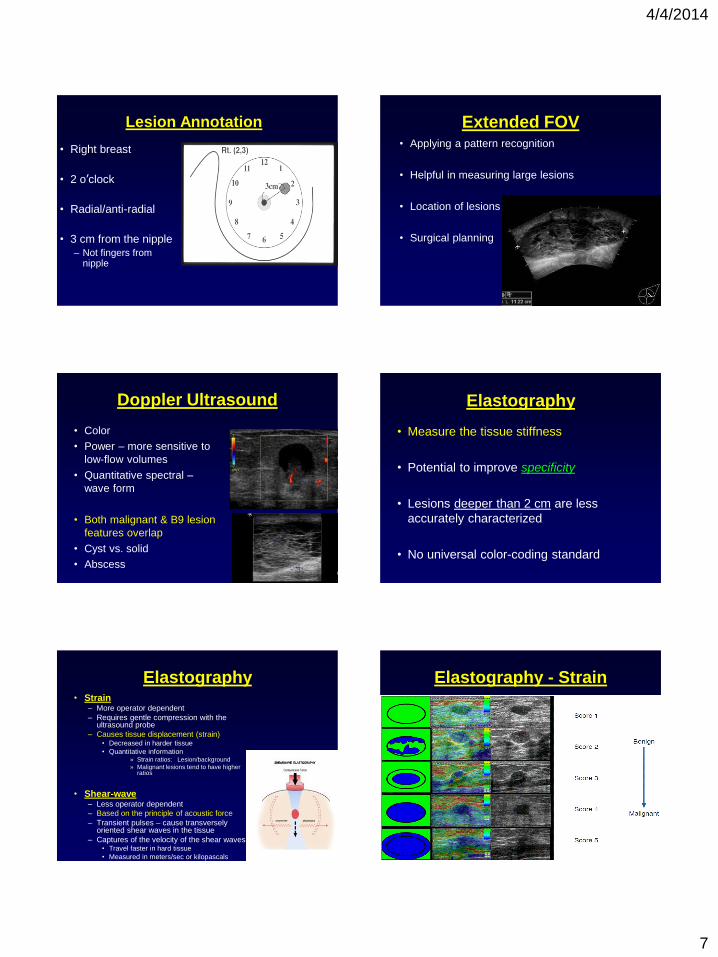
Lesion Annotation
• Right breast
• 2 o’clock
• Radial/anti-radial
• 3 cm from the nipple – Not fingers from
nipple
Extended FOV • Applying a pattern recognition
• Helpful in measuring large lesions
• Location of lesions
• Surgical planning
Doppler Ultrasound
• Color
• Power – more sensitive to
low-flow volumes
• Quantitative spectral –
wave form
• Both malignant & B9 lesion
features overlap
• Cyst vs. solid
• Abscess
Elastography
• Measure the tissue stiffness
• Potential to improve specificity
• Lesions deeper than 2 cm are less
accurately characterized
• No universal color-coding standard
Elastography • Strain
– More operator dependent
– Requires gentle compression with the ultrasound probe
– Causes tissue displacement (strain) • Decreased in harder tissue
• Quantitative information » Strain ratios: Lesion/background
» Malignant lesions tend to have higher ratios
• Shear-wave – Less operator dependent
– Based on the principle of acoustic force
– Transient pulses – cause transversely oriented shear waves in the tissue
– Captures of the velocity of the shear waves • Travel faster in hard tissue
• Measured in meters/sec or kilopascals
Elastography - Strain

4/4/2014
8
Elastography – Shear-wave B9 & Malignant Lesions
• Oval shape
• Circumscribed margin
• Hypoechoic/Iso-echoic
• Parallel to chest wall
• Absence of any malignant features
Cyst Solid Mass
Calcifications Clinical Practice

4/4/2014
9
Imaging Findings
• Asymptomatic patient
– Mammogram
– MRI Screening
• Symptomatic patient
– Diagnostic mammogram
– Diagnostic MRI
– Palpable mass
– Pain
– Discharge
BI-RADS
• Introduced in 2003 for US
• New addition for 2014 – 5th edition
BI-RADS
• Tissue composition (screening only)
1. Homogeneous background echotexture –
fat
2. Homogeneous background echotexture –
fibroglandular
3. Heterogeneous background echotexture
BI-RADS - Mass
• Shape
• Oval
• Round
• Irregular
• Orientation
• Parallel
• Not parallel
• Margin
• Circumscribed
• Not circumscribed
• -Indistinct
• -Angular
• -Microlobulated
• -Spiculated
• Echo pattern
Anechoic
• Hyperechoic
• Complex cystic and solid
• Hypoechoic
• Isoechoic
• Heterogeneous
• Posterior features
• No posterior features
• Enhancement
• Shadowing
• Combined pattern
• Calcifications
• Calcifications in a mass
• Calcifications outside of a mass
BI-RADS - Mass
• Associated features • Architectural distortion
• Duct changes
• Skin changes
• -Skin thickening
• -Skin retraction
• Edema
• Vascularity
• -Absent
• -Internal vascularity
• - Vessels in rim
• Elasticity assessment
• -Soft
• -Intermediate
• -Hard
BI-RADS – Special Cases
• Simple cyst
• Clustered microcysts
• Complicated cyst
• Mass in or on skin
• Foreign body including implants
• Lymph nodes – intramammary
• Lymph nodes – axillary
• Vascular abnormalities
• -AVMs (arteriovenous malformations/
• -Mondor's disease
• Postsurgical fluid
• Fat necrosis

4/4/2014
10
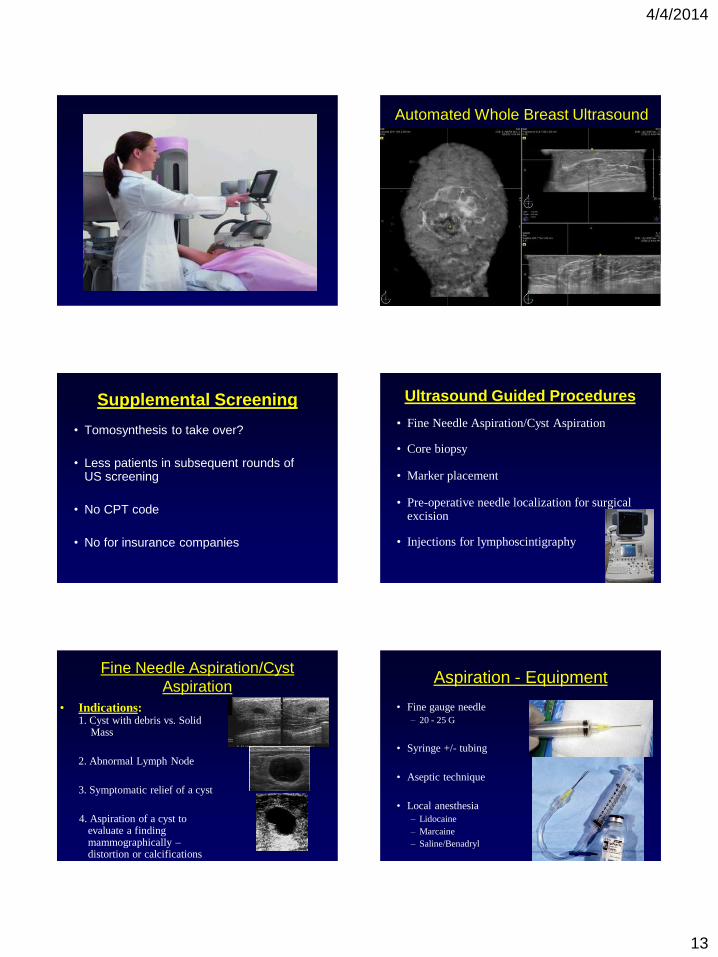
Supplemental Screening
• High-risk patient
• General patient population
– Breast density
Mammography
• Dense tissue limits sensitivity
– 30-48% vs. 80-100% in fatty breasts
• Increased risk of cancer
Ultrasound
• Not limited by breast density – High Sensitivity
• Small hand-held transducer:
– Significant amount of operator subjectivity – Variation in breast ultrasound exams – Time to perform exam can vary
• Limitations:
– Missed lesions – False-positive findings
Occult Cancer in Women with Dense
Breasts: Detection with Screening US:
Diagnostic Yield & Tumor Characteristics
• 11,220 patients – 3,626 patients with dense breasts
– Normal mammogram & PE
• Physician performed whole breast US
• 11 cancers identified with US alone
– Prevalence = 0.30%
• Cancer detection rate increased by 17% (from 63 to 74)
• Conclusion: US can depict small, early-stage, occult cancers similar in size & stage to mammographically identified nonpalpable cancers.
Kolb et al. Radiology 1998; 207:191-199.
Clinical Utility of Bilateral Whole-Breast
US in the Evaluation of Women with
Dense Breast Tissue
• 1,862 patients – Dense breast negative mammogram and PE
• US or mammography technologist performed study – Time of exam: 10 minutes
• 57 biopsies recommended – Data only on 51
– 6 Cancers (cancer detection rate, 0.3%)
• Conclusion: US is useful in detection breast cancer & the cancer detection
rate compares favorably with screening mammography.
Kaplan S. Radiology 2001; 221:641-649.
Initial Studies
• Single center
• Supplemental ultrasound increases detection of
node-negative invasive breast cancer in women
in the 1st prevalence screen
• Increased cancer detection (yield) by 3.5/1000
screened

4/4/2014
11
ACRIN 6666 Study:
Trial of Screening Breast Ultrasound
• Rationale
–No randomized controlled trials
–No multicenter studies
–Reproducibility. . .
Berg et al. JAMA 2012; 307(13):1394-1404.
ACRIN - 6666
• To determine supplemental cancer detection
yield of ultrasound & MRI in women at
elevated risk for breast cancer
– 4/2004-2/2006 dates of study
• 2809 high-risk women with dense breasts @
21 sites
– 612 also underwent MRI at end of study
• 3 annual screens with mammography & US
Berg et al. JAMA 2012; 307(13):1394-1404.
ACRIN - 6666
• Screening mammogram & ultrasound read
independently
• Radiologists did not know the result of the
other study
– Radiologists performed the ultrasound.
• Report & Recommendation……correlation
with mammo
ACRIN - 6666
• 2662 patients had complete data
• 110 had 111 breast cancer events:
– 33 (30%) Mammo detected only
– 32 (29%) US only
– 26 (23%) by both
– 9 (8%) by MRI
– 11 (10%) not detected by any modality
Berg et al. JAMA 2012; 307(13):1394-1404.
ACRIN - 6666
• Additional cancer detection rate:
– US 4.3 cancers/1000 screens
– MRI 14.7 cancers/1000 screens.
• 30/32 US detected only cancers were invasive
– Range 2-40mm (median size 10mm)
– 26 out of 27 staged were node negative.
• US Biopsy rate = 5% (242 of 4814)
– 18/242 (7.4%) were cancer
Berg et al. JAMA 2012; 307(13):1394-1404.
ACRIN - 6666
• Conclusion: Addition of screening US or
MRI to mammography in women at
increased risk of breast cancer resulted in
not only a higher cancer detection yield
but also an increase in false-positive
findings.
Berg et al. JAMA 2012; 307(13):1394-1404.

4/4/2014
12
*Screening US in Patients with Mammographically Dense Breasts: Initial
Experience with CT Public Act 09-41
• Retrospective review of data
– 10/1/2009 through 9/30/10
• Single center - Yale
• Technologists scanned with hand-held transducers
• 935 patients
– 614 low risk
– 149 intermediate risk
– 87 high-risk
Hooley et al. Radiology 2012;265(1):59-69.
CT Public Act 09-41: Results
• 75% (701) BIRADS 1 or 2
• 20% (187) BIRADS 3 – only 82% (145) returned for F/U
• 5% (47) BIRADS 4
• 63 aspirations or biopsies
– 3 cancers!
– Less than 1 cm
– All post-menopausal patients
– 4.7% (44) false-positive
– PPV was 6.5%
– Cancer Detection Rate 3.2/1000
Hooley et al. Radiology 2012;265(1):59-69.
CT Public Act 09-41: Cost
• Estimated cost: CT global Medicare
reimbursement rates for initial screening,
whole-breast ultrasound, F/U,
biopsy/aspiration:
»$180,802.00
»Or
»$60,267.00/cancer diagnosed
Hooley et al. Radiology 2012; 265(1):59-69.
CT Public Act 09-41: Conclusion
• Technologist-performed hand-held
screening breast US offered to women in
the general population of dense breasts
can aid detection of small
mammographically occult breast cancers,
although the overall PPV is low.
Hooley et al. Radiology 2012;265(1):59-69.
Moving Forward
• Breast ultrasound scanning requires a small
hand-held transducer
• Recent advances in technology have lead to the
development of automation
• Automation of ultrasound eliminates operator variation with improved technique standardization
AWBU
• Provides a volume data set of the whole
breast in a standardized manner.
• Short scanning times
– 1 minute per scan projection
• May have AP, Lateral & Medial views of one breast.
• Extra scan projections may be needed for a larger
breast.
4/4/2014
13
Automated Whole Breast Ultrasound
Supplemental Screening
• Tomosynthesis to take over?
• Less patients in subsequent rounds of US screening
• No CPT code
• No for insurance companies
Ultrasound Guided Procedures
• Fine Needle Aspiration/Cyst Aspiration
• Core biopsy
• Marker placement
• Pre-operative needle localization for surgical excision
• Injections for lymphoscintigraphy
Fine Needle Aspiration/Cyst
Aspiration
• Indications: 1. Cyst with debris vs. Solid Mass
2. Abnormal Lymph Node
3. Symptomatic relief of a cyst
4. Aspiration of a cyst to evaluate a finding mammographically – distortion or calcifications
Aspiration - Equipment
• Fine gauge needle
– 20 - 25 G
• Syringe +/- tubing
• Aseptic technique
• Local anesthesia
– Lidocaine
– Marcaine
– Saline/Benadryl
4/4/2014
14
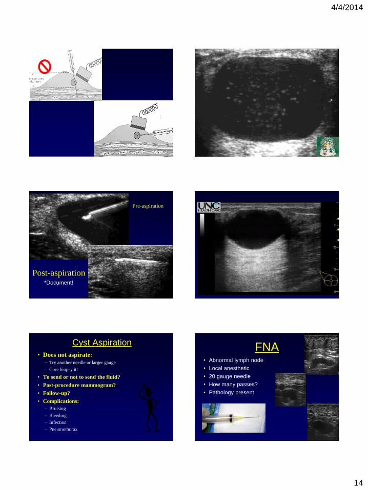
Pre-aspiration
Post-aspiration *Document!
Cyst Aspiration
• Does not aspirate:
– Try another needle or larger gauge
– Core biopsy it!
• To send or not to send the fluid?
• Post-procedure mammogram?
• Follow-up?
• Complications:
– Bruising
– Bleeding
– Infection
– Pneumothorax
FNA • Abnormal lymph node
• Local anesthetic
• 20 gauge needle
• How many passes?
• Pathology present

4/4/2014
15
FNA - Document U/S Core Bx - Technique
US Core Bx - Technique
• Choose proper distance between the probe
edge & planned incision site
– Superficial mass
– Deep mass
• Mark site
• *Tip:
– Proper grip on the transducer
US Core Bx - Technique
• Local anesthesia (skin & future needle track)
• Small incision
• Advance the needle to pre-firing position under US
guidance
• Needle trajectory along the scanning plane, parallel
to the chest wall
• Needle tip at the lesion border and then acquire
sample(s)
• How many samples?

4/4/2014
16
Ultrasound-Guided Core Biopsy
Ultrasound-Guided Core Biopsy
Ultrasound-Guided Core Biopsy

4/4/2014
17
Document - Procedure
Pre-fire Post-fire
Vacuum - Assisted Core Biopsy US - Guided Needle Localization
• Equipment. – Length of needle – 5 cm
– One or more needles?
• Technique. – Shortest distance/same pathway as
core
– Best way to see lesion
– Aseptic technique
– Local anesthetic
– Needle tip in lesion – “shish-kabob”
– Deploy wire
• Post-procedure mammogram
4/4/2014
18
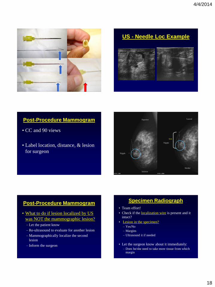
Needle Localization Equipment US - Needle Loc Example
Post-Procedure Mammogram
• CC and 90 views
• Label location, distance, & lesion
for surgeon
4cm
Superior
Inferior
Nipple
Medial
Lateral
Nipple
Post-Procedure Mammogram
• What to do if lesion localized by US
was NOT the mammographic lesion?
– Let the patient know
– Re-ultrasound to evaluate for another lesion
– Mammographically localize the second
lesion
– Inform the surgeon
Specimen Radiograph
• Team effort!
• Check if the localization wire is present and it intact?
• Lesion in the specimen?
– Yes/No
– Margins
– Ultrasound it if needed
• Let the surgeon know about it immediately:
– Does he/she need to take more tissue from which margin

4/4/2014
19
Future Directions
• More complex transducers
• Faster computer processing
• Post-processing algorithms
• Computer aided detection
• Intravenous US microbubble contrast agents – Increase specificity of lesions
Conclusion
• Knowledge & understanding of US
technology, along with meticulous
scanning technique, is imperative for
image optimization & diagnosis.
Conclusion
• Thank You!