brachial plexus
DESCRIPTION
brachial plexusTRANSCRIPT

(https://s100.copyright.com/AppDispatchServlet?publisherName=ELS&orderBeanReset=true&orderSource=ClinicalKey&contentID=B9781416047056000181)
BOOK CHAPTER
Brachial Plexus Elliott L. Mancall MD
Gray's Clinical Neuroanatomy: The Anatomic Basis for Clinical Neuroscience, Chapter 18, 319345
Overview of the Brachial PlexusThe brachial plexus is a union of the ventral rami of the lower four cervical nerves and the greaterpart of the first thoracic ventral ramus ( Figs 18.1, 18.2 (f0010) ). The fourth ramus usually gives abranch to the fifth, and the first thoracic frequently receives one from the second. These ventral ramiare the roots of the plexus; they are almost equal in size but variable in their mode of junction.Contributions to the plexus by C4 and T2 vary. When the branch from C4 is large, that from T2 isfrequently absent and the branch from T1 is reduced, forming a ‘prefixed’ type of plexus. If thebranch from C4 is small or absent, the contribution from C5 is reduced, that from T1 is larger andthere is always a contribution from T2; this arrangement constitutes a ‘postfixed’ type of plexus.
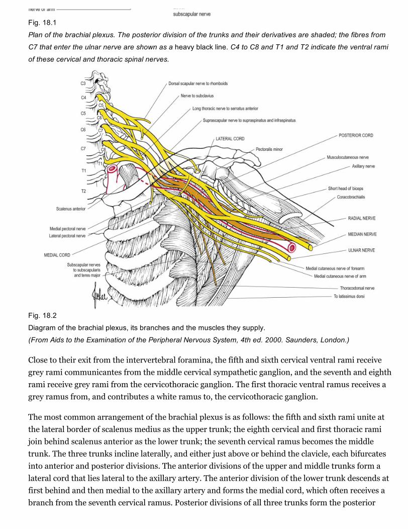
Fig. 18.1
Plan of the brachial plexus. The posterior division of the trunks and their derivatives are shaded; the fibres from
C7 that enter the ulnar nerve are shown as a heavy black line. C4 to C8 and T1 and T2 indicate the ventral rami
of these cervical and thoracic spinal nerves.
Fig. 18.2
Diagram of the brachial plexus, its branches and the muscles they supply.
(From Aids to the Examination of the Peripheral Nervous System, 4th ed. 2000. Saunders, London.)
Close to their exit from the intervertebral foramina, the fifth and sixth cervical ventral rami receivegrey rami communicantes from the middle cervical sympathetic ganglion, and the seventh and eighthrami receive grey rami from the cervicothoracic ganglion. The first thoracic ventral ramus receives agrey ramus from, and contributes a white ramus to, the cervicothoracic ganglion.
The most common arrangement of the brachial plexus is as follows: the fifth and sixth rami unite atthe lateral border of scalenus medius as the upper trunk; the eighth cervical and first thoracic ramijoin behind scalenus anterior as the lower trunk; the seventh cervical ramus becomes the middletrunk. The three trunks incline laterally, and either just above or behind the clavicle, each bifurcatesinto anterior and posterior divisions. The anterior divisions of the upper and middle trunks form alateral cord that lies lateral to the axillary artery. The anterior division of the lower trunk descends atfirst behind and then medial to the axillary artery and forms the medial cord, which often receives abranch from the seventh cervical ramus. Posterior divisions of all three trunks form the posterior
cord, which is at first above and then behind the axillary artery. The posterior division of the lowertrunk is much smaller than the others and contains few, if any, fibres from the first thoracic ramus. Itis frequently derived from the eighth cervical ramus before the trunk is formed.
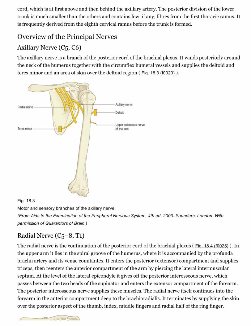
Overview of the Principal NervesAxillary Nerve (C5, C6)The axillary nerve is a branch of the posterior cord of the brachial plexus. It winds posteriorly aroundthe neck of the humerus together with the circumflex humeral vessels and supplies the deltoid andteres minor and an area of skin over the deltoid region ( Fig. 18.3 (f0020) ).
Fig. 18.3
Motor and sensory branches of the axillary nerve.
(From Aids to the Examination of the Peripheral Nervous System, 4th ed. 2000. Saunders, London. With
permission of Guarantors of Brain.)
Radial Nerve (C5–8, T1)The radial nerve is the continuation of the posterior cord of the brachial plexus ( Fig. 18.4 (f0025) ). Inthe upper arm it lies in the spiral groove of the humerus, where it is accompanied by the profundabrachii artery and its venae comitantes. It enters the posterior (extensor) compartment and suppliestriceps, then reenters the anterior compartment of the arm by piercing the lateral intermuscularseptum. At the level of the lateral epicondyle it gives off the posterior interosseous nerve, whichpasses between the two heads of the supinator and enters the extensor compartment of the forearm.The posterior interosseous nerve supplies these muscles. The radial nerve itself continues into theforearm in the anterior compartment deep to the brachioradialis. It terminates by supplying the skinover the posterior aspect of the thumb, index, middle fingers and radial half of the ring finger.

Fig. 18.4
Motor and sensory branches of the radial nerve. Variation exists in the cutaneous innervation of the dorsal
aspects of the digits. Here, the radial nerve is shown supplying all five digits. The dorsum of the ring and little
fingers is frequently innervated by the dorsal branch of the ulnar nerve.
(From Aids to the Examination of the Peripheral Nervous System, 4th ed. 2000. Saunders, London. With
permission of Guarantors of Brain.)

Musculocutaneous Nerve (C5–7)The musculocutaneous nerve is formed from the continuation of the lateral cord of the brachialplexus. It pierces the coracobrachialis; supplies it, biceps and brachialis; then continues into theforearm as the lateral cutaneous nerve of the forearm ( Fig. 18.5 (f0030) ).
Fig. 18.5
Motor and sensory branches of the musculocutaneous nerve.
(From Aids to the Examination of the Peripheral Nervous System, 4th ed. 2000. Saunders, London. With
permission of Guarantors of Brain.)
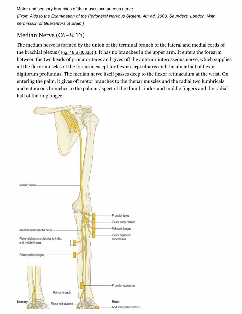
Median Nerve (C6–8, T1)The median nerve is formed by the union of the terminal branch of the lateral and medial cords ofthe brachial plexus ( Fig. 18.6 (f0035) ). It has no branches in the upper arm. It enters the forearmbetween the two heads of pronator teres and gives off the anterior interosseous nerve, which suppliesall the flexor muscles of the forearm except for flexor carpi ulnaris and the ulnar half of flexordigitorum profundus. The median nerve itself passes deep to the flexor retinaculum at the wrist. Onentering the palm, it gives off motor branches to the thenar muscles and the radial two lumbricalsand cutaneous branches to the palmar aspect of the thumb, index and middle fingers and the radialhalf of the ring finger.

Fig. 18.6
Motor and sensory branches of the median nerve. Flexor digitorum profundus to the ring and little fingers is
supplied by the ulnar nerve. Flexor pollicis brevis may be supplied by both the median nerve and the ulnar nerve.
(From Aids to the Examination of the Peripheral Nervous System, 4th ed. 2000. Saunders, London. With
permission of Guarantors of Brain.)
Ulnar Nerve (C7, C8, T1)The ulnar nerve is the continuation of the medial cord of the brachial plexus ( Fig. 18.7 (f0040) ). Likethe median nerve, it has no branches in the upper arm. It enters the posterior compartment of theupper arm midway down its length by piercing the medial intermuscular septum and passes behindthe medial epicondyle of the humerus to enter the forearm. It passes to the wrist deep to flexor carpiulnaris, giving branches to this muscle and to the ulnar half of flexor digitorum profundus. Justproximal to the wrist it gives off a dorsal cutaneous branch that supplies the skin over the dorsalaspect of the little finger and the ulnar half of the ring finger. The ulnar nerve crosses into the palmsuperficial to the flexor retinaculum in Guyon's canal. It divides into a motor branch, which suppliesthe hypothenar muscles, the intrinsics (apart from the radial two lumbricals) and adductor pollicis,and cutaneous branches, which supply the skin of the palmar aspect of the little finger and ulnar halfof the ring finger.

Fig. 18.7
Motor and sensory branches of the ulnar nerve together with the medial cutaneous nerves of the arm and
forearm. Flexor digitorum profundus to index and middle fingers is supplied by the median nerve. Flexor pollicis
brevis may be supplied by both the median nerve and the ulnar nerve.
(From Aids to the Examination of the Peripheral Nervous System, 4th ed. 2000. Saunders, London. With
permission of Guarantors of Brain.)
DermatomesOur knowledge of the extent of individual dermatomes, especially in the limbs, is based largely onclinical evidence ( Fig. 18.8 (f0045) ). The dermatomes of the upper limb arise from spinal nerves C5–8 and T1. C7 supplies the central part of the hand. Considerable overlap exists between adjacentdermatomes innervated by nerves derived from consecutive spinal cord segments.
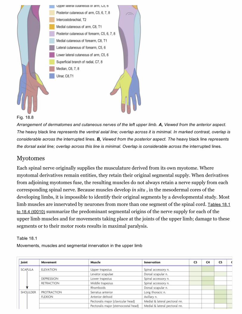
Fig. 18.8
Arrangement of dermatomes and cutaneous nerves of the left upper limb. A, Viewed from the anterior aspect.The heavy black line represents the ventral axial line; overlap across it is minimal. In marked contrast, overlap is
considerable across the interrupted lines. B, Viewed from the posterior aspect. The heavy black line representsthe dorsal axial line; overlap across this line is minimal. Overlap is considerable across the interrupted lines.
MyotomesEach spinal nerve originally supplies the musculature derived from its own myotome. Wheremyotomal derivatives remain entities, they retain their original segmental supply. When derivativesfrom adjoining myotomes fuse, the resulting muscles do not always retain a nerve supply from eachcorresponding spinal nerve. Because muscles develop in situ , in the mesodermal cores of thedeveloping limbs, it is impossible to identify their original segments by a developmental study. Mostlimb muscles are innervated by neurones from more than one segment of the spinal cord. Tables 18.1to 18.4 (t0010) summarize the predominant segmental origins of the nerve supply for each of theupper limb muscles and for movements taking place at the joints of the upper limb; damage to thesesegments or to their motor roots results in maximal paralysis.
Table 18.1
Movements, muscles and segmental innervation in the upper limb


Table 18.2
Segmental innervation of muscles of the upper limb
C3,C4
Trapezius, levator scapulae
C5 Rhomboids, deltoids, supraspinatus, infraspinatus, teres minor, biceps
C6 Serratus anterior, latissimus dorsi, subscapularis, teres major, pectoralis major (clavicularhead), biceps, coracobrachialis, brachialis, brachioradialis, supinator, extensor carpi radialislongus
C7 Serratus anterior, latissimus dorsi, pectoralis major (sternal head), pectoralis minor, triceps,pronator teres, flexor carpi radialis, flexor digitorum superficialis, extensor carpi radialislongus, extensor carpi radialis brevis, extensor digitorum, extensor digiti minimi
C8 Pectoralis major (sternal head), pectoralis minor, triceps, flexor digitorum superficialis, flexordigitorum profundus, flexor pollicis longus, pronator quadratus, flexor carpi ulnaris, extensorcarpi ulnaris, abductor pollicis longus, extensor pollicis longus, extensor pollicis brevis,extensor indicis, abductor pollicis brevis, flexor pollicis brevis, opponens pollicis
T1 Flexor digitorum profundus, intrinsic muscles of the hand (except abductor pollicis brevis,flexor pollicis brevis, opponens pollicis)
Table 18.3
Segmental innervation of joint movements of the upper limb
Shoulder Abductors and lateral rotators C5
Abductors and medial rotators C6–8
Elbow Flexors C5, C6
Extensors C7, C8
Forearm Supinators C6
Pronators C7, C8
Wrist Flexors and extensors C6, C7
Digits Long flexors and extensors C7, C8
Hand Intrinsic muscles C8, T1
Table 18.4
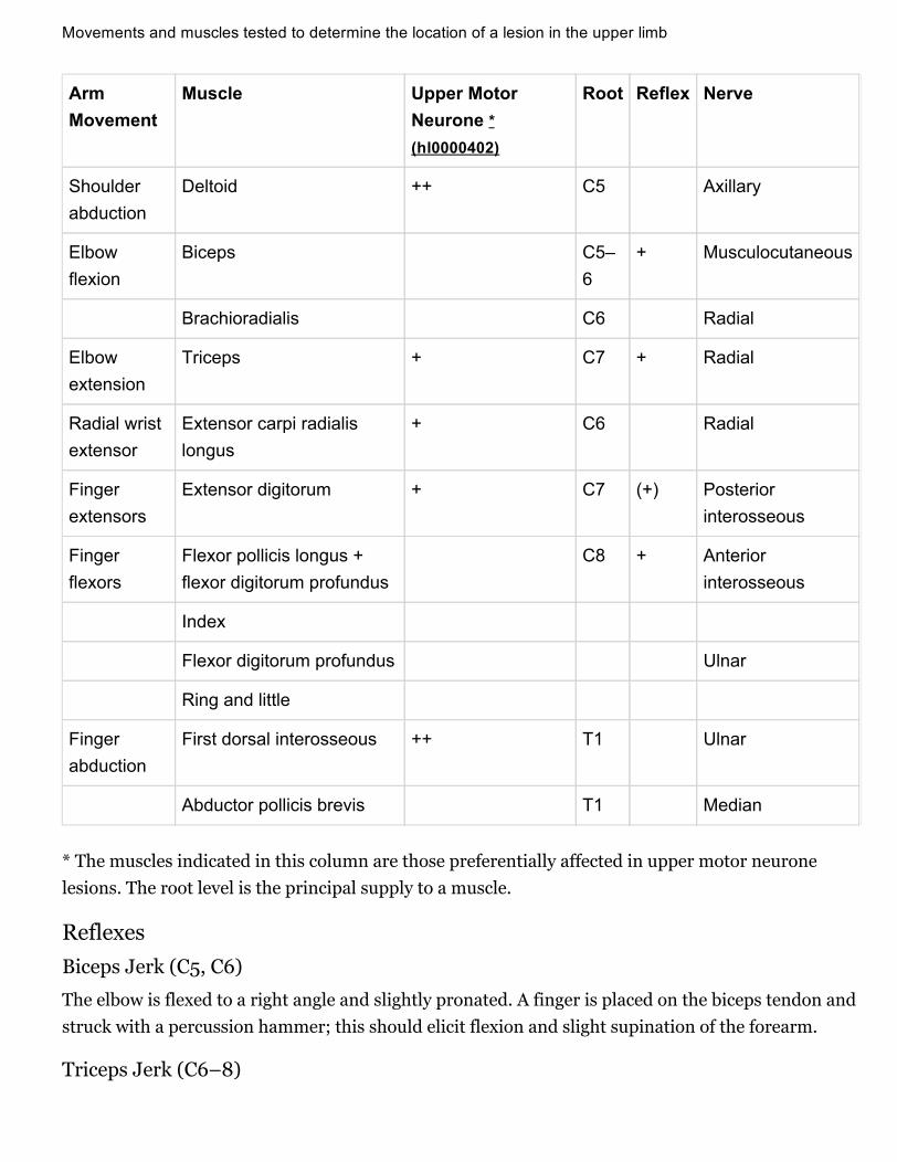
Movements and muscles tested to determine the location of a lesion in the upper limb
ArmMovement
Muscle Upper MotorNeurone *(hl0000402)
Root Reflex Nerve
Shoulderabduction
Deltoid ++ C5 Axillary
Elbowflexion
Biceps C5–6
+ Musculocutaneous
Brachioradialis C6 Radial
Elbowextension
Triceps + C7 + Radial
Radial wristextensor
Extensor carpi radialislongus
+ C6 Radial
Fingerextensors
Extensor digitorum + C7 (+) Posteriorinterosseous
Fingerflexors
Flexor pollicis longus +flexor digitorum profundus
C8 + Anteriorinterosseous
Index
Flexor digitorum profundus Ulnar
Ring and little
Fingerabduction
First dorsal interosseous ++ T1 Ulnar
Abductor pollicis brevis T1 Median
* The muscles indicated in this column are those preferentially affected in upper motor neuronelesions. The root level is the principal supply to a muscle.
ReflexesBiceps Jerk (C5, C6)
The elbow is flexed to a right angle and slightly pronated. A finger is placed on the biceps tendon andstruck with a percussion hammer; this should elicit flexion and slight supination of the forearm.
Triceps Jerk (C6–8)

The arm is supported at the wrist and flexed to a right angle. The triceps is struck with a percussionhammer just proximal to the olecranon; this should elicit extension of the elbow.
Radial Jerk (C7, C8)
The radial jerk is a periosteal, not a tendon, reflex. The elbow is flexed to a right angle, and theforearm is placed in the mid position. The radial styloid is struck with the percussion hammer. Thiselicits contraction of brachioradialis, which causes flexion of the elbow.
Muscle Innvervation and FunctionTable 18.1 (t0010) provides the following information about the innervation and functions of musclesin the upper limb:
Movements
At the central nervous level of control, muscles are recognized not as individual actuators but ascomponents of movement. Muscles may contribute to several types of movement, acting variously asprime movers, antagonists, fixators or synergists. For example, in the movement of the scapulaaround the thorax, serratus anterior acts as an antagonist of trapezius, but in the forward rotation ofthe scapula, the two muscles combine as prime movers. Moreover, a muscle that crosses two jointscan produce more than one movement. Even a muscle that acts across one joint can produce acombination of movements, such as flexion with medial rotation or extension with adduction. Somemuscles have therefore been included in more than one place in Table 18.1 (t0010) , but even theselistings are not exhaustive.
Nerve Roots
The spinal roots listed as contributing to muscle innervation vary in different texts; this is a reflectionof the often unreliable nature of available information. The most positive identifications have beenobtained by electrically stimulating spinal roots and recording the evoked electromyographic activityin the muscles. This is a laborious process, however, and data of this quality are in limited supply.Much of the information in Table 18.1 (t0010) is based on neurological experience gained inexamining the effects of lesions, and some of it is far from new.
Major and Minor Contributions
Spinal roots have been given the same shading in Table 18.1 (t0010) when they innervate a muscle to asimilar extent or when differences in their contribution have not been described. Heavy shadingindicates roots from which there is known to be a dominant contribution. From a clinical viewpoint,some of these roots may be regarded as innervating the muscle almost exclusively: for example,deltoid by C5, brachioradialis by C6, triceps by C7. Minor contributions have been retained in thetable to increase its utility in other contexts, such as electromyography and comparative anatomy.
Clinical Testing

For diagnostic purposes, it is neither necessary nor possible to test every muscle, and the experiencedneurologist can cover every clinical possibility with a much shorter list. In Table 18.1 (t0010) , red hasbeen used to highlight those muscles or movements that have diagnostic value. The emphasis here ison the differentiation of lesions at different root levels. Other lists could be developed to differentiatebetween lesions at the level of the root, plexus or peripheral nerve; at different sites along the lengthof a nerve; or between different peripheral nerves. The preferred criteria for including a given musclein such a list are that it is visible and palpable, that its action is isolated or can be isolated by theexaminer, that it is innervated by one peripheral nerve or (predominantly) one root, that it has aclinically elicitable reflex and that it is useful in differentiating among different nerves, roots or lesionlevels.
Determination of A Lesion's LocationIn clinical practice it is necessary to test only a relatively small number of muscles to determine thelocation of a lesion. For example, abduction of the arm might test shoulder abduction, a C5 rootlesion, the axillary nerve or deltoid.
Any muscle to be tested must satisfy a number of criteria. It should be visible, so that wasting orfasciculation can be observed and the muscle's consistency with contraction can be felt. It shouldhave an isolated action, so that its function can be tested separately. The muscle tested should helpdifferentiate between lesions at different levels in the neuraxis and in peripheral nerves, or betweenperipheral nerves. It should be tested in such a way that normal can be differentiated from abnormal,so that slight weakness can be detected early with reliability. Some preference should be given tomuscles with an easily elicited reflex.
Table 18.4 (t0025) lists movements and muscles chosen according to these criteria. For example, withan upper motor neurone lesion, shoulder abduction, elbow extension, wrist and finger extension andfinger abduction are weaker than their opposing movements. Because this weakness may be moredistal than proximal, or vice versa, normal shoulder abduction and finger abduction excludes anupper motor neurone weakness of the arm. Some muscles are difficult to test but are included forspecial reasons. For example, brachioradialis strength is difficult to assess, but the muscle can beseen and felt, it is innervated mostly by the C6 root, and it has an easily elicited reflex.
To determine the root level of a lesion, it is necessary to know the appropriate muscle to test for eachroot, preferably with an easily elicited reflex.
Knowledge of the sequence in which motor branches leave a peripheral nerve to innervate specificmuscles is very helpful in locating the level of the lesion. For example, with radial nerve lesions, iftriceps is involved, the lesion must be high in the axilla. If, as is usual, triceps is spared butbrachioradialis, wrist extensors, finger extensors and the superficial radial nerve are all involved, thelesion is in the arm, where the radial nerve is vulnerable to pressure against the humerus. If wristextension is normal and the superficial radial nerve is not involved but finger extension is weak, thelesion involves the posterior interosseous branch of the radial nerve.
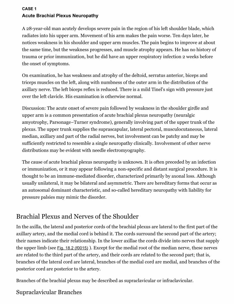
CASE 1Acute Brachial Plexus Neuropathy
A 28yearold man acutely develops severe pain in the region of his left shoulder blade, whichradiates into his upper arm. Movement of his arm makes the pain worse. Ten days later, henotices weakness in his shoulder and upper arm muscles. The pain begins to improve at aboutthe same time, but the weakness progresses, and muscle atrophy appears. He has no history oftrauma or prior immunization, but he did have an upper respiratory infection 2 weeks beforethe onset of symptoms.
On examination, he has weakness and atrophy of the deltoid, serratus anterior, biceps andtriceps muscles on the left, along with numbness of the outer arm in the distribution of theaxillary nerve. The left biceps reflex is reduced. There is a mild Tinel's sign with pressure justover the left clavicle. His examination is otherwise normal.
Discussion: The acute onset of severe pain followed by weakness in the shoulder girdle andupper arm is a common presentation of acute brachial plexus neuropathy (neuralgicamyotrophy, Parsonage–Turner syndrome), generally involving part of the upper trunk of theplexus. The upper trunk supplies the suprascapular, lateral pectoral, musculocutaneous, lateralmedian, axillary and part of the radial nerves, but involvement can be patchy and may besufficiently restricted to resemble a single neuropathy clinically. Involvement of other nervedistributions may be evident with needle electromyography.
The cause of acute brachial plexus neuropathy is unknown. It is often preceded by an infectionor immunization, or it may appear following a nonspecific and distant surgical procedure. It isthought to be an immunemediated disorder, characterized primarily by axonal loss. Althoughusually unilateral, it may be bilateral and asymmetric. There are hereditary forms that occur asan autosomal dominant characteristic, and socalled hereditary neuropathy with liability forpressure palsies may mimic the disorder.
Brachial Plexus and Nerves of the ShoulderIn the axilla, the lateral and posterior cords of the brachial plexus are lateral to the first part of theaxillary artery, and the medial cord is behind it. The cords surround the second part of the artery;their names indicate their relationship. In the lower axillae the cords divide into nerves that supplythe upper limb (see Fig. 18.2 (f0015) ). Except for the medial root of the median nerve, these nervesare related to the third part of the artery, and their cords are related to the second part; that is,branches of the lateral cord are lateral, branches of the medial cord are medial, and branches of theposterior cord are posterior to the artery.
Branches of the brachial plexus may be described as supraclavicular or infraclavicular.
Supraclavicular Branches

Supraclavicular branches arise from roots or from trunks:
From roots 1. Nerves to scaleni and longus colli C5, C6, C7, C8
2. Branch to phrenic nerve C5
3. Dorsal scapular nerve C5
4. Long thoracic nerves C5, C6 (C7)
From trunks 1. Nerve to subclavius C5, C6
2. Suprascapular nerve C5, C6
Branches to the scaleni and longus colli arise from the lower cervical ventral rami near their exit fromthe intervertebral foramina. The phrenic nerve is joined by a branch from the fifth cervical ramusanterior to scalenus anterior.
Dorsal Scapular Nerve
The dorsal scapular nerve comes from the fifth cervical ventral ramus; pierces scalenus medius;passes behind levator scapulae, which it occasionally supplies; and runs with the deep branch of thedorsal scapular artery to the rhomboids, which it supplies.
Long Thoracic Nerve
The long thoracic nerve is usually formed by roots from the fifth to seventh cervical rami, althoughthe last ramus may be absent ( Fig. 18.9 (f0055) ). The upper two roots pierce scalenus mediusobliquely, uniting in or lateral to it. The nerve descends dorsal to the brachial plexus and the firstpart of the axillary artery and crosses the superior border of serratus anterior to reach its lateralsurface. It may be joined by the root from C7, which emerges between scalenus anterior and scalenusmedius, and descends on the lateral surface of medius. The nerve continues downward to the lowerborder of serratus anterior and supplies branches to each of its digitations.

Fig. 18.9
Nerves of the left upper limb, dissected from the anterior aspect.
The long thoracic nerve is the most common nerve to be affected by neuralgic amyotrophy. Wingingof the scapula may be the only clinical manifestation; it is best demonstrated by asking the patient topush against resistance with the arm extended at the elbow and flexed to 90° at the shoulder ( Fig.18.10 (f0060) ; see Case 1 (b0010) ).
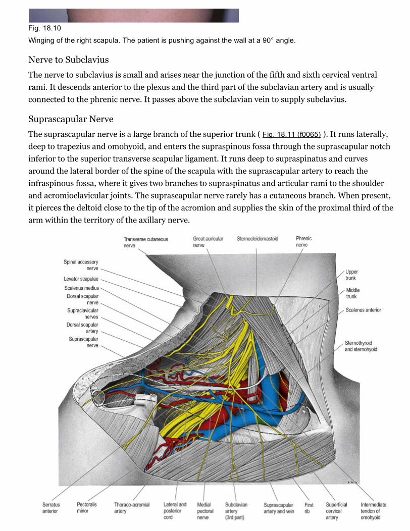
Fig. 18.10
Winging of the right scapula. The patient is pushing against the wall at a 90° angle.
Nerve to Subclavius
The nerve to subclavius is small and arises near the junction of the fifth and sixth cervical ventralrami. It descends anterior to the plexus and the third part of the subclavian artery and is usuallyconnected to the phrenic nerve. It passes above the subclavian vein to supply subclavius.
Suprascapular Nerve
The suprascapular nerve is a large branch of the superior trunk ( Fig. 18.11 (f0065) ). It runs laterally,deep to trapezius and omohyoid, and enters the supraspinous fossa through the suprascapular notchinferior to the superior transverse scapular ligament. It runs deep to supraspinatus and curvesaround the lateral border of the spine of the scapula with the suprascapular artery to reach theinfraspinous fossa, where it gives two branches to supraspinatus and articular rami to the shoulderand acromioclavicular joints. The suprascapular nerve rarely has a cutaneous branch. When present,it pierces the deltoid close to the tip of the acromion and supplies the skin of the proximal third of thearm within the territory of the axillary nerve.

Fig. 18.11
Lower part of the posterior triangle showing the relations of the third part of the right subclavian artery. The
clavicle has been removed, but its outline is indicated by a dashed line. In this dissection, the middle trunk of the
brachial plexus gives an unusual contribution to the medial cord.
Lesions of the Suprascapular Nerve
The most common cause of lesions involving the suprascapular nerve is neuralgic amyotrophy, asdescribed earlier. An entrapment neuropathy may occur in the scapular notch, or the nerve may bedamaged by trauma to the scapula and shoulder. There is pain in the shoulder and wasting andweakness of supraspinatus and infraspinatus.
Infraclavicular BranchesInfraclavicular branches come from the cords, but their axons may be traced back to the spinalnerves detailed below:
Lateral cord Lateral pectoral C5, C6, C7
Musculocutaneous C5, C6, C7
Lateral root of median (C5), C6, C7
Medial cord Medial pectoral C8, T1
Medial cutaneous of forearm C8, T1
Medial cutaneous of arm C8, T1
Ulnar (C7), C8, T1
Medial root of median C8, T1
Posterior cord Upper subscapular C5, C6
Thoracodorsal C6, C7, C8
Lower subscapular C5, C6
Axillary C5, C6
Radial C5, C6, C7, C8, (T1)
Lateral Pectoral Nerve
The lateral pectoral nerve (see Fig. 18.9 (f0055) ) is larger than the medial and may arise from theanterior divisions of the upper and middle trunks or by a single root from the lateral cord. Its axonsare from the fifth to seventh cervical rami. It crosses anterior to the axillary artery and vein, pierces

the clavipectoral fascia and supplies the deep surface of pectoralis major. It sends a branch to themedial pectoral nerve, forming a loop in front of the first part of the axillary artery (see Fig. 18.9(f0055) ), to supply some fibres to pectoralis minor.
Medial Pectoral Nerve
The medial pectoral nerve is derived from the eighth cervical and first thoracic ventral rami andbranches from the medial cord; the latter lies posterior to the axillary artery. It curves forwardbetween the axillary artery and vein. Anterior to the artery it joins a ramus of the lateral pectoralnerve and enters the deep surface of pectoralis minor, which it supplies. Two or three branchespierce pectoralis minor, and others may pass around its inferior border to end in pectoralis major.
Upper (Superior) Subscapular Nerve
The superior subscapular nerve is smaller than the inferior. It arises from the posterior cord (C5 andC6), enters subscapularis at a high level and is frequently double.
Lower (Inferior) Subscapular Nerve
The inferior subscapular nerve arises from the posterior cord (C5 and C6). It supplies the lower partof subscapularis and ends in teres major, which is sometimes supplied by a separate branch.
Thoracodorsal Nerve
The thoracodorsal nerve arises from the posterior cord (C6–8) between the subscapular nerves. Itaccompanies the subscapular artery along the posterior axillary wall and supplies latissimus dorsi,reaching its distal border.
Axillary Nerve
The axillary nerve arises from the posterior cord (C5, C6). It is initially lateral to the radial nerve,posterior to the axillary artery and anterior to subscapularis ( Fig. 18.12 (f0070) ). At the lower borderof subscapularis it curves back inferior to the humeroscapular articular capsule and, with theposterior circumflex humeral vessels, traverses a quadrangular space bounded above bysubscapularis (anterior) and teres minor (posterior), below by teres major, medially by the long headof triceps and laterally by the surgical neck of the humerus. In the space it divides into anterior andposterior branches. The anterior branch curves around the neck of the humerus with the posteriorcircumflex humeral vessels, deep to deltoid. It reaches the anterior border of the muscle, supplies itand gives off a few small cutaneous branches that pierce deltoid and ramify in the skin over its lowerpart. The posterior branch courses medially and posteriorly along the attachment of the lateral headof triceps, inferior to the glenoid rim. It usually lies medial to the anterior branch in thequadrangular space. It gives off the nerve to teres minor and the upper lateral cutaneous nerve of thearm at the lateral edge of the origin of the long head of triceps. The nerve to teres minor enters themuscle on its inferior surface. The posterior branch frequently supplies the posterior aspect ofdeltoid, usually via a separate branch from the main stem, or occasionally from the superior lateralcutaneous nerve of the arm. However, the posterior part of deltoid has a more consistent supply fromthe anterior branch of the axillary nerve, which should be remembered when performing a posterior

deltoidsplitting approach to the shoulder. The upper lateral cutaneous nerve of the arm pierces thedeep fascia at the medial border of the posterior aspect of deltoid and supplies the skin over thelower part of deltoid and upper part of the long head of triceps. The posterior branch is intimatelyrelated to the inferior aspects of the glenoid and shoulder joint capsule, which may place it atparticular risk during capsular plication or thermal shrinkage procedures ( Ball et al 2003 ). There isoften an enlargement or pseudoganglion on the branch to teres minor. The axillary trunk supplies abranch to the shoulder joint below subscapularis.
Fig. 18.12
Dorsal scapular muscles and triceps on the left side. The spine of the scapula has been divided near its lateral
end, and the acromion has been removed along with a large part of deltoid. The humerus is laterally rotated, and
the forearm is pronated.
Lesions of the Axillary Nerve
The most common causes of axillary nerve lesions are trauma (dislocation of the shoulder, fracture ofthe surgical neck of the humerus) and neuralgic amyotrophy. There is deltoid wasting and weakness,which is usually clinically evident, and a patch of sensory loss on the outer aspect of the arm. Thiscan be differentiated from a C5 root lesion by the finding of normal function in the distribution of thesuprascapular nerve.
Musculocutaneous Nerve

The musculocutaneous nerve (see Fig. 18.9 (f0055) ) arises from the lateral cord (C5–7), opposite thelower border of pectoralis minor. It pierces coracobrachialis and descends laterally between bicepsand brachialis to the lateral side of the arm. Just below the elbow it pierces the deep fascia lateral tothe biceps tendon and continues as the lateral cutaneous nerve of the forearm. A line drawn from thelateral side of the third part of the axillary artery across coracobrachialis and biceps to the lateral sideof the biceps tendon is a surface projection for the nerve (but this varies according to its point ofentry into coracobrachialis). It supplies coracobrachialis, both heads of the biceps and most ofbrachialis. The branch to coracobrachialis is given off before the musculocutaneous nerve enters themuscle; its fibres are from the seventh cervical ramus and may branch directly from the lateral cord.Branches to biceps and brachialis leave after the musculocutaneous has pierced coracobrachialis; thebranch to brachialis also supplies the elbow joint. The musculocutaneous nerve supplies a smallbranch to the humerus, which enters the shaft with the nutrient artery.
Lesions of the Musculocutaneous Nerve
An isolated lesion of the musculocutaneous nerve is rare but may occur in injuries to the upper armand shoulder (e.g. fracture of the humerus) and in patients with neuralgic amyotrophy. There ismarked weakness of elbow flexion because biceps brachii and much of the brachialis are paralysed,as well as sensory impairment on the extensor aspect of the forearm in the distribution of the lateralcutaneous nerve of the forearm. Pain and paraesthesia may be aggravated by elbow extension.
Medial Cutaneous Nerve of the Arm
The medial cutaneous nerve of the arm is the smallest and most medial branch of the brachial plexusand arises from the medial cord (C8, T1). It crosses the axilla, either anterior or posterior to theaxillary vein, then passes medial to the axillary vein, communicates with the intercostobrachial nerveand descends medial to the brachial artery and basilic vein. It pierces the deep fascia at the midpointof the upper arm to supply the skin over the medial aspect of the distal third of the upper arm. It isdescribed in further detail below.
Medial Cutaneous Nerve of the Forearm
The medial cutaneous nerve of the forearm arises from the medial cord (C8, T1). It is described inmore detail below.
Median Nerve
The median nerve has two roots from the lateral (C5, C6, C7) and medial (C8, T1) cords, whichembrace the third part of the axillary artery and unite anterior or lateral to it (see Fig. 18.9 (f0055) ).Some fibres from C7 leave the lateral root in the lower part of the axilla and pass distomediallyposterior to the medial root, and usually anterior to the axillary artery, to join the ulnar nerve. Theymay branch from the seventh cervical ventral ramus. Clinically, they are believed to be mainly motorand to supply flexor carpi ulnaris. If the lateral root is small, the musculocutaneous nerve (C5, C6,C7) connects with the median nerve in the arm. It is described in more detail below.
Ulnar Nerve

The ulnar nerve arises from the medial cord (C8, T1) but often receives fibres from the ventral ramusof C7 (see Fig. 18.9 (f0055) ). It runs distally through the axilla medial to the axillary artery, between itand the vein. It is described in more detail below.
Radial Nerve
The radial nerve is the largest branch of the brachial plexus. It arises from the posterior cord (C5, C6,C7, C8, [T1]; see Fig. 18.12 (f0070) ) and descends behind the third part of the axillary artery and theupper part of the brachial artery, anterior to subscapularis and the tendons of latissimus dorsi andteres major. With the arteria profunda brachii it inclines dorsally and passes through the triangularspace below the lower border of teres major, between the long head of triceps and the humerus. It isdescribed in more detail below.
Brachial Plexus LesionsLesions of the brachial plexus commonly affect either the upper part of the plexus (i.e. C5 and C6roots and the upper trunk) or the lower part of the plexus (i.e. C8 and T1 roots and the lower trunk).Lesions affecting the upper part are usually traumatic, whereas those affecting the lower part may becaused by trauma, malignant infiltration or thoracic outlet syndrome. Severe trauma may affect thewhole plexus.
Upper Plexus Palsies
Downward traction on an infant's arm during birth or, in adults, a severe fall on the side of the headand shoulder, forcing the two apart (as frequently occurs in a motorcycle crash), may tear the roots ofC5 and C6. This results in paralysis of the deltoid, short muscles of the shoulder, brachialis andbiceps. The last two are both elbow flexors, and biceps is also a powerful supinator of the superiorradioulnar joint. The arm therefore hangs by the side, with the forearm pronated and the palmfacing backward, like a waiter hinting for a tip (Erb–Duchenne paralysis). There is sensory loss overthe lateral aspect of the upper arm.
Lower Plexus Palsies
Upward traction on the arm, such as in a forcible breech delivery, may tear the lowest root, T1, whichprovides the segmental supply to the intrinsic muscles of the hand. The hand assumes a clawedappearance, reflecting the unopposed action of the long flexors and extensors of the fingers(Klumpke's paralysis). There is sensory loss along the medial aspect of the forearm and often anassociated Horner's syndrome (ptosis and constriction of the pupil), which occurs as a result oftraction on the cervical sympathetic chain.
Malignant infiltration of the brachial plexus may result from extension of an apical lung carcinoma(Pancoast tumour) or from metastatic spread, often from carcinoma of the breast. There is slowlyprogressive weakness that usually starts in the small muscles of the hand (T1) and spreads to involvethe finger flexors (C8). This is usually a painful condition, and the pain may be severe. There issensory loss on the medial aspect of the forearm (T1), extending into the medial side of the hand andto the little finger (C8). Horner's syndrome may occur if there is involvement of the cervical

sympathetic ganglia. A similar syndrome may occur following radiotherapy for breast carcinoma, butthis is usually painless. Thoracic surgery involving a sternal split may cause traction on the brachialplexus and usually affects the lower part of the plexus.
The lower trunk of the brachial plexus (C8, T1), together with the subclavian artery, may beangulated over a cervical rib (thoracic outlet syndrome). Patients may present with vascularsymptoms as a result of kinking of the subclavian artery (this is more likely to occur with large bonyribs), or they may present with neurological deficits (this is more likely in patients with smallrudimentary ribs that extend into a fibrous band that joins the first rib anteriorly). Cervical ribs arequite common and are rarely associated with symptoms. There is a slow, insidious onset of wasting ofthe small muscles of the hand, which often starts on the lateral side with involvement of the thenareminence and first dorsal interosseous. There is pain and paraesthesia in the medial aspect of theforearm, extending to the little finger; this is often aggravated by carrying shopping bags or suitcases.A bruit may be heard over the subclavian artery, and the radial pulse may be easily obliterated bymovements of the arm, particularly with the arm extended and abducted at the shoulder.
CASE 2Pancoast Tumour
A 59yearold man, a heavy smoker for many years, develops posterior left shoulder painradiating down the medial aspect of the left arm into the fourth and fifth digits of the hand,with weakness that ultimately involves the entire left hand. He has lost 35 pounds in the past 3months. Examination demonstrates wasting and weakness of all intrinsic hand muscles on theleft, as well as weakness of wrist flexion. There is decreased sensation in the left medial upperarm, forearm and hand, involving especially the fifth digit ( Fig. 18.13 (f0075) ). He has a mildleft Horner's syndrome, with noticeable ptosis and miosis.
Fig. 18.13
Brachial plexopathy (brachial neuritis). Atrophy of the intrinsic hand muscles due to denervation.
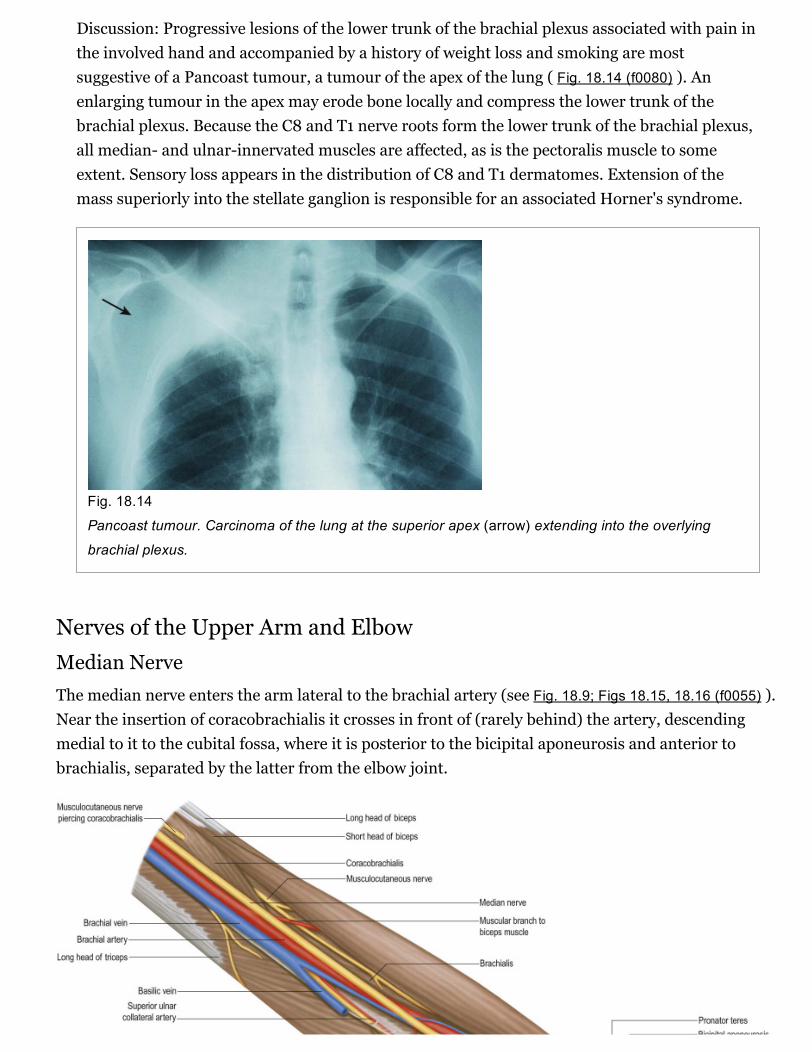
Discussion: Progressive lesions of the lower trunk of the brachial plexus associated with pain inthe involved hand and accompanied by a history of weight loss and smoking are mostsuggestive of a Pancoast tumour, a tumour of the apex of the lung ( Fig. 18.14 (f0080) ). Anenlarging tumour in the apex may erode bone locally and compress the lower trunk of thebrachial plexus. Because the C8 and T1 nerve roots form the lower trunk of the brachial plexus,all median and ulnarinnervated muscles are affected, as is the pectoralis muscle to someextent. Sensory loss appears in the distribution of C8 and T1 dermatomes. Extension of themass superiorly into the stellate ganglion is responsible for an associated Horner's syndrome.
Fig. 18.14
Pancoast tumour. Carcinoma of the lung at the superior apex (arrow) extending into the overlying
brachial plexus.
Nerves of the Upper Arm and ElbowMedian NerveThe median nerve enters the arm lateral to the brachial artery (see Fig. 18.9; Figs 18.15, 18.16 (f0055) ).Near the insertion of coracobrachialis it crosses in front of (rarely behind) the artery, descendingmedial to it to the cubital fossa, where it is posterior to the bicipital aponeurosis and anterior tobrachialis, separated by the latter from the elbow joint.

Fig. 18.15
Muscles, vessels and nerves of the left upper arm viewed from the medial aspect.
Fig. 18.16
Anterior aspect of the left elbow showing deep structures.
It gives off vascular branches to the brachial artery and usually a branch to pronator teres, a variabledistance proximal to the elbow joint.
Pronator Syndrome
This is an uncommon entrapment neuropathy of the median nerve occurring in the elbow region.Entrapment typically occurs at one of four sites. One site is the ligament of Struthers, an anatomicalvariant that, when present, connects a small supracondyloid spur of bone to an accessory origin ofpronator teres. The median nerve can be compressed as it passes under this ligament. The nerve can
also be trapped as it passes deep to the bicipital aponeurosis, the aponeurotic edge of the deep headof pronator teres or the tendinous aponeurotic arch forming the proximal free edge of the radialattachment of flexor digitorum superficialis.
The syndrome presents with pain on the volar aspect of the distal arm and proximal forearm. Thesymptoms may be aggravated by flexing the elbow against resistance, pronating the forearm againstresistance or flexion of superficialis to the middle finger against resistance, depending on the precisecause of the entrapment. If the anterior interosseous nerve is also compressed, there is weakness ofall the muscles innervated by the median nerve, including abductor pollicis brevis and the long fingerflexors, and sensory impairment in the palm of the hand.
Musculocutaneous NerveThe musculocutaneous nerve is the nerve of the anterior compartment of the arm (see Fig. 18.9(f0055) ). It gives a branch to the shoulder joint and then passes through coracobrachialis, which itsupplies, emerging to pass between biceps and brachialis. It sends branches to both these muscles. Inthe cubital fossa it lies at the lateral margin of the biceps tendon, where it continues as the lateralcutaneous nerve of the forearm.
The musculocutaneous nerve has frequent variations. It may run behind coracobrachialis or adherefor some distance to the median nerve and pass behind biceps. Some fibres of the median nerve mayrun in the musculocutaneous nerve, leaving it to join their proper trunk; less frequently, the reverseoccurs, and the median nerve sends a branch to the musculocutaneous. Occasionally it suppliespronator teres and may replace radial branches to the dorsal surface of the thumb.
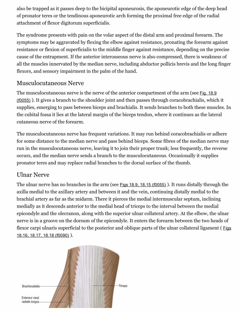
Ulnar NerveThe ulnar nerve has no branches in the arm (see Figs 18.9, 18.15 (f0055) ). It runs distally through theaxilla medial to the axillary artery and between it and the vein, continuing distally medial to thebrachial artery as far as the midarm. There it pierces the medial intermuscular septum, incliningmedially as it descends anterior to the medial head of triceps to the interval between the medialepicondyle and the olecranon, along with the superior ulnar collateral artery. At the elbow, the ulnarnerve is in a groove on the dorsum of the epicondyle. It enters the forearm between the two heads offlexor carpi ulnaris superficial to the posterior and oblique parts of the ulnar collateral ligament ( Figs18.16, 18.17, 18.18 (f0090) ).

Fig. 18.17
Posterior aspect of the left elbow showing superficial structures.
Fig. 18.18
Posterior aspect of the left elbow region showing deep structures.
Articular Branches

Articular branches to the elbow joint issue from the ulnar nerve between the medial epicondyle andolecranon.
Cubital Tunnel Syndrome
Typically, the ulnar nerve can be compressed in the tunnel formed by the tendinous arch connectingthe two heads of flexor carpi ulnaris at their humeral and ulnar attachments. Other local causes ofcompression and neuritis at this site include trauma, compression by the medial head of the triceps,osteophytes, recurrent subluxation of the nerve across the medial epicondyle of the humerus andabnormal muscular variants such as the anconeus epitrochlearis.
The symptoms are pain at the medial aspect of the proximal forearm, together with paraesthesia andnumbness of the little finger and ulnar half of the ring finger and the ulnar side of the dorsum of thehand. These symptoms are typically worse on forced elbow flexion. There may also be associatedweakness of the muscles of the forearm and the intrinsic muscles of the hand innervated by the ulnarnerve. Interestingly, flexor carpi ulnaris and profundus to the ring and little fingers are frequentlyspared, presumably because the fascicles supplying these muscles are located on the deep aspect ofthe nerve. Clawing of the hand is therefore unusual in this syndrome.
Surgical treatment involves decompression of the tunnel by division of the aponeurosis of flexorcarpi ulnaris, with or without subsequent anterior transposition of the ulnar nerve.
Ulnar Nerve Division at the Elbow
The ulnar nerve is in a vulnerable position as it lies between the median epicondyle and theolecranon: it lies on bone covered only by a thin layer of skin. It is easily damaged if the ulnar grooveis shallow, and the nerve may become more prominent than the medial epicondyle or the olecranonwhen the elbow is fully flexed.
Division of the nerve at the elbow paralyses flexor carpi ulnaris, flexor digitorum profundus to thering and little fingers and all the intrinsic muscles of the hand (except for the radial two lumbricals).Clawing of the hand is less intense than that which occurs after division of the ulnar nerve at thewrist, reflecting the imbalance in action between the long flexors and extensors to the ring and littlefingers when digit flexion is produced only by superficialis. In addition, there is sensory loss over thelittle finger and the ulnar half of the ring finger.
CASE 3Ulnar Neuropathy at the Elbow
An 18yearold fractured his distal right humerus while skiing. Following recovery, he was leftwith a mild bony deformity. He continued to play sports but then developed numbness andtingling in the fourth and fifth digits of the right hand, along with pain in the right elbow; hehas also observed wasting of the muscles of his right hand. On examination, there is wastingand decreased strength of the interossei muscles. Flexion of the fourth and fifth digits isimpaired, but wrist flexion is normal, as is strength elsewhere. Sensation is decreased in the
medial half of the fourth digit and in the entire fifth digit on both the dorsal and palmarsurfaces. Reflexes are normal. Percussion over the ulnar nerve proximal to the medialepicondyle elicits a shooting electric shock sensation radiating distally into the little finger(Tinel's sign).
Discussion: The term ‘tardy ulnar palsy’ usually refers to ulnar nerve compression at the elbowcaused, in this case, by prior trauma at the level of the ulnar groove, bony deformity from anold fracture, or inflammation of bursae or subcutaneous tissues with compression of the nervein the cubital tunnel (the nerve is injured at the level of the ulnar groove due to compressionbetween two bony processes or compression within the cubital tunnel itself). The ulnar nervedoes not branch throughout most of its course in the arm, proceeding through the axilla andalong the medial upper arm and into the ulnar groove, between the medial epicondyle and theolecranon; it is here that trauma is such an important pathogenetic event. Distal to the medialepicondyle, the nerve travels in the cubital tunnel below the aponeurosis of the flexor carpiulnaris muscle; its first branch is in fact to the flexor carpi ulnaris itself, which may beunaffected in typical cases of posttraumatic tardy ulnar palsy or cubital tunnel syndrome.
Radial NerveThe branches of the radial nerve in the upper arm are as follows: muscular, cutaneous, articular andsuperficial terminal and posterior interosseous.
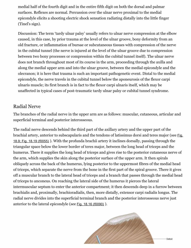
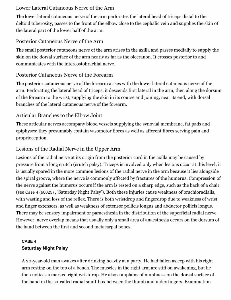
The radial nerve descends behind the third part of the axillary artery and the upper part of thebrachial artery, anterior to subscapularis and the tendons of latissimus dorsi and teres major (see Fig.18.9; Fig. 18.19 (f0055) ). With the profunda brachii artery it inclines dorsally, passing through thetriangular space below the lower border of teres major, between the long head of triceps and thehumerus. There it supplies the long head of triceps and gives rise to the posterior cutaneous nerve ofthe arm, which supplies the skin along the posterior surface of the upper arm. It then spiralsobliquely across the back of the humerus, lying posterior to the uppermost fibres of the medial headof triceps, which separate the nerve from the bone in the first part of the spiral groove. There it givesoff a muscular branch to the lateral head of triceps and a branch that passes through the medial headof triceps to anconeus. On reaching the lateral side of the humerus it pierces the lateralintermuscular septum to enter the anterior compartment; it then descends deep in a furrow betweenbrachialis and, proximally, brachioradialis, then, more distally, extensor carpi radialis longus. Theradial nerve divides into the superficial terminal branch and the posterior interosseous nerve justanterior to the lateral epicondyle (see Fig. 18.16 (f0090) ).
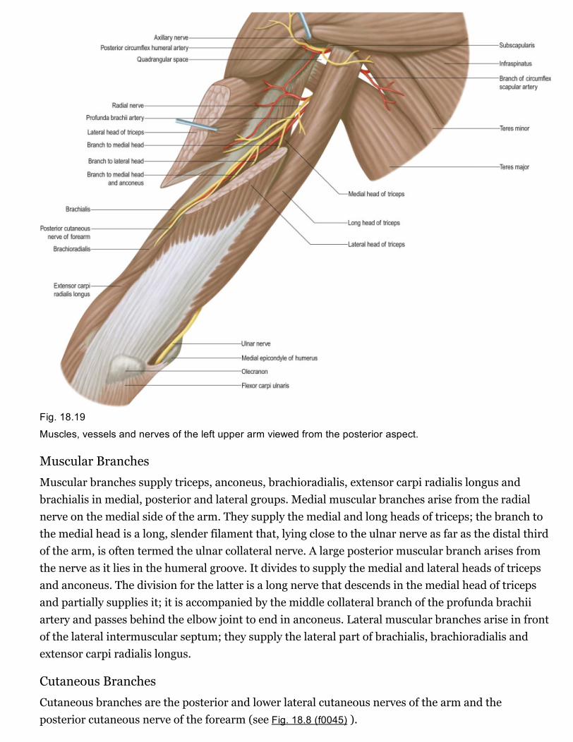
Fig. 18.19
Muscles, vessels and nerves of the left upper arm viewed from the posterior aspect.
Muscular Branches
Muscular branches supply triceps, anconeus, brachioradialis, extensor carpi radialis longus andbrachialis in medial, posterior and lateral groups. Medial muscular branches arise from the radialnerve on the medial side of the arm. They supply the medial and long heads of triceps; the branch tothe medial head is a long, slender filament that, lying close to the ulnar nerve as far as the distal thirdof the arm, is often termed the ulnar collateral nerve. A large posterior muscular branch arises fromthe nerve as it lies in the humeral groove. It divides to supply the medial and lateral heads of tricepsand anconeus. The division for the latter is a long nerve that descends in the medial head of tricepsand partially supplies it; it is accompanied by the middle collateral branch of the profunda brachiiartery and passes behind the elbow joint to end in anconeus. Lateral muscular branches arise in frontof the lateral intermuscular septum; they supply the lateral part of brachialis, brachioradialis andextensor carpi radialis longus.
Cutaneous Branches
Cutaneous branches are the posterior and lower lateral cutaneous nerves of the arm and theposterior cutaneous nerve of the forearm (see Fig. 18.8 (f0045) ).
Lower Lateral Cutaneous Nerve of the Arm
The lower lateral cutaneous nerve of the arm perforates the lateral head of triceps distal to thedeltoid tuberosity, passes to the front of the elbow close to the cephalic vein and supplies the skin ofthe lateral part of the lower half of the arm.
Posterior Cutaneous Nerve of the Arm
The small posterior cutaneous nerve of the arm arises in the axilla and passes medially to supply theskin on the dorsal surface of the arm nearly as far as the olecranon. It crosses posterior to andcommunicates with the intercostobrachial nerve.
Posterior Cutaneous Nerve of the Forearm
The posterior cutaneous nerve of the forearm arises with the lower lateral cutaneous nerve of thearm. Perforating the lateral head of triceps, it descends first lateral in the arm, then along the dorsumof the forearm to the wrist, supplying the skin in its course and joining, near its end, with dorsalbranches of the lateral cutaneous nerve of the forearm.
Articular Branches to the Elbow Joint
These articular nerves accompany blood vessels supplying the synovial membrane, fat pads andepiphyses; they presumably contain vasomotor fibres as well as afferent fibres serving pain andproprioception.
Lesions of the Radial Nerve in the Upper Arm
Lesions of the radial nerve at its origin from the posterior cord in the axilla may be caused bypressure from a long crutch (crutch palsy). Triceps is involved only when lesions occur at this level; itis usually spared in the more common lesions of the radial nerve in the arm because it lies alongsidethe spiral groove, where the nerve is commonly affected by fractures of the humerus. Compression ofthe nerve against the humerus occurs if the arm is rested on a sharp edge, such as the back of a chair(see Case 4 (b0025) , ‘Saturday Night Palsy’). Both these injuries cause weakness of brachioradialis,with wasting and loss of the reflex. There is both wristdrop and fingerdrop due to weakness of wristand finger extensors, as well as weakness of extensor pollicis longus and abductor pollicis longus.There may be sensory impairment or paraesthesia in the distribution of the superficial radial nerve.However, nerve overlap means that usually only a small area of anaesthesia occurs on the dorsum ofthe hand between the first and second metacarpal bones.
CASE 4Saturday Night Palsy
A 20yearold man awakes after drinking heavily at a party. He had fallen asleep with his rightarm resting on the top of a bench. The muscles in the right arm are stiff on awakening, but hethen notices a marked right wristdrop. He also complains of numbness on the dorsal surface ofthe hand in the socalled radial snuffbox between the thumb and index fingers. Examination

demonstrates weakness of the wrist and finger extensors and brachioradialis in the affectedlimb. Triceps strength is normal. There is loss of pinprick sensation in the pattern describedearlier. The brachioradialis reflex is absent, although other reflexes are normal.
Discussion: ‘Saturday night palsy’ is a term used to describe an injury to the radial nerve at thelevel of the spiral groove of the humerus due to compression of the nerve against the bone asthe nerve travels laterally. It often occurs during deep sleep following drug or alcohol abuse,most likely due to pressure as the arm is draped over a hard bench or a chair, as in this case.Sensory changes in the distribution of the radial nerve and wristdrop typically result from suchnerve compression. Because triceps is innervated by a branch of the radial nerve that emergesabove the spiral groove, it is usually spared. The nerve injury may be incomplete, with variableweakness of brachioradialis, as noted above. Reflex changes are also variable. Although thesensory branch of the radial nerve innervates a larger area than described in this patient,significant crossover in innervation restricts the sensory loss.
Medial Cutaneous Nerve of the ArmThe medial cutaneous nerve of the arm supplies the skin of the medial aspect of the arm (see Figs18.8, 18.9 (f0045) ). It is the smallest branch of the brachial plexus, arises from the medial cord andcontains fibres from the eighth cervical and first thoracic ventral rami. It traverses the axilla, crossinganterior or posterior to the axillary vein, to which it is then medial, and communicates with theintercostobrachial nerve; it descends medial to the brachial artery and basilic vein (see Fig. 18.9(f0055) ) to a point midway in the upper arm, where it pierces the deep fascia to supply a medial areain the distal third of the arm, extending to its anterior and posterior aspects. Rami reach the skinanterior to the medial epicondyle and over the olecranon. It connects with the posterior branch of themedial cutaneous nerve of the forearm. Sometimes the medial cutaneous nerve of the arm and theintercostobrachial nerve are connected in a plexiform manner in the axilla. The intercostobrachialnerve may be large and reinforced by part of the lateral cutaneous branch of the third intercostalnerve. It then replaces the medial cutaneous nerve of the arm and receives a connection representingthe latter from the brachial plexus (occasionally, this connection is absent).
Medial Cutaneous Nerve of the ForearmThe medial cutaneous nerve of forearm comes from the medial cord (see Fig. 18.9; Fig. 18.20 (f0055) ).It is derived from the eighth cervical and first thoracic ventral rami. At first it is between the axillaryartery and vein and gives off a ramus that pierces the deep fascia to supply the skin over biceps,almost to the elbow. The nerve descends medial to the brachial artery, pierces the deep fascia withthe basilic vein midway in the arm and divides into anterior and posterior branches. The larger,anterior branch usually passes in front of, or occasionally behind, the median cubital vein,descending anteromedially in the forearm to supply the skin as far as the wrist and connecting withthe palmar cutaneous branch of the ulnar nerve. The posterior branch descends obliquely medial to
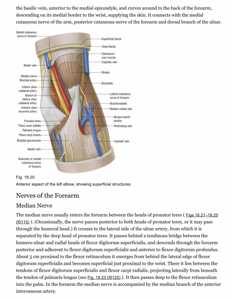
the basilic vein, anterior to the medial epicondyle, and curves around to the back of the forearm,descending on its medial border to the wrist, supplying the skin. It connects with the medialcutaneous nerve of the arm, posterior cutaneous nerve of the forearm and dorsal branch of the ulnar.
Fig. 18.20
Anterior aspect of the left elbow, showing superficial structures.
Nerves of the ForearmMedian NerveThe median nerve usually enters the forearm between the heads of pronator teres ( Figs 18.21–18.25(f0115) ). (Occasionally, the nerve passes posterior to both heads of pronator teres, or it may passthrough the humeral head.) It crosses to the lateral side of the ulnar artery, from which it isseparated by the deep head of pronator teres. It passes behind a tendinous bridge between thehumeroulnar and radial heads of flexor digitorum superficialis, and descends through the forearmposterior and adherent to flexor digitorum superficialis and anterior to flexor digitorum profundus.About 5 cm proximal to the flexor retinaculum it emerges from behind the lateral edge of flexordigitorum superficialis and becomes superficial just proximal to the wrist. There it lies between thetendons of flexor digitorum superficialis and flexor carpi radialis, projecting laterally from beneaththe tendon of palmaris longus (see Fig. 18.23 (f0125) ). It then passes deep to the flexor retinaculuminto the palm. In the forearm the median nerve is accompanied by the median branch of the anteriorinterosseous artery.

Fig. 18.21
Superficial flexor muscles of the left forearm.

Fig. 18.22
Deep flexor muscles of the left forearm.
Fig. 18.23
Transverse section through the left forearm, passing through the distal end of the radius and the styloid process
of the ulna, with the hand and forearm in full supination (distal [inferior] aspect).
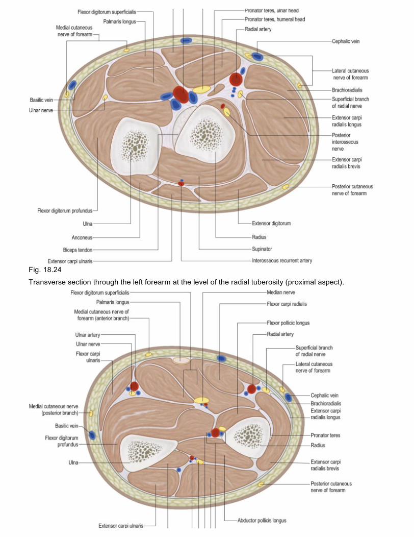
Fig. 18.24
Transverse section through the left forearm at the level of the radial tuberosity (proximal aspect).
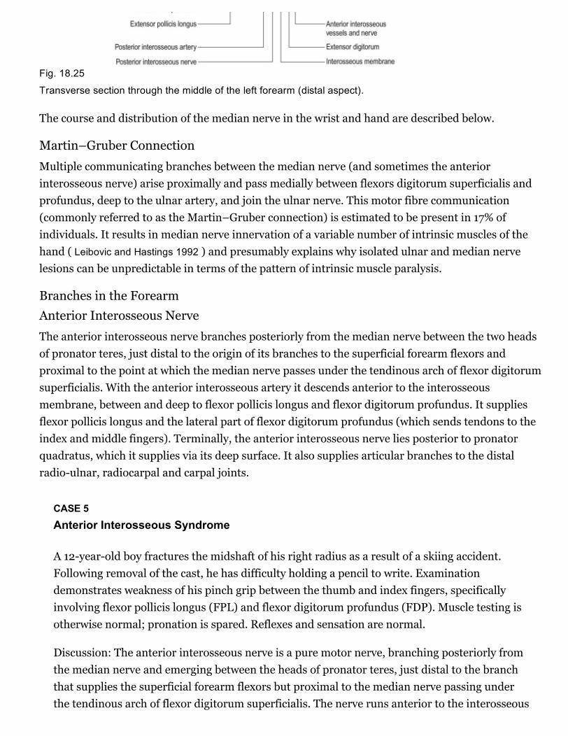
Fig. 18.25
Transverse section through the middle of the left forearm (distal aspect).
The course and distribution of the median nerve in the wrist and hand are described below.
Martin–Gruber Connection
Multiple communicating branches between the median nerve (and sometimes the anteriorinterosseous nerve) arise proximally and pass medially between flexors digitorum superficialis andprofundus, deep to the ulnar artery, and join the ulnar nerve. This motor fibre communication(commonly referred to as the Martin–Gruber connection) is estimated to be present in 17% ofindividuals. It results in median nerve innervation of a variable number of intrinsic muscles of thehand ( Leibovic and Hastings 1992 ) and presumably explains why isolated ulnar and median nervelesions can be unpredictable in terms of the pattern of intrinsic muscle paralysis.
Branches in the ForearmAnterior Interosseous Nerve
The anterior interosseous nerve branches posteriorly from the median nerve between the two headsof pronator teres, just distal to the origin of its branches to the superficial forearm flexors andproximal to the point at which the median nerve passes under the tendinous arch of flexor digitorumsuperficialis. With the anterior interosseous artery it descends anterior to the interosseousmembrane, between and deep to flexor pollicis longus and flexor digitorum profundus. It suppliesflexor pollicis longus and the lateral part of flexor digitorum profundus (which sends tendons to theindex and middle fingers). Terminally, the anterior interosseous nerve lies posterior to pronatorquadratus, which it supplies via its deep surface. It also supplies articular branches to the distalradioulnar, radiocarpal and carpal joints.
CASE 5Anterior Interosseous Syndrome
A 12yearold boy fractures the midshaft of his right radius as a result of a skiing accident.Following removal of the cast, he has difficulty holding a pencil to write. Examinationdemonstrates weakness of his pinch grip between the thumb and index fingers, specificallyinvolving flexor pollicis longus (FPL) and flexor digitorum profundus (FDP). Muscle testing isotherwise normal; pronation is spared. Reflexes and sensation are normal.
Discussion: The anterior interosseous nerve is a pure motor nerve, branching posteriorly fromthe median nerve and emerging between the heads of pronator teres, just distal to the branchthat supplies the superficial forearm flexors but proximal to the median nerve passing underthe tendinous arch of flexor digitorum superficialis. The nerve runs anterior to the interosseous

membrane, between FPL and FDP, supplying FPL and lateral FDP. It also innervates the deeppronator quadratus muscle. Weakness of FPL and FDP, as occurs in anterior interosseoussyndrome, causes a weak pinch between the thumb and index fingers. Pronator quadratusweakness may not be evident on testing owing to the normal strength of pronator teres. Nosensory symptoms are present, distinguishing this syndrome from the socalled pronatorsyndrome, which reflects more proximal involvement of the median nerve, thus implicating asensory branch as well. In the patient described here, a midshaft fracture of the radius resultedin injury to the anterior interosseous nerve distal to its branching from the median nerve.
Muscular Branches
Muscular branches are given off near the elbow to all the superficial flexor muscles except flexorcarpi ulnaris—that is, to pronator teres, flexor carpi radialis, palmaris longus and flexor digitorumsuperficialis. The branch to the part of flexor digitorum superficialis that serves the index finger isgiven off near the midforearm and may be derived from the anterior interosseous nerve.
Other Branches
Articular branches, arising at or just distal to the elbow joint, supply the joint and the proximal radioulnar joint. The palmar cutaneous branch is described below.
Ulnar NerveThe ulnar nerve descends on the medial side of the forearm, lying on flexor digitorum profundus (seeFigs 18.22–18.25 (f0120) ). Proximally, it is covered by flexor carpi ulnaris; its distal half lies lateral tothe muscle and is covered only by skin and fasciae. In the upper third of the forearm, the nerve isdistant from the ulnar artery, but more distally, it comes to lie close to the medial side of the artery.About 5 cm proximal to the wrist it gives off a dorsal branch that continues distally into the hand,anterior to the flexor retinaculum on the lateral side of the pisiform and posteromedial to the ulnarartery. It passes deep to the superficial part of the retinaculum (in Guyon's canal) with the artery anddivides into superficial and deep terminal branches.
The course and distribution of the ulnar nerve in the hand are described below.
Muscular Branches
There are usually two muscular branches. They begin near the elbow and supply flexor carpi ulnarisand the medial half of flexor digitorum profundus.
Palmar Cutaneous Branch
The palmar cutaneous branch arises about midforearm. It descends on the ulnar artery, which itsupplies, and then perforates the deep fascia to end in the palmar skin, after communicating with thepalmar branch of the median nerve. It sometimes supplies palmaris brevis.
Dorsal Branch

The dorsal branch of the ulnar nerve is described below.
Radial NerveThere is some variation in the level at which branches of the radial nerve arise from the main trunkin different subjects ( Fig. 18.26 (f0140) ; see also Figs 18.23, 18.25 (f0125) ). Branches to extensor carpiradialis brevis and supinator may arise from the main trunk of the radial nerve or from the proximalpart of the posterior interosseous nerve, but almost invariably above the arcade of Frohse.
Fig. 18.26
Left supinator muscle (posterolateral aspect).
Radial Tunnel Syndrome
Radial tunnel syndrome is an entrapment neuropathy of the radial nerve near the elbow, where fourstructures can potentially cause compression of the nerve: (1) fibrous bands (which can tether theradial nerve to the radiohumeral joint), (2) the sharp tendinous medial border of extensor carpiradialis brevis, (3) a leash of vessels from the radial recurrent artery as it passes to supplybrachioradialis and extensor carpi radialis longus and (4) the arcade of Frohse, which is the freeaponeurotic proximal edge of the superficial part of supinator (see Fig. 18.16 (f0090) ).

Usually the only presenting symptom is pain over the extensor mass just distal to the elbow. There isno sensory disturbance or motor loss, but there is frequently tenderness along the course of theradial nerve over the radial head. The pain is exacerbated when the elbow is extended and the wrist ispassively flexed and pronated or extended and supinated against resistance. Extension of the middlefinger against resistance when the elbow in fully extended may lead to increased pain. Thesemanoeuvres tighten the anatomical structures, which cause compression.
Superficial Terminal Branch
The superficial terminal branch descends from the lateral epicondyle anterolaterally in the proximaltwothirds of the forearm, initially lying on supinator, lateral to the radial artery and behindbrachioradialis. In the middle third of the forearm it lies behind brachioradialis, close to the lateralside of the artery, and is successively anterior to pronator teres, the radial head of flexor digitorumsuperficialis and flexor pollicis longus. It leaves the artery approximately 7 cm proximal to the wristand passes deep to the brachioradialis tendon. It curves around the lateral side of the radius as itdescends, pierces the deep fascia and divides into five (sometimes four) dorsal digital nerves. On thedorsum of the hand it usually communicates with the posterior and lateral cutaneous nerves of theforearm.
As the nerve crosses the lateral aspect of the radius it is superficial and relatively unprotected; it iseasily compressed here by tight bracelets, watch straps and handcuffs.
Radial Sensory Nerve Entrapment (Wartenberg's Disease)
Entrapment of the superficial radial nerve can occur as it emerges from beneath the edge of thebrachioradialis tendon approximately 6 cm proximal to the radial styloid. The condition is frequentlyassociated with previous trauma in this region. The symptoms are pain and paraesthesia over theradial aspect of the dorsum of the wrist and hand.
Posterior Interosseous Nerve
The posterior interosseous nerve is the deep terminal branch of the radial nerve (see Fig. 18.24 (f0130)). It reaches the back of the forearm by passing around the lateral aspect of the radius between thetwo heads of supinator. It supplies extensor carpi radialis brevis and supinator before enteringsupinator; as it passes through the muscle it supplies it with additional branches. The branch toextensor carpi radialis brevis may arise from the beginning of the superficial branch of the radialnerve. As it emerges from supinator posteriorly, the posterior interosseous nerve gives off three shortbranches to extensor digitorum, extensor digiti minimi and extensor carpi ulnaris; it also gives offtwo longer branches—a medial branch to extensor pollicis longus and extensor indicis, and a lateralbranch that supplies abductor pollicis longus and extensor pollicis brevis. The nerve at first liesbetween the superficial and deep extensor muscles, but at the distal border of extensor pollicis brevisit passes deep to extensor pollicis longus and, diminished to a fine thread, descends on theinterosseous membrane to the dorsum of the carpus. There it presents a flattened and somewhatexpanded termination or ‘pseudoganglion,’ from which filaments supply the carpal ligaments and

articulations. Articular branches from the posterior interosseous nerve supply carpal, distal radioulnar and some intercarpal and intermetacarpal joints. Digital branches supply themetacarpophalangeal and proximal interphalangeal joints.
The distal portion of the nerve lies in a separate fascial sheath in the radial, deep aspect of the fourthdorsal compartment of the extensor retinaculum of the wrist, where it is located deep to extensordigitorum and extensor indicis. This portion of the nerve can be used as a donor nerve for graftingsegmental digital nerve defects, as there is no clinically discernible donor site deficit.
Posterior Interosseous Nerve Palsy
There are many causes of posterior interosseous nerve palsy. These include trauma andinflammatory swelling, as well as entrapment at the same anatomical sites that can cause radialtunnel syndrome. Pain is similar in nature to that of radial tunnel syndrome and is lateraccompanied by weakness and paralysis. When fully developed, there is an inability to extend thefingers at the metacarpophalangeal joints and weakness of thumb extension and abduction. There isalso weakness and radial deviation of wrist extension because extensor carpi ulnaris is usuallyaffected, whereas the radial wrist extensors and brachioradialis are normal (because their nervesupply is given off proximal to the origin of the posterior interosseous nerve). There are no sensorydisturbances, because the superficial radial nerve arises above this level.
Medial Cutaneous Nerve of the ForearmThe medial cutaneous nerve of the forearm has already divided into anterior and posterior branchesbefore it enters the forearm (see Fig. 18.20 (f0110) ). The larger anterior branch usually passes in frontof, or occasionally behind, the median cubital vein and descends anteromedially in the forearm tosupply the skin as far as the wrist. It curves around to the back of the forearm, descending on itsmedial border to the wrist, supplying the skin. It connects with the medial cutaneous nerve of thearm, posterior cutaneous nerve of the forearm and dorsal branch of the ulnar nerve.
Lateral Cutaneous Nerve of the ForearmThe lateral cutaneous nerve of the forearm is a direct continuation of the musculocutaneous nerve asit lies lateral to the biceps tendon in the antecubital fossa (see Figs 18.5, 18.8 (f0030) ). It passes deepto the cephalic vein, descending along the radial border of the forearm to the wrist. It supplies theskin of the anterolateral surface of the forearm and connects with the posterior cutaneous nerve ofthe forearm and the terminal branch of the radial nerve by branches that pass around its radialborder. Its trunk gives rise to a slender recurrent branch that extends along the cephalic vein as far asthe middle third of the upper arm, distributing filaments to the skin over the distal third of theanterolateral surface of the upper arm close to the vein. At the wrist joint the lateral cutaneous nerveof the forearm is anterior to the radial artery. Some filaments pierce the deep fascia and accompanythe artery to the dorsum of the carpus. The nerve then passes to the base of the thenar eminence,where it ends in cutaneous rami. It has branches that connect with the terminal branch of the radialnerve and the palmar cutaneous branch of the median nerve.
Posterior Cutaneous Nerve of the ForearmThe posterior cutaneous nerve of the forearm passes along the dorsum of the forearm to the wrist. Itsupplies the skin along its course and near its end joins the dorsal branches of the lateral cutaneousnerve of the forearm.
Nerves of the Wrist and HandMedian NerveThe median nerve proximal to the flexor retinaculum is lateral to the tendons of flexor digitorumsuperficialis and lies between the tendons of flexor carpi radialis and palmaris longus. It passesunder the retinaculum in the ‘carpal tunnel’ (see below), where its compression may lead to carpaltunnel syndrome. Distal to the retinaculum the nerve enlarges and flattens and usually divides intofive or six branches; the mode and level of division are variable.
Palmar Cutaneous Branch
The palmar cutaneous branch starts just proximal to the flexor retinaculum. It pierces theretinaculum or the deep fascia and divides into lateral branches that supply the thenar skin andconnect with the lateral cutaneous nerve of the forearm. Medial branches supply the central palmarskin and connect with the palmar cutaneous branch of the ulnar nerve.
Communicating branches, which may be multiple, often arise in the proximal forearm, sometimesfrom the anterior interosseous branch. They pass medially between flexors digitorum superficialisand profundus and behind the ulnar artery to join the ulnar nerve. This communication is a factor inexplaining anomalous muscular innervation in the hand (see below).
Muscular Branch (Motor or Recurrent Branch)
The muscular branch is short and thick and arises from the lateral side of the nerve; it may be thefirst palmar branch or a terminal branch that arises level with the digital branches. It runs laterally,just distal to the flexor retinaculum, with a slight recurrent curve beneath the part of the palmaraponeurosis covering the thenar muscles. It turns around the distal border of the retinaculum to liesuperficial to flexor pollicis brevis, which it usually supplies, and continues either superficial to themuscle or traverses it. It gives a branch to abductor pollicis brevis, which enters the medial edge ofthe muscle and then passes deep to it to supply opponens pollicis, entering its medial edge. Itsterminal part occasionally gives a branch to the first dorsal interosseous, which may be its sole orpartial supply. The muscular branch may arise in the carpal tunnel and pierce the flexor retinaculum,which is a point of surgical importance.
Palmar Digital Branches ( Figs 18.27, 18.28 (f0145) )The median nerve usually divides into four or five digital branches. It often divides first into a lateralramus, which provides digital branches to the thumb and radial side of the index finger, and a medialramus, which supplies digital branches to adjacent sides of the index, middle and ring fingers. Othermodes of termination can occur.
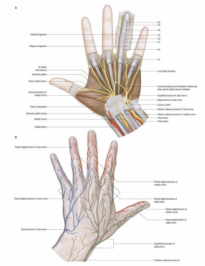

Fig. 18.27
Cutaneous nerves of the hand. A , Palmar aspect. B , Dorsal aspect. The anular and cruciate pulleys are shownschematically in the ring finger.
Fig. 18.28
Palmar aponeurosis and distal fascial complex. A , Schematic diagram of the palmar fascia. B , More detailedview of structures at the web space. C , Fate of the distal longitudinal fibres. D and E , Normal digital fascia.
Digital branches are commonly arranged as follows. They pass distally, deep to the superficial palmararch and its digital vessels, at first anterior to the long flexor tendons. Two proper palmar digitalnerves, sometimes from a common stem, pass to the sides of the thumb; the nerve supplying itsradial side crosses in front of the flexor pollicis longus tendon. The proper palmar digital nerve to thelateral side of the index also supplies the first lumbrical. Two common palmar digital nerves passdistally between the long flexor tendons. The lateral one divides in the distal palm into two properpalmar digital nerves that traverse adjacent sides of the index and middle fingers. The medial onedivides into two proper palmar digital nerves that supply adjacent sides of the middle and ring

fingers. The lateral common digital nerve supplies the second lumbrical, and the medial receives acommunicating twig from the common palmar digital branch of the ulnar nerve and may supply thethird lumbrical. In the distal part of the palm the digital arteries pass deeply between the divisions ofthe digital nerves; the nerves lie anterior to the arteries on the sides of the digits. The median nerveusually supplies palmar cutaneous digital branches to the radial three and a half digits (thumb,index, middle and lateral sides of the ring finger); sometimes the radial side of the ring finger issupplied by the ulnar nerve. Occasionally, there is a communicating branch between the commondigital nerve to the middle and ring fingers (derived from the median nerve) and the common digitalnerve to the ring and little fingers (derived from the ulnar nerve). This can explain variations insensory patterns that do not conform to the classic pattern.
The proper palmar digital nerves pass along the medial side of the index finger, both sides of themiddle finger and the lateral side of the ring finger. They enter these digits in fat between slips of thepalmar aponeurosis. Together with the lumbricals and palmar digital arteries, they pass dorsal to thesuperficial transverse metacarpal ligament and ventral to the deep transverse metacarpal ligament.In the digits, the nerves run distally beside the long flexor tendons (outside their fibrous sheaths),level with the anterior phalangeal surfaces and anterior to the digital arteries, between Grayson's andCleland's ligaments . Each nerve gives off several branches to the skin on the front and sides of thedigit, where many end in Pacinian corpuscles. It also sends branches to the metacarpophalangeal andinterphalangeal joints.
The digital nerves supply the fibrous sheaths of the long flexor tendons, digital arteries (vasomotor)and sweat glands (secretomotor). Distal to the base of the distal phalanx, each digital nerve gives offa branch that passes dorsally to the nail bed. The main nerve frequently trifurcates to supply the pulpand skin of the terminal part of the digit. Distal to the base of the proximal phalanx, each properdigital nerve also gives off a dorsal branch to supply the skin over the back of the middle and distalphalanges. The proper palmar digital nerves to the thumb and lateral side of the index finger emergewith the long flexor tendons from under the lateral edge of the palmar aponeurosis. They arearranged in the digits as described earlier, but in the thumb, small distal branches supply the skin onthe back of the distal phalanx only.
Other Branches
In addition to the branches of the median nerve already described, variable vasomotor branchessupply the radial and ulnar arteries and their branches. Some of the intercarpal, carpometacarpaland intermetacarpal joints are thought to be supplied by the median nerve or its anteriorinterosseous branch; the precise details are uncertain.
CASE 6Carpal Tunnel Syndrome
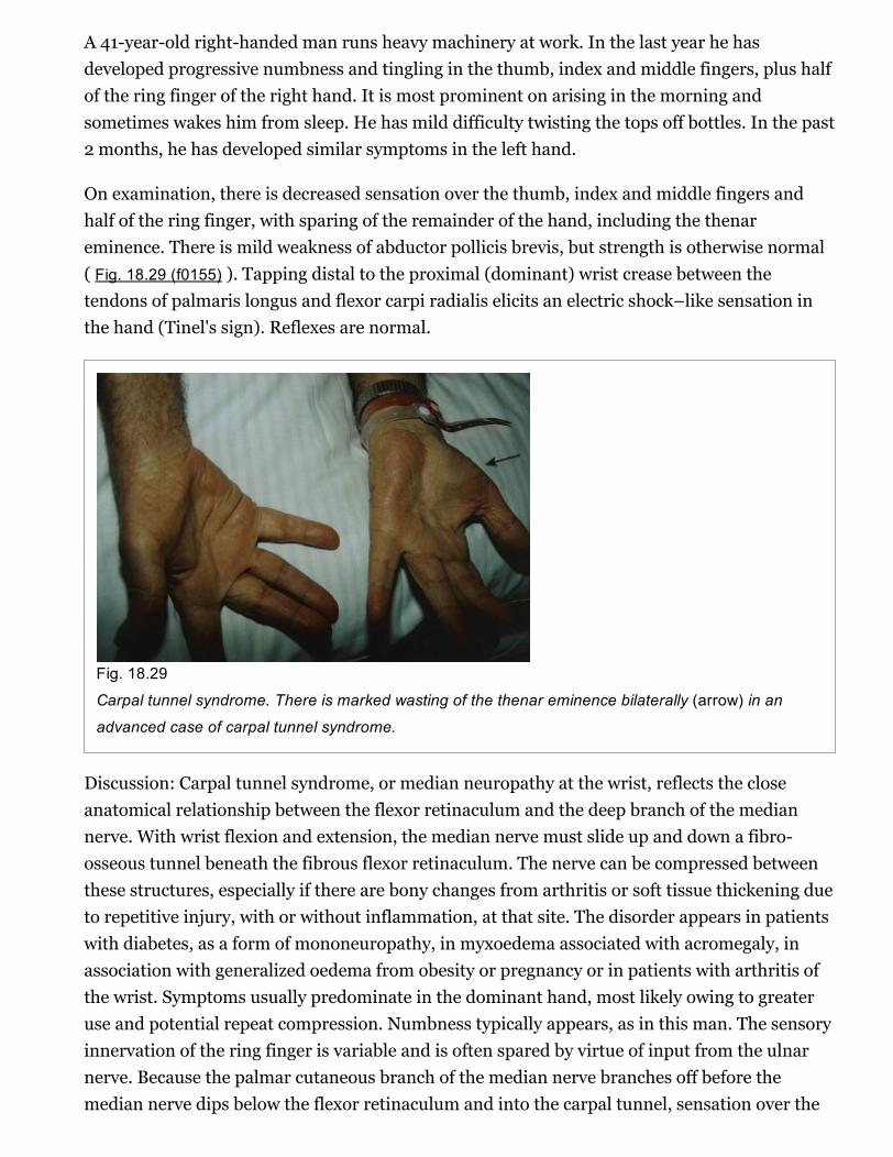
A 41yearold righthanded man runs heavy machinery at work. In the last year he hasdeveloped progressive numbness and tingling in the thumb, index and middle fingers, plus halfof the ring finger of the right hand. It is most prominent on arising in the morning andsometimes wakes him from sleep. He has mild difficulty twisting the tops off bottles. In the past2 months, he has developed similar symptoms in the left hand.
On examination, there is decreased sensation over the thumb, index and middle fingers andhalf of the ring finger, with sparing of the remainder of the hand, including the thenareminence. There is mild weakness of abductor pollicis brevis, but strength is otherwise normal( Fig. 18.29 (f0155) ). Tapping distal to the proximal (dominant) wrist crease between thetendons of palmaris longus and flexor carpi radialis elicits an electric shock–like sensation inthe hand (Tinel's sign). Reflexes are normal.
Fig. 18.29
Carpal tunnel syndrome. There is marked wasting of the thenar eminence bilaterally (arrow) in an
advanced case of carpal tunnel syndrome.
Discussion: Carpal tunnel syndrome, or median neuropathy at the wrist, reflects the closeanatomical relationship between the flexor retinaculum and the deep branch of the mediannerve. With wrist flexion and extension, the median nerve must slide up and down a fibroosseous tunnel beneath the fibrous flexor retinaculum. The nerve can be compressed betweenthese structures, especially if there are bony changes from arthritis or soft tissue thickening dueto repetitive injury, with or without inflammation, at that site. The disorder appears in patientswith diabetes, as a form of mononeuropathy, in myxoedema associated with acromegaly, inassociation with generalized oedema from obesity or pregnancy or in patients with arthritis ofthe wrist. Symptoms usually predominate in the dominant hand, most likely owing to greateruse and potential repeat compression. Numbness typically appears, as in this man. The sensoryinnervation of the ring finger is variable and is often spared by virtue of input from the ulnarnerve. Because the palmar cutaneous branch of the median nerve branches off before themedian nerve dips below the flexor retinaculum and into the carpal tunnel, sensation over the

thenar eminence is spared. Abductor pollicis brevis is innervated by the median nerve, soweakness of this muscle may be found. This indicates a distal median nerve injury; medianinnervated muscles proximal to the flexor retinaculum are unaffected in carpal tunnelsyndrome.
Median Nerve Division at the Wrist
The median nerve is vulnerable to division from lacerations at the wrist. Division leads to paralysis ofthe lumbricals to the index and middle fingers and the thenar muscles (apart from adductor pollicis),as well as loss of sensation to the thumb, index, middle fingers and radial half of the ring finger. Theradial half of the hand becomes flattened as a result of wasting of the thenar muscles and theadducted posture of the thumb.
Ulnar NerveAt the wrist, the ulnar nerve passes under the superficial part of the retinaculum (in Guyon's canal)with the ulnar artery and divides into superficial and deep terminal branches.
Dorsal Branch
The dorsal branch arises approximately 5 cm proximal to the wrist. It passes distally and dorsally,deep to flexor carpi ulnaris; perforates the deep fascia; descends along the medial side of the back ofthe wrist and hand; and then divides into two, or often three, dorsal digital nerves. The first suppliesthe medial side of the little finger; the second, the adjacent sides of the little and ring fingers; and thethird, when present, supplies adjoining sides of the ring and middle fingers. The last may bereplaced, wholly or partially, by a branch of the radial nerve, which always communicates with it onthe dorsum of the hand (see Fig. 18.28 (f0150) ). In the little finger, the dorsal digital nerves extendonly to the base of the distal phalanx; in the ring finger, they extend only to the base of the middlephalanx. The most distal parts of the little finger and of the ulnar side of the ring finger are suppliedby dorsal branches of the proper palmar digital branches of the ulnar nerve. The most distal part ofthe lateral side of the ring finger is supplied by dorsal branches of the proper palmar digital branch ofthe median nerve.
Superficial Terminal Branch
The superficial terminal branch supplies palmaris brevis and the medial palmar skin. It divides intotwo palmar digital nerves, which can be palpated against the hook of the hamate bone. One suppliesthe medial side of the little finger, and the other (a common palmar digital nerve) sends a twig to themedian nerve and divides into two proper digital nerves to supply the adjoining sides of the little andring fingers. The proper digital branches are distributed like those derived from the median nerve.
Deep Terminal Branch
The deep terminal branch accompanies the deep branch of the ulnar artery as it passes betweenabductor digiti minimi and flexor digiti minimi and then perforates the opponens digiti minimi tofollow the deep palmar arch dorsal to the flexor tendons ( Fig. 18.30 (f0160) ). At its origin, it supplies

the three short muscles of the little finger. As it crosses the hand, it supplies the interossei and thethird and fourth lumbricals. It ends by supplying adductor pollicis, first palmar interosseous andusually flexor pollicis brevis. It sends articular filaments to the wrist joint.
Fig. 18.30
Deep structures in the left palm and wrist.
The medial part of flexor digitorum profundus is supplied by the ulnar nerve, as are the third andfourth lumbricals, which are connected with the tendons of this part of the muscle. Similarly, thelateral part of flexor digitorum profundus and the first and second lumbricals are supplied by themedian nerve. The third lumbrical is often supplied by both nerves. The deep terminal branch isthought to give branches to some intercarpal, carpometacarpal and intermetacarpal joints; precisedetails are uncertain. Vasomotor branches, arising in the forearm and hand, supply the ulnar andpalmar arteries.
Ulnar Tunnel Syndrome
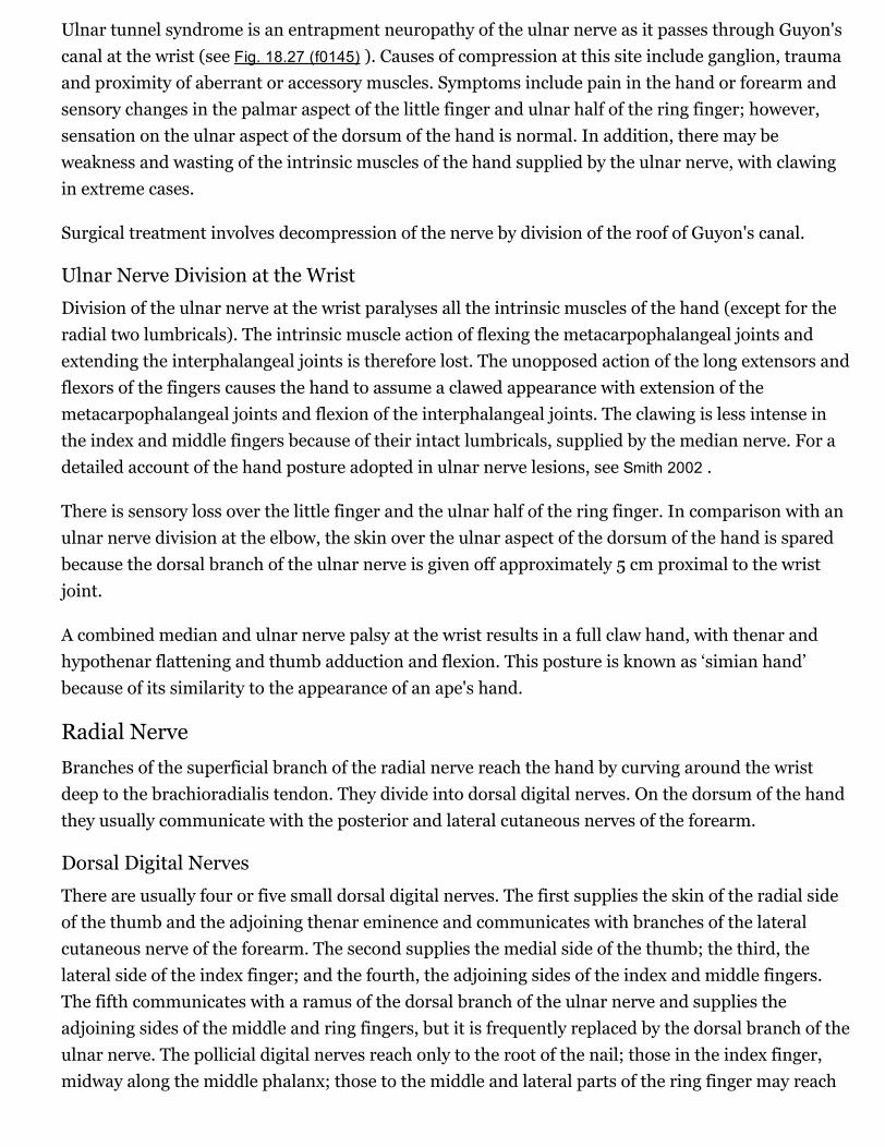
Ulnar tunnel syndrome is an entrapment neuropathy of the ulnar nerve as it passes through Guyon'scanal at the wrist (see Fig. 18.27 (f0145) ). Causes of compression at this site include ganglion, traumaand proximity of aberrant or accessory muscles. Symptoms include pain in the hand or forearm andsensory changes in the palmar aspect of the little finger and ulnar half of the ring finger; however,sensation on the ulnar aspect of the dorsum of the hand is normal. In addition, there may beweakness and wasting of the intrinsic muscles of the hand supplied by the ulnar nerve, with clawingin extreme cases.
Surgical treatment involves decompression of the nerve by division of the roof of Guyon's canal.
Ulnar Nerve Division at the Wrist
Division of the ulnar nerve at the wrist paralyses all the intrinsic muscles of the hand (except for theradial two lumbricals). The intrinsic muscle action of flexing the metacarpophalangeal joints andextending the interphalangeal joints is therefore lost. The unopposed action of the long extensors andflexors of the fingers causes the hand to assume a clawed appearance with extension of themetacarpophalangeal joints and flexion of the interphalangeal joints. The clawing is less intense inthe index and middle fingers because of their intact lumbricals, supplied by the median nerve. For adetailed account of the hand posture adopted in ulnar nerve lesions, see Smith 2002 .
There is sensory loss over the little finger and the ulnar half of the ring finger. In comparison with anulnar nerve division at the elbow, the skin over the ulnar aspect of the dorsum of the hand is sparedbecause the dorsal branch of the ulnar nerve is given off approximately 5 cm proximal to the wristjoint.
A combined median and ulnar nerve palsy at the wrist results in a full claw hand, with thenar andhypothenar flattening and thumb adduction and flexion. This posture is known as ‘simian hand’because of its similarity to the appearance of an ape's hand.
Radial NerveBranches of the superficial branch of the radial nerve reach the hand by curving around the wristdeep to the brachioradialis tendon. They divide into dorsal digital nerves. On the dorsum of the handthey usually communicate with the posterior and lateral cutaneous nerves of the forearm.
Dorsal Digital Nerves
There are usually four or five small dorsal digital nerves. The first supplies the skin of the radial sideof the thumb and the adjoining thenar eminence and communicates with branches of the lateralcutaneous nerve of the forearm. The second supplies the medial side of the thumb; the third, thelateral side of the index finger; and the fourth, the adjoining sides of the index and middle fingers.The fifth communicates with a ramus of the dorsal branch of the ulnar nerve and supplies theadjoining sides of the middle and ring fingers, but it is frequently replaced by the dorsal branch of theulnar nerve. The pollicial digital nerves reach only to the root of the nail; those in the index finger,midway along the middle phalanx; those to the middle and lateral parts of the ring finger may reach
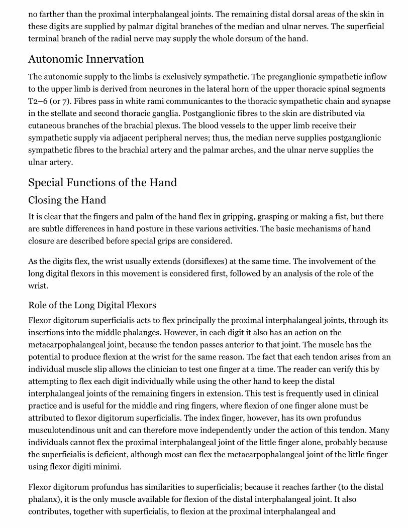
no farther than the proximal interphalangeal joints. The remaining distal dorsal areas of the skin inthese digits are supplied by palmar digital branches of the median and ulnar nerves. The superficialterminal branch of the radial nerve may supply the whole dorsum of the hand.
Autonomic InnervationThe autonomic supply to the limbs is exclusively sympathetic. The preganglionic sympathetic inflowto the upper limb is derived from neurones in the lateral horn of the upper thoracic spinal segmentsT2–6 (or 7). Fibres pass in white rami communicantes to the thoracic sympathetic chain and synapsein the stellate and second thoracic ganglia. Postganglionic fibres to the skin are distributed viacutaneous branches of the brachial plexus. The blood vessels to the upper limb receive theirsympathetic supply via adjacent peripheral nerves; thus, the median nerve supplies postganglionicsympathetic fibres to the brachial artery and the palmar arches, and the ulnar nerve supplies theulnar artery.
Special Functions of the HandClosing the HandIt is clear that the fingers and palm of the hand flex in gripping, grasping or making a fist, but thereare subtle differences in hand posture in these various activities. The basic mechanisms of handclosure are described before special grips are considered.
As the digits flex, the wrist usually extends (dorsiflexes) at the same time. The involvement of thelong digital flexors in this movement is considered first, followed by an analysis of the role of thewrist.
Role of the Long Digital Flexors
Flexor digitorum superficialis acts to flex principally the proximal interphalangeal joints, through itsinsertions into the middle phalanges. However, in each digit it also has an action on themetacarpophalangeal joint, because the tendon passes anterior to that joint. The muscle has thepotential to produce flexion at the wrist for the same reason. The fact that each tendon arises from anindividual muscle slip allows the clinician to test one finger at a time. The reader can verify this byattempting to flex each digit individually while using the other hand to keep the distalinterphalangeal joints of the remaining fingers in extension. This test is frequently used in clinicalpractice and is useful for the middle and ring fingers, where flexion of one finger alone must beattributed to flexor digitorum superficialis. The index finger, however, has its own profundusmusculotendinous unit and can therefore move independently under the action of this tendon. Manyindividuals cannot flex the proximal interphalangeal joint of the little finger alone, probably becausethe superficialis is deficient, although most can flex the metacarpophalangeal joint of the little fingerusing flexor digiti minimi.
Flexor digitorum profundus has similarities to superficialis; because it reaches farther (to the distalphalanx), it is the only muscle available for flexion of the distal interphalangeal joint. It alsocontributes, together with superficialis, to flexion at the proximal interphalangeal and
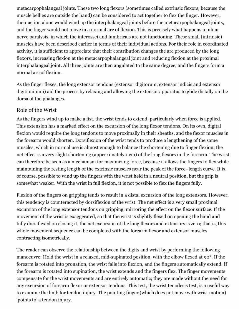
metacarpophalangeal joints. These two long flexors (sometimes called extrinsic flexors, because themuscle bellies are outside the hand) can be considered to act together to flex the finger. However,their action alone would wind up the interphalangeal joints before the metacarpophalangeal joints,and the finger would not move in a normal arc of flexion. This is precisely what happens in ulnarnerve paralysis, in which the interossei and lumbricals are not functioning. These small (intrinsic)muscles have been described earlier in terms of their individual actions. For their role in coordinatedactivity, it is sufficient to appreciate that their contribution changes the arc produced by the longflexors, increasing flexion at the metacarpophalangeal joint and reducing flexion at the proximalinterphalangeal joint. All three joints are then angulated to the same degree, and the fingers form anormal arc of flexion.
As the finger flexes, the long extensor tendons (extensor digitorum, extensor indicis and extensordigiti minimi) aid the process by relaxing and allowing the extensor apparatus to glide distally on thedorsa of the phalanges.
Role of the Wrist
As the fingers wind up to make a fist, the wrist tends to extend, particularly when force is applied.This extension has a marked effect on the excursion of the long flexor tendons. On its own, digitalflexion would require the long tendons to move proximally in their sheaths, and the flexor muscles inthe forearm would shorten. Dorsiflexion of the wrist tends to produce a lengthening of the samemuscles, which in normal use is almost enough to balance the shortening due to finger flexion; thenet effect is a very slight shortening (approximately 1 cm) of the long flexors in the forearm. The wristcan therefore be seen as a mechanism for maximizing force, because it allows the fingers to flex whilemaintaining the resting length of the extrinsic muscles near the peak of the force–length curve. It is,of course, possible to wind up the fingers with the wrist held in a neutral position, but the grip issomewhat weaker. With the wrist in full flexion, it is not possible to flex the fingers fully.
Flexion of the fingers on gripping tends to result in a distal excursion of the long extensors. However,this tendency is counteracted by dorsiflexion of the wrist. The net effect is a very small proximalexcursion of the long extensor tendons on gripping, mirroring the effect on the flexor surface. If themovement of the wrist is exaggerated, so that the wrist is slightly flexed on opening the hand andfully dorsiflexed on closing it, the net excursion of the long flexors and extensors is zero; that is, thiswhole movement sequence can be completed with the forearm flexor and extensor musclescontracting isometrically.
The reader can observe the relationship between the digits and wrist by performing the followingmanoeuvre: Hold the wrist in a relaxed, midsupinated position, with the elbow flexed at 90°. If theforearm is rotated into pronation, the wrist falls into flexion, and the fingers automatically extend. Ifthe forearm is rotated into supination, the wrist extends and the fingers flex. The finger movementscompensate for the wrist movements and are entirely automatic; they are made without the need forany excursion of forearm flexor or extensor tendons. This test, the wrist tenodesis test, is a useful wayto examine the limb for tendon injury. The pointing finger (which does not move with wrist motion)‘points to’ a tendon injury.

Wrist motion is controlled principally by two wrist flexors (flexor carpi radialis and flexor carpiulnaris) and three extensors (extensors carpi radialis longus and brevis, and extensor carpi ulnaris).Although the radiocarpal joint has some functional similarity to a ball and socket joint, it is possibleto conceive of the wrist as a variable hinge joint, the axis of which may be set in a number ofinclinations. For example, when using a hammer, it is useful to rotate the wrist backward andforward about an axis that permits not only wrist flexion but also ulnar deviation. It would be veryrestricting to have a pure hinge joint with collateral ligaments of fixed length. In this context, thewrist flexors and extensors may be regarded as variable collateral ligaments that allow the joint to beset about a number of different axes.
For movement about major axes, the wrist tendons can be considered to act in pairs:
Wrist flexion: flexon carpi radialis and flexor carpi ulnaris
Wrist extension: extensor carpi radialis longus and brevis, and extensor carpi ulnaris
Ulnar deviation: extensor carpi ulnaris and flexor carpi ulnaris
Radial deviation: flexor carpi radialis, extensor carpi radialis longus and brevis, extensorpollicis and abductor pollicis longus
Making a Tight FistIt is possible to observe and palpate the muscle groups that are active in making a tight fist. Theflexor compartment of the forearm is contracted tightly, and electromyographic evidence confirmsthat flexor digitorum profundus and flexor digitorum superficialis are active. Flexor carpi ulnarismay be seen and felt to contract strongly. The extensor compartment is tightly contracted, and thewrist extensors would certainly be expected to be active. Palpation of the long digital extensors on theback of the wrist shows that these are contracting as well. It seems that when the fingers are heldtightly closed, the long digital extensors are unable to move the extensor apparatus; they haveacquired a new fixed point on which to act—namely, the proximal limit of the extensor apparatusover the metacarpophalangeal joint. They therefore perform the only task available to them and acttogether as an additional wrist extensor.
In the thumb web, palpation confirms that the first dorsal interosseous is contracting, as are all theother interossei and the thenar and hypothenar muscles. As the firm fist is swung forward in anger,the brachioradialis stands out, and at the moment of impact, virtually every muscle in the limb is in astate of contraction, with the exception of the lumbricals.
Opening the HandThe hand is opened from its relaxed balanced posture, such as when stretching out to reach anobject. This motion is made up of extension of the distal interphalangeal, proximal interphalangealand metacarpophalangeal joints. The hand is provided with an ingenious mechanism that allows thisto happen. The laws of mechanics suggests that one motor would be required for every joint in a
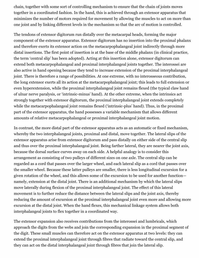
chain, together with some sort of controlling mechanism to ensure that the chain of joints movestogether in a coordinated fashion. In the hand, this is achieved through an extensor apparatus thatminimizes the number of motors required for movement by allowing the muscles to act on more thanone joint and by linking different levels in the mechanism so that the arc of motion is controlled.
The tendons of extensor digitorum run distally over the metacarpal heads, forming the majorcomponent of the extensor apparatus. Extensor digitorum has no insertion into the proximal phalanxand therefore exerts its extensor action on the metacarpophalangeal joint indirectly through moredistal insertions. The first point of insertion is at the base of the middle phalanx (in clinical practice,the term ‘central slip’ has been adopted). Acting at this insertion alone, extensor digitorum canextend both metacarpophalangeal and proximal interphalangeal joints together. The interossei arealso active in hand opening because they tend to increase extension of the proximal interphalangealjoint. There is therefore a range of possibilities. At one extreme, with no interosseous contribution,the long extensor exerts all its action at the metacarpophalangeal joint; this leads to full extension oreven hyperextension, while the proximal interphalangeal joint remains flexed (the typical claw handof ulnar nerve paralysis, or ‘intrinsicminus’ hand). At the other extreme, when the intrinsics actstrongly together with extensor digitorum, the proximal interphalangeal joint extends completelywhile the metacarpophalangeal joint remains flexed (‘intrinsicplus’ hand). Thus, in the proximalpart of the extensor apparatus, the hand possesses a variable mechanism that allows differentamounts of relative metacarpophalangeal or proximal interphalangeal joint motion.
In contrast, the more distal part of the extensor apparatus acts as an automatic or fixed mechanism,whereby the two interphalangeal joints, proximal and distal, move together. The lateral slips of theextensor apparatus arise from extensor digitorum and pass distally on either side of the central slipand thus over the proximal interphalangeal joint. Being farther lateral, they are nearer the joint axis,because the dorsal surface curves away on each side. A helpful analogy is to consider thisarrangement as consisting of two pulleys of different sizes on one axle. The central slip can beregarded as a cord that passes over the larger wheel, and each lateral slip as a cord that passes overthe smaller wheel. Because these latter pulleys are smaller, there is less longitudinal excursion for agiven rotation of the wheel, and this allows some of the excursion to be used for another function—namely, extension at the distal joint. There is an additional mechanism by which the lateral slipsmove laterally during flexion of the proximal interphalangeal joint. The effect of this lateralmovement is to further reduce the distance between the lateral slips and the joint axis, therebyreducing the amount of excursion at the proximal interphalangeal joint even more and allowing moreexcursion at the distal joint. When the hand flexes, this mechanical linkage system allows bothinterphalangeal joints to flex together in a coordinated way.
The extensor expansion also receives contributions from the interossei and lumbricals, whichapproach the digits from the webs and join the corresponding expansion in the proximal segment ofthe digit. These small muscles can therefore act on the extensor apparatus at two levels: they canextend the proximal interphalangeal joint through fibres that radiate toward the central slip, andthey can act on the distal interphalangeal joint through fibres that join the lateral slip.

Apart from the components of the extensor expansion concerned with joint function, the wholestructure requires additional anchorage. This must be arranged in such a way that it is not displacedfrom the underlying skeleton, yet it must not restrict longitudinal movement. These difficultrequirements are met by transverse retinacular ligaments at the level of the joints, the transverseligaments running to relatively fixed attachment points in the region of the joint axis. As theexpansion glides backward and forward, the transverse fibres move like bucket handles. Smoothgliding layers are required under the expansion and retinacular ligaments to allow motion to occurwithout friction.
One final component of the extensor apparatus provides an additional automatic function. This is afibrous anchorage system, Landsmeer's oblique retinacular ligament, which anchors the distalexpansion to the middle phalanx. The role of the oblique retinacular ligament is controversial(reviewed by Bendz 1985 ). Some argue that it may act in a dynamic tenodesis effect to synchronizethe movements of the interphalangeal joints; that is, it may initiate extension of the distalinterphalangeal joint as the proximal interphalangeal joint is extended from a fully flexed position,and it may relax with proximal interphalangeal joint flexion to allow full distal interphalangeal jointflexion. Others argue that it becomes taut only when the proximal interphalangeal joint is fullyextended and the distal interphalangeal joint is flexed, so that it functions as a restraining force tostabilize the fingertip when it is flexed against resistance (e.g. in the hook grip). Another possibility isthat the ligament is merely a secondary lateral stabilizer of the proximal interphalangeal joint andthat it acts to centralize the extensor components over the dorsum of the middle phalanx.
Movements of the ThumbAn opposable thumb requires a different system of control from the other digits. Because themetacarpal is much more mobile than in the digits, muscles are needed to control the extra freedomof movement.
The thumb does not easily assume the classic anatomical position. Therefore, the normal descriptiveterms—anterior, posterior, medial and lateral—do not readily apply. The terms palmar, dorsal, ulnarand radial have been adopted in clinical practice.
The basic active movements are flexion–extension, abduction–adduction, rotation andcircumduction. In the resting position of the first metacarpal, flexion and extension are parallel withthe palmar plane, and abduction and adduction occur at right angles to this.
Flexion and extension should be confined to motion at the interphalangeal or metacarpophalangealjoints ( Fig. 18.31A–C (f0165) ). Palmar abduction ( Fig. 18.31D, E (f0165) ), in which the first metacarpalmoves away from the second at right angles to the plane of the palm, and radial abduction ( Fig.18.31D, F (f0165) ), in which the first metacarpal moves away from the second with the thumb in theplane of the palm, occur at the carpometacarpal joint. The opposite of radial abduction is ulnaradduction, or transpalmar adduction, in which the thumb crosses the palm toward its ulnar border.In clinical practice, the term adduction is generally used without qualification. Circumductiondescribes the angular motion of the first metacarpal, solely at the carpometacarpal joint, from a
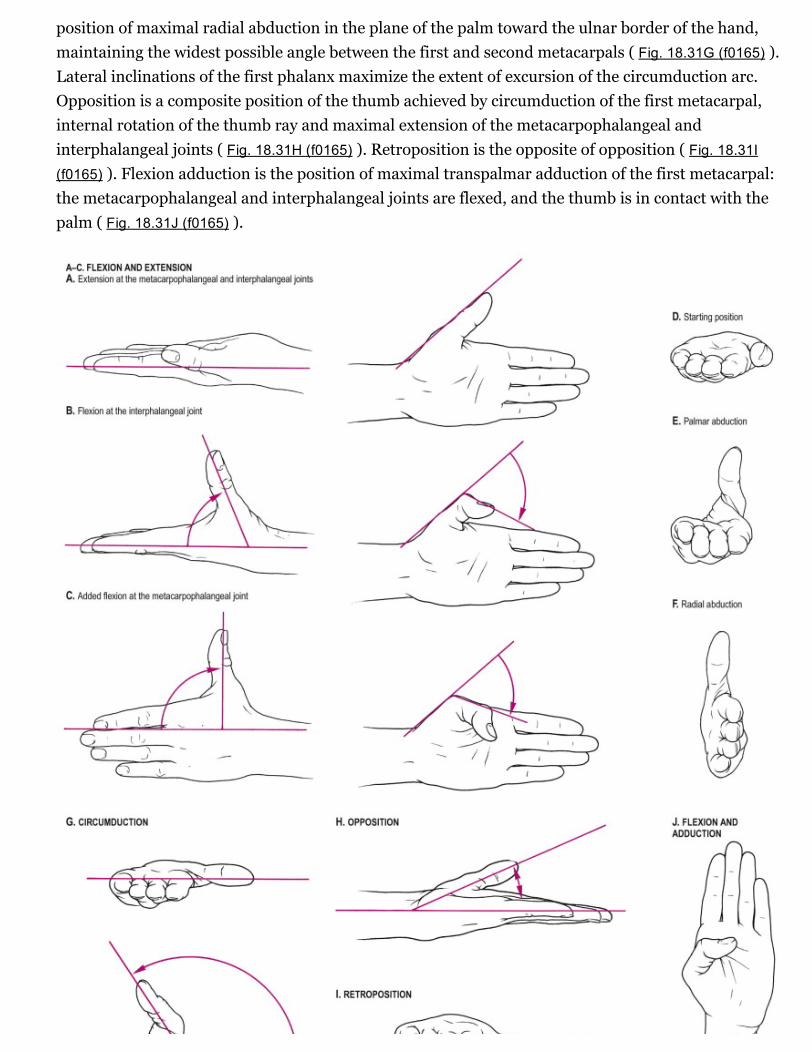
position of maximal radial abduction in the plane of the palm toward the ulnar border of the hand,maintaining the widest possible angle between the first and second metacarpals ( Fig. 18.31G (f0165) ).Lateral inclinations of the first phalanx maximize the extent of excursion of the circumduction arc.Opposition is a composite position of the thumb achieved by circumduction of the first metacarpal,internal rotation of the thumb ray and maximal extension of the metacarpophalangeal andinterphalangeal joints ( Fig. 18.31H (f0165) ). Retroposition is the opposite of opposition ( Fig. 18.31I(f0165) ). Flexion adduction is the position of maximal transpalmar adduction of the first metacarpal:the metacarpophalangeal and interphalangeal joints are flexed, and the thumb is in contact with thepalm ( Fig. 18.31J (f0165) ).
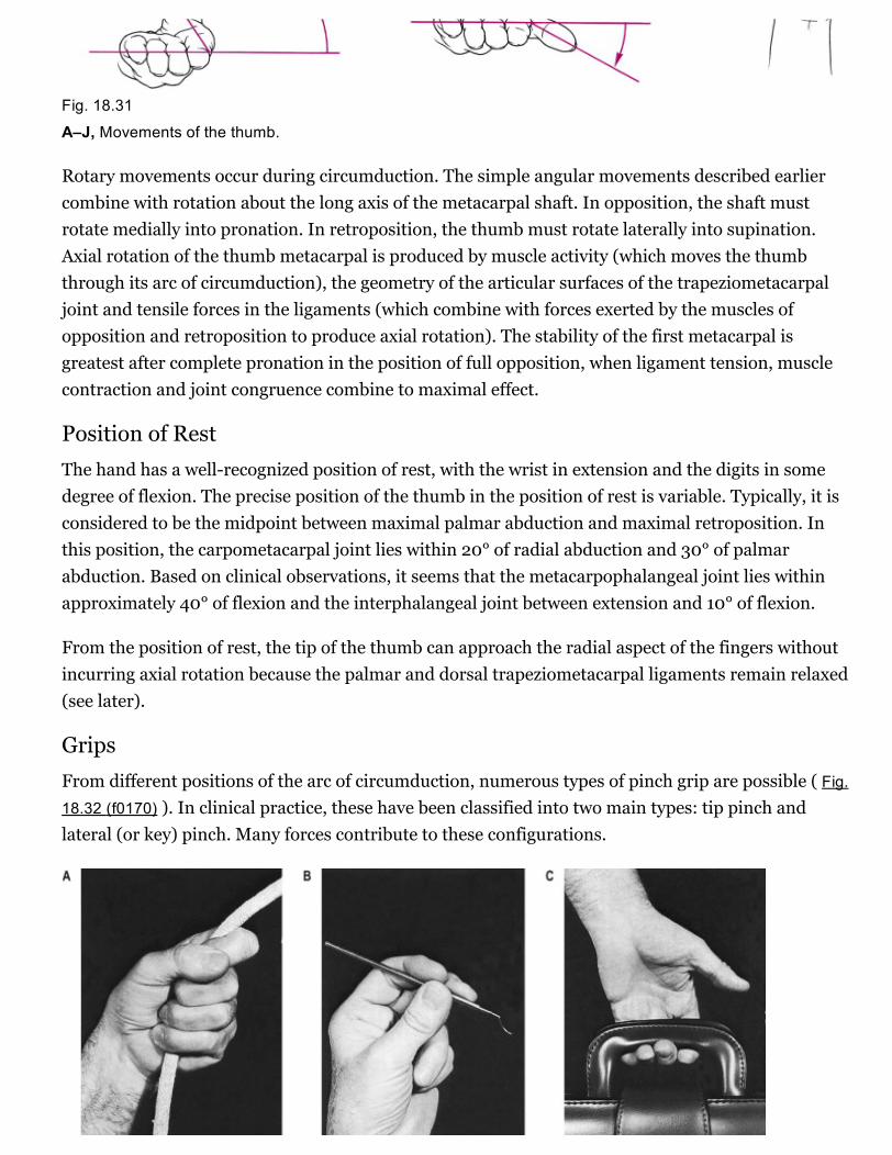
Fig. 18.31
A–J, Movements of the thumb.
Rotary movements occur during circumduction. The simple angular movements described earliercombine with rotation about the long axis of the metacarpal shaft. In opposition, the shaft mustrotate medially into pronation. In retroposition, the thumb must rotate laterally into supination.Axial rotation of the thumb metacarpal is produced by muscle activity (which moves the thumbthrough its arc of circumduction), the geometry of the articular surfaces of the trapeziometacarpaljoint and tensile forces in the ligaments (which combine with forces exerted by the muscles ofopposition and retroposition to produce axial rotation). The stability of the first metacarpal isgreatest after complete pronation in the position of full opposition, when ligament tension, musclecontraction and joint congruence combine to maximal effect.
Position of RestThe hand has a wellrecognized position of rest, with the wrist in extension and the digits in somedegree of flexion. The precise position of the thumb in the position of rest is variable. Typically, it isconsidered to be the midpoint between maximal palmar abduction and maximal retroposition. Inthis position, the carpometacarpal joint lies within 20° of radial abduction and 30° of palmarabduction. Based on clinical observations, it seems that the metacarpophalangeal joint lies withinapproximately 40° of flexion and the interphalangeal joint between extension and 10° of flexion.
From the position of rest, the tip of the thumb can approach the radial aspect of the fingers withoutincurring axial rotation because the palmar and dorsal trapeziometacarpal ligaments remain relaxed(see later).
GripsFrom different positions of the arc of circumduction, numerous types of pinch grip are possible ( Fig.18.32 (f0170) ). In clinical practice, these have been classified into two main types: tip pinch andlateral (or key) pinch. Many forces contribute to these configurations.

Fig. 18.32
Some of the many functional postures that can be adopted by the human hand. A , In the power grip, the fingersare flexed around an object, with counterpressure from the thumb. Any skill in wielding the object derives from
the limb, including the wrist; relative movements of the thumb and fingers are not involved. B , The precisiongrip, which varies considerably with the task, stabilizes the object between the tips of one or more fingers and the
thumb. The gross position of the object can be adjusted by movements at the wrist, elbow or even shoulder, but
the most skilled manipulations are carried out by the digits themselves, such as when advancing a thread
through the eye of a needle. C , The hook grip is used to suspend or to pull open objects. The fingers are flexedaround the object; the thumb may or may not be involved. It is a grip for the transmission of forces, not for skillful
manipulation. D , Powerful opposition of the thumb to the radial side of the index finger produces a lateral pinchgrip, such as to hold a door key; here the object is larger than a key, and all the fingers are involved. E , Manyactivities involve a combination of grips. Here a fountain pen is stabilized in a power grip by flexion of digits 4 and
5 against the palm, while the index finger and thumb, used in a precision grip, unscrew the cap. F , Complexmanipulation.
The thumb is a triarticular system, unlike the finger, which is a biarticular system. The thumb isactivated by monoarticular muscles (abductor pollicis longus and opponens pollicis), biarticularmuscles (extensor pollicis brevis, adductor pollicis, abductor pollicis brevis and flexor pollicis brevis),and triarticular muscles (extensor pollicis longus and flexor pollicis longus). It appears, however, thateven a monoarticular muscle can change posture in all three joints by altering the overall balance offorces, making it very difficult to attribute function to the individual intrinsic muscles. However, thethumb muscles seem to provide two broad functions: they control metacarpal positioning (the guyrope function), an activity that is automatically accompanied by rotation, and they control the axialstability of the skeleton of the thumb.
The thumb muscles can be classified into those used for retroposition, opposition and pinch grip.
Retroposition Muscles
The muscles that bring about retroposition are extensor pollicis longus, extensor pollicis brevis andabductor pollicis longus. As the thumb moves into retroposition, automatic axial rotation producessupination of the first metacarpal. This is produced by the offaxis action of two parallel butoppositely directed forces—one exerted by extensors pollicis longus and brevis, and the other byabductor pollicis longus and the anterior oblique carpometacarpal ligament.
Opposition Muscles
A succession of activity occurs in the thenar muscles during the movement of opposition. Threesubgroups of radial (abductor pollicis longus and extensor pollicis brevis), central (abductor pollicisbrevis and opponens pollicis) and ulnar (flexor pollicis brevis) muscles are involved.
These forces act simultaneously but with different intensities, depending on the situation of thethumb. As the thumb moves into opposition, there is automatic axial rotation of the first metacarpalshaft to produce pronation. This is produced by the paired action of oppositely directed forces: theopposition muscles provide one force, and the posterior oblique carpometacarpal ligament providesthe other.
Pinch Grip Muscles
The muscles of pinch grip can be divided into lateral, medial and intermediate subgroups. The lateralsubgroup (opposition muscles) moves the first metacarpal into palmar abduction. The metacarpalshaft rotates medially into pronation. Radial angulation at the metacarpophalangeal joint increasesthe span of the hand. The metacarpophalangeal joint is stabilized principally by extensor pollicisbrevis and flexor pollicis brevis. Flexion of the proximal and distal phalanges is controlled. Musclesof the medial subgroup (abductor pollicis brevis and first dorsal interosseous) produce an approachof the first metacarpal toward the palm. Because they act with the lateral group, they have a strongcontrolling effect on the position and rotation of the first metacarpal. The intermediate subgroupconsists simply of flexor pollicis longus, which flexes the interphalangeal or metacarpophalangealjoint, as described earlier. Palpating the thenar eminence during tip and lateral pinch provides someappreciation of the action of the pinch grip muscles.
References
Ball, Steger, Galatz, Yamaguchi, 2003. Ball C.M., Steger T, Galatz L.M., and Yamaguchi K.:The posterior branch of the axillary nerve: an anatomic study. J. Bone Joint Surg. 2003; 85:pp. 14971501View In Article (refInSitubib1)
Bendz, 1985. Bendz P.: The functional significance of the oblique retinacular ligament ofLandsmeer: a review and new proposals. J. Hand Surg. 1985; 10: pp. 2529View In Article (refInSitubib2) | Cross Ref (http://proxy.library.upenn.edu:2092/10.1016/S02667681(85)800097)
Leibovic, Hastings, 1992. Leibovic S.J., and Hastings H.: Martin–Gruber revisited. J. Hand
Surg. 1992; 17A: pp. 4753View In Article (refInSitubib4) | Cross Ref (http://proxy.library.upenn.edu:2092/10.1016/03635023(92)901123)
Smith, 2002. Smith P.J.: Lister's The Hand: Diagnosis and Indications. Edinburgh:Churchill Livingstone, 2002.View In Article (refInSitubib5)
Copyright © 2015 Elsevier, Inc. All rights reserved.