bmc urology biomed central - home - springer · biomed central page 1 of 4 (page ... partial...
TRANSCRIPT

BioMed CentralBMC Urology
ss
Open AcceCase reportPrimary PEComa of the bladder treated with primary excision and adjuvant interferon-alpha immunotherapy: a case reportJeremy R Parfitt*1, Anthony J Bella2, Bret M Wehrli1 and Jonathan I Izawa2Address: 1Department of Pathology, London Health Sciences Centre and University of Western Ontario, London, Canada and 2Department of Surgery, Division of Urology, London Health Sciences Centre and University of Western Ontario, London, Canada
Email: Jeremy R Parfitt* - [email protected]; Anthony J Bella - [email protected]; Bret M Wehrli - [email protected]; Jonathan I Izawa - [email protected]
* Corresponding author
AbstractBackground: Perivascular epithelioid cell tumors (PEComas) are rare mesenchymal neoplasms ofuncertain malignant potential, which have in common the co-expression of muscle and melanocyticimmunohistochemical markers.
Case presentation: A 48-year-old man presented with dysuria, passage of urinary sediment andlower abdominal discomfort. A three centimeter mass was identified by cystoscopy in the posteriormidline of the bladder. Computerized tomography suggested an enterovesical fistula. The patientunderwent laparotomy, partial cystectomy and partial small bowel resection. Pathologicalexamination revealed PEComa of the bladder. The patient underwent adjuvant interferon-αimmunotherapy. Subsequent follow-up procedures, including cystoscopy and imaging, have notrevealed evidence of recurrence. The patient is clinically free of disease 48 months after surgery.
Conclusion: This case represents the second documented PEComa of bladder and demonstratesthat adjuvant therapies, including anti-angiogenic and immunotherapy, may be considered forpatients with locally advanced or metastatic genitourinary PEComa.
BackgroundPrimary perivascular epithelioid cell tumors (PEComas)are a rare and unusual group of mesenchymal neoplasmswith unpredictable malignant potential. The term"PEComa" was originally coined by Zamboni et al and isthe current nomenclature for tumors composed of PECsother than angiomyolipoma (AML), clear cell sugar tumorof lung (CCST) and lymphangioleiomyomatosis (LAM),which are related lesions with distinct clinical features [1].Consequent to the World Health Organization's (WHO)endorsement of PEComa as a bonafide entity, an increas-ing number of reports have documented PEComas arisingin varied anatomic locations, including bladder, kidney
and prostate [2-8]. Despite increasing awareness of thisentity, accurately predicting the biological behavior ofPEComas remains difficult and contemporary reports arelimited by short clinical follow-up. Herein, we report thediagnosis, management and four-year follow-up of thesecond documented case of primary PEComa of the uri-nary bladder [8].
Case presentationA 48-year-old man presented with dysuria, passage of uri-nary sediment and lower abdominal discomfort. A three-centimeter (cm) smooth, lobular mass with mild bullousedema was identified by cystoscopy in the posterior mid-
Published: 22 August 2006
BMC Urology 2006, 6:20 doi:10.1186/1471-2490-6-20
Received: 18 April 2006Accepted: 22 August 2006
This article is available from: http://www.biomedcentral.com/1471-2490/6/20
© 2006 Parfitt et al; licensee BioMed Central Ltd.This is an Open Access article distributed under the terms of the Creative Commons Attribution License (http://creativecommons.org/licenses/by/2.0), which permits unrestricted use, distribution, and reproduction in any medium, provided the original work is properly cited.
Page 1 of 4(page number not for citation purposes)
BMC Urology 2006, 6:20 http://www.biomedcentral.com/1471-2490/6/20
line of the bladder. Laboratory and staging investigationswere negative and computerized tomography (CT) sug-gested an enterovesical fistula. The patient underwentlaparotomy, partial cystectomy and partial small bowelresection. Pathological examination revealed PEComa ofthe bladder. A search for primary melanoma was negativeand there were no stigmata of tuberous sclerosis. Thepatient underwent adjuvant interferon (IFN)-α immuno-therapy for primary PEComa of the bladder. Subsequentfollow-up evaluations were performed 3 monthly for thefirst 12 months, then 6 monthly thereafter. Routine fol-low-up procedures included clinical examination, cystos-copy, chest roentgenography, CT of abdomen and pelvisand routine blood work. On one occasion, positron emis-sion tomography was performed in order to detect neo-plastic metabolic activity. None of these follow-upprocedures revealed evidence of recurrence. The patient isclinically free of disease 48 months after surgery.
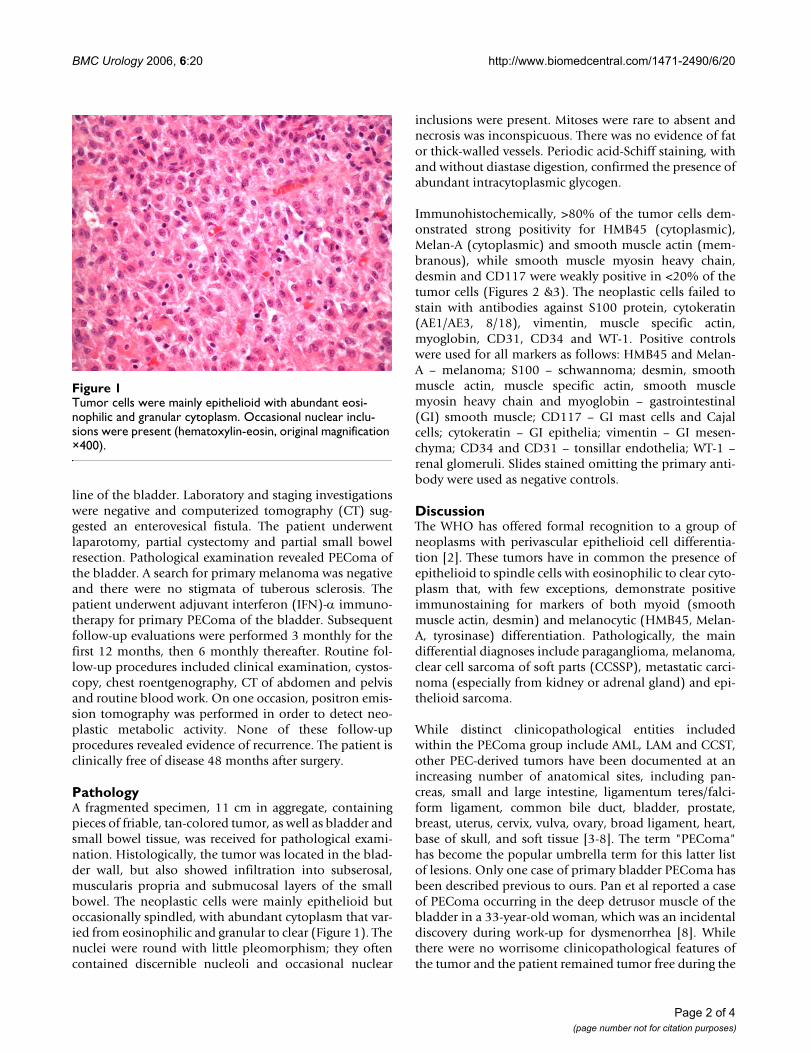
PathologyA fragmented specimen, 11 cm in aggregate, containingpieces of friable, tan-colored tumor, as well as bladder andsmall bowel tissue, was received for pathological exami-nation. Histologically, the tumor was located in the blad-der wall, but also showed infiltration into subserosal,muscularis propria and submucosal layers of the smallbowel. The neoplastic cells were mainly epithelioid butoccasionally spindled, with abundant cytoplasm that var-ied from eosinophilic and granular to clear (Figure 1). Thenuclei were round with little pleomorphism; they oftencontained discernible nucleoli and occasional nuclear
inclusions were present. Mitoses were rare to absent andnecrosis was inconspicuous. There was no evidence of fator thick-walled vessels. Periodic acid-Schiff staining, withand without diastase digestion, confirmed the presence ofabundant intracytoplasmic glycogen.
Immunohistochemically, >80% of the tumor cells dem-onstrated strong positivity for HMB45 (cytoplasmic),Melan-A (cytoplasmic) and smooth muscle actin (mem-branous), while smooth muscle myosin heavy chain,desmin and CD117 were weakly positive in <20% of thetumor cells (Figures 2 &3). The neoplastic cells failed tostain with antibodies against S100 protein, cytokeratin(AE1/AE3, 8/18), vimentin, muscle specific actin,myoglobin, CD31, CD34 and WT-1. Positive controlswere used for all markers as follows: HMB45 and Melan-A – melanoma; S100 – schwannoma; desmin, smoothmuscle actin, muscle specific actin, smooth musclemyosin heavy chain and myoglobin – gastrointestinal(GI) smooth muscle; CD117 – GI mast cells and Cajalcells; cytokeratin – GI epithelia; vimentin – GI mesen-chyma; CD34 and CD31 – tonsillar endothelia; WT-1 –renal glomeruli. Slides stained omitting the primary anti-body were used as negative controls.
DiscussionThe WHO has offered formal recognition to a group ofneoplasms with perivascular epithelioid cell differentia-tion [2]. These tumors have in common the presence ofepithelioid to spindle cells with eosinophilic to clear cyto-plasm that, with few exceptions, demonstrate positiveimmunostaining for markers of both myoid (smoothmuscle actin, desmin) and melanocytic (HMB45, Melan-A, tyrosinase) differentiation. Pathologically, the maindifferential diagnoses include paraganglioma, melanoma,clear cell sarcoma of soft parts (CCSSP), metastatic carci-noma (especially from kidney or adrenal gland) and epi-thelioid sarcoma.
While distinct clinicopathological entities includedwithin the PEComa group include AML, LAM and CCST,other PEC-derived tumors have been documented at anincreasing number of anatomical sites, including pan-creas, small and large intestine, ligamentum teres/falci-form ligament, common bile duct, bladder, prostate,breast, uterus, cervix, vulva, ovary, broad ligament, heart,base of skull, and soft tissue [3-8]. The term "PEComa"has become the popular umbrella term for this latter listof lesions. Only one case of primary bladder PEComa hasbeen described previous to ours. Pan et al reported a caseof PEComa occurring in the deep detrusor muscle of thebladder in a 33-year-old woman, which was an incidentaldiscovery during work-up for dysmenorrhea [8]. Whilethere were no worrisome clinicopathological features ofthe tumor and the patient remained tumor free during the
Tumor cells were mainly epithelioid with abundant eosi-nophilic and granular cytoplasmFigure 1Tumor cells were mainly epithelioid with abundant eosi-nophilic and granular cytoplasm. Occasional nuclear inclu-sions were present (hematoxylin-eosin, original magnification ×400).
Page 2 of 4(page number not for citation purposes)

BMC Urology 2006, 6:20 http://www.biomedcentral.com/1471-2490/6/20
entire six-year follow-up period, there was no descriptionof post-operative treatment.
PEComas demonstrate uncertain tumor biology andunpredictable clinical behavior. While the majority ofreported "PEComas" have behaved in a benign fashion,an important minority have demonstrated malignantbehavior with locally destructive recurrences, distantmetastases and patient death, underscoring the need for
accurate identification and effective treatment strategies[4,5,7]. When Folpe et al combined results of 24 of theirown cases of PEComa of soft tissue and gynecological ori-gin with data from 45 previously reported cases ofPEComa, they found that recurrence and metastasis wereassociated with tumor size >5 cm, infiltrative growth pat-tern, high nuclear grade, necrosis and a mitotic index of>1 per 50 high power fields [9,10]. However, otherauthors feel that accurate criteria which reliably predictthe behavior of PEComas remain lacking [10]. In thepresent case, the surgical margins were not evaluable, dueto the fragmented nature of the specimen, and lymphnodes were not sampled, since the intraoperative impres-sion was that of a benign enterovesical fistula, rather thana potentially malignant neoplasm.
Optimal treatment for PEComas is not known at thistime. Primary excision is usually curative, as most tumorsare benign. However, locally advanced or metastatic dis-ease portends a poor prognosis and strategies incorporat-ing chemotherapy, radiation and immunotherapy havebeen reported. In this patient, a one-year course of adju-vant IFN-α 2b at 10 million units given subcutaneouslythree times per week was initiated based on the vascularnature of this tumor and IFN-α's additional anti-ang-iogenic effect [11]. While IFN-α 2b therapy for the man-agement of PEComa remains experimental, other authorshave described the efficacy of IFN-α 2a in causing regres-sion of life-threating hemangiomas in infants [12,13].While fever, neutropenia and skin necrosis have beenreported as uncommon, short-term side effects of IFN-α2a, no such effects were seen in the present case [12]. Asthere was no evidence of residual tumor in the presentcase, a limitation of our report would be that the effect ofIFN-α on PEComa morphology could not be docu-mented. Thus, further studies are needed to clarify theclinical and pathological effects of IFN-α therapy inpatients with PEComa and the risks of IFN therapy shouldbe weighed against the potential benefits in any patientlacking detectable residual tumor. Partial, complete andabsent responses have also been noted for dacarbazine,vincristine and imatinib mesylate, a tyrosine-kinaseinhibitor [14]. Adjuvant radiation for CCSSP has alsobeen reported following wide surgical excision, with pri-mary site irradiation appearing to confer a survival benefitfor disease located in soft tissue of the extremities [15].
ConclusionIn summary, we report the first case of PEComa of thebladder treated with adjuvant IFN-α immunotherapy,with long-term follow-up. Given the uncertainty ofPEComa tumor biology, adjuvant therapies, includinganti-angiogenic and immunotherapy, may be consideredfor patients with locally advanced or metastatic genitouri-nary PEComa.
Tumor cells showed strong and diffuse membranous positiv-ity for smooth muscle actin (smooth muscle actin immu-noperoxidase, original magnification ×400)Figure 3Tumor cells showed strong and diffuse membranous positiv-ity for smooth muscle actin (smooth muscle actin immu-noperoxidase, original magnification ×400).
Tumor cells demonstrated strong and diffuse cytoplasmic positivity for HMB45 and Melan-A (Melan-A immunoperoxi-dase, original magnification ×400)Figure 2Tumor cells demonstrated strong and diffuse cytoplasmic positivity for HMB45 and Melan-A (Melan-A immunoperoxi-dase, original magnification ×400).
Page 3 of 4(page number not for citation purposes)

BMC Urology 2006, 6:20 http://www.biomedcentral.com/1471-2490/6/20
Publish with BioMed Central and every scientist can read your work free of charge
"BioMed Central will be the most significant development for disseminating the results of biomedical research in our lifetime."
Sir Paul Nurse, Cancer Research UK
Your research papers will be:
available free of charge to the entire biomedical community
peer reviewed and published immediately upon acceptance
cited in PubMed and archived on PubMed Central
yours — you keep the copyright
Submit your manuscript here:http://www.biomedcentral.com/info/publishing_adv.asp
BioMedcentral
Competing interestsThe author(s) declare that they have no competing inter-ests.
Authors' contributionsJRP prepared the manuscript.
AJB helped to provide patient history and helped in thewriting of the manuscript.
BMW was involved with diagnosis of the pathologic spec-imen and contributed to the writing of the manuscript.
JII performed the surgery, provided patient history,obtained patient consent and contributed to the writing ofthe manuscript.
All authors read and approved the final manuscript.
AcknowledgementsConsent was obtained from the patient for publication.
References1. Zamboni G, Pea M, Martignoni G, Zancanaro C, Faccioli G, Gilioli E,
Pederzoli P, Bonetti F: Clear cell "sugar" tumor of the pancreas.A novel member of the family of lesions characterized by thepresence of perivascular epithelioid cells. Am J Surg Pathol 1996,20:722-730.
2. Folpe AL: Neoplasms with perivascular epithelioid cell differ-entiation (PEComas). In World Health Organization Classification ofTumors: Pathology and Genetics of Tumors of Soft Tissue and Bone Editedby: Fletcher CDM, Unni KK, Mertens F. Lyon: IARC Press;2002:221-222.
3. Evert M, Wardelmann E, Nestler G, Schulz HU, Roessner A, RockenC: Abdominopelvic perivascular epithelioid cell sarcoma(malignant PEComa) mimicking gastrointestinal stromaltumor of the rectum. Histopathology 2005, 46:115-117.
4. Harris GC, McCulloch TA, Perks G, Fisher C: Malignant perivas-cular epithelioid cell tumor ("PEComa") of soft tissue: aunique case. Am J Surg Pathol 2004, 28:1655-1658.
5. Lehman NL: Malignant PEComa of the skull base. Am J SurgPathol 2004, 28:1230-1232.
6. Sadeghi S, Krigman H, Maluf H: Perivascular epithelioid clear celltumor of the common bile duct. Am J Surg Pathol 2004,28:1107-1110.
7. Pan CC, Yang AH, Chiang H: Malignant perivascular epithelioidcell tumor involving the prostate. Arch Pathol Lab Med 2003,127:E96-98.
8. Pan CC, Yu IT, Yang AH, Chiang H: Clear cell myomelanocytictumor of the urinary bladder. Am J Surg Pathol 2003, 27:689-692.
9. Folpe AL, Mentzel T, Lehr HA, Fisher C, Balzer BL, Weiss SW:Perivascular epithelioid cell neoplasms of soft tissue andgynecologic origin: a clinicopathologic study of 26 cases andreview of the literature. Am J Surg Pathol 2005, 29:1558-1575.
10. Hornick JL, Fletcher CDM: PEComa: what do we know so far?Histopathology 2006, 48:75-82.
11. Dreau D, Foster M, Hogg M, Swiggett J, Holder WD, White RL: Ang-iogenic and immune parameters during recombinant inter-feron-alpha2b adjuvant treatment in patients withmelanoma. Oncol Res 2000, 12:241-51.
12. Ezekowitz RA, Mulliken JB, Folkman J: Interferon alfa-2a therapyfor life-threatening hemangiomas of infancy. N Engl J Med1992, 326:1456-1463.
13. Ezekowitz RA, Mulliken JB, Folkman J: Additional corrections:interferon alfa-2a for hemangiomas of infancy. N Engl J Med1995, 333:595-596.
14. Rigby H, Yu W, Schmidt MH, Fernandez CV: Lack of response of ametastatic renal perivascular epithelial cell tumor
(PEComa) to successive courses of DTIC based-therapy andimatinib mesylate. Pediatr Blood Cancer 2005, 45:202-6.
15. Deenik W, Mooi WJ, Rutgers EJ, Peterse JL, Hart AA, Kroon BB:Clear cell sarcoma (malignant melanoma) of soft parts: aclinicopathologic study of 30 cases. Cancer 1999, 86:969-75.
Pre-publication historyThe pre-publication history for this paper can be accessedhere:
http://www.biomedcentral.com/1471-2490/6/20/prepub
Page 4 of 4(page number not for citation purposes)