bmc biotechnology biomed central - springer · bmc biotechnology research article open ... cees...
TRANSCRIPT

BioMed CentralBMC Biotechnology
ss
Open AcceResearch articlePeroxicretion: a novel secretion pathway in the eukaryotic cellCees MJ Sagt*1, Peter J ten Haaft1, Ingeborg M Minneboo1, Miranda P Hartog1, Robbert A Damveld1, Jan Metske van der Laan1, Michiel Akeroyd1, Thibaut J Wenzel1, Francisca A Luesken2, Marten Veenhuis3, Ida van der Klei3 and Johannes H de Winde1,4Address: 1DSM Biotechnology Center, Beijerinck Laboratory, PO Box 1, 2600MA Delft, the Netherlands, 2Department of Microbiology Radboud University Nijmegen Toernooiveld 1, 6525ED Nijmegen, the Netherlands, 3Groningen University department of microbiology, Groningen, the Netherlands and 4Kluyver Centre for Genomics of Industrial Fermentation, Delft University of Technology, Department for Biotechnology, Julianalaan 67, 2628BC Delft, the Netherlands
Email: Cees MJ Sagt* - [email protected]; Peter J ten Haaft - [email protected]; Ingeborg M Minneboo - [email protected]; Miranda P Hartog - [email protected]; Robbert A Damveld - [email protected]; Jan Metske van der Laan - [email protected]; Michiel Akeroyd - [email protected]; Thibaut J Wenzel - [email protected]; Francisca A Luesken - [email protected]; Marten Veenhuis - [email protected]; Ida van der Klei - [email protected]; Johannes H de Winde - [email protected]
* Corresponding author
AbstractBackground: Enzyme production in microbial cells has been limited to secreted enzymes orintracellular enzymes followed by expensive down stream processing. Extracellular enzymesconsists mainly of hydrolases while intracellular enzymes exhibit a much broader diversity. If theseintracellular enzymes could be secreted by the cell the potential of industrial applications ofenzymes would be enlarged. Therefore a novel secretion pathway for intracellular proteins wasdeveloped, using peroxisomes as secretion vesicles.
Results: Peroxisomes were decorated with a Golgi derived v-SNARE using a peroxisomalmembrane protein as an anchor. This allowed the peroxisomes to fuse with the plasma membrane.Intracellular proteins were transported into the peroxisomes by adding a peroxisomal import signal(SKL tag). The proteins which were imported in the peroxisomes, were released into the extra-cellular space through this artificial secretion pathway which was designated peroxicretion. Thisconcept was supported by electron microscopy studies.
Conclusion: Our results demonstrate that it is possible to reroute the intracellular trafficking ofvesicles by changing the localisation of SNARE molecules, this approach can be used in in vivobiological studies to clarify the different control mechanisms regulating intracellular membranetrafficking. In addition we demonstrate peroxicretion of a diverse set of intracellular proteins.Therefore, we anticipate that the concept of peroxicretion may revolutionize the production ofintracellular proteins from fungi and other microbial cells, as well as from mammalian cells.
Published: 20 May 2009
BMC Biotechnology 2009, 9:48 doi:10.1186/1472-6750-9-48
Received: 28 April 2009Accepted: 20 May 2009
This article is available from: http://www.biomedcentral.com/1472-6750/9/48
© 2009 Sagt et al; licensee BioMed Central Ltd. This is an Open Access article distributed under the terms of the Creative Commons Attribution License (http://creativecommons.org/licenses/by/2.0), which permits unrestricted use, distribution, and reproduction in any medium, provided the original work is properly cited.
Page 1 of 11(page number not for citation purposes)

BMC Biotechnology 2009, 9:48 http://www.biomedcentral.com/1472-6750/9/48
BackgroundThe specificity of intracellular membrane trafficking isdetermined by multiple layers of control mechanisms thatensure that only appropriate organelles fuse with specifictarget compartments. These include Rab-GTPases [1]operating in conjunction with polyphosphoinositides [2]and Rab effectors [3] that frequently include multiproteincomplexes. In eukaryotes, membrane fusion of secretoryvesicles is mediated by SNARE-proteins [4] and specificityof membrane fusion is obtained by specific SNARE-pro-tein interactions. In yeast, fusion of post-Golgi traffickingvesicles requires at least 10 genes including the Rab-GTPase Sec4 [5] the Exocyst multiprotein complex [6] andthe SNAREs Snc1/2 [7] on the transport vesicle and Sso1/2 [8] and Sec9 [9] on the plasma membrane. Moreoverorganelles can only fuse with target membranes once theyare transported into close proximity, involving directedtransport along cytoskeletal tracts [10]. The formation ofthe resulting SNARE-pin subsequently triggers membranefusion [11]. The ER supplies the secretory route withmembrane enclosed vesicles which travel from the ER viathe Golgi towards the cell membrane.
The ER is very different from the cytosol regarding post-translational protein modifications. N-glycosylation ofproteins in the ER is important for folding, degradation andquality control [12]. The cytosol does not contain an N-gly-cosylation machinery and as a consequence, solublecytosolic proteins are not N-glycosylated [13]. In addition,the reducing environment of the cytosol is very differentfrom that in the ER and Golgi, where oxidizing conditionsand specialized folding enzymes like Pdi1 and Ero1 facili-tate disulfide bridge formation [14]. These fundamentaldifferences between secretory pathway and cytosol compli-cate the routing of cytosolic proteins through the secretorypathway to yield active, secreted enzymes. In fact, literaturedoes not describe any successful extracellular production ofan intracellular protein through the secretory pathway.Cytosolic proteins preferentially fold into their active con-formation with the aid of specific chaperones and foldingenzymes, under the reducing conditions which are normalto the cytosol [15]. Recently it has been described that per-oxisomes also may have their origin in the ER [16]. How-ever they do not fuse with other compartments and SNAREmolecules have not been detected on peroxisomes [17].The peroxisome has all the necessary features to enableimport of completely folded and mature intracellular pro-teins [18]. Proteins of the peroxisomal lumen containeither a PTS1 [19] or a PTS2 signal [20]. The PTS1 signal isa specific tripeptide located at the C-terminal end of theprotein, and is recognized by the Pex5 receptor, a transloca-tor for PTS1 containing proteins [21]. The ER origin of per-oxisomes, combined with their capacity to importcompletely folded proteins, would render them ideallysuited for secretion of intracellular proteins.
To enable this we have decorated Aspergillus niger peroxi-somes with the A. niger ortholog of the v-SNARE Snc1(SncA), by expressing it as a chimera with the A. nigerortholog of the peroxisomal membrane protein Pmp22(PmpA) [22]. In Figure 1 panel C a schematic representa-tion of the fusion of peroxisomes with the plasmame-brane is shown. The modified peroxisomes were able tofuse with the plasma membrane as evidenced by electronmicroscopy and extracellular secretion of peroxisomalaccumulated proteins, which were tagged with the PTS1-signal peptide -SKL. We have named this novel technol-ogy peroxicretion, for peroxisome-mediated intracellularprotein secretion.
ResultsPTS 1 mediated peroxisomal import in A. nigerThe PTS1 receptor Pex5 is responsible for recognition andtransport of PTS1-containing proteins into peroxisomes[21]. To confirm that PTS1 signals will result in peroxiso-mal localization in A. niger we have identified a Pex5ortholog in the genome of A. niger (Genbank 4989140).Amino acids important for PTS1 recognition are con-served in the Pex5 ortholog (figure 1), suggesting that thepresence of an -SKL sequence at the C-terminus of modelproteins will lead to peroxisomal localization. Indeed,expressing -SKL tagged eGFP in A. niger (figure 2) causeda punctated pattern typical for peroxisomal localization.This result indicated that -SKL mediated peroxisomal tar-geting occurs in A. niger as expected based on the presenceof a Pex5 receptor ortholog.
Decoration of A. niger peroxisomes with v-SNARE moleculesTo enable fusion of peroxisomes with the plasma mem-brane it was necessary to identify a peroxisomal mem-brane anchor, which could be used to place the v-SNARESncA on the peroxisome. This peroxisomal membraneanchor should have the N-terminus positioned towardsthe cytosol, enabling N-terminal fusions. The resultingchimeric protein is anchored in the peroxiomal mem-brane with the N-terminal fused SncA positioned at thecytosolic side of the peroxisome. Using the CBS predictionserver http://www.cbs.dtu.dk/services/ we predicted thetopology of the A. niger ortholog of Pmp22, which hasbeen studied in Arabidopsis and in mammalian cells[22,23]. A membrane topology was predicted of 4 TMD'swith the N-terminus positioned at the cytosolic side of theperoxisome. PmpA contains two peroxisomal targetingregions with similar clusters of basic amino acids, interact-ing with Pex19p [22]. This prediction is in agreement withexperimental evidence determining the topology ofPmp22 in Arabidopsis and mammalian cells [22,23]. Todetermine whether the Pmp22p ortholog of A. niger local-ises to the peroxisomes we constructed an eGFP-pmpA chi-mera and expressed this fusion gene in A. niger. We
Page 2 of 11(page number not for citation purposes)

BMC Biotechnology 2009, 9:48 http://www.biomedcentral.com/1472-6750/9/48
Page 3 of 11(page number not for citation purposes)
Alignment of the PEX5 orthologs of S. cerevisiae, A. niger and K. lactisFigure 1Alignment of the PEX5 orthologs of S. cerevisiae, A. niger and K. lactis. Identical aminoacids are indicated with *, highly similar aminoacids are indicated with :, similar aminoacids are indicated with •. The conserved aminoacids which are important for PTS1 recognition are colored in yellow.
K. lactis ---------------------------------MSADCSVGSNP-LAQLNKHAQQNPALR S. cerevisiae ---MDVGSCSVGNNPLAQLHKHTQQNKSLQFN-QKNNGRLNESP-LQGTNKPGISEAFIS A. niger MSFLGGAECSTAGNPLTQFTKHVQDDKSLQRDRLVGRGPGGMQEGMRSRGMMGGQDQMMD . . : . . .: :
QVGYQN-PASNVAQNFKTHVNEVSNANRFQMDQFMNRSPGFS---DGQLGMAPVPSAILS NVNAISQENMANMQRFINGEPLIDDKRRMEIGPSSGRLPPFSNVHSLQTSANPTQIKGVN EFAQQPGQIPGAPPQPFAMEQLRRELDQFQTTPPRTGSPGWAAEFDAGEHARMEAAFAGP :. . : ::: * :: .
HGPRFGLK--KQDSGSSNMSAGDTAQHSRSWGNEFNSRSPQQGLASRVNNVERISNTNSM DISHWSQE--FQGSNSIQNRNADTGNSEKAWQRGSTTASSRFQYPNTMMNNYAYASMNSL QGPMMNNASGFTPAEFARFQQQSRAGMPQTANHVASAPSPMMAGYQRPMGMGGYMGMGGM . . . : . . . :: . .: *. . . . ..:
SSYRPGMSRIGRPMMHTGISSLHN------YSHMSQQTPQMSSDDGVLADKQWNEQFEAL SGSRLQSPAFMNQQQSGRSKEGVN------EQEQQPWTDQFEKLEKEVSENLDINDEIEK GMMPQTFNPMAMQQQPAEATTQDKGKGRMVELDDENWEAQFAEMETADTQKLDDEANAAV . : . : . . *: . : ::: :
EKAVAENLTMEDNKEETKEEIVVEDGYQADFQEVWD---KLQAETADNNLETSDS-QWEK EENVSEVEQNKPETVE-KEEGVYGDQYQSDFQEVWDSIHKDAEEVLPSELVNDDL-NLGE EAELNDLDRSVPQDSGDSAFESVWQRVQAETATNRKLAEGETDFNIDDNLHMGEMGEWDG * : : : . : *:: . .:* .: :
DYARYMTGKATHIPPYRFDNDNQYMHNPNAYEIGCILMENGAKLSEAALAFEAAVQEDPA DYLKYLGGRVNGNIEYAFQSNNEYFNNPNAYKIGCLLMENGAKLSEAALAFEAAVKEKPD FDTLNTRFRNPQLGDYMFEEDNVFRSVSNPFEEGVKIMREGGNLSLAALAFEAAVQKDPQ : * *:.:* : .*.:: * :*.:*.:** *********::.*
HVDAWLKLGLVQTQNEKEMNGISALEQCLSLDPTNQQALMTISISYINEGYDLTAFSMLN HVDAWLRLGLVQTQNEKELNGISALEECLKLDPKNLEAMKTLAISYINEGYDMSAFTMLD HVQAWTMLGSAQAQNEKELPAIRALEQALKIDANNLDALMGLAVSYTNEGYDSTSYRTLE **:** ** .*:*****: .* ***:.*.:*..* :*: :::** ***** ::: *:
RWLDSKYPELT--RSPTID-----EANIDRFNLSKQVITKYLQVANALPQVDPEVQLGLG KWAETKYPEIWS-RIKQQDDKFQKEKGFTHIDMNAHITKQFLQLANNLSTIDPEIQLCLG RWLSVKYPQIINPNDVSSEADLGFTDRQLLHDRVTDLFIQAAQLSPSGEQMDPDVQVGLG :* . ***:: . : : .: : *:: :**::*: **
TLFYANEEFGKTIDCFRTALEVNP----NDE----LMWNRLGASLANSNRSEEAIQAYHK LLFYTKDDFDKTIDCFESALRVNP----NDE----LMWNRLGASLANSNRSEEAIQAYHR VLFYCAEEYDKAVDCFSAALASTESGTSNQQEQLHLLWNRLGATLANSGRSEEAIEAYEQ *** :::.*::*** :** . *:: *:******:****.******:**.:
ALALKPSFVRARYNLAISSMNIGCYKEAAESLLSALSMHEVEN----------VPITGSV ALQLKPSFVRARYNLAVSSMNIGCFKEAAGYLLSVLSMHEVNT----------NNKKGDV ALNINPNFVRARYNLGVSCINIGCYPEAAQHLLGALSMHRVVEQEGRERAREIVGGEGGI ** ::*.********.:*.:****: *** **..****.* *.:
-------------VQSNNILETLKRSFVAMDRRDLLEKVMPGMDLQQFRNEFNF GS--------LLNTYNDTVIETLKRVFIAMNRDDLLQEVKPGMDLKRFKGEFSF DDEQLDRMIHVSQNQSTNLYDTLRRVFSQMGRRDLADQVVAGMDVNVFRREFEF
. .: :**:* * *.* ** ::* .***:: *: **.*
BMC Biotechnology 2009, 9:48 http://www.biomedcentral.com/1472-6750/9/48
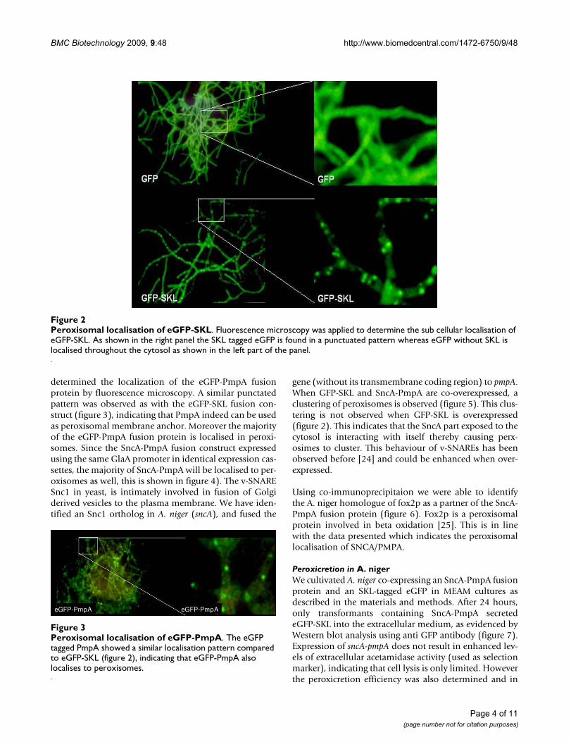
determined the localization of the eGFP-PmpA fusionprotein by fluorescence microscopy. A similar punctatedpattern was observed as with the eGFP-SKL fusion con-struct (figure 3), indicating that PmpA indeed can be usedas peroxisomal membrane anchor. Moreover the majorityof the eGFP-PmpA fusion protein is localised in peroxi-somes. Since the SncA-PmpA fusion construct expressedusing the same GlaA promoter in identical expression cas-settes, the majority of SncA-PmpA will be localised to per-oxisomes as well, this is shown in figure 4). The v-SNARESnc1 in yeast, is intimately involved in fusion of Golgiderived vesicles to the plasma membrane. We have iden-tified an Snc1 ortholog in A. niger (sncA), and fused the
gene (without its transmembrane coding region) to pmpA.When GFP-SKL and SncA-PmpA are co-overexpressed, aclustering of peroxisomes is observed (figure 5). This clus-tering is not observed when GFP-SKL is overexpressed(figure 2). This indicates that the SncA part exposed to thecytosol is interacting with itself thereby causing perx-osimes to cluster. This behaviour of v-SNAREs has beenobserved before [24] and could be enhanced when over-expressed.
Using co-immunoprecipitaion we were able to identifythe A. niger homologue of fox2p as a partner of the SncA-PmpA fusion protein (figure 6). Fox2p is a peroxisomalprotein involved in beta oxidation [25]. This is in linewith the data presented which indicates the peroxisomallocalisation of SNCA/PMPA.
Peroxicretion in A. nigerWe cultivated A. niger co-expressing an SncA-PmpA fusionprotein and an SKL-tagged eGFP in MEAM cultures asdescribed in the materials and methods. After 24 hours,only transformants containing SncA-PmpA secretedeGFP-SKL into the extracellular medium, as evidenced byWestern blot analysis using anti GFP antibody (figure 7).Expression of sncA-pmpA does not result in enhanced lev-els of extracellular acetamidase activity (used as selectionmarker), indicating that cell lysis is only limited. Howeverthe peroxicretion efficiency was also determined and in
Peroxisomal localisation of eGFP-SKLFigure 2Peroxisomal localisation of eGFP-SKL. Fluorescence microscopy was applied to determine the sub cellular localisation of eGFP-SKL. As shown in the right panel the SKL tagged eGFP is found in a punctuated pattern whereas eGFP without SKL is localised throughout the cytosol as shown in the left part of the panel.
Peroxisomal localisation of eGFP-PmpAFigure 3Peroxisomal localisation of eGFP-PmpA. The eGFP tagged PmpA showed a similar localisation pattern compared to eGFP-SKL (figure 2), indicating that eGFP-PmpA also localises to peroxisomes.
eGFP-PmpA eGFP-PmpA
Page 4 of 11(page number not for citation purposes)

BMC Biotechnology 2009, 9:48 http://www.biomedcentral.com/1472-6750/9/48
the peroxicretion strain, overexpressing SncA-PmpA andeGFP-SKL, 55% of the total GFP was extracellular. Whenwe expressed only GFP-SKL we determined 25% of thetotal GFP in the supernatant. This indicates that less than50% of the extracellular GFP is due to lysis and more than50% due to actual peroxicretion. We conclude that deco-ration of peroxisomes with the v-SNARE SncA resulted infusion of peroxisomes with the plasma membrane, caus-ing release of peroxisomal content in the extracellularmedium. The applicability of this approach to secreteintracellular enzymes was further investigated using a setof enzymes indicated in figure 8. We have expressed theindicated proteins in wild type A. niger (panel A) and in aperoxicreting A. niger (panel B), when indicated an SKLtag was placed at the C terminus of the indicated proteins.The amylase proteins (amyA and amyB) are also visible,the presence of amyA is pH dependent. This explains whyamyA is not always visible. The peroxicretion strain showsa slightly different acidification profile compared to thewild type strain. Results clearly showed peroxicretion of 3overexpressed putative peroxisomal proteins from a SncA-PmpA expressing strain (figure 8). Using MS/MS we couldcorroborate peroxicretion of at least one of those 3 pro-teins (strong similarity to catalase/peroxidase CpeB,(An01g01830)) and in addition identified one extra per-oxicreted protein (similarity to endo-1,4-beta-xylanaseXynD, (An11g03120)). The relatively low abundance ofthese proteins is likely to be caused by exposure to oxi-
Expression of the fusion peptide SncA-PmpAFigure 4Expression of the fusion peptide SncA-PmpA. Cell free extracts were obtained from untransformed A. niger (lane 1) and from SncA-PmpA transformed A. niger (lane 2). The Cell free extracts were subjected to SDS-PAGE and western blotting according the manufacturers instructions (Invitrogen) and detection was performed using a custom made antibody against SncA protein (Eurogentec). The expected size of the SncA-PmpA fusion protein is 35 kDa.
188
28
98
49
62
38
14
188
28
98
49
62
38
14
M 1 2
17
188
28
98
49
62
38
14
M 1 2
1717
188
28
98
49
62
38
14
188
28
98
49
62
38
14
M 1 2
17
188
28
98
49
62
38
14
188
28
98
49
62
38
14
M 1 2
188
28
98
49
62
38
14
188
28
98
49
62
38
14
M 1 2
188
28
98
49
62
38
14
188
28
98
49
62
38
14
M 1 2
17
188
28
98
49
62
38
14
188
28
98
49
62
38
14
M 1 2
1717
188
28
98
49
62
38
14
188
28
98
49
62
38
14
M 1 2
17
188
28
98
49
62
38
14
188
28
98
49
62
38
14
M 1 2
188
28
98
49
62
38
14
188
28
98
49
62
38
14
M 1 2
Co-expression of eGFP-SKL and SncA-PmpA in A. nigerFigure 5Co-expression of eGFP-SKL and SncA-PmpA in A. niger. A. niger transformants were grown on MEAM as described in materials and methods. After 48 hours biomass was transferred to glass slides and subjected to fluorescence microscopy. The decoration of peroxisomes with the v-SNARE SncA results in clustering of peroxisomes.
GFPskl , PMP/SNCGFP-SKL , SNC1-PMP22GFPskl , PMP/SNCGFP-SKL , SNC1-PMP22eGFP-SKL and SncA-PmpA
Analysis of co-immunoprecipitation complexes using anti-SncA by SDS-PAGE and Sypro rubyFigure 6Analysis of co-immunoprecipitation complexes using anti-SncA by SDS-PAGE and Sypro ruby. SDS-PAGE (4–12% Bis-Tris) gel loaded with the immunoprecipitation samples of wild type strain (lane 1) and peroxicretion strain (wild type strain transformed with SncA-PmpA fusion con-struct) (lane 2) was stained with Sypro Ruby. The heavy and light chain of the used antibodies are running at 55 kDa and 28 kDa. Proteins Differential bands were identified by MS/MS.
62
188
28
38
49
98
kDa
62
188
28
38
49
98
kDa 1 2
Page 5 of 11(page number not for citation purposes)
BMC Biotechnology 2009, 9:48 http://www.biomedcentral.com/1472-6750/9/48
dised conditions combined with the presence of extracel-lular proteases. It is evident that putative peroxisomalproteins can be peroxicreted as well as cytosolic proteinslike the XynD orthologue. However the peroxisomalenzymes have a higher success rate probably because theyare adapted to peroxisomal conditions in contrast tocytosolic proteins. Simple C-terminal SKL addition is suf-ficient to peroxicrete the XynD orthologue in SncA-PmpAexpressing cells. Proteins which contain a putative PTS1sequence like the catalase/peroxidase CpeB orthologue(An01g01830) and the alcohol oxidase orthologue(An18g05480) could be peroxicreted without modifica-tions. The peroxicreted alcohol oxidase shows enzymaticactivity in an H2O2 degrading assay, described in [26], anddepicted in figure 9. The wild type strain shows almost noH2O2 degrading activity in the supernatant whereas theperoxicretion strain shows an increasing in H2O2 degrad-ing activity in the supernatant. This is most likely due toperoxicretion of endogenous catalases/peroxidases local-ised in peroxisomes.
Finetuning of peroxicretionFusion of peroxisomes with the plasma membrane wassupported using electron microscopy. Inspection ofultrathin section of KMnO4-fixed cells revealed that per-oxisomes were frequently located in close vicinity of the
cell membrane and often showed continuation with thismembrane (figure 10A, B, C.). This was never observed inwild type cells without SncA-PmpA expression in whichthe organelles are scattered throughout the cytosol but arenot seen in close proximity of the cell membrane (figure10D). The efficiency of peroxicretion is likely to be con-trolled at the level of SNARE-pin formation during mem-brane fusion. In order to increase this efficiency of SNAREpin formation we have truncated the cytoplasmic tail ofPmpA in order to place the v-SNARE SncA in closer prox-imity to the peroxisomal membrane. The peroxicretionefficiency is reduced when the N-terminus of PmpA istruncated with more than 18 amino acids, probably dueto mislocalization (figure 11).
Another way of increasing the peroxicretion efficiency isto use C2 ceramide. Activation of CAPP by adding C2-ceramide is known to result in increased availability of t-SNARE, Sso1p, which is important for SNARE-pin forma-tion [26,27]. Indeed, addition of C2-ceramide slightlyenhanced the peroxicretion efficiency (figure 12). A third
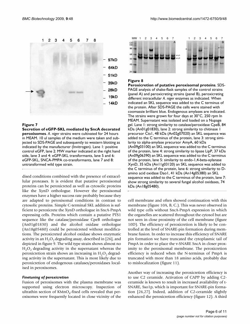
Secretion of eGFP-SKL mediated by SncA decorated peroxi-somesFigure 7Secretion of eGFP-SKL mediated by SncA decorated peroxisomes. A. niger strains were cultivated for 24 hours in MEAM. 10 ul samples of the medium were taken and sub-jected to SDS-PAGE and subsequently to western blotting as indicated by the manufacturer (Invitrogen). Lane 1: positive control eGFP, lane 2: MW marker indicated at the right hand side, lane 3 and 4: eGFP-SKL transformants, lane 5 and 6: eGFP-SKL, SNCA-PMPA co-transformants, lane 7 and 8: untransformed wild type strain.
97kD
64kD
51kD
39kD
28kD
19kD
14kD
1 2 3 4 5 6 7 8
97kD
64kD
51kD
39kD
28kD
19kD
14kD
97kD
64kD
51kD
39kD
28kD
19kD
14kD
1 2 3 4 5 6 7 8
Peroxicretion of putative peroxisomal proteinsFigure 8Peroxicretion of putative peroxisomal proteins. SDS-PAGE analysis of shake-flask samples of the control strains (panel A) and peroxicreting strains (panel B), peroxicreting different intracellular A. niger enzymes as indicated. When indicated an SKL sequence was added to the C terminus of the protein. After SDS-PAGE the cells were stained with coomassie brilliant blue. Endogenous amylases are indicated. The strains were grown for four days at 30°C, 250 rpm In MEAM. Supernatant was isolated and loaded on a Nupage gel. Lane 1: strong similarity to catalase/peroxidase CpeB, 84 kDa (An01g01830), lane 2: strong similarity to chitinase 1 precursor Cts1, 48 kDa (An02g07020) an SKL sequence was added to the C terminus of the protein, lane 3: strong simi-larity to alpha-amylase precursor AmyA, 60 kDa (An09g03100) an SKL sequence was added to the C terminus of the protein, lane 4: strong similarity to lipase LipP, 37 kDa (An09g06390) an SKL sequence was added to the C terminus of the protein, lane 5: similarity to endo-1,4-beta-xylanase XynD, 35 kDa (An11g03120) an SKL sequence was added to the C terminus of the protein, lane 6: strong similarity to D-amino acid oxidase Dao1, 41 kDa (An14g05380) an SKL sequence was added to the C terminus of the protein, lane 7: show strong similarity to several fungal alcohol oxidases, 74 kDa (An18g05480).
A B1 2 3 4 5 6 7 1 2 3 4 5 6 7MW
amyA
amyB
Page 6 of 11(page number not for citation purposes)

BMC Biotechnology 2009, 9:48 http://www.biomedcentral.com/1472-6750/9/48
approach to increase the efficiency of peroxicreion is toincrease the number of peroxisomes. Overexpression ofthe A. niger ortholog of Pex11, which is known to beinvolved in peroxisomal proliferation [21] only resultedin minor peroxisomal proliferation in the A. niger trans-formants, while peroxicretion was not enhanced (figure13). This may be explained by the fact that the increase inorganelle numbers is not associated with a concomitantincrease in matrix protein levels.
DiscussionIn this study the v-SNARE SncA without its transmem-brane domain was placed on the peroxisome using PmpAas a membrane anchor. The transmembrane domain ofSnc1p is reported to be important for its function [28].However, replacing the SncA-TMD by PmpA did notdiminish the potential of SncA to enforce membranefusion, since the peroxisomal content was released in theextracellular environment. PmpA as membrane anchor issufficient for SncA to bring the membrane bilayers in closecontact so that spontaneous membrane fusion occurs. Wehave selected peroxisomes because they can import com-pletely folded proteins, which makes them ideally suitedto transport and secrete proteins which are normally arelocalized intracellular. Recently, convincing evidence waspresented that peroxisomes may originate from the ER[16]. This is important since the lipid composition of per-
Enzymatic activity of peroxicreted alcohol oxidaseFigure 9Enzymatic activity of peroxicreted alcohol oxidase. Using 100 μl of supernatant of shakeflask grown A. niger cul-tures the degradation of 0.1% H2O2 was monitored in 2 ml milliQ by measuring absorption at 240 nm. (black triangle) represents the H2O2 degrading activity of 100 μl supernatant of the wild type strain, (black circle) represents the H2O2 degrading activity of 100 μl supernatant of the peroxicretion strain and (black square) represents the H2O2 degrading activity of 100 μl supernatant of the peroxicretion strain overexpressing gene ID 4990113 which shows strong similar-ity with several alcohol oxidases.
H2O2 degradation
1.6
1.65
1.7
1.75
1.8
1.85
1.9
1.95
2
2.05
0 1 2 3 4 5
time (minutes)
A 2
40 n
m
Survey of hyphal cells showing the position of a peroxisome (arrow) in the vicinity of the cell membrane in SncA-PmpA expressing A. nigerFigure 10Survey of hyphal cells showing the position of a peroxisome (arrow) in the vicinity of the cell membrane in SncA-PmpA expressing A. niger. Panel A. Lengthwise overview of cellular membranes in SncA-PmpA expressing A. niger. Arrow indicates continuity of peroxisomes with the cell membrane (high magnification in panel A1). Panel B. Crosswise over-view of cellular membranes in SncA-PmpA expressing A. niger. Arrow indicates continuity of peroxisomes with the cell mem-brane (high magnification in panel B1). Panel C. Schematic representation of the release of the peroxisomal content due to fusion of the peroxisome with the plasmamembrane. The target SNARE (Sso1 ortholog) is shown in yellow as a transmem-brane protein. The chimeric protein SncA-PmpA is depicted in blue (PmpA part) and red (SncA part). For simplicity Sec9 ortholog is not shown. The release of peroxisomal content is depicted, demonstrating peroxicretion of SKL tagged proteins (in green). Panel D. Detail of a glucose-grown A. niger WT cell, showing the presence of peroxisomes that are randomly scattered in the cytosol. M-mitochondrion, N – nucleus, P – peroxisomes, V-vacuole.
C
D
scalebar = 2 m
Page 7 of 11(page number not for citation purposes)

BMC Biotechnology 2009, 9:48 http://www.biomedcentral.com/1472-6750/9/48
oxisomes now is likely to be comparable to that of otherER/Golgi derived organelles like other secretory vesicles.Because of this similarity in lipid composition it appearsunlikely that lipid incompatibility between peroxisomesand the plasma membrane would present a major hurdlefor peroxicretion. Moreover, the small GTPase Rho1 islocalized on peroxisomes through interaction with theperoxisomal membrane protein Pex25 [29]. Rho1p isknown to play a role in actin reorganization and mem-brane dynamics. In yeast, Rho1 regulates polarizedgrowth, and in the filamentous fungus A. niger polarizedgrowth is even more predominant. In Arabidopsis, trans-portation of peroxisomes by actin filaments is reported[30] similar to transportation of secretory vesicles. Thiswould be a possible mechanism how peroxisomes areable to be transported intracellular, Snc1 decorated perox-isomes are able to fuse with the plasmamembrane. Theexamples of Peroxicretion as described in this paper,shows that positioning of SncA on the peroxisomal mem-brane is sufficient for fusion of the peroxisome with theplasma membrane and subsequent release of its cargo.Interestingly, all these findings strengthen the notion thatperoxisomes are derivatives of the secretory pathway. Thisrenders peroxisomes as attractive vehicles for the transportof intracellular proteins towards the plasma membrane
and secretion into the extracellular medium. It is howevernot excluded that peroxismes fuse to other intracellularcompartments like endosomes before actual fusion withthe plasmamebrane takes place. It is very likely that not allperoxisomes will be fused to the cellmembrane since thelocalization of PmpA fused proteins is not absolutely per-oxisomal. In addition we do not observe decreasedgrowth under conditions were peroxisomes are expectedto be important. It is more likely that the peroxicretionconcept as presented in this paper is not yet very efficientand that several key steps in the process like fusion of per-oxisomes with the cell membrane, peroxisomal import of
Overexpression of PEX11 ortholog leads to increased per-oxisomal proliferation in A. nigerFigure 13Overexpression of PEX11 ortholog leads to increased peroxisomal proliferation in A. niger. PEX11 ortholog overexpression leads to a minor increase in the number of peroxisomes however the peroxisomes seems to be smaller compared to the strain where only eGFP-SKL was expressed.
GFP-SKL GFP-SKL,PEX11GFP-SKL GFP-SKL,PEX11
Mean fluorescence of eGFP-SKL overexpressing strains con-taining different truncated SncA-PmpA constructsFigure 11Mean fluorescence of eGFP-SKL overexpressing strains containing different truncated SncA-PmpA constructs. Fluorescence of individual samples was meas-ured in supernatant of MTP cultures. SD is indicated as well as the number of transformants which were analysed.
0
200
400
600
800
1000
1200
1400
1600
N=93 N=21 N=169 N=157
318 AA 300 AA 285 AA 270 AA
Ave
rag
e fl
uo
resc
ence
(re
l. u
nit
s)
Ceramide stimulates peroxicretion, overexpression of PEX11 ortholog (An11g02590) does notFigure 12Ceramide stimulates peroxicretion, overexpression of PEX11 ortholog (An11g02590) does not. 10 μl of supernatant after 48 hours growth of the strains transformed with the indicated constructs at 30°C, 250 rpm in MEAM. Lane 1; eGFP-SKL, lane 2; eGFP-SKL and C2 ceramide, lane 3; eGFP-SKL (duplo of lane 1), lane 4; MW, lane 5; eGFP-SKL and SncA-PmpA, lane 6; eGFP-SKL, C2 ceramide and SNCA-PMPA, lane 7; eGFP-SKL, SncA-PmpA and PEX11 ortholog, lane 8; eGFP-SKL, C2 ceramide, SncA-PmpA and PEX11 ortholog lane 9; WT, lane 10; WT and C2 ceramide.
GFP
97kD
64kD
51kD
39kD
28kD
19kD
14kDGFPGFP
97kD
64kD
51kD
39kD
28kD
19kD
14kD
97kD
64kD
51kD
39kD
28kD
19kD
14kD
1 2 3 4 5 6 7 8 9 10
GFP
97kD
64kD
51kD
39kD
28kD
19kD
14kDGFPGFP
97kD
64kD
51kD
39kD
28kD
19kD
14kD
97kD
64kD
51kD
39kD
28kD
19kD
14kD
1 2 3 4 5 6 7 8 9 10
Page 8 of 11(page number not for citation purposes)
BMC Biotechnology 2009, 9:48 http://www.biomedcentral.com/1472-6750/9/48
proteins to be peroxicreted and stability of these proteinshave to be improved.
ConclusionThis paper shows that it is possible to redirect intracellulartrafficking of organelles by re-localizing v-SNARE mole-cules. The decoration of target organelles with selected v-SNARE proteins is facilitated by using a modified mem-brane anchor which positions the v-SNARE onto themembrane of the target vesicle. This technology opens upnumerous possibilities for studying intracellular vesicletrafficking in vivo. Besides this fundamental applicationthe redirection of intracellular organelles also can be usedin industrial biotechnology. As demonstrated in thispaper, the targeting of proteins towards peroxisomes byusing a C-terminal SKL sequence followed by fusion ofmodified peroxisomes with the plasmamembrane resultsin secretion of intracellular proteins. This process is desig-nated peroxicretion. Although the levels of the peroxi-creted products are still very low it shows the potential ofthe peroxicretion technology.
MethodsConstructs, strains and transformationStandard molecular cloning techniques were performed. Theconstructs pGBFINSNP-2 to 5 contain the v-SNARE namedsncA (An12g07570), fused to the N-terminal 318, 300, 285,or 275 amino acids of the peroxisomal membrane proteinpmpA (An04g09130) respectively. The sncA-pmpA fusiongene is deposited as: GenBank DQ768214. Both genes wereobtained by PCR on genomic DNA and the obtained nucle-otide sequence was confirmed by sequencing (Baseclear, Lei-den). The vector pGBFINGFS-1 contains eGFP with theamino acids SKL added to the C-terminus. The nucleotidesequence (5'-TCCAAGCTC-3') encoding for the amino acidsSKL was introduced at the C-terminus of eGFP by PCR. Theconstruct pGBFINGFM-2 (GenBank DQ768213) wasobtained by translational fusion of the eGFP and pmpA ORFthrough PCR. The pGBFIN vector was also used for overex-pression of different genes (An01g01830, An02g07020,An09g03100, An09g06390, An11g03120, An14g05380 andAn18g05480). Using primers which were extended by 9nucleotides encoding SKL the protein encoding sequences ofAn02g07020, An09g03100, An09g06390, An11g03120 andAn14g05380 were modified at the C terminus. All of theabove A. niger expression constructs are driven by the strongglucoamylase promoter and harbour flanking regions toensure convenient targeting and expression in the fungalhost as described previously [31]. Subsequent transforma-tion of A. niger strain CBS 513.88 with the expression con-structs was performed as previously described [32].
Culture methodsA. niger strains were inoculated at 5 × 10⁶ spores/mlMEAM consisting of: 6 g NaNO3; 0.52 g KCl; 1.52 g
KH2PO4; 1.12 ml 4 M KOH; 0.52 g MgSO4 .7H2O; 10 g
glucose; 1 g casaminoacids; 22 mg g ZnSO4 .7H2O; 11 mg
H3BO3; 5 mg FeSO4 .7H2O; 1.7 mg CoCl2. 6H2O; 1.6 mg
CuSO4. 5H2O; 1.5 mg Na2MoO4. 2H2O; 50 mg EDTA; 5mg MnCl2. 2H2O; 2 mg riboflavin; 2 mg thiamin-HCl; 2mg nicotinamid; 1 mg pyrodoxin-HCl; 0.2 mg pan-tothenic acid; 4 μg biotin;10 ml penicillin/streptomycine(Invitrogen) per liter. The strains were grown at 30°C and250 rpm for 1–3 days. When required conidiospores wereobtained by growth on Potato Dextrose Agar (PDA,Oxoid, England) for 5 days at 30°C, and isolated withMilliQ and a spatula.
Western blot analysisSDS-PAGE was performed using NuPAGE Novex Bis-Trisprecast gels (Invitrogen) according to the supplier's man-ual. Proteins were visualized by staining with SimplyBlueSafeStain (Invitrogen). Western analysis was performedwith the XCell II semi-wet blotting module (Invitrogen)using MOPS buffer and nitrocellulose membrane (0.45μm pore size) according to the supplier's manual. GFPwas specifically detected by using 1:1,000 fold dilutedGFP monoclonal antibody (Covance, California). Afterincubation with secondary antibody conjugated to horse-radish peroxidase (anti-mouse, 1:1,000 dilution,PIERCE), immunoreactive proteins were detected by theenhanced chemiluminescence system (ECL, AmershamPharmacia) and exposed to radiographic film (Kodak).The SNC 1 westerns were prepared similar to the proce-dure described above with the expection that the anti-SNC1 antibody was custom made by Eurogentec.
Immunoprecipitation of SncA-pmpAFrozen cells were disrupted in a mortar filled with liquidnitrogen and suspended in 0.5 ml 20 mM Sodium-Phos-phate, 1% TritonX-100, 1 mM EDTA, and protease inhib-iter. Cell lysate was voraciously vortexed and placed on icefor 10 minutes. TritonX-100 induces lyses of A. niger. Thecell lysate was centrifuged for 5 minutes at 13.000 g andsupernatant was taken. A pre-clearance step with 25 μl wasused to reduce the background of a-specific bonding toProtein-A-Sepharose (10% Protein-A-Sepharose in 20mM Sodium phosphate pH 7,4, 1 mM EDTA, 0,1% TritonX-100). The soluble fraction was incubated with Protein-A-Sepharose and rotated head over head for 1 hour at4°C. The supernatant was incubated with 2.5 μl 10-5
diluted antibody SNC α-SNC (serum 2e booster NL03077, rabbit no = SN1391) for 1 hour at 4°C to bind theSNC for 1 hour at 4°C. In total 25 μl Protein-A-Sepharosewas added to the sample to bind antibody SNC withbounded SNC and the incubation was extended for againone hour. Protein-A-Sepharose was used to facilitate spin-down antibody SNC with bounded SNC. To remove theremaining antibody, Protein-A-Sepharose and the notbound proteins, the sample was washed once for 5 min-
Page 9 of 11(page number not for citation purposes)

BMC Biotechnology 2009, 9:48 http://www.biomedcentral.com/1472-6750/9/48
utes with 1,0 ml 20 mM SodiumPhosphate, 0,1% Tri-tonX-100, 1 mM EDTA, and protease inhibiter. Thesamples were treated with 25 μl sample buffer and 5 μlreducing agent. After heating the samples for 5 minutes at95°C and centrifuging, 20 μl supernatant was loaded onthe 4%–12% SDS-PAGE gel. After electrophoresis, SyproRuby staining was performed. For 24 hours the gel wasstained in 100 ml Sypro Ruby staining. After staining, thegel was washed once with MilliQ. Instead of a Coomassieblue staining the gel was stained with a Sypro Ruby,because this is more sensitive. Sypro ruby does not needto be destained, because the dye does not bind irreversibleto proteins and is therefore washable from the sample(Patton, 2000). The fragments were cut out of the gel andidentified by mass-spectrometry.
MicroscopyFor analysis of eGFP localization the fungal cells weregrown at 30°C in MEAM for 1–2 days. Mycelium wastransferred to microscope coverslides and studied under aLeica DMLA microscope connected to a CTRMIC unit. Theapparatus was controlled by Qwin software from Leica.Electron microscopy was performed as described before[33].
Digestion and LC-MS/MS analysisThe sups were filtered over centrifugal devices (Pall) intubes. Proteins with MW >100 kDa were filtered over 100kDa centrifugal devices, proteins with MW 30–100 kDawere filtered over 30 kDa centrifugal devices and proteinswith MW 10–30 kDa were filtered over 10 kDa centrifugaldevices. 500 μL MQ was added on the filters and again thesamples were centrifuged at 13000 rpm 4°C for 15 min-utes. 150 μL 80 mM NH4HCO3 was added to the retentateof each of the samples after filtration and the retentate wastransferred to 1.5 mL eppendorf tubes after pipetting upand down on the filter a couple of times. The proteinswere denatured by incubation at 97°C for 10 minutes.350 μL 80 mM NH4HCO3 and 20 μL 250 μg/mL trypsinwere added and the proteins were digested by incubationat 37°C over night. 6 μL 100 mM DTT was added and thesamples were incubated at room temperature for 30 min-utes. LC-MS/MS was performed on the CapLC-QTOFII(Waters) system. For each of the samples a different MS/MS method was made with the selected precursors for theover-expressed proteins. For each of the precursors theo-retical fragmentation (MS/MS) spectra were made usingMasslynx software (Waters) and the LC-MS/MS data wascompared to these theoretical fragmentation spectra.
Quantification of fluorescenceApproximately 1 × 10⁵ conidiospores were inoculated in300 μl MEAM (vitamins were omitted because of interfer-ence with the fluorescence measurements) per MTP well.After 5 days incubation at 30°C in an MTP (Nunc) the
medium was separated from the mycelium and 200 μlwas transferred to a new MTP (Greiner, Fluotrac 200).Subsequently the fluorescence was measured on a Geminispectra MAX (Molecular devices) controlled by SOFT maxPRO v3.1.1 (Molecular Devices) using an excitation wave-length of 490 nm and an emission wavelength of 510 nm.Additional settings: cut-off 495 nm, PMT auto, calibrateon, 6 reads per well, 9 points per well. The amount ofeGFP-SKL was determined in RFUs (relative fluorescentunits).
AbbreviationsSNARE: Soluble Nsf-Attachment protein Receptors; ER:Endoplasmic Reticulum; PTS: peroxisomal TargetingSequence; eGFP: enhanced Green Fluorescent Protein;TMD: Trans Membrane Domain; CAPP: Ceramide Acti-vated Protein Phosphatase; MEAM: Minimal EnrichedAspergillus Medium.
Authors' contributionsThe experiments were conceived and designed by CS, TW,RD and PtH. MH and JdW performed the PmpA trunca-tion studies and PtH performed the peroxicretion experi-ments. IM constructed the sncA-pmpA fusion gene whichwas used throughout this study. FL performed the peroxi-cretion experiments with endogenous proteins, whichwere designed by RD. RB performed electron microscopy.MV and IK interpreted the electron microscopy experi-ments. RD wrote the materials and methods section andsubmitted the sequences to Genbank. JM v/d L composedthe peroxicretion protein test set. MA performed MS/MSexperiments. All authors contributed to editing and writ-ing of the paper.
AcknowledgementsWe thank Rana Al-Majidi, Martine Steenbeek, Martine Spaans, Emmie Heeren, Brenda Vonk, Siebe Hartmans, Panagiotis Sarantinopoulos, Aldo Greeve, Lydia Schild and Ron Booy for experimental assistance. We specif-ically thank prof. Reinhard Jahn (Max Planck Institute, Goettingen) for sci-entific discussions.
References1. Olkkonen VM, Stenmark H: Role of Rab GTPases in membrane
traffic. Int Rev Cytol 1997, 176:1-85.2. Wurmser AE, Emr SD: Phosphoinositide signaling and turno-
ver: PtdIns(3)P, a regulator of membrane traffic, is trans-ported to the vacuole and degraded by a process thatrequires lumenal vacuolar hydrolase activities. EMBO J 1998,17:4930-4942.
3. Spang A: Vesicle transport: a close collaboration of Rabs andeffectors. Curr Biol 2004, 14:R33-R34.
4. Rothman JE: Intracellular membrane fusion. Adv Second Messen-ger Phosphoprotein Res 1994, 29:81-96.
5. Kabcenell AK, Goud B, Northup JK, Novick PJ: Binding and hydrol-ysis of guanine nucleotides by Sec4p, a yeast protein involvedin the regulation of vesicular traffic. J Biol Chem 1990,265:9366-9372.
6. TerBush DR, Maurice T, Roth D, Novick P: The Exocyst is a mul-tiprotein complex required for exocytosis in Saccharomycescerevisiae. EMBO J 1996, 15:6483-6494.
Page 10 of 11(page number not for citation purposes)
BMC Biotechnology 2009, 9:48 http://www.biomedcentral.com/1472-6750/9/48
Publish with BioMed Central and every scientist can read your work free of charge
"BioMed Central will be the most significant development for disseminating the results of biomedical research in our lifetime."
Sir Paul Nurse, Cancer Research UK
Your research papers will be:
available free of charge to the entire biomedical community
peer reviewed and published immediately upon acceptance
cited in PubMed and archived on PubMed Central
yours — you keep the copyright
Submit your manuscript here:http://www.biomedcentral.com/info/publishing_adv.asp
BioMedcentral
7. Protopopov V, Govindan B, Novick P, Gerst JE: Homologs of thesynaptobrevin/VAMP family of synaptic vesicle proteinsfunction on the late secretory pathway in S. cerevisiae. Cell1993, 74:855-861.
8. Katz L, Hanson PI, Heuser JE, Brennwald P: Genetic and morpho-logical analyses reveal a critical interaction between the C-termini of two SNARE proteins and a parallel four helicalarrangement for the exocytic SNARE complex. EMBO J 1998,17:6200-6209.
9. Couve A, Gerst J: Yeast Snc proteins complex with Sec9. Func-tional interactions between putative SNARE proteins. J BiolChem 1994, 269(38):23391-23394.
10. Rudolf R, Salm T, Rustom A, Gerdes HH: Dynamics of immaturesecretory granules: role of cytoskeletal elements duringtransport, cortical restriction, and F-actin-dependent teth-ering. Mol Biol Cell 2001, 12:1353-1365.
11. Weber T, Zemelman BV, McNew JA, Westermann B, Gmachl M, Par-lati F, Söllner TH, Rothman JE: SNAREpins: minimal machineryfor membrane fusion. Cell 1998, 92(6):759-772.
12. Helenius A, Aebi M: Roles of N-linked glycans in the endoplas-mic reticulum. Annu Rev Biochem 2004, 73:1019-1049.
13. Spiro RG: Protein glycosylation: nature, distribution, enzy-matic formation, and disease implications of glycopeptidebonds. Glycobiology 2002, 12(4):43R-56R.
14. Frand AR, Cuozzo JW, Kaiser CA: Pathways for protein disul-phide bond formation. Trends Cell Biol 2000, 10:203-210.
15. Østergaard H, Tachibana C, Winther JR: Monitoring disulfidebond formation in the eukaryotic cytosol. J Cell Biol 2004,166:337-345.
16. Hoepfner D, Schildknegt DM, Braakman I, Philippsen P, Tabak HF:Contribution of the Endoplasmic reticulum to the peroxi-some formation. Cell 2005, 122:85-95.
17. Kikuchi M, Hatano N, Yokota S, Shimozawa N, Imanaka T, TaniguchiH: Proteomic analysis of rat liver peroxisome: presence ofperoxisome-specific isozyme of Lon protease. J Biol Chem2004, 279:421-428.
18. Walton PA, Hill PE, Subramani S: Import of stably folded proteinsinto peroxisomes. Mol Biol Cell 1995, 6:675-683.
19. Gould SG, Keller GA, Subramani S: Identification of a peroxiso-mal targeting signal at the carboxy terminus of firefly luci-ferase. J Cell Biol 1987, 105:2923-2931.
20. Swinkels BW, Gould SJ, Bodnar AG, Rachubinski RA, Subramani S: Anovel, cleavable peroxisomal targeting signal at the amino-terminus of the rat 3-ketoacyl-CoA thiolase. EMBO J 1991,10:3255-3262.
21. Subramani S: Components involved in peroxisome import,biogenesis, proliferation, turnover and movement. Physiologi-cal reviews 1998, 78:171-184.
22. Brosius U, Dehmel T, Gartner J: Two different targeting signalsdirect human peroxisomal membrane protein 22 to peroxi-somes. J Biol Chem 2002, 277:774-784.
23. Tugal HB, Pool M, Baker A: Arabidopsis 22-kilodalton peroxiso-mal membrane protein. Nucleotide sequence analysis andbiochemical characterization. Plant Physiol 1999, 120:309-20.
24. Wendler F, Tooze S: Syntaxin 6: the promiscuous behaviour ofa SNARE protein. Traffic 2001, 2(9):606-611.
25. Hiltunen JK, Wenzel B, Beyer A, Erdmann R, Fosså A, Kunau WH:Peroxisomal multifunctional beta-oxidation protein of Sac-charomyces cerevisiae. Molecular analysis of the fox2 geneand gene product. J Biol Chem 1992, 267(10):6646-6653.
26. Beers RF Jr, Sizer IW: A spectrophotometric method for meas-uring the breakdown of hydrogen peroxide by catalase. J BiolChem 1952, 195(1):133-140.
27. Gurunathan S, Marash M, Weinberger A, Gerst JE: t-SNARE phos-phorylation regulates endocytosis in yeast. Mol Biol Cell 2002,13(5):1594-1607.
28. Grote E, Baba M, Ohsumi Y, Novick PJ: GeranylgeranylatedSNAREs Are Dominant Inhibitors of Membrane Fusion. J CellBiol 2000, 151:453-466.
29. Marelli M, Smith JJ, Jung S, Yi E, Nesvizhskii AI, Christmas RH, SaleemRA, Tam YY, Fagarasanu A, Goodlett DR, Aebersold R, RachubinskiRA, Aitchison JD: Quantitative mass spectrometry reveals arole for the GTPase Rho1p in actin organization on the per-oxisome membrane. J Cell Biol 2004, 167:1099-1112.
30. Hashimoto K, Igarashi H, Mano S, Nishimura M, Shimmen T, YokotaE: Peroxisomal localization of a myosin XI isoform in Arabi-dopsis thaliana. Plant Cell Physiol 2005, 46:782-789.
31. van Dijck PW, Selten GC, Hempenius RA: On the safety of a newgeneration of DSM Aspergillus niger enzyme productionstrains. Regul Toxicol Pharmacol 2003, 38:27-35.
32. Kelly JM, Hynes MJ: Transformation of Aspergillus niger by theamdS gene of Aspergillus nidulans. EMBO J 1985, 4:475-479.
33. Waterham HR, Titorenko VI, Haima P, Cregg JM, Harder W, Veen-huis M: The Hansenula polymorpha PER1 gene is essential forperoxisome biogenesis and encodes a peroxisomal matrixprotein with both carboxy- and amino-terminal targetingsignals. J Cell Biol 1994, 127(3):737-749.
Page 11 of 11(page number not for citation purposes)