blood vessel distribution
DESCRIPTION
vesselsTRANSCRIPT

6/16/14 11:20 AMBlood Vessel Distribution
Page 1 of 11http://droualb.faculty.mjc.edu/Lecture%20Notes/Unit%204/link%20blood_vessel_distribution%20with%20figures.htm
Blood Vessel Distribution
Pulmonary Circuit The pulmonary circuit begins at the semilunar valve of the pulmonary trunk that carries deoxygenatedblood to the lungs. The pulmonary trunk divides into left and right pulmonary arteries that go to each lung.Blood is oxygenated in capillaries that flow through the alveoli of the lungs. Oxygenated blood is then returnedto the left atrium of the heart by four pulmonary veins.
Systemic Circuit
The systemic circuit begins at the aortic semilunar valve with the ascending aorta. As previously noted,the coronary arteries come off at base of the ascending aorta. The curving aortic arch connects the ascendingaorta with the descending aorta.
Aortic Arch
Three elastic arteries come of aortic arch:
1. brachiocephalic trunk (a.k.a. innominate artery) divides into the right common carotid and rightsubclavian arteries.
2. left common carotid a. 3. left subclavian a. These arteries supply blood to the head, neck, shoulders and arms. Subclavian Arteries The subclavian a. give off three major arteries before leaving the thoracic cavity: 1. Thyrocervical trunk 2. Internal thoracic a. 3. Vertebral a.
6/16/14 11:20 AMBlood Vessel Distribution
Page 2 of 11http://droualb.faculty.mjc.edu/Lecture%20Notes/Unit%204/link%20blood_vessel_distribution%20with%20figures.htm
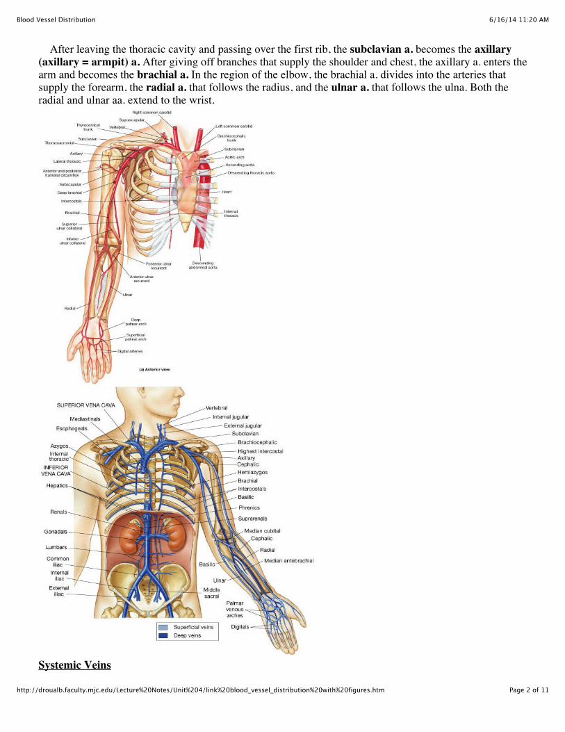
After leaving the thoracic cavity and passing over the first rib, the subclavian a. becomes the axillary(axillary = armpit) a. After giving off branches that supply the shoulder and chest, the axillary a. enters thearm and becomes the brachial a. In the region of the elbow, the brachial a. divides into the arteries thatsupply the forearm, the radial a. that follows the radius, and the ulnar a. that follows the ulna. Both theradial and ulnar aa. extend to the wrist.
Systemic Veins
6/16/14 11:20 AMBlood Vessel Distribution
Page 3 of 11http://droualb.faculty.mjc.edu/Lecture%20Notes/Unit%204/link%20blood_vessel_distribution%20with%20figures.htm
The systemic veins return deoxygenated blood to the right atrium of the heart via the superior venacava and inferior vena cava. Many systemic veins run alongside systemic arteries and have the same name as the accompanyingartery. In the neck and limbs, in addition to the deep veins that run alongside deep arteries there aresuperficial veins that are much more variable. The superior vena cava receives blood from the head, neck, shoulder, chest and arms.
Venous return from upper limb The superficial veins of the upper limb include the cephalic v. that ascends along radial side of forearmand stays on the lateral side of the arm until it fuses with the axillary v. The basilic v. ascends along theulnar side of the forearm and stays on the medial side of the arm until it fuses with the brachial v andcontinues as the axillary v. The median cubital v. interconnects cephalic and basilic veins. The deep veins are alongside the arteries and have the same names. The radial and ulnar veins fuse toform brachial v. The brachial v. receives blood from basilic v and becomes axillary v. and the axillary v.receives blood from the cephalic v. and becomes the subclavian v.
Carotid Arteries and the Blood Supply to the Brain The common carotid a. ascends the neck and divides at the larynx into internal carotid a. and externalcarotid a. The external carotid a. supplies blood to the neck, pharynx, esophagus, larynx, lower jaw and face.The internal carotid a. enters the cranium through the carotid canal and divides into the: 1. Ophthalmic a. 2. Anterior cerebral a. 3. Middle cerebral a.
Blood is also supplied to the brain by the vertebral a. The vertebral a. comes off the subclavian a., ascendsthrough the transverse foramina of the cervical vertebrae and enters cranium through foramen magnum. Onthe medulla oblongata it fuses to form the basilar a. At the pons the basilar a. divides into the posteriorcerebral arteries.
Although the internal carotid a. supplies the anterior brain and the vertebral a. supplies the posterior brain,blood supply to the entire brain is ensured by anastomoses between the vessels from these two sources. Thiscomplex of anastomosing arteries is called the cerebral arterial circle (circle of Willis) and includes: Internal carotids Anterior cerebral arteries linked by anterior communicating artery Posterior cerebral arteries linked to internal carotids by the posterior communicating arteries

6/16/14 11:20 AMBlood Vessel Distribution
Page 4 of 11http://droualb.faculty.mjc.edu/Lecture%20Notes/Unit%204/link%20blood_vessel_distribution%20with%20figures.htm
Veins returning blood from cranium The superficial veins drain blood from cerebrum into dural sinuses. These dural sinuses form within the duramater of the brain. The location of some of these sinuses can be easily seen by the impressions they leave on theinner surface of the skull. These include the superior sagittal sinus, transverse sinus and sigmoid sinus. The deep veins that drain blood from the brain, also ultimately drain into the dural sinuses. The bloodcollected from the brain drains into the sigmoid sinus which exits the cranium by the jugular foramen to becomethe internal jugular vein. The vertebral v. collects blood from the cervical spinal cord and rear of the skull. It travels alongside thevertebral a. through transverse foramina of cervical vertebrae.

6/16/14 11:20 AMBlood Vessel Distribution
Page 5 of 11http://droualb.faculty.mjc.edu/Lecture%20Notes/Unit%204/link%20blood_vessel_distribution%20with%20figures.htm
Superficial veins of head and neck The facial, temporal and maxillary veins drain blood from the superficial tissues of the head. The temporaland maxillary veins drain into the external jugular v. and the facial vein drains into the internal jugular v.There is a broad anastomosis between the internal and external jugular veins at the angle of the mandible. The external jugular vein drains into the subclavian v. and the internal jugular v. joins the subclavian v. toform the brachiocephalic v.
Veins within thorax In the thorax, the external and internal jugular veins fuse with the subclavian v. and become thebrachiocephalic v. The right and left brachiocephalic veins fuse to form the superior vena cava. The azygos v. ascends from lumbar region on right side of vertebral column and drains into the superior venacava at the level of the second thoracic vertebra. The smaller hemiazygos v. collects blood from the left side ofthe thorax and drains into the azygos v. The azygos v. and hemiazygos v are the chief collecting vessels of thethorax. The inferior vena cava collects blood from organs inferior to diaphragm.
6/16/14 11:20 AMBlood Vessel Distribution
Page 6 of 11http://droualb.faculty.mjc.edu/Lecture%20Notes/Unit%204/link%20blood_vessel_distribution%20with%20figures.htm
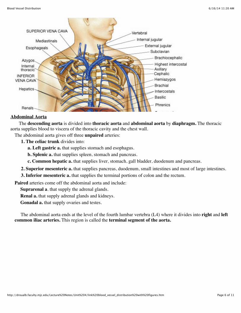
Abdominal Aorta The descending aorta is divided into thoracic aorta and abdominal aorta by diaphragm. The thoracicaorta supplies blood to viscera of the thoracic cavity and the chest wall.
The abdominal aorta gives off three unpaired arteries:
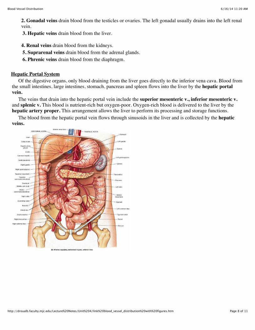
1. The celiac trunk divides into: a. Left gastric a. that supplies stomach and esophagus. b. Splenic a. that supplies spleen, stomach and pancreas. c. Common hepatic a. that supplies liver, stomach, gall bladder, duodenum and pancreas.
2. Superior mesenteric a. that supplies pancreas, duodenum, small intestines and most of large intestines. 3. Inferior mesenteric a. that supplies the terminal portions of colon and the rectum.
Paired arteries come off the abdominal aorta and include: Suprarenal a. that supply the adrenal glands. Renal a. that supply adrenal glands and kidneys. Gonadal a. that supply ovaries and testes.
The abdominal aorta ends at the level of the fourth lumbar vertebra (L4) where it divides into right and leftcommon iliac arteries. This region is called the terminal segment of the aorta.
6/16/14 11:20 AMBlood Vessel Distribution
Page 7 of 11http://droualb.faculty.mjc.edu/Lecture%20Notes/Unit%204/link%20blood_vessel_distribution%20with%20figures.htm
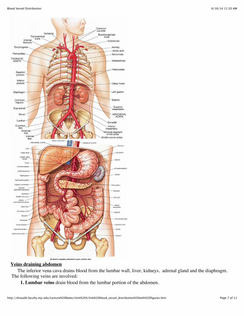
Veins draining abdomen The inferior vena cava drains blood from the lumbar wall, liver, kidneys, adrenal gland and the diaphragm.The following veins are involved: 1. Lumbar veins drain blood from the lumbar portion of the abdomen.
6/16/14 11:20 AMBlood Vessel Distribution
Page 8 of 11http://droualb.faculty.mjc.edu/Lecture%20Notes/Unit%204/link%20blood_vessel_distribution%20with%20figures.htm
2. Gonadal veins drain blood from the testicles or ovaries. The left gonadal usually drains into the left renalvein.
3. Hepatic veins drain blood from the liver.
4. Renal veins drain blood from the kidneys. 5. Suprarenal veins drain blood from the adrenal glands.
6. Phrenic veins drain blood from the diaphragm.
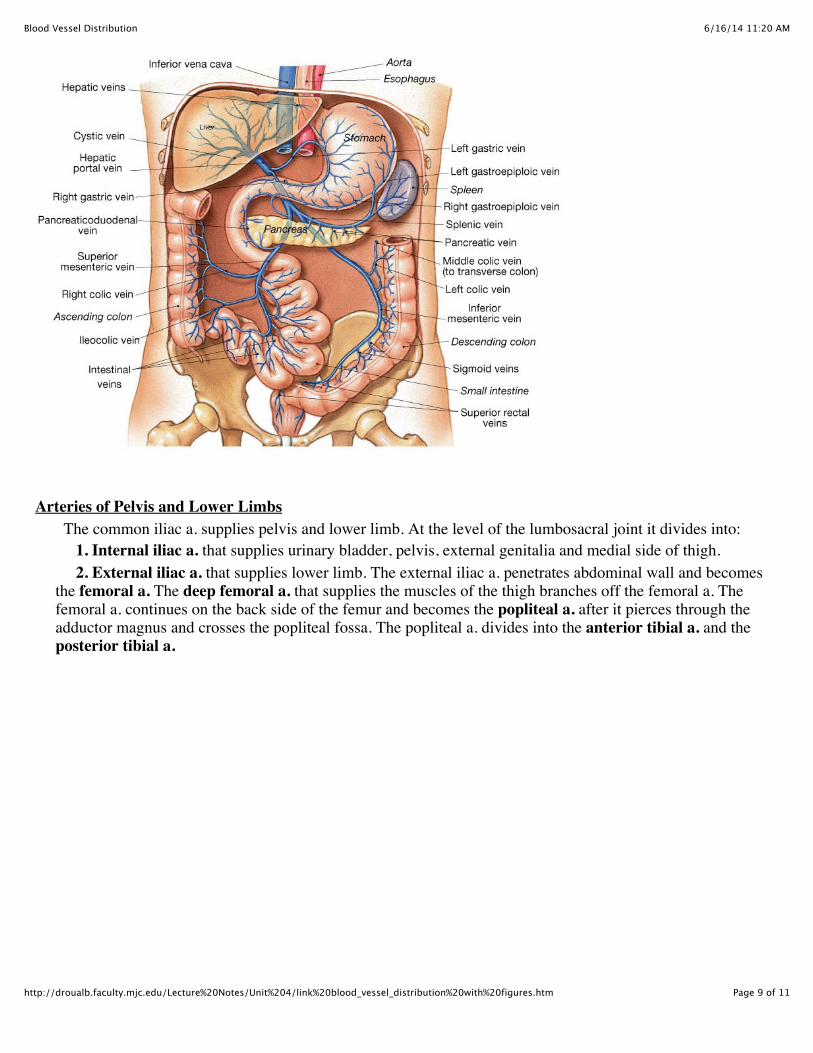
Hepatic Portal System Of the digestive organs, only blood draining from the liver goes directly to the inferior vena cava. Blood fromthe small intestines, large intestines, stomach, pancreas and spleen flows into the liver by the hepatic portalvein. The veins that drain into the hepatic portal vein include the superior mesenteric v., inferior mesenteric v.and splenic v. This blood is nutrient-rich but oxygen-poor. Oxygen-rich blood is delivered to the liver by thehepatic artery proper. This arrangement allows the liver to perform its processing and storage functions. The blood from the hepatic portal vein flows through sinusoids in the liver and is collected by the hepaticveins.
6/16/14 11:20 AMBlood Vessel Distribution
Page 9 of 11http://droualb.faculty.mjc.edu/Lecture%20Notes/Unit%204/link%20blood_vessel_distribution%20with%20figures.htm
Arteries of Pelvis and Lower Limbs The common iliac a. supplies pelvis and lower limb. At the level of the lumbosacral joint it divides into: 1. Internal iliac a. that supplies urinary bladder, pelvis, external genitalia and medial side of thigh.
2. External iliac a. that supplies lower limb. The external iliac a. penetrates abdominal wall and becomesthe femoral a. The deep femoral a. that supplies the muscles of the thigh branches off the femoral a. Thefemoral a. continues on the back side of the femur and becomes the popliteal a. after it pierces through theadductor magnus and crosses the popliteal fossa. The popliteal a. divides into the anterior tibial a. and theposterior tibial a.

6/16/14 11:20 AMBlood Vessel Distribution
Page 10 of 11http://droualb.faculty.mjc.edu/Lecture%20Notes/Unit%204/link%20blood_vessel_distribution%20with%20figures.htm
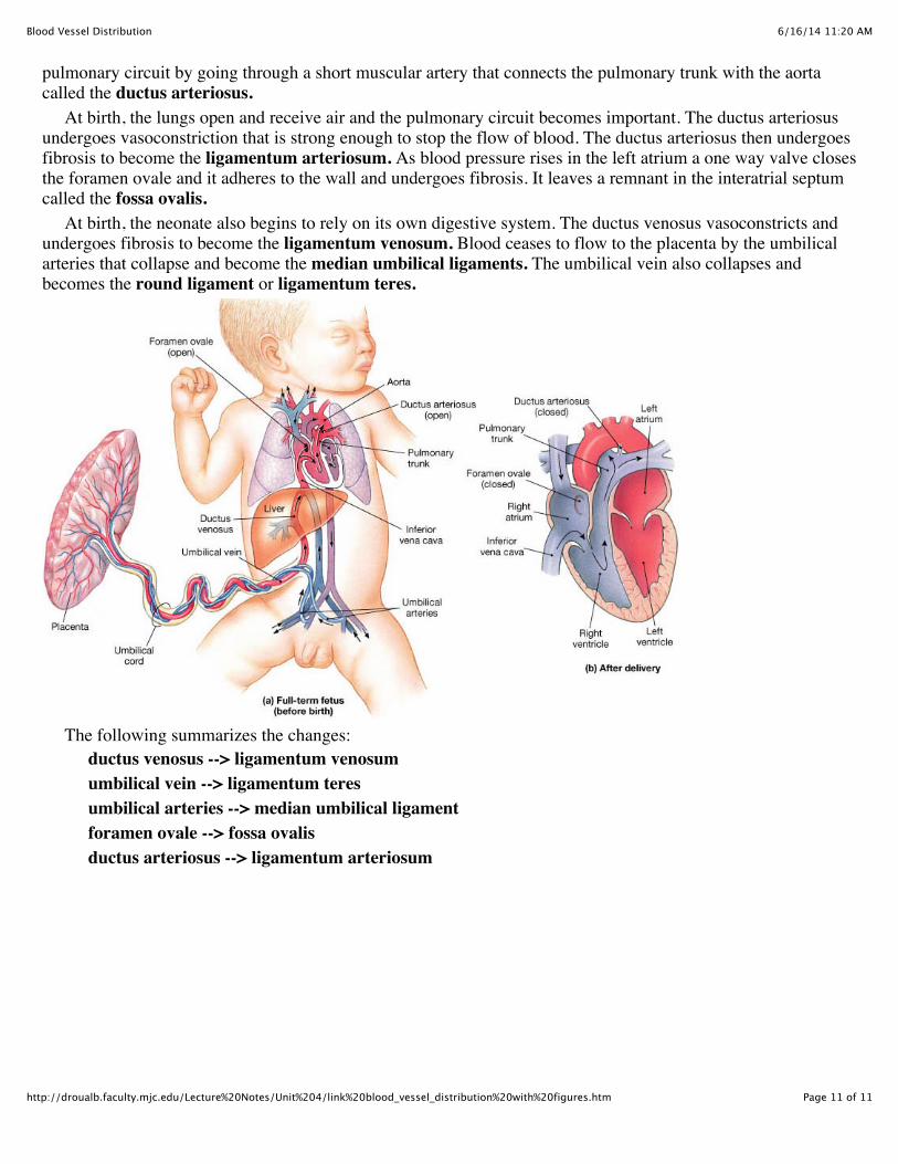
Veins draining lower limbs The deep veins of the lower limbs are again alongside the arteries of the same name. The anterior tibial v.and posterior tibial v. drain blood from lower leg and fuse to form the popliteal v. The ascends alongside thefemur and becomes the femoral v. The deep femoral v. that collects blood from the inner thigh drains into thefemoral v. When the femoral v. penetrates the abdominal wall it is called the internal iliac v. The superficial veins of the lower limb include the great saphenous v. that ascend along the medial aspect ofthe leg and thigh and drains into the femoral v. and the small saphenous v. that ascends along the posterior andlateral side of the leg and joins the popliteal v. Veins draining pelvis Inside the pelvis external iliac v. merges with internal iliac v. to form common iliac v. The common iliacveins fuse at the 5th lumbar vertebra to form the inferior vena cava. Fetal Circulation Differences between the adult and fetal circulation reflect differences in the nutritional and respiratorysupport. The fetus depends upon the maternal circulation for oxygen, nutrients and waste removal. Exchangetakes place at the placenta. In the fetus, two umbilical arteries branch off internal iliac arteries and go to theplacenta and a single umbilical vein drains the placenta. The umbilical vein drains oxygenated and nutrient-rich blood into ductus venosus in liver. The ductusvenosus also receives deoxygenated blood from the liver and drains into the inferior vena cava. The fetal pulmonary circuit is not as important to the fetus as the lung is collapsed. Some of the blood thatenters the right atrium bypasses the pulmonary circuit by going through an opening in the interatrial septumcalled the foramen ovale. As blood leaves the right ventricle another major portion of blood bypasses the
6/16/14 11:20 AMBlood Vessel Distribution
Page 11 of 11http://droualb.faculty.mjc.edu/Lecture%20Notes/Unit%204/link%20blood_vessel_distribution%20with%20figures.htm
pulmonary circuit by going through a short muscular artery that connects the pulmonary trunk with the aortacalled the ductus arteriosus. At birth, the lungs open and receive air and the pulmonary circuit becomes important. The ductus arteriosusundergoes vasoconstriction that is strong enough to stop the flow of blood. The ductus arteriosus then undergoesfibrosis to become the ligamentum arteriosum. As blood pressure rises in the left atrium a one way valve closesthe foramen ovale and it adheres to the wall and undergoes fibrosis. It leaves a remnant in the interatrial septumcalled the fossa ovalis. At birth, the neonate also begins to rely on its own digestive system. The ductus venosus vasoconstricts andundergoes fibrosis to become the ligamentum venosum. Blood ceases to flow to the placenta by the umbilicalarteries that collapse and become the median umbilical ligaments. The umbilical vein also collapses andbecomes the round ligament or ligamentum teres.
The following summarizes the changes: ductus venosus --> ligamentum venosum umbilical vein --> ligamentum teres umbilical arteries --> median umbilical ligament foramen ovale --> fossa ovalis ductus arteriosus --> ligamentum arteriosum