blood - palm beach state college · functions of blood transport dissolved gases, nutrients, ......
TRANSCRIPT
Lecture Presentation by Lori Garrett
17Blood
© 2018 Pearson Education, Inc.
Section 1: Plasma and Formed Elements
Learning Outcomes
17.1 List the components of the cardiovascular system, and describe several important functions of blood.
17.2 Describe the important components and major properties of blood.
17.3 Explain the origins and differentiation of the formed elements.
© 2018 Pearson Education, Inc.
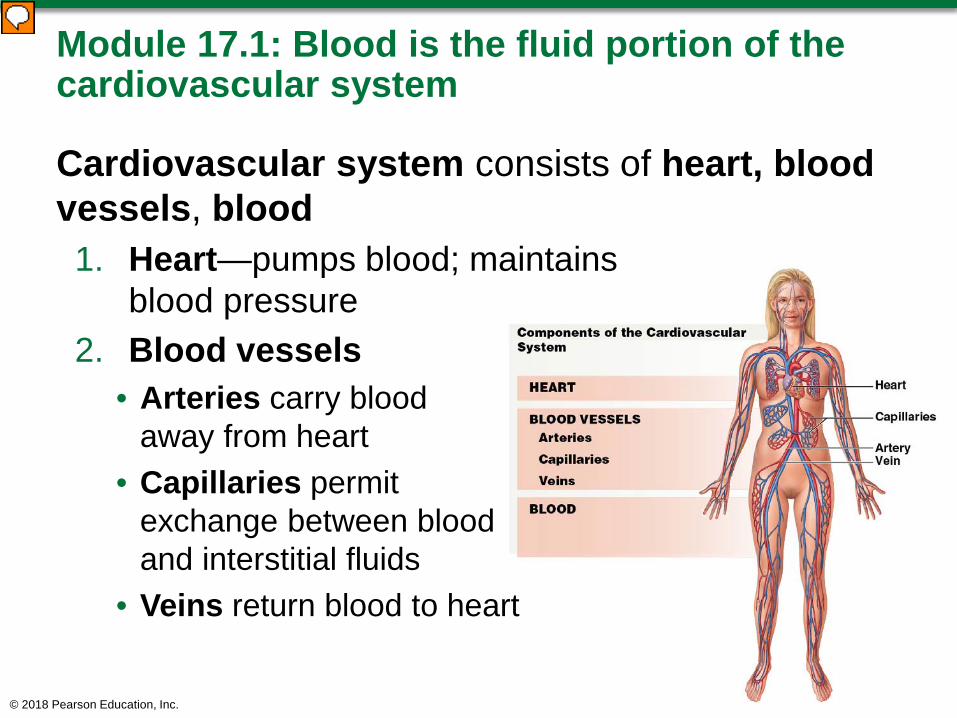
Module 17.1: Blood is the fluid portion of the cardiovascular system
Cardiovascular system consists of heart, blood vessels, blood1. Heart—pumps blood; maintains
blood pressure2. Blood vessels
• Arteries carry blood away from heart
• Capillaries permit exchange between blood and interstitial fluids
• Veins return blood to heart
© 2018 Pearson Education, Inc.
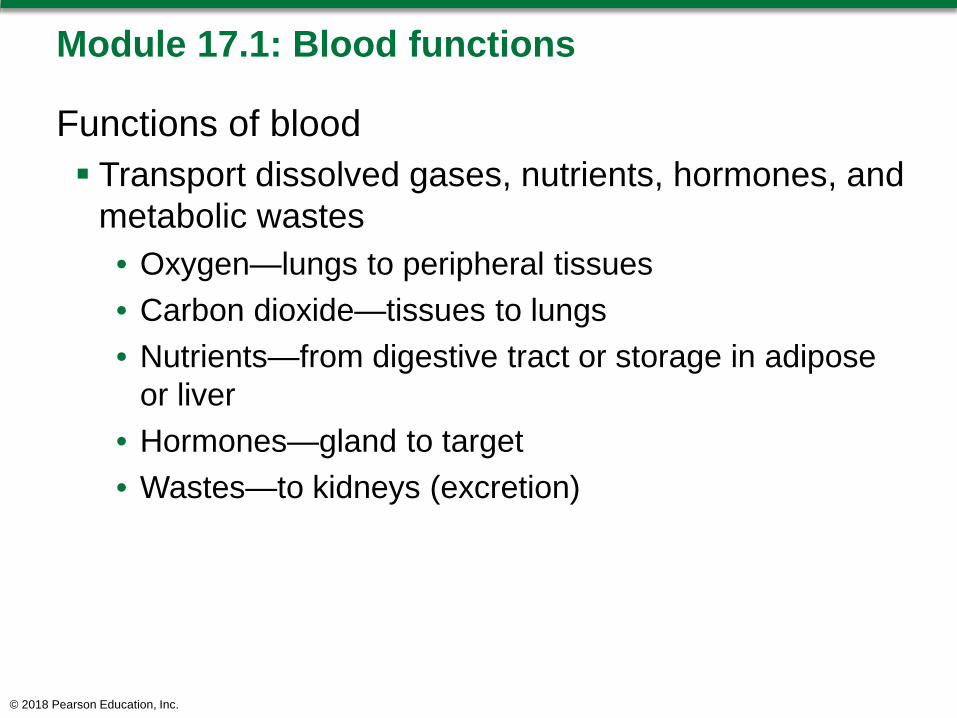
Module 17.1: Blood functions
Functions of blood Transport dissolved gases, nutrients, hormones, and
metabolic wastes• Oxygen—lungs to peripheral tissues • Carbon dioxide—tissues to lungs• Nutrients—from digestive tract or storage in adipose
or liver• Hormones—gland to target• Wastes—to kidneys (excretion)
© 2018 Pearson Education, Inc.

Module 17.1: Blood functions
Functions of blood (continued) Regulate pH and ion composition of interstitial fluids
(IF)• Absorbs/neutralizes acids • Diffusion between blood and IF balances ion
concentrations Restrict fluid loss at injury sites
• Enzymes/other substances initiate clotting when vessel wall is broken; clot = temporary patch
Defend against toxins and pathogens• Transports white blood cells and antibodies to fight
infection
© 2018 Pearson Education, Inc.
Module 17.1: Blood functions
Functions of blood (continued) Stabilize body temperature
• Absorbs heat generated in one area; distributes to other tissues
• High body temperature—blood directed closer to skin• Low body temperature—blood directed to brain,
internal organs
© 2018 Pearson Education, Inc.

© 2018 Pearson Education, Inc.
Module 17.1: Review
A. Identify the components of the cardiovascular system.
B. What are the functions of blood?
Learning Outcome: List the components of the cardiovascular system, and describe several important functions of blood.
© 2018 Pearson Education, Inc.
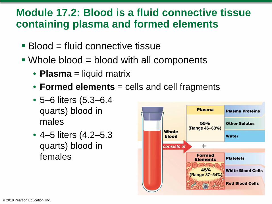
Module 17.2: Blood is a fluid connective tissue containing plasma and formed elements
Blood = fluid connective tissueWhole blood = blood with all components
• Plasma = liquid matrix• Formed elements = cells and cell fragments• 5–6 liters (5.3–6.4
quarts) blood in males
• 4–5 liters (4.2–5.3 quarts) blood in females
© 2018 Pearson Education, Inc.

© 2018 Pearson Education, Inc.
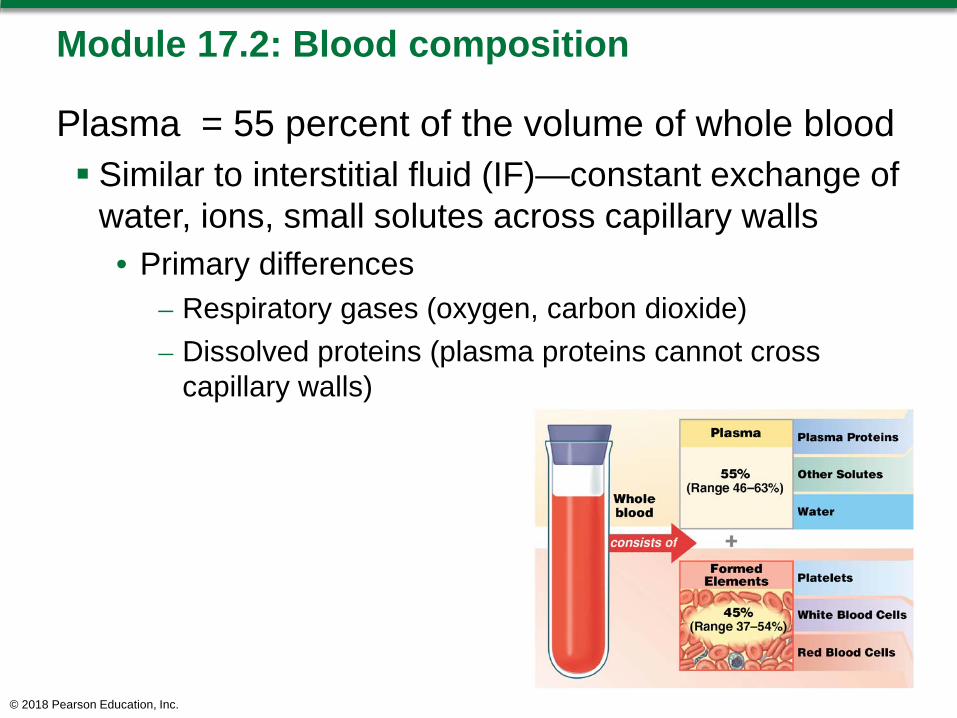
Module 17.2: Blood composition
Plasma = 55 percent of the volume of whole blood Similar to interstitial fluid (IF)—constant exchange of
water, ions, small solutes across capillary walls• Primary differences
– Respiratory gases (oxygen, carbon dioxide)– Dissolved proteins (plasma proteins cannot cross
capillary walls)
© 2018 Pearson Education, Inc.

Module 17.2: Blood composition
Formed elements = blood cells/fragments; ~45 percent of whole blood 99 percent of formed elements are red blood cells Hematocrit (packed cell volume, PCV)
• Percentage of whole blood from formed elements • Overall average
45 percent (range 37–54 percent)
• Males average 47 percent; females average 42 percent (androgens stimulate RBC production)
© 2018 Pearson Education, Inc.
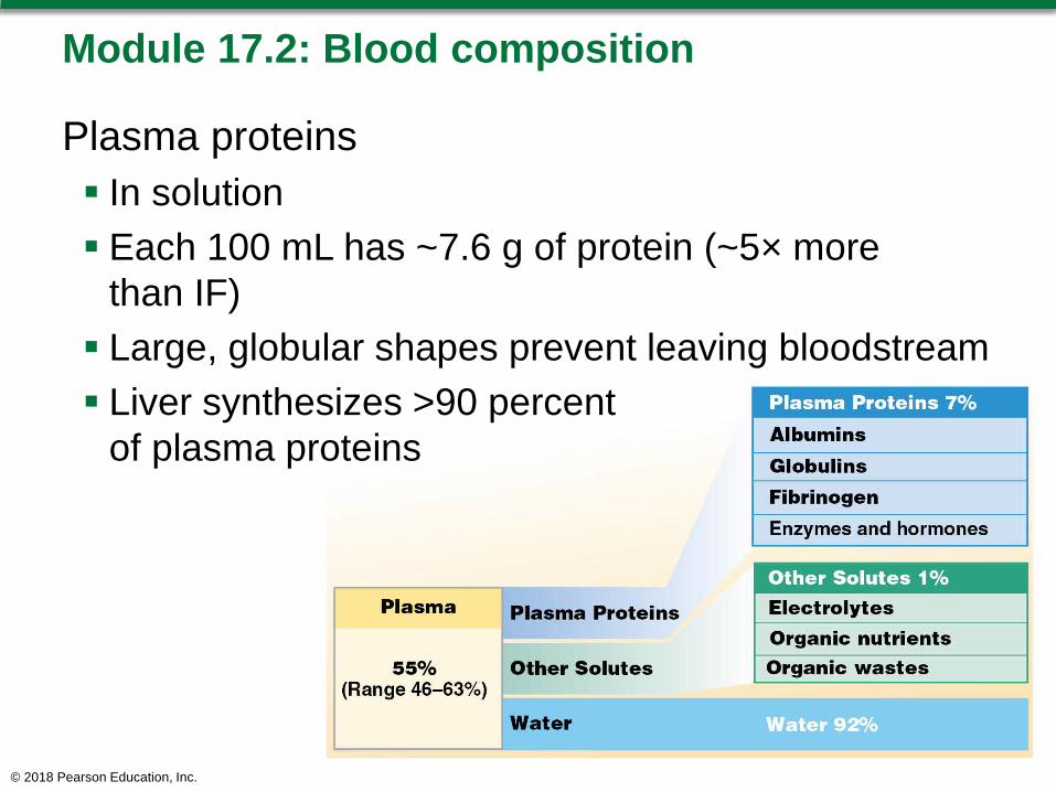
Module 17.2: Blood composition
Plasma proteins In solution Each 100 mL has ~7.6 g of protein (~5× more
than IF) Large, globular shapes prevent leaving bloodstream Liver synthesizes >90 percent
of plasma proteins
© 2018 Pearson Education, Inc.
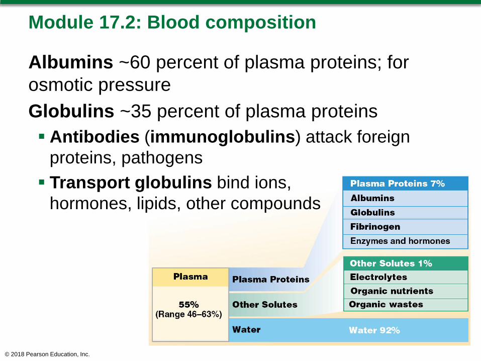
Module 17.2: Blood composition
Albumins ~60 percent of plasma proteins; for osmotic pressureGlobulins ~35 percent of plasma proteins Antibodies (immunoglobulins) attack foreign
proteins, pathogens Transport globulins bind ions,
hormones, lipids, other compounds
© 2018 Pearson Education, Inc.
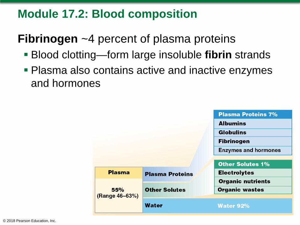
Module 17.2: Blood composition
Fibrinogen ~4 percent of plasma proteins Blood clotting—form large insoluble fibrin strands Plasma also contains active and inactive enzymes
and hormones
© 2018 Pearson Education, Inc.
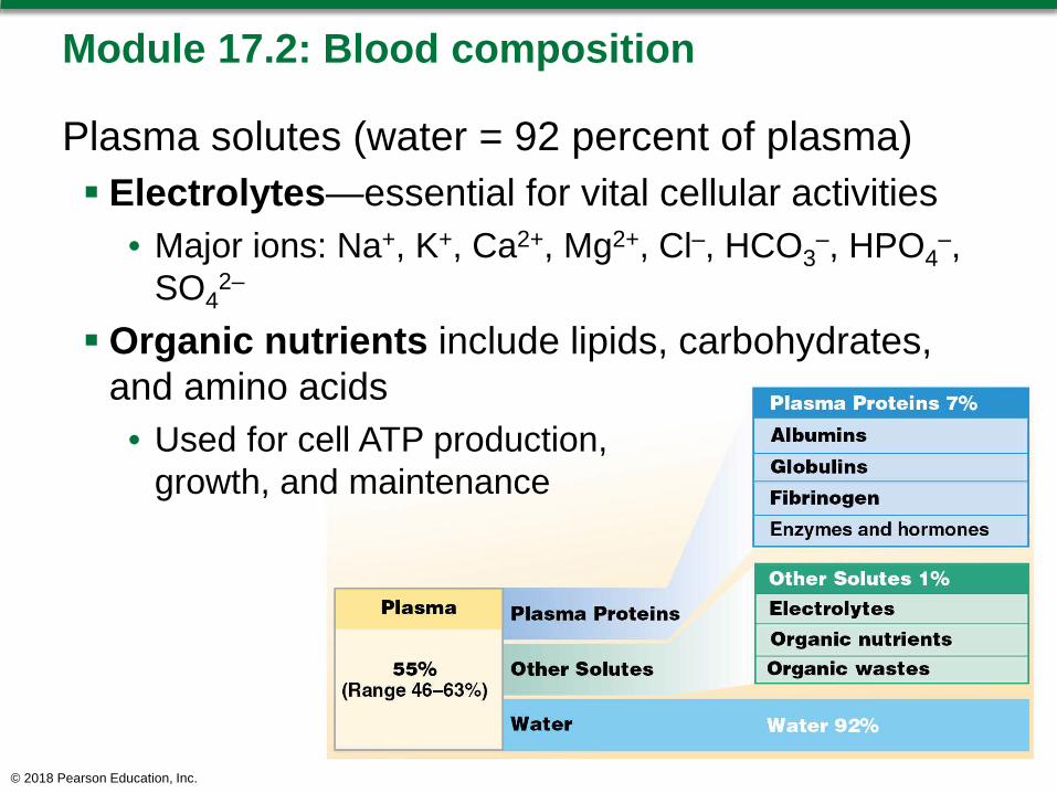
Module 17.2: Blood composition
Plasma solutes (water = 92 percent of plasma) Electrolytes—essential for vital cellular activities
• Major ions: Na+, K+, Ca2+, Mg2+, Cl–, HCO3–, HPO4
–, SO4
2–
Organic nutrients include lipids, carbohydrates, and amino acids• Used for cell ATP production,
growth, and maintenance
© 2018 Pearson Education, Inc.
Module 17.2: Blood composition
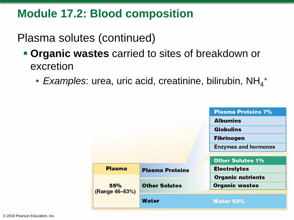
Plasma solutes (continued) Organic wastes carried to sites of breakdown or
excretion• Examples: urea, uric acid, creatinine, bilirubin, NH4
+
© 2018 Pearson Education, Inc.
Module 17.2: Blood composition
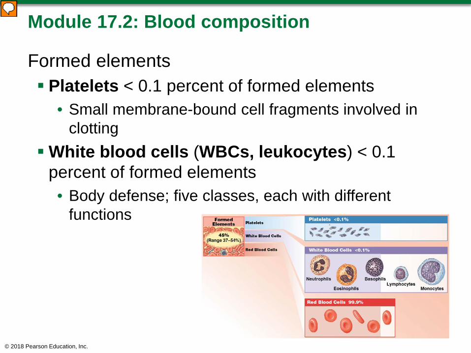
Formed elements Platelets < 0.1 percent of formed elements
• Small membrane-bound cell fragments involved in clotting
White blood cells (WBCs, leukocytes) < 0.1 percent of formed elements• Body defense; five classes, each with different
functions
© 2018 Pearson Education, Inc.

Module 17.2: Blood composition
Formed elements (continued) Red blood cells (RBCs, erythrocytes) = 99.9
percent of formed elements• Oxygen transport
© 2018 Pearson Education, Inc.

Summary of the composition of whole blood
© 2018 Pearson Education, Inc.
Module 17.2: Review
A. Identify the two components making up whole blood, and list the composition of each.
B. Define hematocrit.C. Which specific plasma proteins would you
expect to be elevated during an infection?
Learning Outcome: Describe the important components and major properties of blood.
© 2018 Pearson Education, Inc.

Module 17.3: Formed elements are produced by stem cells in red bone marrow
Development of formed elements = hemopoiesis/hematopoiesisOccurs in red bone marrow
Hemocytoblasts from hematopoietic stem cells (HSCs) Produce two types of stem cells
1. Lymphoid stem cells produce lymphocytes (type of WBC)
2. Myeloid stem cells produce RBCs, other WBCs
© 2018 Pearson Education, Inc.
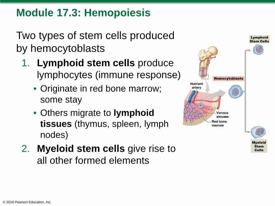
Module 17.3: Hemopoiesis
Two types of stem cells produced by hemocytoblasts1. Lymphoid stem cells produce
lymphocytes (immune response)• Originate in red bone marrow;
some stay• Others migrate to lymphoid
tissues (thymus, spleen, lymph nodes)
2. Myeloid stem cells give rise to all other formed elements
© 2018 Pearson Education, Inc.
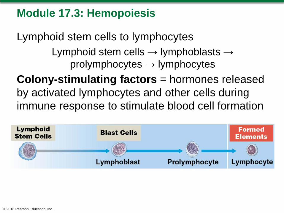
Module 17.3: Hemopoiesis
Lymphoid stem cells to lymphocytesLymphoid stem cells → lymphoblasts →
prolymphocytes → lymphocytesColony-stimulating factors = hormones released by activated lymphocytes and other cells during immune response to stimulate blood cell formation
© 2018 Pearson Education, Inc.
Module 17.3: Hemopoiesis
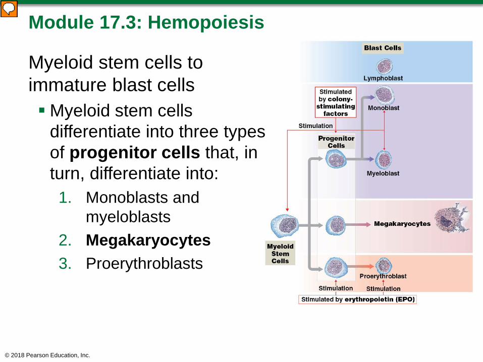
Myeloid stem cells to immature blast cellsMyeloid stem cells
differentiate into three types of progenitor cells that, in turn, differentiate into:1. Monoblasts and
myeloblasts2. Megakaryocytes3. Proerythroblasts
© 2018 Pearson Education, Inc.
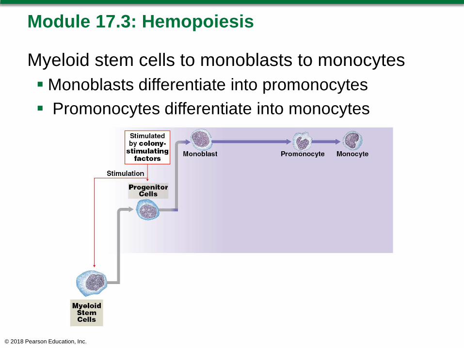
Module 17.3: Hemopoiesis
Myeloid stem cells to monoblasts to monocytesMonoblasts differentiate into promonocytes Promonocytes differentiate into monocytes
© 2018 Pearson Education, Inc.
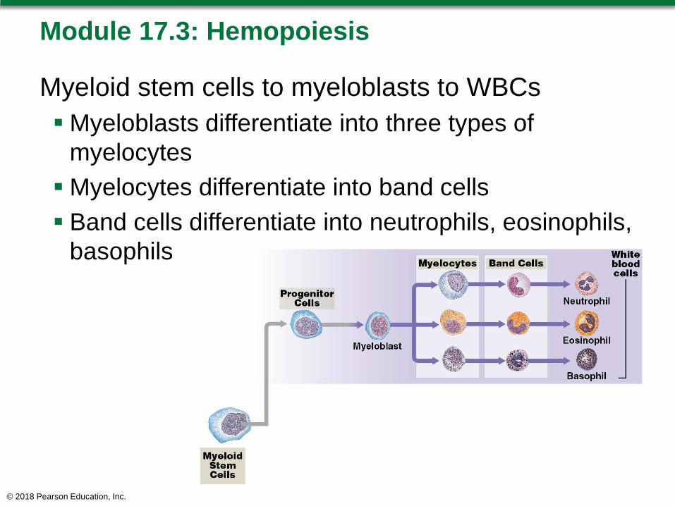
Module 17.3: Hemopoiesis
Myeloid stem cells to myeloblasts to WBCsMyeloblasts differentiate into three types of
myelocytesMyelocytes differentiate into band cells Band cells differentiate into neutrophils, eosinophils,
basophils
© 2018 Pearson Education, Inc.

Module 17.3: Hemopoiesis
Myeloid stem cells to megakaryocytes to plateletsMyeloid stem cells differentiate into
megakaryocytes• Megakaryocytes are enormous cells with large nuclei• Eventually shed cytoplasm in small, membrane-
enclosed packets = platelets
© 2018 Pearson Education, Inc.

Module 17.3: Hemopoiesis
Myeloid stem cells to proerythroblasts to RBCsMyeloid stem cells differentiate into proerythroblasts Proerythroblasts differentiate to erythroblasts, which
shed their nuclei, and then on to anucleatereticulocytes Reticulocytes differentiate to erythrocytes (red blood
cells/RBCs)
© 2018 Pearson Education, Inc.
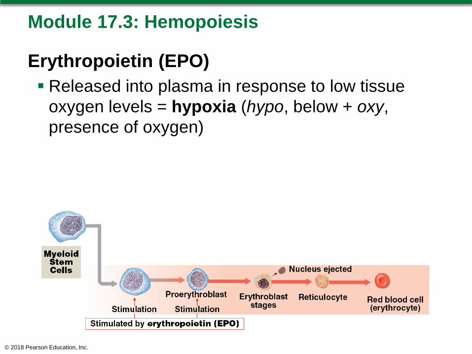
Module 17.3: Hemopoiesis
Erythropoietin (EPO) Released into plasma in response to low tissue
oxygen levels = hypoxia (hypo, below + oxy, presence of oxygen)
© 2018 Pearson Education, Inc.
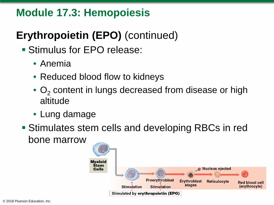
Module 17.3: Hemopoiesis
Erythropoietin (EPO) (continued) Stimulus for EPO release:
• Anemia• Reduced blood flow to kidneys• O2 content in lungs decreased from disease or high
altitude• Lung damage
Stimulates stem cells and developing RBCs in red bone marrow
© 2018 Pearson Education, Inc.
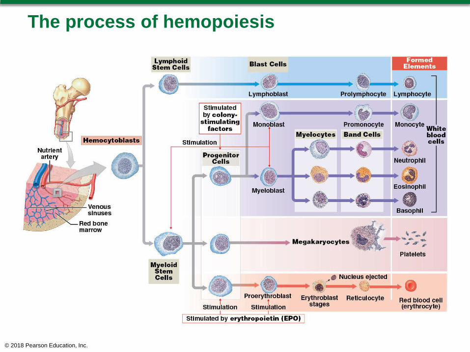
The process of hemopoiesis
© 2018 Pearson Education, Inc.
Module 17.3: Review
A. Define hemocytoblasts.B. Describe platelets and their origin.C. Compare the types of cells that lymphoid stem
cells and myeloid stem cells produce.
Learning Outcome: Explain the origins and differentiation of the formed elements.
© 2018 Pearson Education, Inc.

Section 2: Structure and Function of Formed Elements
Learning Outcomes
17.4 Define hematology, describe the elements of a complete blood count (CBC), and give examples of red blood cell lab tests.
17.5 List the characteristics and functions of red blood cells, and describe the structure and functions of hemoglobin.
17.6 Describe how the components of aged or damaged red blood cells are recycled.
© 2018 Pearson Education, Inc.
Section 2: Structure and Function of Formed Elements
Learning Outcomes (continued)
17.7 Explain the importance of blood typing and the basis for ABO and Rh incompatibilities.
17.8 Clinical Module: Describe hemolytic disease of the newborn, explain the clinical significance of the cross-reaction between fetal and maternal blood types, and cite preventive measures.
© 2018 Pearson Education, Inc.
Section 2: Structure and Function of Formed Elements
Learning Outcomes (continued)
17.9 Categorize the various types of white blood cells on the basis of their structures and functions.
17.10 Discuss the mechanisms that control blood loss after an injury, and describe the reaction sequences responsible for blood clotting.
17.11 Clinical Module: Explain how blood disorders are detected, and describe examples of the various categories of blood disorders.
© 2018 Pearson Education, Inc.

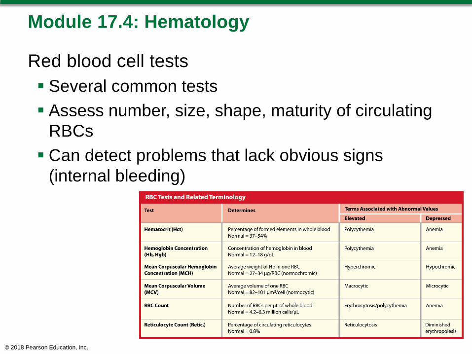
Module 17.4: Hematology is the study of blood and blood-forming tissues
Hematology Important information about a person’s health Can detect disorders (anemia, infection, clotting
disorders) Dyscrasias = blood disorders; can have systemic
effectsReasons for performing blood tests Determine blood type Evaluate types/numbers of RBCs, WBCs, and
platelets Abnormal values may indicate underlying medical
conditions© 2018 Pearson Education, Inc.

Module 17.4: Hematology
Complete blood count (CBC) Determines following in 1 cubic
millimeter (1 microliter or μL) of blood:• RBC count• WBC count• Erythrocyte indices (hemoglobin)• Hematocrit• Others
WBC differential count Identifies numbers of each
type of white blood cell
© 2018 Pearson Education, Inc.
Module 17.4: Hematology
Red blood cell tests Several common tests Assess number, size, shape, maturity of circulating
RBCs Can detect problems that lack obvious signs
(internal bleeding)
© 2018 Pearson Education, Inc.

Module 17.4: Review
A. What is hematology?B. Describe a complete blood count (CBC).C. Which condition would a patient have if she had
a depressed hematocrit level?
Learning Outcome: Define hematology, describe the elements of a complete blood count (CBC), and give examples of red blood cell lab tests.
© 2018 Pearson Education, Inc.
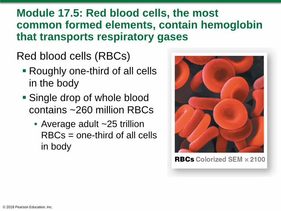
Module 17.5: Red blood cells, the most common formed elements, contain hemoglobin that transports respiratory gases
Red blood cells (RBCs) Roughly one-third of all cells
in the body Single drop of whole blood
contains ~260 million RBCs• Average adult ~25 trillion
RBCs = one-third of all cells in body
© 2018 Pearson Education, Inc.
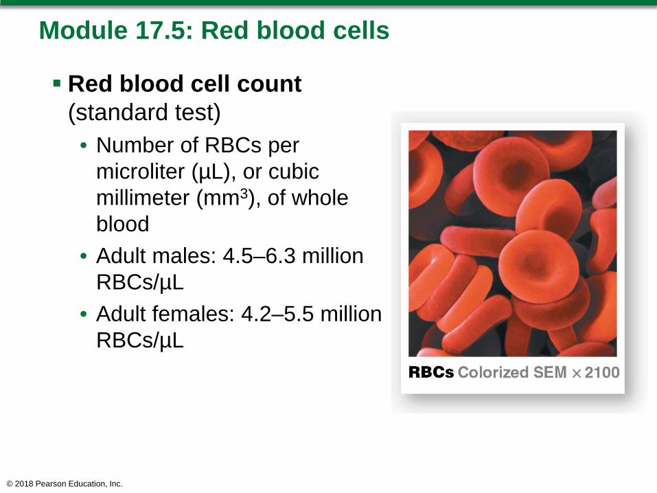
Module 17.5: Red blood cells
Red blood cell count(standard test)• Number of RBCs per
microliter (µL), or cubic millimeter (mm3), of whole blood
• Adult males: 4.5–6.3 million RBCs/µL
• Adult females: 4.2–5.5 million RBCs/µL
© 2018 Pearson Education, Inc.
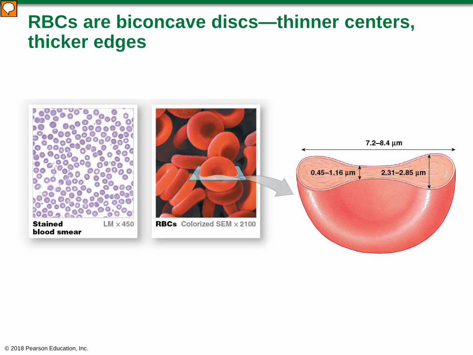
RBCs are biconcave discs—thinner centers, thicker edges
© 2018 Pearson Education, Inc.
Module 17.5: Red blood cells
Functional aspects of red blood cells Large surface area–to–volume ratio
• Packed with hemoglobin (= protein that carries oxygen)
• Allows more oxygen exchange • Total RBC surface area (adult) equals ~2000 times
total surface area of body Form stacks (rouleaux)—facilitate transport in small
vessels Flexible—RBCs can move through narrowest
capillaries with diameters smaller than RBC
© 2018 Pearson Education, Inc.

© 2018 Pearson Education, Inc.
Module 17.5: Red blood cells
Red blood cell characteristics Lose most organelles during developmentMature RBCs lack nuclei (anucleate) and
ribosomes• Cannot divide/repair
Life span < 120 days Primary function—transport respiratory gases
• 95 percent of RBC intracellular proteins are hemoglobin molecules
• Hemoglobin (Hb) content, whole blood:– 14–18 g/dL males– 12–16 g/dL females
© 2018 Pearson Education, Inc.

Module 17.5: Red blood cells
Hemoglobin Complex quaternary
structure Each Hb molecule has:
• Two alpha (α) chains• Two beta (β) chains
Similar to myoglobin in muscle cells Each chain has a single
heme molecule
© 2018 Pearson Education, Inc.
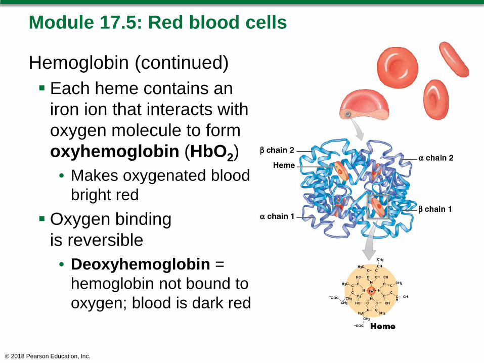
Module 17.5: Red blood cells
Hemoglobin (continued) Each heme contains an
iron ion that interacts with oxygen molecule to form oxyhemoglobin (HbO2)• Makes oxygenated blood
bright redOxygen binding
is reversible• Deoxyhemoglobin =
hemoglobin not bound to oxygen; blood is dark red
© 2018 Pearson Education, Inc.

Module 17.5: Red blood cells
Hemoglobin (continued) An RBC has ~280 million
Hb molecules Each Hb molecule has
four heme units So, each RBC can carry
>1 billion oxygen molecules ~98.5 percent of oxygen
in blood is bound to Hb; rest is dissolved in plasma
© 2018 Pearson Education, Inc.

Module 17.5: Review
A. Describe the functional aspects of RBCs.B. Describe hemoglobin.C. Compare oxyhemoglobin with
deoxyhemoglobin.
Learning Outcome: List the characteristics and functions of red blood cells, and describe the structure and functions of hemoglobin.
© 2018 Pearson Education, Inc.
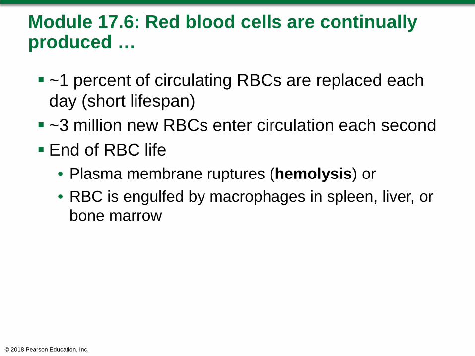
Module 17.6: Red blood cells are continually produced …
~1 percent of circulating RBCs are replaced each day (short lifespan) ~3 million new RBCs enter circulation each second End of RBC life
• Plasma membrane ruptures (hemolysis) or• RBC is engulfed by macrophages in spleen, liver, or
bone marrow
© 2018 Pearson Education, Inc.
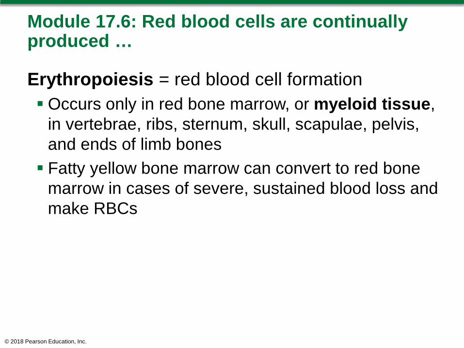
Module 17.6: Red blood cells are continually produced …
Erythropoiesis = red blood cell formationOccurs only in red bone marrow, or myeloid tissue,
in vertebrae, ribs, sternum, skull, scapulae, pelvis, and ends of limb bones Fatty yellow bone marrow can convert to red bone
marrow in cases of severe, sustained blood loss and make RBCs
© 2018 Pearson Education, Inc.

Module 17.6: RBC production and breakdown
Erythroblasts begin producing Hb Normoblasts lose their nuclei and become
reticulocytes Reticulotyes contain
~80 percent of the Hb of mature RBCs; enter bloodstream after 2 days
© 2018 Pearson Education, Inc.
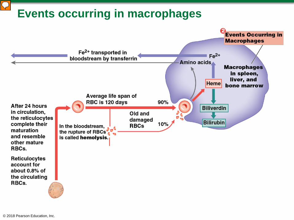
Events occurring in macrophages
© 2018 Pearson Education, Inc.

Module 17.6: RBC production and breakdown
Events occurring in macrophages (continued)Macrophages monitor condition of circulating RBCs
• Engulf old RBCs before rupture (hemolyze)• Remove Hb molecules/cell fragments
Heme units stripped of iron• Iron is stored in phagocyte or enters blood and binds
to transferrin (plasma protein)• Heme → biliverdin → bilirubin → bloodstream →
liverGlobular proteins disassembled and amino acids
recycled
© 2018 Pearson Education, Inc.
Module 17.6: RBC production and breakdown
Events occurring in macrophages (continued) Hemoglobin that is not phagocytized breaks down
into its protein chains and is excreted in urine Breakdown of an abnormally large number of RBCs
results in red or brown urine; condition is hemoglobinuria
© 2018 Pearson Education, Inc.
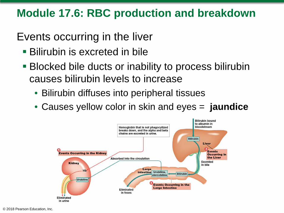
Module 17.6: RBC production and breakdown
Events occurring in the liver Bilirubin is excreted in bile Blocked bile ducts or inability to process bilirubin
causes bilirubin levels to increase• Bilirubin diffuses into peripheral tissues• Causes yellow color in skin and eyes = jaundice
© 2018 Pearson Education, Inc.
Module 17.6: RBC production and breakdown
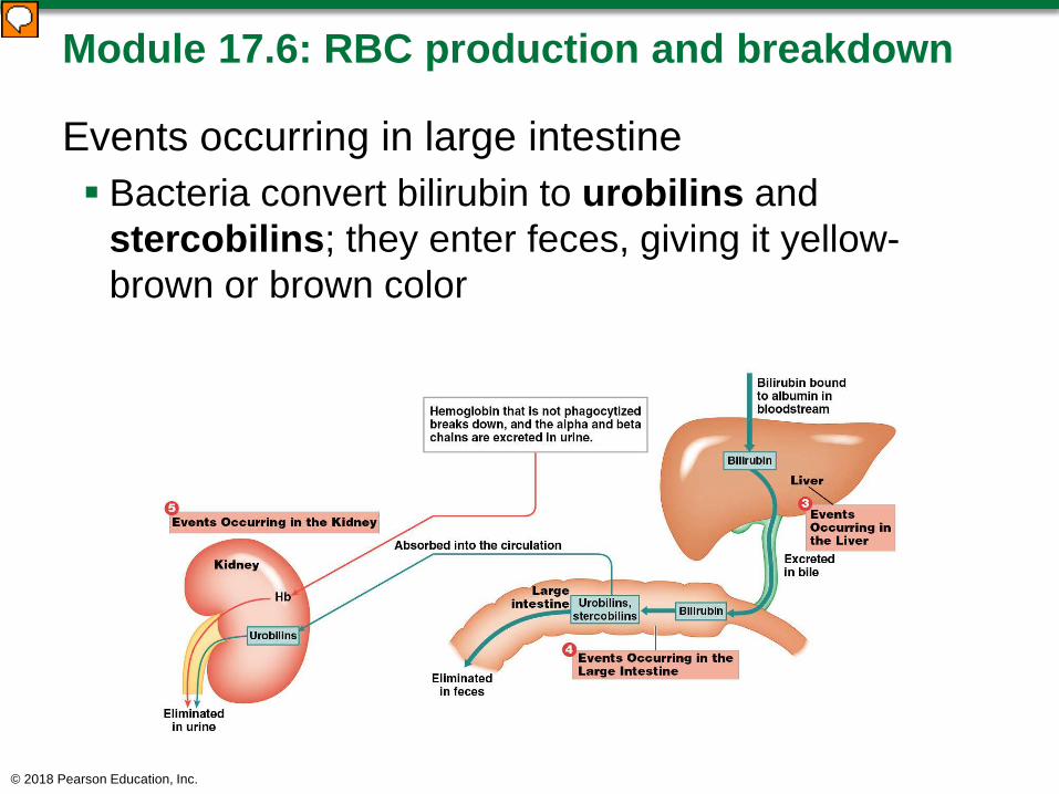
Events occurring in large intestine Bacteria convert bilirubin to urobilins and
stercobilins; they enter feces, giving it yellow-brown or brown color
© 2018 Pearson Education, Inc.
Module 17.6: RBC production and breakdown
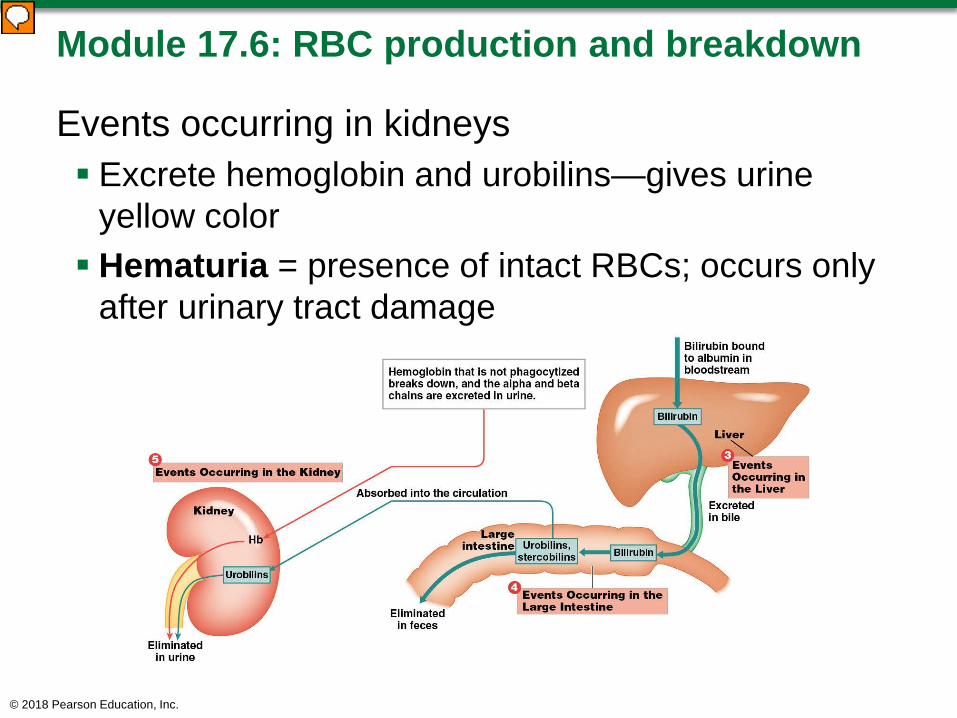
Events occurring in kidneys Excrete hemoglobin and urobilins—gives urine
yellow color Hematuria = presence of intact RBCs; occurs only
after urinary tract damage
© 2018 Pearson Education, Inc.

Summary of the production and clearance of RBCs
© 2018 Pearson Education, Inc.
Module 17.6: Review
A. In what way would a liver disease affect the level of bilirubin in the blood?
Learning Outcome: Describe how the components of aged or damaged red blood cells are recycled.
© 2018 Pearson Education, Inc.

Module 17.7: Blood type is determined by the presence or absence of specific surface antigens on RBCsBlood types Antigens = substances that can elicit immune
responseOur cells have surface antigens embedded in their
plasma membranes; recognized as normal, or self, by immune system
© 2018 Pearson Education, Inc.

Module 17.7: Blood type
Blood types (continued) Blood type determined genetically by which surface
antigens are present in RBC plasma membranes• > 50 blood cell surface antigens• Three most important:
– A– B– Rh (or D)
© 2018 Pearson Education, Inc.
Module 17.7: Blood type
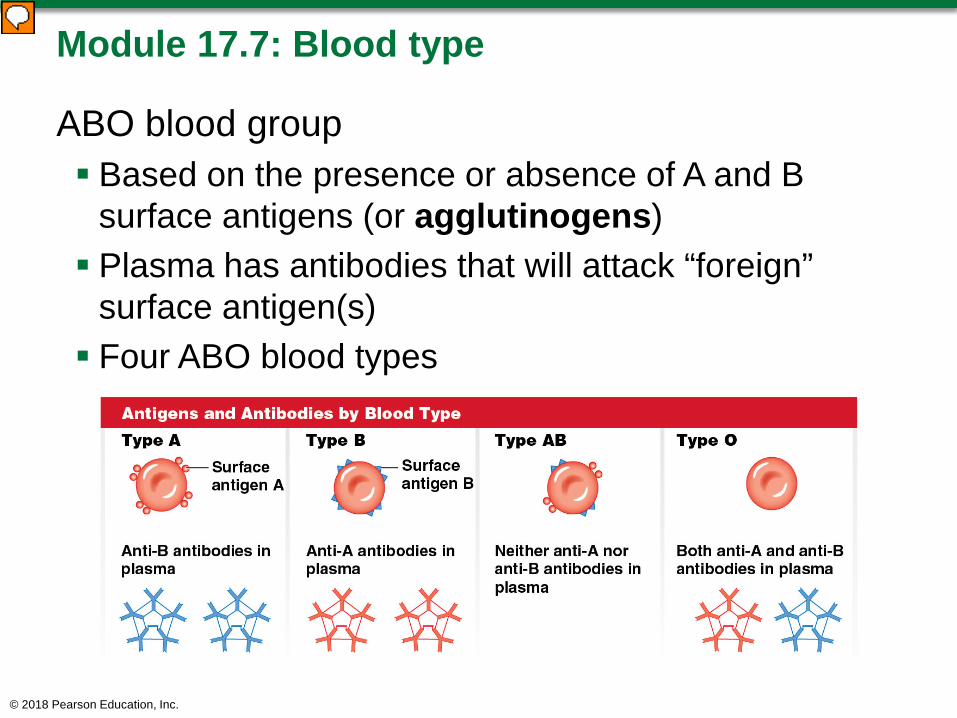
ABO blood group Based on the presence or absence of A and B
surface antigens (or agglutinogens) Plasma has antibodies that will attack “foreign”
surface antigen(s) Four ABO blood types
© 2018 Pearson Education, Inc.
Module 17.7: Blood type
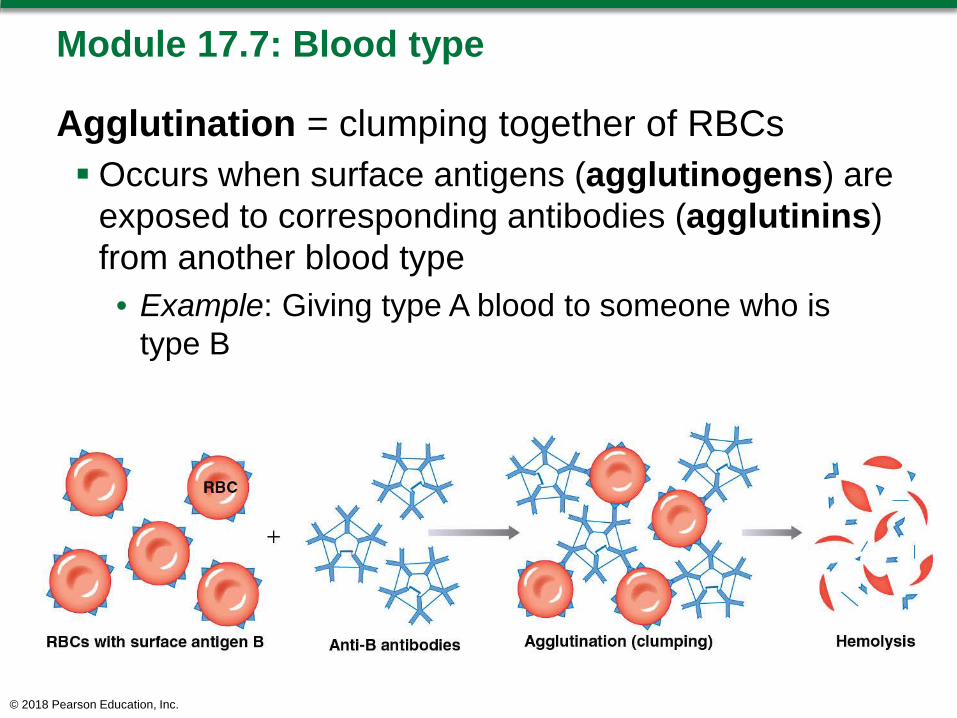
Agglutination = clumping together of RBCs Occurs when surface antigens (agglutinogens) are
exposed to corresponding antibodies (agglutinins) from another blood type• Example: Giving type A blood to someone who is
type B
© 2018 Pearson Education, Inc.
Module 17.7: Blood type
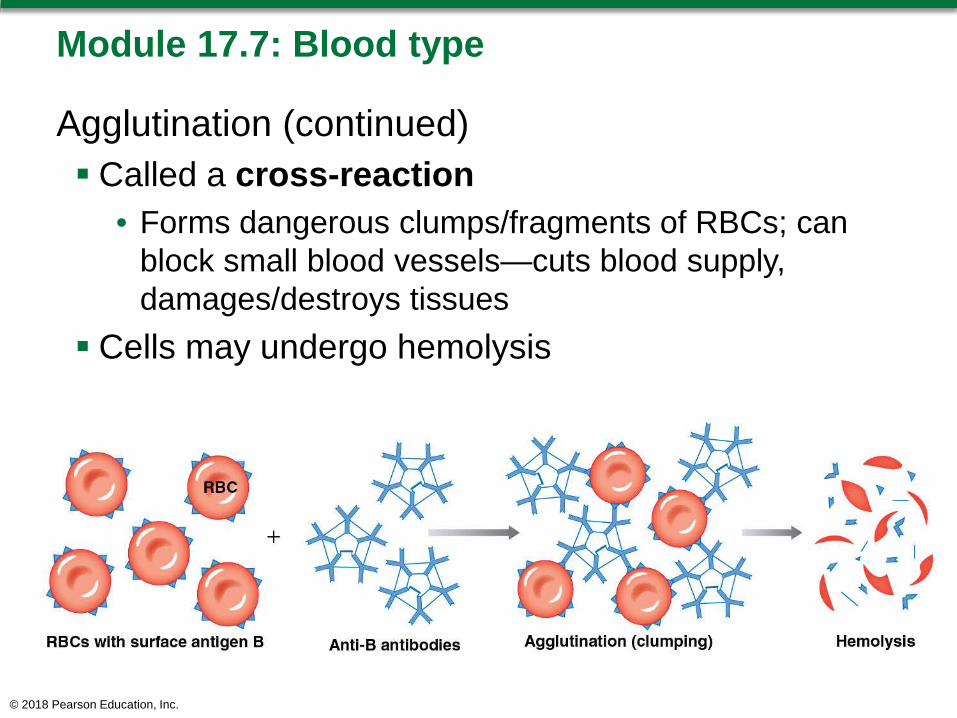
Agglutination (continued) Called a cross-reaction
• Forms dangerous clumps/fragments of RBCs; can block small blood vessels—cuts blood supply, damages/destroys tissues
Cells may undergo hemolysis
© 2018 Pearson Education, Inc.
Module 17.7: Blood type
Rh blood group Based on presence or absence of Rh surface
antigen on RBCs “Rh” comes from original discovery in Rhesus
monkeys Rh positive (Rh+) has Rh surface antigen
• Indicates presence of Rh surface antigen Rh negative (Rh–) lacks Rh surface antigen Included in full blood type as + or –
Examples: O negative (O–), AB positive (AB+)
© 2018 Pearson Education, Inc.

© 2018 Pearson Education, Inc.
Module 17.7: Blood type
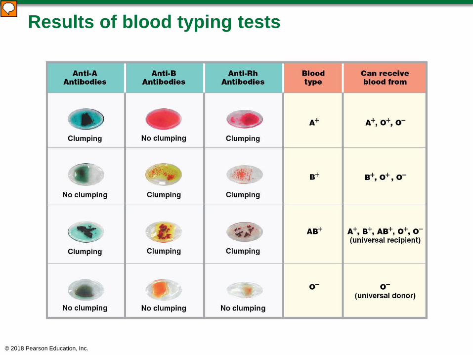
Blood typing tests Drops of person’s blood are mixed with solutions
containing antibodies to surface antigens A, B, and Rh Clumping (agglutination) occurs where sample
contains the corresponding antigen• Example: Type A blood will clump with anti-A
antibodies Typing is necessary to avoid transfusion reactions
(cross-reactions occurring from transfusing mismatched blood)• Donor and recipient blood types must be compatible
(will not cross-react)© 2018 Pearson Education, Inc.
Results of blood typing tests
© 2018 Pearson Education, Inc.
Module 17.7: Blood type
Genetics of blood type Presence of anti-A and/or anti-B antibodies is
genetically determined• Exposure to foreign RBCs not needed to develop the
antibodies Anti-Rh antibodies are not automatically present
• Rh-negative person will not have any anti-Rh antibodies until exposed to Rh-positive RBCs (sensitized); then develops anti-Rh antibodies
© 2018 Pearson Education, Inc.

Module 17.7: Review
A. What is determined by the surface antigens on RBCs?
B. What is the most common blood type in the United States?
C. Which blood type(s) can be safely transfused into a person with type O− blood?
D. Why can’t a person with type A blood safely receive blood from a person with type B blood?
Learning Outcome: Explain the importance of blood typing and the basis for ABO and Rh incompatibilities.
© 2018 Pearson Education, Inc.
Module 17.8: Clinical Module: Hemolytic disease of the newborn is an RBC-related disorder caused by a cross-reaction between fetal and maternal blood types
Background Blood type is determined by combining genes from
both parents• Child can have blood type different from either parent
During pregnancy, mother’s antibodies may cross the placenta and attack/destroy fetal RBCs. Condition is hemolytic diseases of the newborn
(HDN)Many forms—some very dangerous, others
undetectable© 2018 Pearson Education, Inc.
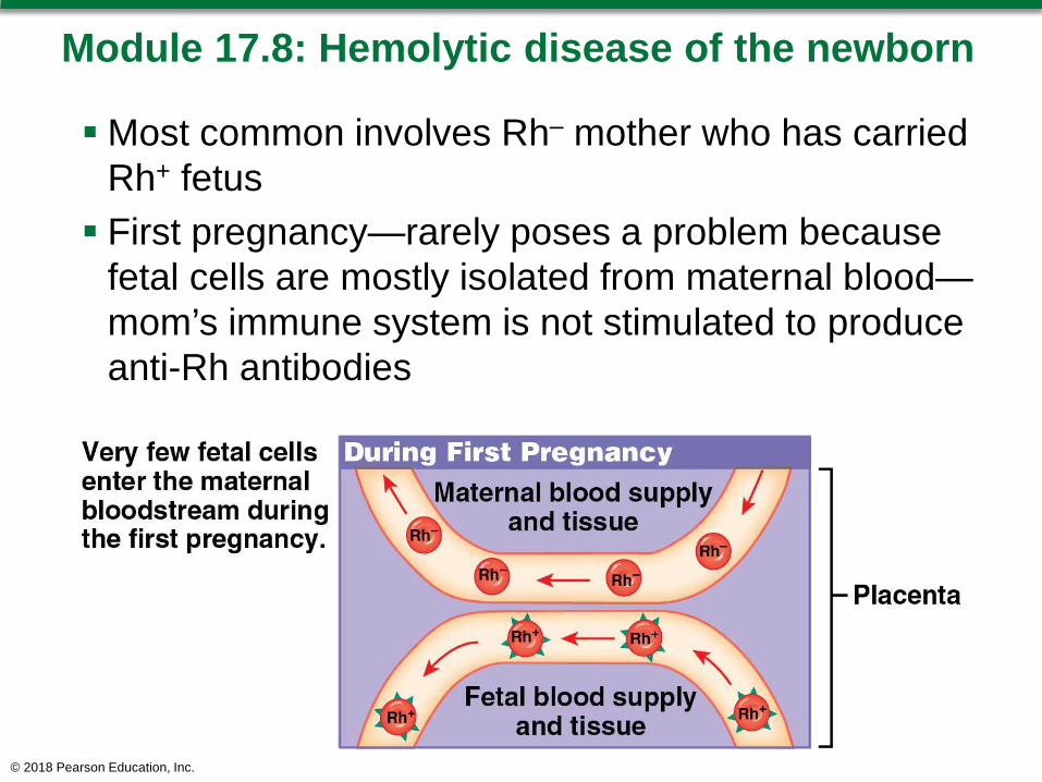
Module 17.8: Hemolytic disease of the newborn
Most common involves Rh– mother who has carried Rh+ fetus First pregnancy—rarely poses a problem because
fetal cells are mostly isolated from maternal blood—mom’s immune system is not stimulated to produce anti-Rh antibodies
© 2018 Pearson Education, Inc.
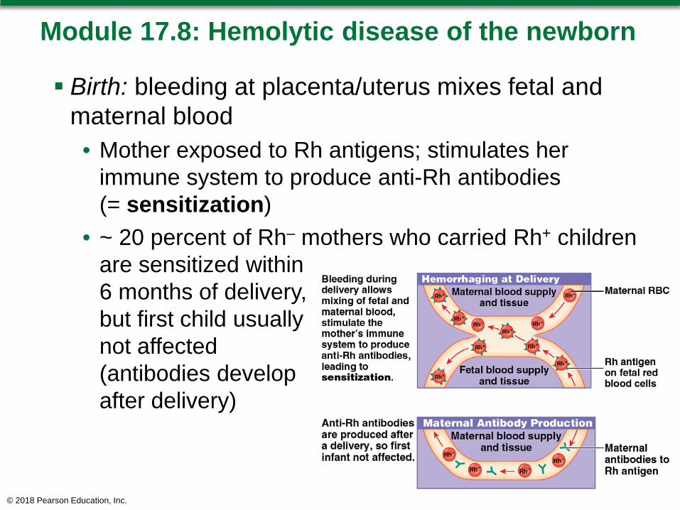
Module 17.8: Hemolytic disease of the newborn
Birth: bleeding at placenta/uterus mixes fetal and maternal blood• Mother exposed to Rh antigens; stimulates her
immune system to produce anti-Rh antibodies (= sensitization)
• ~ 20 percent of Rh– mothers who carried Rh+ children are sensitized within 6 months of delivery,but first child usuallynot affected (antibodies develop after delivery)
© 2018 Pearson Education, Inc.

Module 17.8: Hemolytic disease of the newborn
Subsequent pregnancy with Rh+ fetus:Maternal anti-Rh antibodies can cross placenta;
destroy fetal RBCs, causing severe anemia Fetal demand for RBCs increases; erythroblasts
enter bloodstream before maturity—leads to alternative name, erythroblastosis fetalis High fatality rate without treatment Newborn with severe HDN is anemic/jaundiced
(from high bilirubin concentrations)
© 2018 Pearson Education, Inc.
Module 17.8: Hemolytic disease of the newborn
Maternal antibodies remain active 1–2 months after delivery• May require replacement of infant’s entire blood
volume HDN can be prevented by giving mother anti-Rh
antibodies (RhoGAM) at weeks 26–28 of pregnancy and during/after delivery The antibodies destroy fetal RBCs that crossed
placenta before RBCs stimulate maternal immune response (= no sensitization)
© 2018 Pearson Education, Inc.
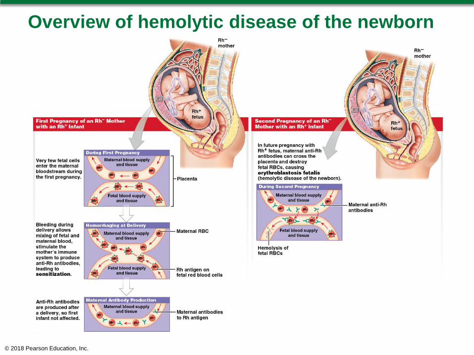
Overview of hemolytic disease of the newborn
© 2018 Pearson Education, Inc.

Module 17.8: Review
A. Define hemolytic disease of the newborn (HDN).
B. Why is RhoGAM administered to pregnant Rh–
women?C. Does an Rh+ mother carrying an Rh– fetus
require a RhoGAM injection? Explain your answer.
Learning Outcome: Describe hemolytic disease of the newborn, explain the clinical significance of the cross-reaction between fetal and maternal blood types, and cite preventive measures.
© 2018 Pearson Education, Inc.
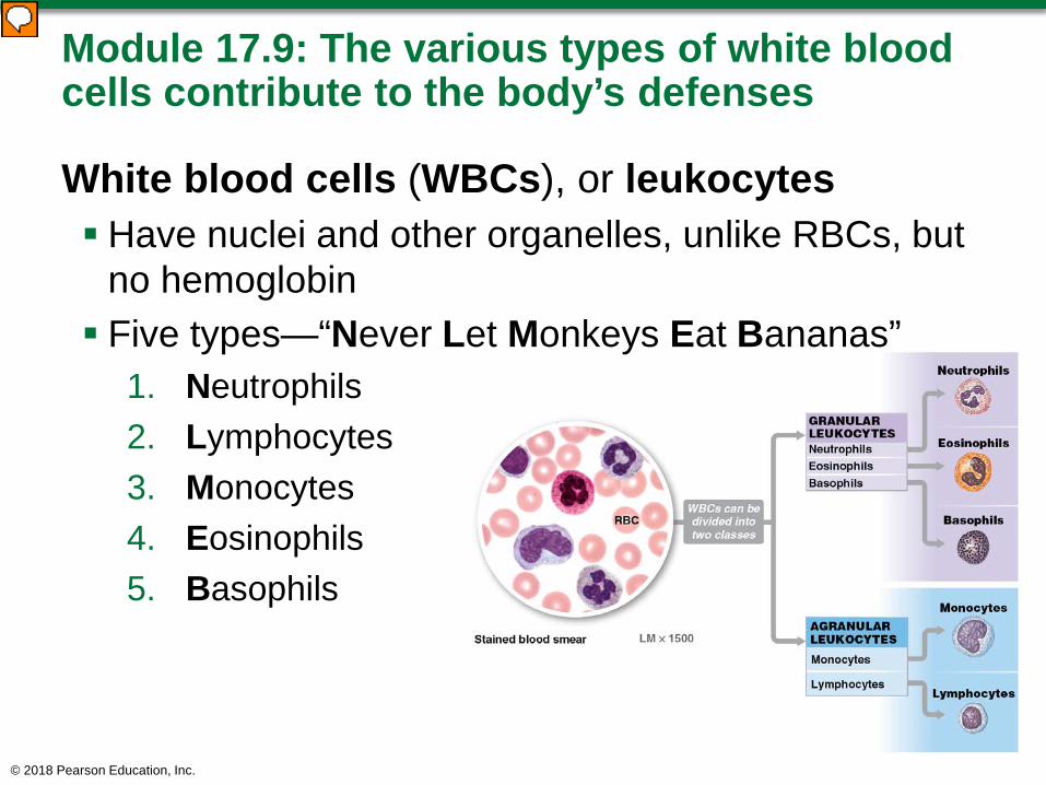
Module 17.9: The various types of white blood cells contribute to the body’s defenses
White blood cells (WBCs), or leukocytes Have nuclei and other organelles, unlike RBCs, but
no hemoglobin Five types—“Never Let Monkeys Eat Bananas”
1. Neutrophils2. Lymphocytes3. Monocytes4. Eosinophils5. Basophils
© 2018 Pearson Education, Inc.
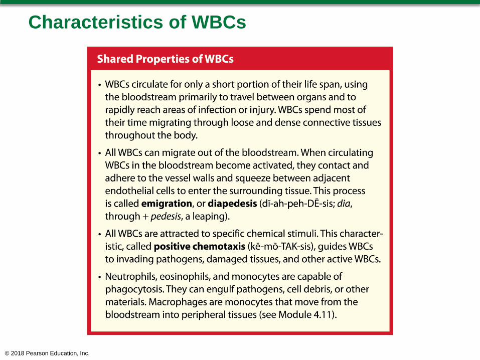
Characteristics of WBCs
© 2018 Pearson Education, Inc.

Module 17.9: White blood cells
White blood cell counts Number can be increased in response to infection,
inflammation, or allergic responsesWBC differential count identifies types and
numbers of WBCs in a blood sample; reported as percentage (per 100 WBCs)
© 2018 Pearson Education, Inc.
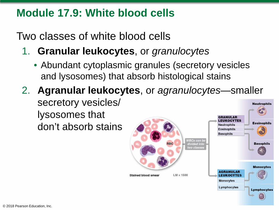
Module 17.9: White blood cells
Two classes of white blood cells1. Granular leukocytes, or granulocytes
• Abundant cytoplasmic granules (secretory vesicles and lysosomes) that absorb histological stains
2. Agranular leukocytes, or agranulocytes—smaller secretory vesicles/lysosomes that don’t absorb stains
© 2018 Pearson Education, Inc.
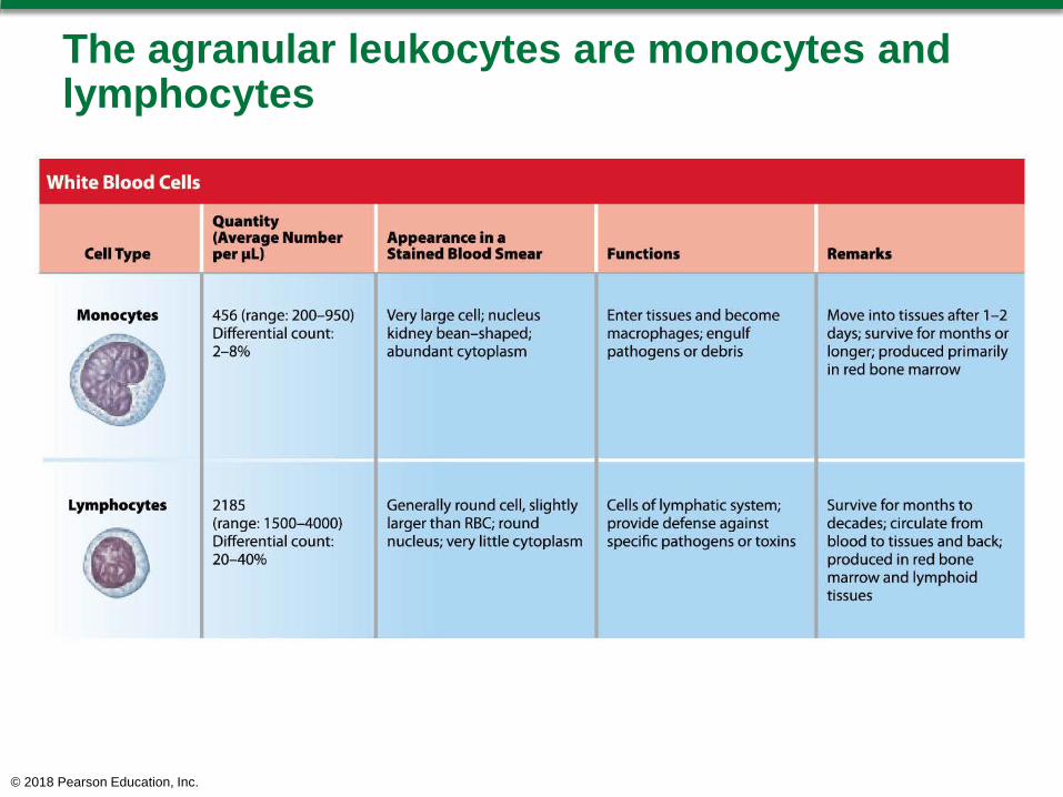
The agranular leukocytes are monocytes and lymphocytes
© 2018 Pearson Education, Inc.
Module 17.9: Review
A. Identify the five types of white blood cells.B. How do basophils respond to tissue damage?C. Which type of white blood cell would you find in
the greatest numbers in an infected cut?
Learning Outcome: Categorize the various types of white blood cells on the basis of their structures and functions.
© 2018 Pearson Education, Inc.
Module 17.10: The clotting response is a complex cascade of events that reduces blood lossHemostasis (haima, blood + stasis, halt) Process responsible for stopping blood loss through
walls of damaged blood vessels Establishes framework for tissue repairs Three Phases
1. Vascular phase2. Platelet phase3. Coagulation phase
© 2018 Pearson Education, Inc.
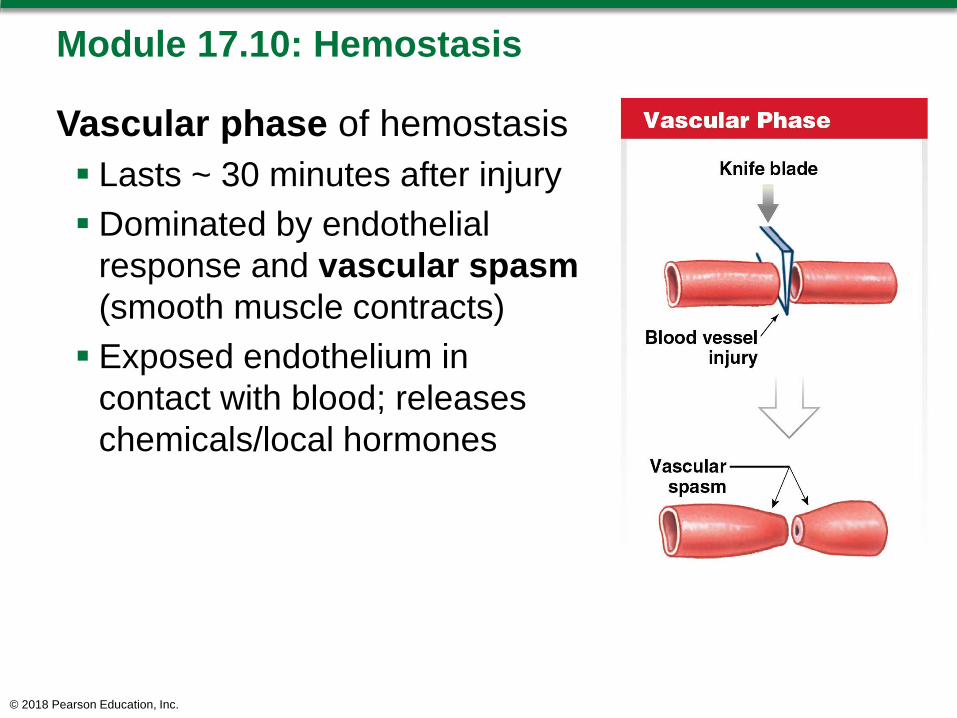
Module 17.10: Hemostasis
Vascular phase of hemostasis Lasts ~ 30 minutes after injury Dominated by endothelial
response and vascular spasm(smooth muscle contracts) Exposed endothelium in
contact with blood; releases chemicals/local hormones
© 2018 Pearson Education, Inc.
Module 17.10: Hemostasis
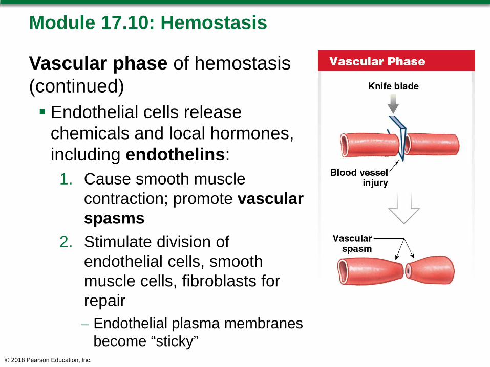
Vascular phase of hemostasis (continued) Endothelial cells release
chemicals and local hormones, including endothelins:1. Cause smooth muscle
contraction; promote vascularspasms
2. Stimulate division of endothelial cells, smooth muscle cells, fibroblasts for repair – Endothelial plasma membranes
become “sticky”© 2018 Pearson Education, Inc.
Module 17.10: Hemostasis
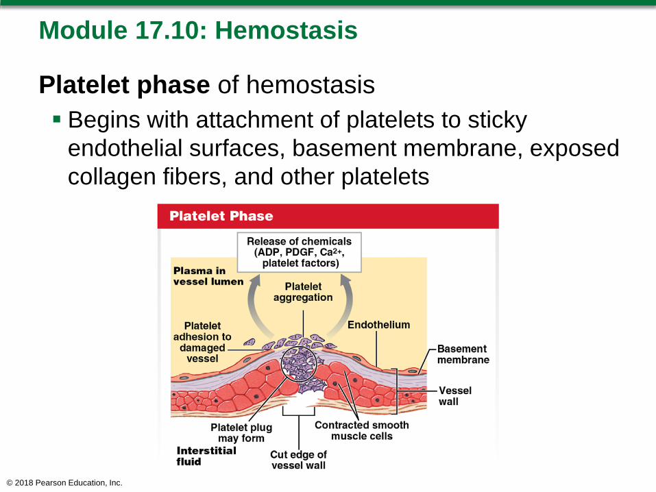
Platelet phase of hemostasis Begins with attachment of platelets to sticky
endothelial surfaces, basement membrane, exposed collagen fibers, and other platelets
© 2018 Pearson Education, Inc.
Module 17.10: Hemostasis
Chemicals released by platelets: Adenosine diphosphate (ADP) stimulates platelet
aggregation and secretion Chemicals that stimulate vascular spasms (smooth
muscle contraction) Platelet factors—proteins that play role in clotting Platelet-derived growth factor (PDGF)—promotes
vessel repair Calcium ions—required for platelet aggregation and
clotting process
© 2018 Pearson Education, Inc.
Module 17.10: Hemostasis
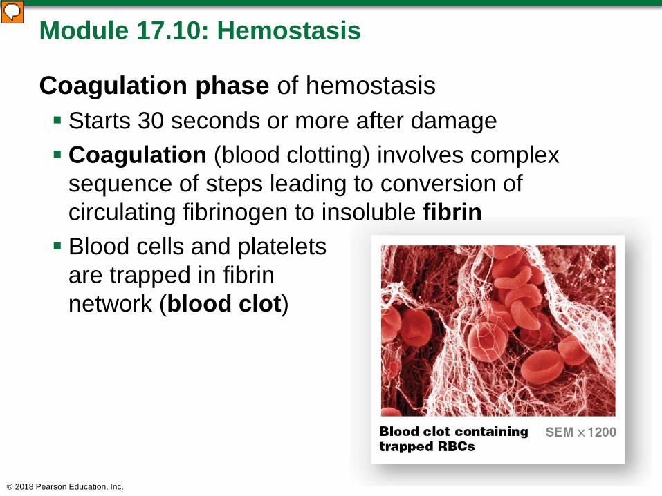
Coagulation phase of hemostasis Starts 30 seconds or more after damage Coagulation (blood clotting) involves complex
sequence of steps leading to conversion of circulating fibrinogen to insoluble fibrin Blood cells and platelets
are trapped in fibrin network (blood clot)
© 2018 Pearson Education, Inc.
Module 17.10: Hemostasis
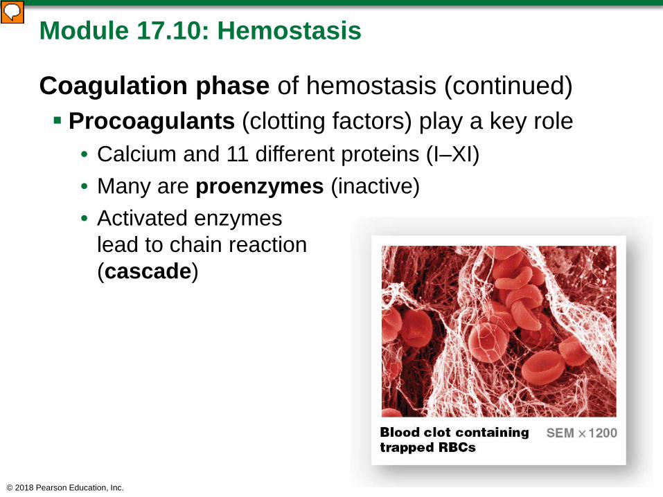
Coagulation phase of hemostasis (continued) Procoagulants (clotting factors) play a key role
• Calcium and 11 different proteins (I–XI)• Many are proenzymes (inactive)• Activated enzymes
lead to chain reaction (cascade)
© 2018 Pearson Education, Inc.
Module 17.10: Hemostasis
Coagulation phase of hemostasis (continued) Two pathways lead to common pathway—extrinsic
and intrinsic1. Extrinsic pathway
– Begins with release of tissue factor (factor III) from damaged endothelial cells or peripheral tissues
– Tissue factor combines with Ca2+ and another clotting factor to activate factor X (first step in common pathway)
© 2018 Pearson Education, Inc.
Module 17.10: Hemostasis
Coagulation phase of hemostasis (continued) Two pathways (continued)
2. Intrinsic pathway– Begins with activation of proenzymes exposed to
collagen fibers at injury site– Pathway proceeds with assistance of PF-3 (factor
released by aggregating platelets)– Sequence of enzyme activations leads to factor X
© 2018 Pearson Education, Inc.
Module 17.10: Hemostasis
Coagulation phase of hemostasis (continued) Common pathway
• Activated factor X activates prothrombin activator, a complex that converts the prothrombin (a proenzyme) to the enzyme thrombin
• Thrombin converts fibrinogen to fibrin – Completes clotting process
Clot retraction• RBCs and platelets stick to the fibrin threads• Platelets contract to form tighter clot and pull edges
together• Continues for 30–60 minutes
© 2018 Pearson Education, Inc.

© 2018 Pearson Education, Inc.

Module 17.10: Hemostasis
Fibrinolysis Process of clot dissolving Begins with activation of:
• Plasminogen by thrombin (from common pathway)• Tissue plasminogen activator, or t-PA, from
damaged tissues Produces plasmin—erodes the clot
© 2018 Pearson Education, Inc.

Module 17.10: Review
A. Define hemostasis.B. Briefly describe the vascular, platelet, and
coagulation phases of hemostasis.C. Describe the events that follow the coagulation
phase.
Learning Outcome: Discuss the mechanisms that control blood loss after an injury, and describe the reaction sequences responsible for blood clotting.
© 2018 Pearson Education, Inc.

Module 17.11: Blood disorders can be classified by their origins and the changes in blood characteristicsObtaining blood for diagnosis Venipuncture (vena,
vein + punctura, a piercing)• Withdrawal of whole blood from superficial vein, such
as median cubital vein
© 2018 Pearson Education, Inc.

Module 17.11: Blood disorders
Obtaining blood for diagnosis (continued) Commonly used because:
1. Easy to locate superficial veins2. Vein walls are thinner than walls of comparable
arteries3. Venous blood
pressure is relatively low, so vein seals quickly
© 2018 Pearson Education, Inc.
Module 17.11: Blood disorders
Nutritional blood disorders Iron deficiency anemia
• Insufficient iron intake (needed to form functional hemoglobin)
• Resulting RBCs are small (microcytic); transport less oxygen
• More common in women—iron reserves half the reserves of a typical man
© 2018 Pearson Education, Inc.
Module 17.11: Blood disorders
Nutritional blood disorders (continued) Pernicious anemia
• Vitamin B12 deficiency prevents normal stem cell divisions
• Fewer RBCs produced; often misshaped, large (macrocytic)
• Can be from lack of intrinsic factor—secreted in stomach; needed for vitamin B12 absorption
Ca2+ and vitamin K deficiencies• Calcium is required for all clotting pathways• Vitamin K is required by liver to synthesize clotting
factors
© 2018 Pearson Education, Inc.

Module 17.11: Blood disorders
Congenital blood disorders Sickle cell disease (SDC)—
group of inherited RBC disorders• Genetic mutation affects amino
acid sequence of the beta globin subunit in hemoglobin
• RBCs take on sickle shape when they release oxygen
– RBCs more fragile, easily damaged
– Can get stuck in smaller vessels, block flow
© 2018 Pearson Education, Inc.

Module 17.11: Blood disorders
Congenital blood disorders (continued) Sickle cell anemia—most severe type of sickle cell
disease • Requires two copies of the sickling gene
– People with one copy have the sickling trait, but not disorder
– Having the trait increases person’s resistance to malariao Infected RBCs sickleo Sickled cells are destroyed by macrophages—
malarial pathogen destroyed with infected cells
© 2018 Pearson Education, Inc.

Module 17.11: Blood disorders
Congenital blood disorders (continued) Hemophilia—Inherited bleeding disorder
• Affects 1 person in 10,000; ~80–90 percent are males• Caused by missing or reduced production of a clotting
factor• Severity of disorder varies• In severe cases, extensive bleeding occurs with minor
contact – Bleeding also occurs at joints and around muscles
© 2018 Pearson Education, Inc.
Module 17.11: Blood disorders
Congenital blood disorders (continued) Thalassemias—diverse group of inherited disorders
• Unable to adequately produce normal Hb protein subunits
• Severity depends on which/how many subunits are abnormal
© 2018 Pearson Education, Inc.

Module 17.11: Blood disorders
Blood infections Caused by pathogens entering the blood through
wound or infection• Bacteremia—bacteria circulate in blood, do not
multiply there• Viremia—viruses circulating in blood, but not
multiplying there
© 2018 Pearson Education, Inc.

Module 17.11: Blood disorders
Blood infections (continued)• Sepsis
– Widespread infection of body tissue
– Septicemiao Sepsis of the blood (“blood
poisoning”)o Pathogens present,
multiplying in blood, and spreading
© 2018 Pearson Education, Inc.
Module 17.11: Blood disorders
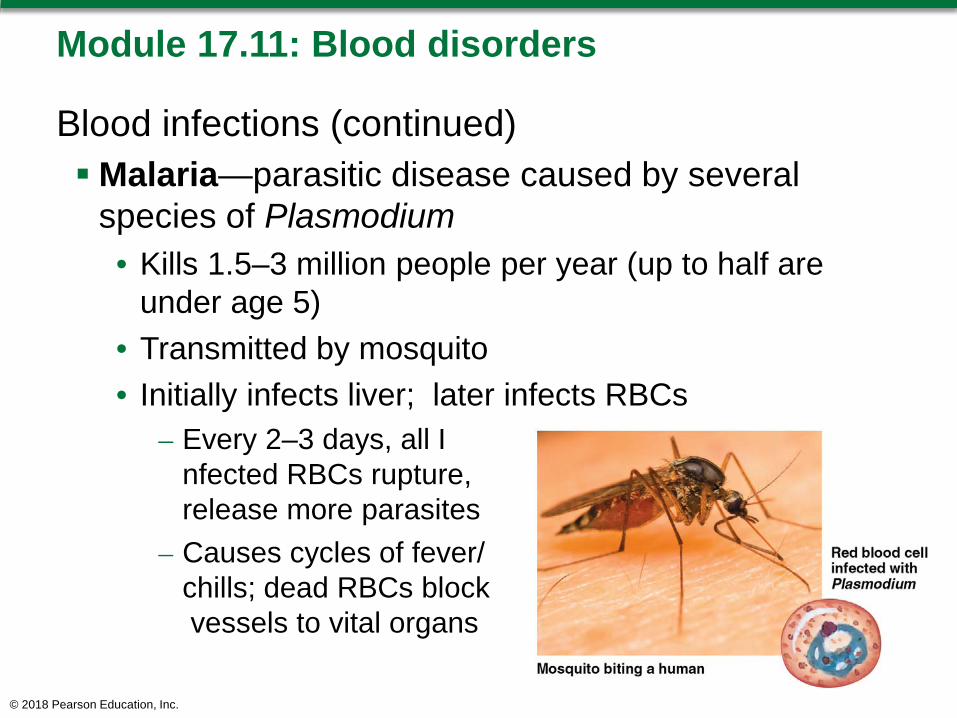
Blood infections (continued)Malaria—parasitic disease caused by several
species of Plasmodium• Kills 1.5–3 million people per year (up to half are
under age 5)• Transmitted by mosquito• Initially infects liver; later infects RBCs
– Every 2–3 days, all Infected RBCs rupture, release more parasites
– Causes cycles of fever/chills; dead RBCs blockvessels to vital organs
© 2018 Pearson Education, Inc.
Module 17.11: Blood disorders
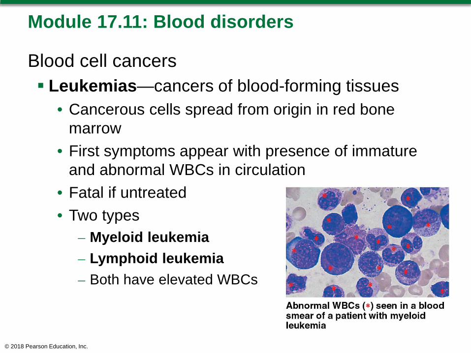
Blood cell cancers Leukemias—cancers of blood-forming tissues
• Cancerous cells spread from origin in red bone marrow
• First symptoms appear with presence of immature and abnormal WBCs in circulation
• Fatal if untreated• Two types
– Myeloid leukemia – Lymphoid leukemia– Both have elevated WBCs
© 2018 Pearson Education, Inc.
Module 17.11: Blood disorders
Degenerative blood disorders Disseminated intravascular coagulation (DIC)
• Bacterial toxins activate steps in coagulation process– Converts too much fibrinogen to fibrin; small clots may
block small vessels and damage nearby tissue– Phagocytes and plasmin work to remove fibrin
• Liver tries to maintain adequate fibrinogen levels; if cannot keep up, uncontrolled bleeding may occur
© 2018 Pearson Education, Inc.
Module 17.11: Review
A. Compare pernicious anemia with iron deficiency anemia.
B. Identify the two types of leukemia.C. Explain why venipuncture is a common clinical
procedure for obtaining blood for examination.
Learning Outcome: Explain how blood disorders are detected, and describe examples of the various categories of blood disorders.
© 2018 Pearson Education, Inc.