biomimetic fabrication of hydroxyapatite microcapsules by using apatite nuclei 13
TRANSCRIPT

Biomimetic Fabrication of Hydroxyapatite Microcapsules by using Apatite Nuclei 273
Biomimetic Fabrication of Hydroxyapatite Microcapsules by using Apatite Nuclei
Takeshi Yao and Takeshi Yabutsuka
X
Biomimetic Fabrication of Hydroxyapatite Microcapsules by using Apatite Nuclei
Takeshi Yao and Takeshi Yabutsuka
Kyoto University (Japan)
1. Introduction
1.1 Hydroxyapatite Living bone consists of 69 wt % of inorganic substances whose main component is hydroxyapatite (Ca10(PO4)6(OH)2), 22 wt% of organic substances whose main component is collagen and 9 wt% of water (Park & Lakes, 1992; Bhat, 2005). It has a skillful woven structure constructed with collagen fiber on which hydroxyapatite nano-crystals are precipitated. Hydroxyapatite is a main inorganic component of living bone and has attracted much attention as a biomaterial with high bioaffinity. It has high affinity to living bone (Jarcho et al., 1977; LeGeros & LeGeros, 1993; LeGeros & LeGeros, 2008; Oonishi et al., 2008) and cells (Deligianni et al., 2001; Rizzi et al., 2001) and an ability to absorb biopolymer such as protein (Tiselius et al., 1956). From these properties, hydroxyapatite is considered as one of the most important biomaterials.
1.2 Bioactivity Generally, most of artificial materials implanted into living body are encapsulated with non-calcified fibrous tissue and isolated from surrounding tissue (Park & Lakes, 1992). This reaction is a normal protective reaction of living body against foreign substances. In early 1970s, Hench et al. discovered that glass in the system Na2O-CaO-SiO2-P2O5, called Bioglass®, spontaneously bonds to living bone without encapsulated with fibrous tissues (Hench et al., 1971; Hench, 1991; Hench & Andersson, 1993). Since the discovery of Bioglass®, ceramic materials such as glass-ceramic Ceravital® containing crystalline hydroxyapatite (Gross et al., 1993), sintered hydroxyapatite (Jarcho et al., 1977; LeGeros & LeGeros, 1993), glass-ceramics Cerabone® A-W containing crystalline hydroxyapatite and wollastonite (CaO·SiO2) (Kokubo et al., 1982; Kokubo, 1990a; Kokubo, 1993a; Kokubo, 2008), glass-ceramic Bioverit® containing crystalline hydroxyapatite and phlogopite ((Na,K)Mg3(AlSiO10)F2) (Höland & Vogel, 1993) and sintered β-tricalcium phosphate (3CaO·P2O5) (Rejda et al., 1977) have been found to bond to living bone. Most of the ceramics mentioned above forms hydroxyapatite layer on their surface and can avoid the protective reaction in living body (Hench, 1991; Höland et al., 1985; Kitsugi et al., 1987; Kitsugi et al., 1989; Kokubo, 1990d; Ohura et al., 1991; Ohtsuki et al., 1991; Neo et al., 1992; Neo et al., 1993). This hydroxyapatite layer consists of minute crystals containing carbonate ions in chemical composition (Kokubo et al., 1990b) and is similar to
13
www.intechopen.com

Biomimetics, Learning from Nature274
hydroxyapatite which composes living bone (Kim et al., 1999; Kim et al., 2000). On the hydroxyapatite layer, osteoblast actively proliferates and differentiates (Neo et al., 1992; Loty et al., 2000). As a result, a living bone is formed on the hydroxyapatite layer and the materials spontaneously bond to the surrounding living bone thorough the layer. This special property of the materials is termed bioactivity among researchers of ceramic-based biomaterials.
1.3 Simulated body fluid Kokubo et al. invented a simulated body fluid (SBF) with ion concentrations nearly equal to those of human blood plasma (Kokubo et al., 1990c; Kokubo & Takadama, 2006; Takadama & Kokubo, 2008). It became possible to imitate the reaction of hydroxyapatite formation in living body by using SBF. Kokubo, Yao and Tanahashi applied the biomimetic reaction in SBF and formed hydroxyapatite thin film on the surface of various kinds of substrates (Tanahashi et al., 1992; Kokubo et al., 1993b; Tanahashi et al., 1994a; Tanahashi et al., 1994b, Tanahashi et al., 1994c; Tanahashi et al., 1995a; Tanahashi et al., 1995b).
1.4 Apatite Nuclei When the pH or temperature of SBF is raised, fine particles of calcium phosphate are precipitated from the fluid. Yao discovered that the fine particles show high activity for forming hydroxyapatite in SBF and he named the particles Apatite Nuclei (Yao et al., 2006). The function of Apatite Nuclei is very attractive for development of various kinds of biomaterials and environmental materials in micron or nano scale. Applying the function of Apatite Nuclei, the authors fabricated bioactive polyethylene (PE)-Apatite Nuclei composite (Yabutsuka et al., 2007) and titanium (Ti)-Apatite Nuclei composite (Yabutsuka et al., 2008a). They soaked porous PE or Ti plate formed many micropores by sulfuric acid treatment in SBF and precipitated Apatite Nuclei in the pores by raising pH or tremperature of SBF. By soaking in SBF, Apatite Nuclei precipitated in the pores induce hydroxyapatite and the composites show high bioactivity. Also, formed hydroxyapatite showed high adhesive strength to the composite by a mechanical interlocking effect. The authors also fabricated hydroxyapatite micropattern by using Apatite Nuclei (Yao, 2000; Yamaguchi et al., 2007). Resist pattern was developed on a cathode for electrophoretic deposition and a polytetrafluoroethylene (PTFE) film was set on the cathode. Then electrophoretic deposition was performed with a suspension of Apatite Nuclei in ethanol. In this process, Apatite Nuclei were deposited on a porous PTFE film so as to transcribe the resist pattern. The substrate was soaked in SBF and hydroxyapatite was selectively induced on Apatite Nuclei. As a result, apatite pattern whose resolution was as high as the resist pattern was fabricated.
1.5 Fabrication of hydroxyapatite microcapsule by biomimetic method In living body, hydroxyapatite is not recognized as a foreign material and can avoid protective reaction of living body because hydroxyapatite induces bonelike hydroxyapatite from body fluid and forms its layer in living body. Therefore, microcapsules possessing high bioaffinity can be formed by using hydroxyapatite and the hydroxyapatite microcapsules are thought to be useful to drug delivery systems.
Yao et al. proposed that hydroxyapatite microcapsules can be fabricated by using biomimetic method (Adachi, Takeuchi, Ozawa & Yao, 2002). For the first process, Apatite Nuclei are attached to the surfaces of microspheres. For the second process, the microspheres are soaked in SBF. By this treatment, hydroxyapatite is induced from the Apatite Nuclei and grows over the whole surface area of the microspheres. As a result, hydroxyapatite is coated on the whole surface of the microspheres and hydroxyapatite microcapsules can be obtained. By this method, it is expected to encapsulate various kinds of microspheres with hydroxyapatite.
2. Fabrication of hollow hydroxyapatite microcapsule
Hollow microcapsule is expected to have many applications to the chemotherapy because it can be filled with various medical agents. In this chapter, we fabricated hollow hydroxyapatite microcapsules by using biomimetic method (Tabe et al., 2007). First, Apatite Nuclei were attached to the surfaces of polylactic acid (PLA) microspheres used as molds of hollow microcapsules. When these PLA microspheres were soaked in SBF, hydroxyapatite was induced from Apatite Nuclei on the PLA microspheres and covered the whole surface of the PLA microspheres. As a result, encapsulated PLA microspheres with hydroxyapatite were fabricated. Finally, the PLA was dissolved out in acetone and hollow hydroxyapatite microcapsules were fabricated.
2.1 Materials & Methods
2.1.1 Preparation of SBF SBF was prepared by dissolving reagent-grade NaCl, NaHCO3, KCl, K2HPO4·3H2O, MgCl2·6H2O, CaCl2 and Na2SO4 in ultrapure water with the composition as shown in Table 1 and buffered at pH 7.40 with tris(hydroxymethyl)aminomethane ((CH2OH)3CNH2) and hydrochloric acid at 36.5 °C (Kokubo & Takadama, 2006).
Ion concentration / mmol·dm-3 SBF Blood plasma Na+ 142.0 142.0 K+ 5.0 5.0 Ca2+ 2.5 2.5 Mg2+ 1.5 1.5 Cl– 147.8 103.0 HCO3– 4.2 27.0 HPO42– 1.0 1.0 SO42– 0.5 0.5
Table 1. Ion concentrations of simulated body fluid (SBF) and human blood plasma.
2.1.2 Precipitation of Apatite Nuclei The pH of SBF was raised to pH 8.50 by dissolving (CH2OH)3CNH2 at 25.0 °C, and precipitated Apatite Nuclei in the SBF, which were collected by filtration using a 50 nm polytetrafluoroethylene (PTFE) membrane filter (Millipore, USA) and washed with distilled
www.intechopen.com

Biomimetic Fabrication of Hydroxyapatite Microcapsules by using Apatite Nuclei 275
hydroxyapatite which composes living bone (Kim et al., 1999; Kim et al., 2000). On the hydroxyapatite layer, osteoblast actively proliferates and differentiates (Neo et al., 1992; Loty et al., 2000). As a result, a living bone is formed on the hydroxyapatite layer and the materials spontaneously bond to the surrounding living bone thorough the layer. This special property of the materials is termed bioactivity among researchers of ceramic-based biomaterials.
1.3 Simulated body fluid Kokubo et al. invented a simulated body fluid (SBF) with ion concentrations nearly equal to those of human blood plasma (Kokubo et al., 1990c; Kokubo & Takadama, 2006; Takadama & Kokubo, 2008). It became possible to imitate the reaction of hydroxyapatite formation in living body by using SBF. Kokubo, Yao and Tanahashi applied the biomimetic reaction in SBF and formed hydroxyapatite thin film on the surface of various kinds of substrates (Tanahashi et al., 1992; Kokubo et al., 1993b; Tanahashi et al., 1994a; Tanahashi et al., 1994b, Tanahashi et al., 1994c; Tanahashi et al., 1995a; Tanahashi et al., 1995b).
1.4 Apatite Nuclei When the pH or temperature of SBF is raised, fine particles of calcium phosphate are precipitated from the fluid. Yao discovered that the fine particles show high activity for forming hydroxyapatite in SBF and he named the particles Apatite Nuclei (Yao et al., 2006). The function of Apatite Nuclei is very attractive for development of various kinds of biomaterials and environmental materials in micron or nano scale. Applying the function of Apatite Nuclei, the authors fabricated bioactive polyethylene (PE)-Apatite Nuclei composite (Yabutsuka et al., 2007) and titanium (Ti)-Apatite Nuclei composite (Yabutsuka et al., 2008a). They soaked porous PE or Ti plate formed many micropores by sulfuric acid treatment in SBF and precipitated Apatite Nuclei in the pores by raising pH or tremperature of SBF. By soaking in SBF, Apatite Nuclei precipitated in the pores induce hydroxyapatite and the composites show high bioactivity. Also, formed hydroxyapatite showed high adhesive strength to the composite by a mechanical interlocking effect. The authors also fabricated hydroxyapatite micropattern by using Apatite Nuclei (Yao, 2000; Yamaguchi et al., 2007). Resist pattern was developed on a cathode for electrophoretic deposition and a polytetrafluoroethylene (PTFE) film was set on the cathode. Then electrophoretic deposition was performed with a suspension of Apatite Nuclei in ethanol. In this process, Apatite Nuclei were deposited on a porous PTFE film so as to transcribe the resist pattern. The substrate was soaked in SBF and hydroxyapatite was selectively induced on Apatite Nuclei. As a result, apatite pattern whose resolution was as high as the resist pattern was fabricated.
1.5 Fabrication of hydroxyapatite microcapsule by biomimetic method In living body, hydroxyapatite is not recognized as a foreign material and can avoid protective reaction of living body because hydroxyapatite induces bonelike hydroxyapatite from body fluid and forms its layer in living body. Therefore, microcapsules possessing high bioaffinity can be formed by using hydroxyapatite and the hydroxyapatite microcapsules are thought to be useful to drug delivery systems.
Yao et al. proposed that hydroxyapatite microcapsules can be fabricated by using biomimetic method (Adachi, Takeuchi, Ozawa & Yao, 2002). For the first process, Apatite Nuclei are attached to the surfaces of microspheres. For the second process, the microspheres are soaked in SBF. By this treatment, hydroxyapatite is induced from the Apatite Nuclei and grows over the whole surface area of the microspheres. As a result, hydroxyapatite is coated on the whole surface of the microspheres and hydroxyapatite microcapsules can be obtained. By this method, it is expected to encapsulate various kinds of microspheres with hydroxyapatite.
2. Fabrication of hollow hydroxyapatite microcapsule
Hollow microcapsule is expected to have many applications to the chemotherapy because it can be filled with various medical agents. In this chapter, we fabricated hollow hydroxyapatite microcapsules by using biomimetic method (Tabe et al., 2007). First, Apatite Nuclei were attached to the surfaces of polylactic acid (PLA) microspheres used as molds of hollow microcapsules. When these PLA microspheres were soaked in SBF, hydroxyapatite was induced from Apatite Nuclei on the PLA microspheres and covered the whole surface of the PLA microspheres. As a result, encapsulated PLA microspheres with hydroxyapatite were fabricated. Finally, the PLA was dissolved out in acetone and hollow hydroxyapatite microcapsules were fabricated.
2.1 Materials & Methods
2.1.1 Preparation of SBF SBF was prepared by dissolving reagent-grade NaCl, NaHCO3, KCl, K2HPO4·3H2O, MgCl2·6H2O, CaCl2 and Na2SO4 in ultrapure water with the composition as shown in Table 1 and buffered at pH 7.40 with tris(hydroxymethyl)aminomethane ((CH2OH)3CNH2) and hydrochloric acid at 36.5 °C (Kokubo & Takadama, 2006).
Ion concentration / mmol·dm-3 SBF Blood plasma Na+ 142.0 142.0 K+ 5.0 5.0 Ca2+ 2.5 2.5 Mg2+ 1.5 1.5 Cl– 147.8 103.0 HCO3– 4.2 27.0 HPO42– 1.0 1.0 SO42– 0.5 0.5
Table 1. Ion concentrations of simulated body fluid (SBF) and human blood plasma.
2.1.2 Precipitation of Apatite Nuclei The pH of SBF was raised to pH 8.50 by dissolving (CH2OH)3CNH2 at 25.0 °C, and precipitated Apatite Nuclei in the SBF, which were collected by filtration using a 50 nm polytetrafluoroethylene (PTFE) membrane filter (Millipore, USA) and washed with distilled
www.intechopen.com

Biomimetics, Learning from Nature276
water, were dispersed in 200 ml of ethanol with ultrasonic vibration, and Apatite Nuclei-dispersed ethanol was obtained.
2.1.3 Fabrication of encapsulated PLA microspheres with hydroxyapatite The ethanol contained in the Apatite Nuclei-dispersed ethanol was replaced with ultrapure water by an evaporator for the purpose of prevention of elution of PLA microsphere. By this treatment, Apatite Nuclei-dispersed water was obtained. 0.2 mg of commercially obtained PLA microspheres with 2 µm of average diameter (Corefront, Japan) were soaked in the Apatite Nuclei-dispersed water mentioned above and held for 1 d. The PLA microspheres were collected by filtration using a 100 nm PTFE membrane filter. These PLA microspheres were soaked in SBF at pH 7.40 at 36.5 °C for 7 d. After that, the PLA microspheres were collected by filtration, washed with ultrapure water, and dried at 36.5 °C. The surfaces of the PLA microspheres were analyzed by scanning electron microscopy (SEM: ESEM-2700, Nikon, Japan) and energy dispersive X-ray analysis (EDX: DX-4, EDAX International, USA). For the reference, the PLA microspheres not soaked in Apatite Nuclei suspension were also soaked in 1.0 SBF. The surfaces of these PLA microspheres were also analyzed by SEM and EDX.
2.1.3 Fabrication of hollow hydroxyapatite microcapsules The encapsulated PLA microspheres with hydroxyapatite were soaked in acetone for 1 d. The samples thus obtained were analyzed by SEM. For the reference, not-treated PLA microspheres were also analyzed by SEM and EDX.
2.2 Results & Discussion
2.2.1 Observation of the encapsulated PLA microspheres Fig. 1 shows (a) SEM micrograph and (b) the result of EDX analysis of not-treated PLA microsphere after the soak in SBF for 7 d. In Fig. 1(a), it was observed that the not-treated PLA microsphere have smooth surface, maybe due to the production process, and no evidence of hydroxyapatite were detected. In Fig. 1(b), no peaks of P and Ca were detected. Fig. 2 shows (a) SEM micrograph and (b) the result of EDX analysis of PLA microspheres soaked in the Apatite Nuclei-dispersed water for 1 d, and then soaked in SBF for 7 d. In Fig. 2(a), it was observed that needle like crystals characteristic to hydroxyapatite coated whole surface of the PLA microsphere. In Fig. 2(b), peaks of P and Ca, constituents of hydroxyapatite, were detected on the surface. These results indicate that hydroxyapatite was induced from the Apatite Nuclei attached to the surface of the PLA microsphere and spread over whole surface area of the PLA microsphere in SBF.
0 1 2 3 4 5
AuO
F
C
Inte
nsi
ty
Energy / keV (a) (b)
Fig. 1. (a) SEM micrograph and (b) the result of EDX analysis of the not-treated PLA microsphere after the soak in SBF for 7 d.
0 1 2 3 4 5
Ca
AuCa
PO
F
C
Inte
nsi
ty
Energy / keV (a) (b)
Fig. 2. (a) SEM micrograph and (b) the result of EDX analysis of PLA microsphere soaked in the Apatite Nuclei-dispersed ethanol for 1 d, and then soaked in SBF for 7 d.
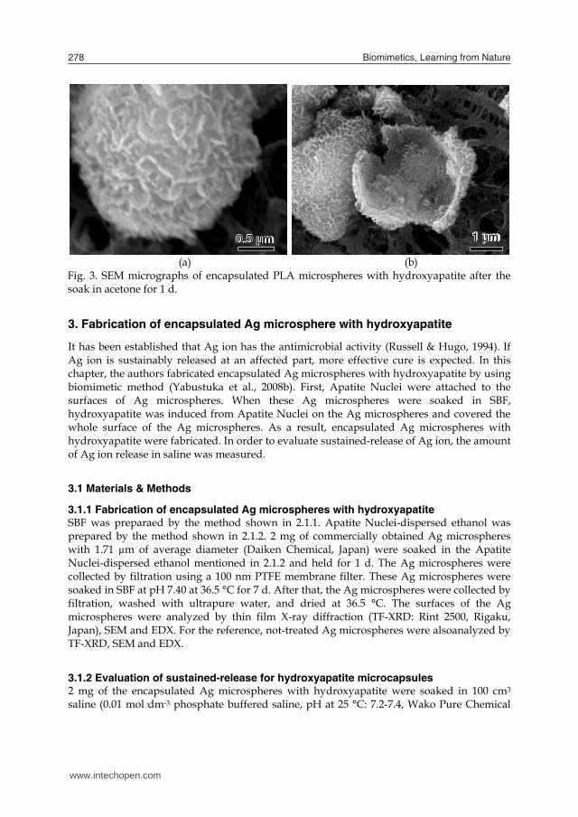
2.2.2 Observation of hollow hydroxyapatite microcapsules Fig. 3(a) and (b) show SEM micrographs of the above mentioned encapsulated PLA microspheres with hydroxyapatite after the soak in acetone for 1 d. By the soak in acetone, the PLA microsphere was dissolved out and a spherical hollow hydroxyapatite microcapsule was obtained. In Fig. 3(a), the spherical microcapsule constructed with hydroxyapatite was observed. In Fig. 3(b), a broken spherical microcapsule of hydroxyapatite was also observed. Fig. 3(b) is shown that PLA microspheres were completely dissolved by acetone and this result confirmed that the microsphere shown in Fig. 3(a) have a hollow structure. Consequently, the microcapsule constructed of hydroxyapatite was fabricated.
2 µm 2 µm 2 µm
1 µm 1 µm 1 µm 1 µm 1 µm
www.intechopen.com

Biomimetic Fabrication of Hydroxyapatite Microcapsules by using Apatite Nuclei 277
water, were dispersed in 200 ml of ethanol with ultrasonic vibration, and Apatite Nuclei-dispersed ethanol was obtained.
2.1.3 Fabrication of encapsulated PLA microspheres with hydroxyapatite The ethanol contained in the Apatite Nuclei-dispersed ethanol was replaced with ultrapure water by an evaporator for the purpose of prevention of elution of PLA microsphere. By this treatment, Apatite Nuclei-dispersed water was obtained. 0.2 mg of commercially obtained PLA microspheres with 2 µm of average diameter (Corefront, Japan) were soaked in the Apatite Nuclei-dispersed water mentioned above and held for 1 d. The PLA microspheres were collected by filtration using a 100 nm PTFE membrane filter. These PLA microspheres were soaked in SBF at pH 7.40 at 36.5 °C for 7 d. After that, the PLA microspheres were collected by filtration, washed with ultrapure water, and dried at 36.5 °C. The surfaces of the PLA microspheres were analyzed by scanning electron microscopy (SEM: ESEM-2700, Nikon, Japan) and energy dispersive X-ray analysis (EDX: DX-4, EDAX International, USA). For the reference, the PLA microspheres not soaked in Apatite Nuclei suspension were also soaked in 1.0 SBF. The surfaces of these PLA microspheres were also analyzed by SEM and EDX.
2.1.3 Fabrication of hollow hydroxyapatite microcapsules The encapsulated PLA microspheres with hydroxyapatite were soaked in acetone for 1 d. The samples thus obtained were analyzed by SEM. For the reference, not-treated PLA microspheres were also analyzed by SEM and EDX.
2.2 Results & Discussion
2.2.1 Observation of the encapsulated PLA microspheres Fig. 1 shows (a) SEM micrograph and (b) the result of EDX analysis of not-treated PLA microsphere after the soak in SBF for 7 d. In Fig. 1(a), it was observed that the not-treated PLA microsphere have smooth surface, maybe due to the production process, and no evidence of hydroxyapatite were detected. In Fig. 1(b), no peaks of P and Ca were detected. Fig. 2 shows (a) SEM micrograph and (b) the result of EDX analysis of PLA microspheres soaked in the Apatite Nuclei-dispersed water for 1 d, and then soaked in SBF for 7 d. In Fig. 2(a), it was observed that needle like crystals characteristic to hydroxyapatite coated whole surface of the PLA microsphere. In Fig. 2(b), peaks of P and Ca, constituents of hydroxyapatite, were detected on the surface. These results indicate that hydroxyapatite was induced from the Apatite Nuclei attached to the surface of the PLA microsphere and spread over whole surface area of the PLA microsphere in SBF.
0 1 2 3 4 5
AuO
F
C
Inte
nsi
ty
Energy / keV (a) (b)
Fig. 1. (a) SEM micrograph and (b) the result of EDX analysis of the not-treated PLA microsphere after the soak in SBF for 7 d.
0 1 2 3 4 5
Ca
AuCa
PO
F
C
Inte
nsi
ty
Energy / keV (a) (b)
Fig. 2. (a) SEM micrograph and (b) the result of EDX analysis of PLA microsphere soaked in the Apatite Nuclei-dispersed ethanol for 1 d, and then soaked in SBF for 7 d.
2.2.2 Observation of hollow hydroxyapatite microcapsules Fig. 3(a) and (b) show SEM micrographs of the above mentioned encapsulated PLA microspheres with hydroxyapatite after the soak in acetone for 1 d. By the soak in acetone, the PLA microsphere was dissolved out and a spherical hollow hydroxyapatite microcapsule was obtained. In Fig. 3(a), the spherical microcapsule constructed with hydroxyapatite was observed. In Fig. 3(b), a broken spherical microcapsule of hydroxyapatite was also observed. Fig. 3(b) is shown that PLA microspheres were completely dissolved by acetone and this result confirmed that the microsphere shown in Fig. 3(a) have a hollow structure. Consequently, the microcapsule constructed of hydroxyapatite was fabricated.
2 µm 2 µm 2 µm
1 µm 1 µm 1 µm 1 µm 1 µm
www.intechopen.com
Biomimetics, Learning from Nature278
(a) (b)
Fig. 3. SEM micrographs of encapsulated PLA microspheres with hydroxyapatite after the soak in acetone for 1 d.
3. Fabrication of encapsulated Ag microsphere with hydroxyapatite
It has been established that Ag ion has the antimicrobial activity (Russell & Hugo, 1994). If Ag ion is sustainably released at an affected part, more effective cure is expected. In this chapter, the authors fabricated encapsulated Ag microspheres with hydroxyapatite by using biomimetic method (Yabustuka et al., 2008b). First, Apatite Nuclei were attached to the surfaces of Ag microspheres. When these Ag microspheres were soaked in SBF, hydroxyapatite was induced from Apatite Nuclei on the Ag microspheres and covered the whole surface of the Ag microspheres. As a result, encapsulated Ag microspheres with hydroxyapatite were fabricated. In order to evaluate sustained-release of Ag ion, the amount of Ag ion release in saline was measured.
3.1 Materials & Methods
3.1.1 Fabrication of encapsulated Ag microspheres with hydroxyapatite SBF was preparaed by the method shown in 2.1.1. Apatite Nuclei-dispersed ethanol was prepared by the method shown in 2.1.2. 2 mg of commercially obtained Ag microspheres with 1.71 µm of average diameter (Daiken Chemical, Japan) were soaked in the Apatite Nuclei-dispersed ethanol mentioned in 2.1.2 and held for 1 d. The Ag microspheres were collected by filtration using a 100 nm PTFE membrane filter. These Ag microspheres were soaked in SBF at pH 7.40 at 36.5 °C for 7 d. After that, the Ag microspheres were collected by filtration, washed with ultrapure water, and dried at 36.5 °C. The surfaces of the Ag microspheres were analyzed by thin film X-ray diffraction (TF-XRD: Rint 2500, Rigaku, Japan), SEM and EDX. For the reference, not-treated Ag microspheres were alsoanalyzed by TF-XRD, SEM and EDX.
3.1.2 Evaluation of sustained-release for hydroxyapatite microcapsules 2 mg of the encapsulated Ag microspheres with hydroxyapatite were soaked in 100 cm3 saline (0.01 mol·dm-3 phosphate buffered saline, pH at 25 °C: 7.2-7.4, Wako Pure Chemical
0.5 µm 0.5 µm 0.5 µm 0.5 µm 0.5 µm 1 µm 1 µm 1 µm 1 µm 1 µm
Industries, Japan). The saline was continued to shake by using shaking apparatus for up to 192 h in an incubator held at 36.5 °C. Changes in Ag ion concentration in saline were measured by inductively coupled plasma atomic emission spectroscopy (ICP: ICPS-7500, Shimadzu, Japan). For the reference, not-treated Ag microspheres were also dispersed in saline and conducted the same measurement.
3.2 Results & Discussion
3.2.1 TF-XRD measurement Fig.4 shows (a) TF-XRD profile of the not-treated Ag microspheres and (b) that of the Ag microspheres soaked in the Apatite Nuclei-dispersed ethanol for 1 d, and then soaked in SBF for 7 d. After the soak in SBF for 7 d, diffraction peaks of hydroxyapatite were detected. This result indicates that hydroxyapatite was induced from the Apatite Nuclei attached to the surfaces of Ag microspheres.
25 30 35 40 45 50
Ag
(b)
...
(a)
Inte
nsi
ty
2 (CuK) / o
Hydroxyapatite.
Fig. 4. TF-XRD profiles of the surface of (a) not-treated Ag microspheres and (b) Ag microspheres soaked in the Apatite Nuclei-dispersed ethanol for 1 d, and then soaked in SBF for 7 d.
3.2.2 Observation by SEM and EDX Fig. 5 shows (a) SEM micrograph and (b) the result of EDX analysis of not-treated Ag microsphere. In Fig. 5(a), it was observed that the not-treated Ag microsphere have
www.intechopen.com

Biomimetic Fabrication of Hydroxyapatite Microcapsules by using Apatite Nuclei 279
(a) (b)
Fig. 3. SEM micrographs of encapsulated PLA microspheres with hydroxyapatite after the soak in acetone for 1 d.
3. Fabrication of encapsulated Ag microsphere with hydroxyapatite
It has been established that Ag ion has the antimicrobial activity (Russell & Hugo, 1994). If Ag ion is sustainably released at an affected part, more effective cure is expected. In this chapter, the authors fabricated encapsulated Ag microspheres with hydroxyapatite by using biomimetic method (Yabustuka et al., 2008b). First, Apatite Nuclei were attached to the surfaces of Ag microspheres. When these Ag microspheres were soaked in SBF, hydroxyapatite was induced from Apatite Nuclei on the Ag microspheres and covered the whole surface of the Ag microspheres. As a result, encapsulated Ag microspheres with hydroxyapatite were fabricated. In order to evaluate sustained-release of Ag ion, the amount of Ag ion release in saline was measured.
3.1 Materials & Methods
3.1.1 Fabrication of encapsulated Ag microspheres with hydroxyapatite SBF was preparaed by the method shown in 2.1.1. Apatite Nuclei-dispersed ethanol was prepared by the method shown in 2.1.2. 2 mg of commercially obtained Ag microspheres with 1.71 µm of average diameter (Daiken Chemical, Japan) were soaked in the Apatite Nuclei-dispersed ethanol mentioned in 2.1.2 and held for 1 d. The Ag microspheres were collected by filtration using a 100 nm PTFE membrane filter. These Ag microspheres were soaked in SBF at pH 7.40 at 36.5 °C for 7 d. After that, the Ag microspheres were collected by filtration, washed with ultrapure water, and dried at 36.5 °C. The surfaces of the Ag microspheres were analyzed by thin film X-ray diffraction (TF-XRD: Rint 2500, Rigaku, Japan), SEM and EDX. For the reference, not-treated Ag microspheres were alsoanalyzed by TF-XRD, SEM and EDX.
3.1.2 Evaluation of sustained-release for hydroxyapatite microcapsules 2 mg of the encapsulated Ag microspheres with hydroxyapatite were soaked in 100 cm3 saline (0.01 mol·dm-3 phosphate buffered saline, pH at 25 °C: 7.2-7.4, Wako Pure Chemical
0.5 µm 0.5 µm 0.5 µm 0.5 µm 0.5 µm 1 µm 1 µm 1 µm 1 µm 1 µm
Industries, Japan). The saline was continued to shake by using shaking apparatus for up to 192 h in an incubator held at 36.5 °C. Changes in Ag ion concentration in saline were measured by inductively coupled plasma atomic emission spectroscopy (ICP: ICPS-7500, Shimadzu, Japan). For the reference, not-treated Ag microspheres were also dispersed in saline and conducted the same measurement.
3.2 Results & Discussion
3.2.1 TF-XRD measurement Fig.4 shows (a) TF-XRD profile of the not-treated Ag microspheres and (b) that of the Ag microspheres soaked in the Apatite Nuclei-dispersed ethanol for 1 d, and then soaked in SBF for 7 d. After the soak in SBF for 7 d, diffraction peaks of hydroxyapatite were detected. This result indicates that hydroxyapatite was induced from the Apatite Nuclei attached to the surfaces of Ag microspheres.
25 30 35 40 45 50
Ag
(b)
...
(a)
Inte
nsi
ty
2 (CuK) / o
Hydroxyapatite.
Fig. 4. TF-XRD profiles of the surface of (a) not-treated Ag microspheres and (b) Ag microspheres soaked in the Apatite Nuclei-dispersed ethanol for 1 d, and then soaked in SBF for 7 d.
3.2.2 Observation by SEM and EDX Fig. 5 shows (a) SEM micrograph and (b) the result of EDX analysis of not-treated Ag microsphere. In Fig. 5(a), it was observed that the not-treated Ag microsphere have
www.intechopen.com

Biomimetics, Learning from Nature280
characteristic wrinkle surface, maybe due to the production process. In Fig. 5(b), no peak other than Ag, except C due to a carbon tape, was detected. Fig.6 shows SEM micrograph of the Ag microspheres soaked in the Apatite Nuclei-dispersed ethanol for 1 d, and then soaked in SBF for 7 d of low magnification. In Fig. 6, many encapsulated Ag microspheres with hydroxyapatite were observed. This result indicates that this method has high reproducibility. Fig. 7 shows (a) picture of a microcapsule by magnification and (b) the result of EDX analysis of the Ag microspheres soaked in the Apatite Nuclei-dispersed ethanol for 1 d, and then soaked in SBF for 7 d. In Fig. 7(a), it was observed that needle like crystals characteristic to hydroxyapatite coated whole surface of the Ag microsphere. In Fig. 7(b), peaks of P and Ca, constituents of hydroxyapatite, were detected on the surface. These results indicate that hydroxyapatite was induced from the Apatite Nuclei attached to the surface of the Ag microsphere and spread over whole surface area of the Ag microsphere in SBF.
0 1 2 3 4 5 6
C
AgIn
ten
sity
Energy / keV
Ag
(a) (b)
Fig. 5. (a) SEM micrograph and (b) the result of EDX analysis of the not-treated Ag microsphere.
Fig. 6. SEM micrograph of Ag microspheres soaked in the Apatite Nuclei-dispersed ethanol for 1 d, and then soaked in SBF for 7 d of low magnification.
1 µm 1 µm 1 µm 1 µm 1 µm
10 µm 10 µm 10 µm 10 µm 10 µm
0 1 2 3 4 5
Ca
CaP
O
C
Inte
nsi
ty
Energy / keV (a) (b)
Fig. 7. (a) SEM micrograph and (b) the result of EDX analysis of Ag microsphere soaked in the Apatite Nuclei-dispersed ethanol for 1 d, and then soaked in SBF for 7 d.
4.2.3 Amount of Ag ion release Fig.8 shows (a) the amount of Ag ion release for the not-treated Ag microspheres and (b) that for the above mentioned encapsulated Ag microspheres with hydroxyapatite in saline up to 192 h at 36.5 °C. The concentration of Ag ion for the encapsulated Ag microspheres with hydroxyapatite was approximately one over ten of that for not-treated ones. This result indicates that sustained-release of Ag ion is achieved by encapsulating Ag microsphere with hydroxyapatite.
Fig. 8. Amount of Ag ion release for (a) not-treated Ag microspheres and (b) encapsulated Ag microspheres with hydroxyapatite in saline up to 192 h at 36.5 oC.
4. Fabrication of encapsulated silicagel microsphere with hydroxyapatite
Silicagel has porous structure. If medical agents are absorbed in silicagel microspheres and the silicagel microspheres are coated with hydroxyapatite, the encapsulated silicagel microspheres with hydroxyapatite are expected as carriers of drug delivery system. When
1 µm 1 µm 1 µm 1 µm 1 µm
www.intechopen.com

Biomimetic Fabrication of Hydroxyapatite Microcapsules by using Apatite Nuclei 281
characteristic wrinkle surface, maybe due to the production process. In Fig. 5(b), no peak other than Ag, except C due to a carbon tape, was detected. Fig.6 shows SEM micrograph of the Ag microspheres soaked in the Apatite Nuclei-dispersed ethanol for 1 d, and then soaked in SBF for 7 d of low magnification. In Fig. 6, many encapsulated Ag microspheres with hydroxyapatite were observed. This result indicates that this method has high reproducibility. Fig. 7 shows (a) picture of a microcapsule by magnification and (b) the result of EDX analysis of the Ag microspheres soaked in the Apatite Nuclei-dispersed ethanol for 1 d, and then soaked in SBF for 7 d. In Fig. 7(a), it was observed that needle like crystals characteristic to hydroxyapatite coated whole surface of the Ag microsphere. In Fig. 7(b), peaks of P and Ca, constituents of hydroxyapatite, were detected on the surface. These results indicate that hydroxyapatite was induced from the Apatite Nuclei attached to the surface of the Ag microsphere and spread over whole surface area of the Ag microsphere in SBF.
0 1 2 3 4 5 6
C
Ag
Inte
nsi
ty
Energy / keV
Ag
(a) (b)
Fig. 5. (a) SEM micrograph and (b) the result of EDX analysis of the not-treated Ag microsphere.
Fig. 6. SEM micrograph of Ag microspheres soaked in the Apatite Nuclei-dispersed ethanol for 1 d, and then soaked in SBF for 7 d of low magnification.
1 µm 1 µm 1 µm 1 µm 1 µm
10 µm 10 µm 10 µm 10 µm 10 µm
0 1 2 3 4 5
Ca
CaP
O
C
Inte
nsi
ty
Energy / keV (a) (b)
Fig. 7. (a) SEM micrograph and (b) the result of EDX analysis of Ag microsphere soaked in the Apatite Nuclei-dispersed ethanol for 1 d, and then soaked in SBF for 7 d.
4.2.3 Amount of Ag ion release Fig.8 shows (a) the amount of Ag ion release for the not-treated Ag microspheres and (b) that for the above mentioned encapsulated Ag microspheres with hydroxyapatite in saline up to 192 h at 36.5 °C. The concentration of Ag ion for the encapsulated Ag microspheres with hydroxyapatite was approximately one over ten of that for not-treated ones. This result indicates that sustained-release of Ag ion is achieved by encapsulating Ag microsphere with hydroxyapatite.
Fig. 8. Amount of Ag ion release for (a) not-treated Ag microspheres and (b) encapsulated Ag microspheres with hydroxyapatite in saline up to 192 h at 36.5 oC.
4. Fabrication of encapsulated silicagel microsphere with hydroxyapatite
Silicagel has porous structure. If medical agents are absorbed in silicagel microspheres and the silicagel microspheres are coated with hydroxyapatite, the encapsulated silicagel microspheres with hydroxyapatite are expected as carriers of drug delivery system. When
1 µm 1 µm 1 µm 1 µm 1 µm
www.intechopen.com

Biomimetics, Learning from Nature282
porous material is soaked in SBF and the pH is raised, Apatite Nuclei precipitated in the pores. Thus treated porous material has high bioactivity because precipitated Apatite Nuclei in the pores induce hydroxyapatite (Yao et al., 2007). In this chapter, the authors fabricated encapsulated silicagel microspheres with hydroxyapatite by using biomimetic method (Yamane et al., 2009). First, Apatite Nuclei were precipitated in the pores of silicagel microspheres. When these silicagel microspheres were soaked in SBF, hydroxyapatite was induced from Apatite Nuclei in the pores of the silicagel microspheres and covered the whole surface of the microspheres. As a result, encapsulated silicagel microspheres with hydroxyapatite were fabricated.
4.1 Materials & Methods SBF was preparaed by the method shown in 2.1.1. Silicagel microspheres (4.4 µm of average diameter, 6 nm of average pore diameter, Fuji Silysia Chemical, Japan) were soaked in SBF. The pH of SBF was raised to pH 8.60 by using (CH2OH)3CNH2 at 25.0 °C. By this treatment, Apatite Nuclei were precipitated in the pores of the silicagel microspheres. The silicagel microspheres were collected by filtration using a 0.1 μm PTFE membrane filter (Millipore, USA), washed with ultrapure water and soaked in SBF at pH 7.40 at 36.5 °C for 7 d. The SBF was renewed every 4 d. After that, the silicagel microspheres were collected by filtration, washed with ultrapure water, and dried at 36.5 °C. The surface of the silicagel microspheres were analyzed by TF-XRD, SEM and EDX.
4.2 Results and Discussion
4.2.1 TF-XRD measurement Fig. 9 shows TF-XRD profiles of the not-treated silicagel microspheres and silicagel microspheres Apatite Nuclei precipitated then soaked in SBF for 7 d. After the soak for 7 d, diffraction peaks of hydroxyapatite were detected. This result indicates that hydroxyapatite was induced from Apatite Nuclei precipitated in the pores of silicagel microspheres.
4.2.2 Observation by SEM and EDX Fig. 10(a) and (b) show SEM micrographs and (c) shows the result of EDX analysis of not-treated silicagel microspheres. Fig. 10(b) is higher magnification. In Fig. 10(c), peaks of Si, constituent of silicagel was detected by the EDX analysis. Fig. 11(a) and (b) show the SEM micrographs and (c) shows the result of EDX analysis of silicagel microspheres Apatite Nuclei precipitated and soaked in SBF for 7 d. In Fig. 11(a), many encapusulated silicagel microspheres with hydroxyapatite were observed. This indicates that this method has high reproducibility. In Fig. 11(b), higher magnification, it was observed that needle like crystals characteristic to hydroxyapatite coated whole surface of the silicagel microsphere. In Fig. 11(c), peaks of P and Ca, constituents of hydroxyapatite, were detected on the surface. From these results, it is considered that hydroxyapatite was induced from the Apatite Nuclei precipitated in the pores of the silicagel microspheres and spread over whole surface area of the silicagel microspheres in SBF.
25 30 35 40 45 50
(a)
..(b)
Inte
nsi
ty
2 (CuK) / o
Hydroxyapatite.
Fig. 9. TF-XRD profiles of the (a) not-treated silicagel microspheres and (b) silicagel microspheres Apatite Nuclei precipitated then soaked in SBF for 7 d.
(a) (b)
0 1 2 3 4 5
Si
O
C
Inte
nsi
ty
Energy / keV (c)
Fig. 10. SEM micrographs of (a) not-treated silicagel microspheres, (b) higher magnification of (a), and (c) result of EDX of (b).
2 μ
10 µm 10 µm 10 µm 10 µm 10 µm 1 µm 1 µm 1 µm 1 µm 1 µm
www.intechopen.com

Biomimetic Fabrication of Hydroxyapatite Microcapsules by using Apatite Nuclei 283
porous material is soaked in SBF and the pH is raised, Apatite Nuclei precipitated in the pores. Thus treated porous material has high bioactivity because precipitated Apatite Nuclei in the pores induce hydroxyapatite (Yao et al., 2007). In this chapter, the authors fabricated encapsulated silicagel microspheres with hydroxyapatite by using biomimetic method (Yamane et al., 2009). First, Apatite Nuclei were precipitated in the pores of silicagel microspheres. When these silicagel microspheres were soaked in SBF, hydroxyapatite was induced from Apatite Nuclei in the pores of the silicagel microspheres and covered the whole surface of the microspheres. As a result, encapsulated silicagel microspheres with hydroxyapatite were fabricated.
4.1 Materials & Methods SBF was preparaed by the method shown in 2.1.1. Silicagel microspheres (4.4 µm of average diameter, 6 nm of average pore diameter, Fuji Silysia Chemical, Japan) were soaked in SBF. The pH of SBF was raised to pH 8.60 by using (CH2OH)3CNH2 at 25.0 °C. By this treatment, Apatite Nuclei were precipitated in the pores of the silicagel microspheres. The silicagel microspheres were collected by filtration using a 0.1 μm PTFE membrane filter (Millipore, USA), washed with ultrapure water and soaked in SBF at pH 7.40 at 36.5 °C for 7 d. The SBF was renewed every 4 d. After that, the silicagel microspheres were collected by filtration, washed with ultrapure water, and dried at 36.5 °C. The surface of the silicagel microspheres were analyzed by TF-XRD, SEM and EDX.
4.2 Results and Discussion
4.2.1 TF-XRD measurement Fig. 9 shows TF-XRD profiles of the not-treated silicagel microspheres and silicagel microspheres Apatite Nuclei precipitated then soaked in SBF for 7 d. After the soak for 7 d, diffraction peaks of hydroxyapatite were detected. This result indicates that hydroxyapatite was induced from Apatite Nuclei precipitated in the pores of silicagel microspheres.
4.2.2 Observation by SEM and EDX Fig. 10(a) and (b) show SEM micrographs and (c) shows the result of EDX analysis of not-treated silicagel microspheres. Fig. 10(b) is higher magnification. In Fig. 10(c), peaks of Si, constituent of silicagel was detected by the EDX analysis. Fig. 11(a) and (b) show the SEM micrographs and (c) shows the result of EDX analysis of silicagel microspheres Apatite Nuclei precipitated and soaked in SBF for 7 d. In Fig. 11(a), many encapusulated silicagel microspheres with hydroxyapatite were observed. This indicates that this method has high reproducibility. In Fig. 11(b), higher magnification, it was observed that needle like crystals characteristic to hydroxyapatite coated whole surface of the silicagel microsphere. In Fig. 11(c), peaks of P and Ca, constituents of hydroxyapatite, were detected on the surface. From these results, it is considered that hydroxyapatite was induced from the Apatite Nuclei precipitated in the pores of the silicagel microspheres and spread over whole surface area of the silicagel microspheres in SBF.
25 30 35 40 45 50
(a)
..(b)
Inte
nsi
ty
2 (CuK) / o
Hydroxyapatite.
Fig. 9. TF-XRD profiles of the (a) not-treated silicagel microspheres and (b) silicagel microspheres Apatite Nuclei precipitated then soaked in SBF for 7 d.
(a) (b)
0 1 2 3 4 5
Si
O
C
Inte
nsi
ty
Energy / keV (c)
Fig. 10. SEM micrographs of (a) not-treated silicagel microspheres, (b) higher magnification of (a), and (c) result of EDX of (b).
2 μ
10 µm 10 µm 10 µm 10 µm 10 µm 1 µm 1 µm 1 µm 1 µm 1 µm
www.intechopen.com

Biomimetics, Learning from Nature284
(a) (b)
0 1 2 3 4 5
Ca
CaPO
C
Inte
nsi
ty
Energy / keV (c)
Fig. 11. SEM micrographs of (a) silicagel microspheres Apatite Nuclei precipitated and soaked in SBF for 7 d, (b) higher magnification, and (c) result of EDX of (b).
5. Conclusion
When the pH or the temperature of SBF is raised, fine particles of calcium phosphate are precipitated in the fluid. It was found that these particles are very active for forming hydroxyapatite from SBF and these particles were named Apatite Nuclei. By the discovery of Apatite Nuclei, it became possible to develop various multifunctional biomaterials possesing high bioaffinity in micron or nano scale by using biomimetic method. The authors have successfully encapsulated Ag, PLA and silicagel microspheres with hydroxyapatite by biomimetic method. For encapsulated Ag and PLA microspheres, Apatite Nuclei were synthesized by raising pH of SBF. Hydroxyapatite was formed from Apatite Nuclei attached on the microspheres by soaking in SBF, and then the encapsulated Ag and PLA microspheres with hydroxyapatite were obtained. For encapsulated silicagel microspheres, silicagel microspheres were soaked in SBF and precipitated Apatite Nuclei in the pores of the microspheres by raising pH of SBF. Hydroxyapatite was formed from Apatite Nuclei precipitated in the pores of the microspheres by soaking in SBF, and then the encapsulated silicagel microspheres with hydroxyapatite were obtained. For the
10 µm 10 µm 10 µm 10 µm 10 µm 1 µm 1 µm 1 µm 1 µm 1 µm
encapsulated Ag microspheres with hydroxyapatite, sustained-release of Ag ion was achieved. Hollow hydroxyapatite microcapsules were obtained by soaking the encapsulated PLA microsphere with hydroxyapatite in acetone. These hydroxyapatite microcapsules mentioned above possessed high bioaffinity. This method is promising for fabrication of carriers for drug delivery systems.
6. References
Adachi, M.; Takeuchi, I.; Ozawa, N. & Yao, T. (2002). Calcium phosphate capsule and method for fabrication thereof. Japan Patent, 2002-277662
Bhat, S. V. (2005). Biomaterials 2nd ed., Alpha Science, ISBN 1-84265-207-9, Harrow Deligianni, D. D.; Katsala, N. D.; Koutsoukos, P. G. & Missirlis, Y. F. (2001). Effect of surface
roughness of hydroxyapatite on human bone marrow cell adhesion, proliferation, differentiation and detachment strength. Biomaterials, 22, 1, (Jan 2001) 87-96, ISSN 0142-9612
Gross, U. M.; Müller-Mai, C. & Voigt, C. (1993). Ceravital® bioactive ceramics, In: An Introduction to Bioceramics, Hench, L. L. & Wilson, J., (Ed.), 105-124, World Scientific, ISBN 981-02-1400-6, Singapore
Hench, L. L.; Splinter, R. J.; Allen, W. C. & Greenlee, T. K. (1971). Bonding mechanisms at the interface of ceramic prosthetic materials. J. Biomed. Mater. Res., 5, 6, (Nov 1971) 117-141, ISSN 0021-9304
Hench, L. L. (1991). Bioceramics: from concept to clinic. J. Am. Ceram. Soc., 74, 7, (Jul 1991) 1487-1510, ISSN 0002-7820
Hench, L. L. & Andersson, Ö. (1993). Bioactive glasses, In: An Introduction to Bioceramics, Hench, L. L. & Wilson, J., (Ed.), 42-62, World Scientific, ISBN 981-02-1400-6, Singapore
Höland, W.; Vogel, W.; Nauman, K. & Gummel, J. (1985) Interface reactions between machinable bioactive glass-ceramics and bone. J. Biomed. Mater. Res., 19, 3, (Mar 1985) 303-312, ISSN 0021-9304
Höland, W. & Vogel, W. (1993). Machineable and phosphate glass-ceramics, In: An Introduction to Bioceramics, Hench, L. L. & Wilson, J., (Ed.), 125-137, World Scientific, ISBN 981-02-1400-6, Singapore
Jarcho, M. J; Kay, J. L.; Gumaer R. H. & Drobeck, H. P. (1977). Tissue, cellular and subcellular events at bone-ceramic hydroxyapatite interface. J. Bioeng., 1, 2, (Jan 1977) 79-92, ISSN 0145-3068
Kim, H. M.; Kishimoto, K.; Miyaji, F.; Kokubo, T.; Yao, T., Suetsugu, Y., Tanaka, J. & Nakamura, T. (1999). Composition and structure of the apatite formed on PET substrates in SBF modified with various ionic activity products. J. Biomed. Mater. Res., 46, 2, (Aug 1999) 228-235, ISSN 0021-9304
Kim, H. M.; Kishimoto, K.; Miyaji, F.; Kokubo, T.; Yao, T.; Suetsugu, Y.; Tanaka, J. & Nakamura, T. (2000). Composition and structure of apatite formed on organic polymer in simulated body fluid with a high content of carbonate ion. J. Mater. Sci.: Mater. Med., 11, 7, (Jul 2000) 421-426, ISSN 0957-4530
www.intechopen.com

Biomimetic Fabrication of Hydroxyapatite Microcapsules by using Apatite Nuclei 285
(a) (b)
0 1 2 3 4 5
Ca
CaPO
C
Inte
nsi
ty
Energy / keV (c)
Fig. 11. SEM micrographs of (a) silicagel microspheres Apatite Nuclei precipitated and soaked in SBF for 7 d, (b) higher magnification, and (c) result of EDX of (b).
5. Conclusion
When the pH or the temperature of SBF is raised, fine particles of calcium phosphate are precipitated in the fluid. It was found that these particles are very active for forming hydroxyapatite from SBF and these particles were named Apatite Nuclei. By the discovery of Apatite Nuclei, it became possible to develop various multifunctional biomaterials possesing high bioaffinity in micron or nano scale by using biomimetic method. The authors have successfully encapsulated Ag, PLA and silicagel microspheres with hydroxyapatite by biomimetic method. For encapsulated Ag and PLA microspheres, Apatite Nuclei were synthesized by raising pH of SBF. Hydroxyapatite was formed from Apatite Nuclei attached on the microspheres by soaking in SBF, and then the encapsulated Ag and PLA microspheres with hydroxyapatite were obtained. For encapsulated silicagel microspheres, silicagel microspheres were soaked in SBF and precipitated Apatite Nuclei in the pores of the microspheres by raising pH of SBF. Hydroxyapatite was formed from Apatite Nuclei precipitated in the pores of the microspheres by soaking in SBF, and then the encapsulated silicagel microspheres with hydroxyapatite were obtained. For the
10 µm 10 µm 10 µm 10 µm 10 µm 1 µm 1 µm 1 µm 1 µm 1 µm
encapsulated Ag microspheres with hydroxyapatite, sustained-release of Ag ion was achieved. Hollow hydroxyapatite microcapsules were obtained by soaking the encapsulated PLA microsphere with hydroxyapatite in acetone. These hydroxyapatite microcapsules mentioned above possessed high bioaffinity. This method is promising for fabrication of carriers for drug delivery systems.
6. References
Adachi, M.; Takeuchi, I.; Ozawa, N. & Yao, T. (2002). Calcium phosphate capsule and method for fabrication thereof. Japan Patent, 2002-277662
Bhat, S. V. (2005). Biomaterials 2nd ed., Alpha Science, ISBN 1-84265-207-9, Harrow Deligianni, D. D.; Katsala, N. D.; Koutsoukos, P. G. & Missirlis, Y. F. (2001). Effect of surface
roughness of hydroxyapatite on human bone marrow cell adhesion, proliferation, differentiation and detachment strength. Biomaterials, 22, 1, (Jan 2001) 87-96, ISSN 0142-9612
Gross, U. M.; Müller-Mai, C. & Voigt, C. (1993). Ceravital® bioactive ceramics, In: An Introduction to Bioceramics, Hench, L. L. & Wilson, J., (Ed.), 105-124, World Scientific, ISBN 981-02-1400-6, Singapore
Hench, L. L.; Splinter, R. J.; Allen, W. C. & Greenlee, T. K. (1971). Bonding mechanisms at the interface of ceramic prosthetic materials. J. Biomed. Mater. Res., 5, 6, (Nov 1971) 117-141, ISSN 0021-9304
Hench, L. L. (1991). Bioceramics: from concept to clinic. J. Am. Ceram. Soc., 74, 7, (Jul 1991) 1487-1510, ISSN 0002-7820
Hench, L. L. & Andersson, Ö. (1993). Bioactive glasses, In: An Introduction to Bioceramics, Hench, L. L. & Wilson, J., (Ed.), 42-62, World Scientific, ISBN 981-02-1400-6, Singapore
Höland, W.; Vogel, W.; Nauman, K. & Gummel, J. (1985) Interface reactions between machinable bioactive glass-ceramics and bone. J. Biomed. Mater. Res., 19, 3, (Mar 1985) 303-312, ISSN 0021-9304
Höland, W. & Vogel, W. (1993). Machineable and phosphate glass-ceramics, In: An Introduction to Bioceramics, Hench, L. L. & Wilson, J., (Ed.), 125-137, World Scientific, ISBN 981-02-1400-6, Singapore
Jarcho, M. J; Kay, J. L.; Gumaer R. H. & Drobeck, H. P. (1977). Tissue, cellular and subcellular events at bone-ceramic hydroxyapatite interface. J. Bioeng., 1, 2, (Jan 1977) 79-92, ISSN 0145-3068
Kim, H. M.; Kishimoto, K.; Miyaji, F.; Kokubo, T.; Yao, T., Suetsugu, Y., Tanaka, J. & Nakamura, T. (1999). Composition and structure of the apatite formed on PET substrates in SBF modified with various ionic activity products. J. Biomed. Mater. Res., 46, 2, (Aug 1999) 228-235, ISSN 0021-9304
Kim, H. M.; Kishimoto, K.; Miyaji, F.; Kokubo, T.; Yao, T.; Suetsugu, Y.; Tanaka, J. & Nakamura, T. (2000). Composition and structure of apatite formed on organic polymer in simulated body fluid with a high content of carbonate ion. J. Mater. Sci.: Mater. Med., 11, 7, (Jul 2000) 421-426, ISSN 0957-4530
www.intechopen.com

Biomimetics, Learning from Nature286
Kitsugi, T.; Nakamura, T.; Yamamuro, T.; Kokubo, T.; Shibuya, T. & Takagi, M. (1987). SEM-EPMA observation of three types of apatite-containing glass-ceramics: the variance of a Ca-P-rich layer. J. Biomed. Mater. Res., 21, 10, (Oct 1987) 1255-1271, ISSN 0021-9304
Kitsugi, T.; Yamamuro, T.; Nakamura, T. & Kokubo, T. (1989) Bone-bonding behavior of MgO-CaO-SiO2-P2O5-CaF2 glass (mother glass of A/W-glass ceramics). J. Biomed. Mater. Res., 23, 6, (Jun 1989) 631-648, ISSN 0021-9304
Kokubo, T.; Shigematsu, M.; Nagashima, Y.; Tashiro, M.; Yamamuro, T. & Higashi, S. (1982). Apatite and wollastonite-containing glass-ceramics for prosthetic application. Bull. Inst. Chem. Res., Kyoto Univ., 60, 3-4, (Oct 1982) 260-268, ISSN 0023-6071
Kokubo, T. (1990a). Surface chemistry of bioactive glass-ceramics. J. Non-Cryst. Solids, 120, 1-3, (Apr 1990) 138-150, ISSN 0022-3093
Kokubo, T.; Ito, S.; Huang, Z. T.; Hayashi, T.; Sakka, S.; Kitsugi, T. & Yamamuro, T. (1990b). Ca, P-rich layer formed on high-strength bioactive glass-ceramic A-W. J. Biomed. Mater. Res., 24, 3, (Mar 1990) 331-343, ISSN 0021-9304
Kokubo, T.; Kushitani, H.; Sakka, S.; Kitsugi, T. & Yamanuro, T. (1990c). Solutions able to reproduce in vivo surface-structure changes in bioactive glass-ceramic A-W. J. Biomed. Mater. Res., 24, 6, (Jun 1990) 721-734, ISSN 0021-9304
Kokubo, T. (1990d). Bonding mechanism of bioactive glass-ceramics A-W to living bone, In: Handbook of Bioactive Ceramics, Vol. 1: Bioactive Glasses and Glass-Ceramics, Yamamuro, T.; Hench, L. L. & Wilson, J., (Ed.), 41-49, CRC Press, ISBN 0-8493-3241-9, Boca Raton
Kokubo, T. (1993a). A/W glass ceramics: processing and properties, In: An Introduction to Bioceramics, Hench, L. L. & Wilson, J., (Ed.), 75-88, World Scientific, ISBN 981-02-1400-6, Singapore
Kokubo, T.; Yao, T.; Ogawa, M. & Shibutani, T. (1993b). Method for hydroxyapatite coating. Japan Patent, 1993-100380
Kokubo, T. & Takadama, H. (2006). How useful is SBF in predicting in vivo bone bioactivity?. Biomaterials, 27, 15, (May 2006) 2907-2915, ISSN 0142-9612
Kokubo, T. (2008). Bioactive glass-ceramics, In: Bioceramics and their clinical applications, Kokubo, T., (Ed.), 284-301, Woodhead Publishing, ISBN 1-84569-204-7, Cambridge
LeGeros, R. Z. & LeGeros, J. P. (1993). Dense Hydroxyapatite, In: An Introduction to Bioceramics, Hench, L. L. & Wilson, J., (Ed.), 139-180, World Scientific, ISBN 981-02-1400-6, Singapore
LeGeros, R. Z.; & LeGeros, J. P. (2008). Hydroxyapatite, In: Bioceramics and their clinical applications, Kokubo, T., (Ed.), 367-394, Woodhead Publishing, ISBN 1-84569-204-7, Cambridge
Loty, C.; Sautier, J. M.; Boulekbache, H.; Kokubo, T.; Kim H. M. & Forest, N. (2000). In vitro bone formation on a bone-like apatite layer prepared by a biomimetic process on a bioactive glass-ceramic. J. Biomed. Mater. Res., 49, 4, (Mar 2000) 423-434, ISSN 0021-9304
Neo, M.; Kotani, S.; Nakamura, T.; Yamamuro, T.; Ohtsuki, C.; Kokubo, T. & Bando, Y. (1992). A comparative study of ultrastructure of the interface between four kinds of surface-active ceramic and bone. J. Biomed. Mater. Res., 26, 11, (Nov 1992) 1419-1432, ISSN 0021-9304
Neo, M.; Nakamura, T.; Ohtsuki, C.; Kokubo, T. & Yamamuro, T. (1993). Apatite formation on three kinds of bioactive materials at an early stage in vivo: a comparative study by transmission electron microscopy,” J. Biomed. Mater. Res., 27, 8, (Aug 1993) 999-1006, ISSN 0021-9304
Ohura, K.; Yamamuro, T.; Nakamura, T.; Kokubo, T.; Ebisawa, Y.; Kotoura, Y. & Oka, M. (1991). Bone-bonding ability of P2O5-free CaO-SiO2 glasses. J. Biomed. Mater. Res., 25, 3, (Mar 1991) 357-365, ISSN 0021-9304
Ohtsuki, C.; Kushitani, H.; Kokubo, T.; Kotani, S.; & Yamamuro, T. (1991). Apatite formation on the surface of Ceravital-type glass-ceramic in the body. J. Biomed. Mater. Res., 25, 11, (Nov 1991) 1363-1370, ISSN 0021-9304
Oonishi, H.; Oonishi Jr., H.; & Kim, S. C. (2008). Clinical application of hydroxyapatite, In: Bioceramics and their clinical applications, Kokubo, T., (Ed.), 606-687, Woodhead Publishing, ISBN 1-84569-204-7, Cambridge
Park, J. B. & Lakes, R. S. (1992). Biomaterials: An Introduction, 2nd ed., Plenum Press, ISBN 0-306-43992-1, New York
Rejda, B. V.; Peelen J. G. J. & Groot, K. D. (1977). Tri-calcium phosphate as a bone substitute. J. Bioeng., 1, 2, (Jan 1977) 93-97, ISSN 0145-3068
Rizzi, S. C.; Heath, D. J.; Coombes, A. G. A.; Bock, N.; Textor, M.; & Downes, S. (2001). Biodegradable polymer/hydroxyapatite composites: surface analysis and initial attachment of human osteoblasts. J. Biomed. Mater. Res., 55, 4, (Jun 2001) 475-486, ISSN 0021-9304
Russell, A. D. & Hugo, W. B. (1994). Antimicrobial activity and action of silver, Prog. Med. Chem., 31, (Apr 1994) 351-370, ISSN 0079-6468
Tabe, Y.; Hibino, M. & Yao, T. (2007). Fabrication of hydroxyapatite microcapsules by biomimetic method. Key Eng. Mater., 330-332, (Feb 2007) 1029-1032, ISSN 1013-9826
Takadama, H. & Kokubo, T. (2008). In vitro evaluation of bone bioactivity, In: Bioceramics and their clinical applications, Kokubo, T., (Ed.), 165-182, Woodhead Publishing, ISBN 1-84569-204-7, Cambridge
Tanahashi, M.; Hata, K.; Kokubo, T.; Minoda, M.; Miyamoto, T.; Nakamura, T. & Yamamuro, T. (1992). Effect of substrate on apatite forming by a biomimetic process, In: Bioceramics, Vol. 5, Yamamuro, T.; Kokubo, T. & Nakamura, T., (Ed.), 57-64, Kobunshi Kankokai, ISBN 4-7702-0060-9, Kyoto
Tanahashi, M; Yao, T.; Kokubo, T., Minoda, M.; Miyamoto, T.; Nakamura T. & Yamamuro, T. (1994a). Apatite formation on organic polymers by biomimetic process using Na2O-SiO2 glasses as nucleating-agent. J. Ceram. Soc. Jpn., 102, 9, (Sep 1994) 822-829, ISSN 0914-5400
Tanahashi, M; Yao, T.; Kokubo, T., Minoda, M.; Miyamoto, T.; Nakamura T. & Yamamuro, T. (1994b). Apatite coating on organic polymer by a biomimetic process. J. Am. Ceram. Soc., 77, 11, (Nov 1994), 2805-2808, ISSN 0002-7820
Tanahashi, M; Yao, T.; Kokubo, T., Minoda, M.; Miyamoto, T.; Nakamura T. & Yamamuro, T. (1994c). Apatite coated on organic polymers by biomimetic process: improvement in its adhesion to substrate by NaOH treatment. J. Appl. Biomater., 5, 4, (Win 1994) 339-347, ISSN 1045-4861
www.intechopen.com

Biomimetic Fabrication of Hydroxyapatite Microcapsules by using Apatite Nuclei 287
Kitsugi, T.; Nakamura, T.; Yamamuro, T.; Kokubo, T.; Shibuya, T. & Takagi, M. (1987). SEM-EPMA observation of three types of apatite-containing glass-ceramics: the variance of a Ca-P-rich layer. J. Biomed. Mater. Res., 21, 10, (Oct 1987) 1255-1271, ISSN 0021-9304
Kitsugi, T.; Yamamuro, T.; Nakamura, T. & Kokubo, T. (1989) Bone-bonding behavior of MgO-CaO-SiO2-P2O5-CaF2 glass (mother glass of A/W-glass ceramics). J. Biomed. Mater. Res., 23, 6, (Jun 1989) 631-648, ISSN 0021-9304
Kokubo, T.; Shigematsu, M.; Nagashima, Y.; Tashiro, M.; Yamamuro, T. & Higashi, S. (1982). Apatite and wollastonite-containing glass-ceramics for prosthetic application. Bull. Inst. Chem. Res., Kyoto Univ., 60, 3-4, (Oct 1982) 260-268, ISSN 0023-6071
Kokubo, T. (1990a). Surface chemistry of bioactive glass-ceramics. J. Non-Cryst. Solids, 120, 1-3, (Apr 1990) 138-150, ISSN 0022-3093
Kokubo, T.; Ito, S.; Huang, Z. T.; Hayashi, T.; Sakka, S.; Kitsugi, T. & Yamamuro, T. (1990b). Ca, P-rich layer formed on high-strength bioactive glass-ceramic A-W. J. Biomed. Mater. Res., 24, 3, (Mar 1990) 331-343, ISSN 0021-9304
Kokubo, T.; Kushitani, H.; Sakka, S.; Kitsugi, T. & Yamanuro, T. (1990c). Solutions able to reproduce in vivo surface-structure changes in bioactive glass-ceramic A-W. J. Biomed. Mater. Res., 24, 6, (Jun 1990) 721-734, ISSN 0021-9304
Kokubo, T. (1990d). Bonding mechanism of bioactive glass-ceramics A-W to living bone, In: Handbook of Bioactive Ceramics, Vol. 1: Bioactive Glasses and Glass-Ceramics, Yamamuro, T.; Hench, L. L. & Wilson, J., (Ed.), 41-49, CRC Press, ISBN 0-8493-3241-9, Boca Raton
Kokubo, T. (1993a). A/W glass ceramics: processing and properties, In: An Introduction to Bioceramics, Hench, L. L. & Wilson, J., (Ed.), 75-88, World Scientific, ISBN 981-02-1400-6, Singapore
Kokubo, T.; Yao, T.; Ogawa, M. & Shibutani, T. (1993b). Method for hydroxyapatite coating. Japan Patent, 1993-100380
Kokubo, T. & Takadama, H. (2006). How useful is SBF in predicting in vivo bone bioactivity?. Biomaterials, 27, 15, (May 2006) 2907-2915, ISSN 0142-9612
Kokubo, T. (2008). Bioactive glass-ceramics, In: Bioceramics and their clinical applications, Kokubo, T., (Ed.), 284-301, Woodhead Publishing, ISBN 1-84569-204-7, Cambridge
LeGeros, R. Z. & LeGeros, J. P. (1993). Dense Hydroxyapatite, In: An Introduction to Bioceramics, Hench, L. L. & Wilson, J., (Ed.), 139-180, World Scientific, ISBN 981-02-1400-6, Singapore
LeGeros, R. Z.; & LeGeros, J. P. (2008). Hydroxyapatite, In: Bioceramics and their clinical applications, Kokubo, T., (Ed.), 367-394, Woodhead Publishing, ISBN 1-84569-204-7, Cambridge
Loty, C.; Sautier, J. M.; Boulekbache, H.; Kokubo, T.; Kim H. M. & Forest, N. (2000). In vitro bone formation on a bone-like apatite layer prepared by a biomimetic process on a bioactive glass-ceramic. J. Biomed. Mater. Res., 49, 4, (Mar 2000) 423-434, ISSN 0021-9304
Neo, M.; Kotani, S.; Nakamura, T.; Yamamuro, T.; Ohtsuki, C.; Kokubo, T. & Bando, Y. (1992). A comparative study of ultrastructure of the interface between four kinds of surface-active ceramic and bone. J. Biomed. Mater. Res., 26, 11, (Nov 1992) 1419-1432, ISSN 0021-9304
Neo, M.; Nakamura, T.; Ohtsuki, C.; Kokubo, T. & Yamamuro, T. (1993). Apatite formation on three kinds of bioactive materials at an early stage in vivo: a comparative study by transmission electron microscopy,” J. Biomed. Mater. Res., 27, 8, (Aug 1993) 999-1006, ISSN 0021-9304
Ohura, K.; Yamamuro, T.; Nakamura, T.; Kokubo, T.; Ebisawa, Y.; Kotoura, Y. & Oka, M. (1991). Bone-bonding ability of P2O5-free CaO-SiO2 glasses. J. Biomed. Mater. Res., 25, 3, (Mar 1991) 357-365, ISSN 0021-9304
Ohtsuki, C.; Kushitani, H.; Kokubo, T.; Kotani, S.; & Yamamuro, T. (1991). Apatite formation on the surface of Ceravital-type glass-ceramic in the body. J. Biomed. Mater. Res., 25, 11, (Nov 1991) 1363-1370, ISSN 0021-9304
Oonishi, H.; Oonishi Jr., H.; & Kim, S. C. (2008). Clinical application of hydroxyapatite, In: Bioceramics and their clinical applications, Kokubo, T., (Ed.), 606-687, Woodhead Publishing, ISBN 1-84569-204-7, Cambridge
Park, J. B. & Lakes, R. S. (1992). Biomaterials: An Introduction, 2nd ed., Plenum Press, ISBN 0-306-43992-1, New York
Rejda, B. V.; Peelen J. G. J. & Groot, K. D. (1977). Tri-calcium phosphate as a bone substitute. J. Bioeng., 1, 2, (Jan 1977) 93-97, ISSN 0145-3068
Rizzi, S. C.; Heath, D. J.; Coombes, A. G. A.; Bock, N.; Textor, M.; & Downes, S. (2001). Biodegradable polymer/hydroxyapatite composites: surface analysis and initial attachment of human osteoblasts. J. Biomed. Mater. Res., 55, 4, (Jun 2001) 475-486, ISSN 0021-9304
Russell, A. D. & Hugo, W. B. (1994). Antimicrobial activity and action of silver, Prog. Med. Chem., 31, (Apr 1994) 351-370, ISSN 0079-6468
Tabe, Y.; Hibino, M. & Yao, T. (2007). Fabrication of hydroxyapatite microcapsules by biomimetic method. Key Eng. Mater., 330-332, (Feb 2007) 1029-1032, ISSN 1013-9826
Takadama, H. & Kokubo, T. (2008). In vitro evaluation of bone bioactivity, In: Bioceramics and their clinical applications, Kokubo, T., (Ed.), 165-182, Woodhead Publishing, ISBN 1-84569-204-7, Cambridge
Tanahashi, M.; Hata, K.; Kokubo, T.; Minoda, M.; Miyamoto, T.; Nakamura, T. & Yamamuro, T. (1992). Effect of substrate on apatite forming by a biomimetic process, In: Bioceramics, Vol. 5, Yamamuro, T.; Kokubo, T. & Nakamura, T., (Ed.), 57-64, Kobunshi Kankokai, ISBN 4-7702-0060-9, Kyoto
Tanahashi, M; Yao, T.; Kokubo, T., Minoda, M.; Miyamoto, T.; Nakamura T. & Yamamuro, T. (1994a). Apatite formation on organic polymers by biomimetic process using Na2O-SiO2 glasses as nucleating-agent. J. Ceram. Soc. Jpn., 102, 9, (Sep 1994) 822-829, ISSN 0914-5400
Tanahashi, M; Yao, T.; Kokubo, T., Minoda, M.; Miyamoto, T.; Nakamura T. & Yamamuro, T. (1994b). Apatite coating on organic polymer by a biomimetic process. J. Am. Ceram. Soc., 77, 11, (Nov 1994), 2805-2808, ISSN 0002-7820
Tanahashi, M; Yao, T.; Kokubo, T., Minoda, M.; Miyamoto, T.; Nakamura T. & Yamamuro, T. (1994c). Apatite coated on organic polymers by biomimetic process: improvement in its adhesion to substrate by NaOH treatment. J. Appl. Biomater., 5, 4, (Win 1994) 339-347, ISSN 1045-4861
www.intechopen.com

Biomimetics, Learning from Nature288
Tanahashi, M; Yao, T.; Kokubo, T., Minoda, M.; Miyamoto, T.; Nakamura T. & Yamamuro, T. (1995a). Apatite coated on organic polymers by biomimetic process: improvement in its adhesion to substrate by glow-discharge treatment. J. Biomed. Mater. Res., 29, 3, (Mar 1995) 349-357, ISSN 0021-9304
Tanahashi, M; Yao, T.; Kokubo, T., Minoda, M.; Miyamoto, T.; Nakamura T. & Yamamuro, T. (1995b). Apatite coated on organic polymers by biomimetic process: improvement in its adhesion to substrate by HCl treatment. J. Mater. Sci.: Mater. Med., 6, 6, (Jun 1995) 319-326 ISSN 0957-4530
Tiselius, A.; Hjerten, S. & Levin; O. (1956). Protein chromatography on calcium phosphate columns. Arch. Biochem. Biophys., 65, 1, (Nov 1956) 132-155, ISSN 0003-9861
Yabutsuka, T.; Yamaguchi, S.; Hibino, M. & Yao, T. (2007). Development of bioactive polyethylene-apatite nuclei composite. Key Eng. Mater., 330-332, (Feb 2007) 467-470, ISSN 1013-9826
Yabutsuka, T.; Hibino, M. & Yao, T. (2008a). Development of bioactive titanium-apatite nuclei composite. Key Eng. Mater., 361-363, (Feb 2008) 709-712, ISSN 1013-9826
Yabutsuka, T.; Tsuboi, S., Hibino, M. & Yao, T. (2008b). Fabrication of encapsulated Ag microsphere with hydroxyapatite for sustained-release. Key Eng. Mater., 361-363, (Feb 2008) 1199-1202, ISSN 1013-9826
Yamaguchi, S.; Yabutsuka, T.; Hibino, M. & Yao, T. (2007). Formation of apatite pattern by electrophoretic deposition of apatite nuclei. Key Eng. Mater., 330-332, (Feb 2007) 3-6, ISSN 1013-9826
Yamane, S.; Yabustuka, T.; Hibino, M. & Yao, T. (2009). Fabrication of encapsulated silicagel microsphere with hydroxyapatite for sustained-release. Key Eng. Mater., 396-398, (Feb 2009) 519-522, ISSN 1013-9826
Yao, T. (2000). Apatite structures and method for forming apatite patterns. Japan Patent, 2000-104926
Yao, T.; Hibino, M.; Yamaguchi, S. & Okada, H. (2006). Method for stabilizing calcium phosphate fine particles, process for production of calcium phosphate fine particles by utilizing the method, and use thereof. PCT Patent, PCT/JP2006/316054
Yao, T.; Hibino, M. & Yabutsuka, T. (2007). Method of producing bioactive complex material. PCT Patent, PCT/JP2007/062301
www.intechopen.com

Biomimetics Learning from NatureEdited by Amitava Mukherjee
ISBN 978-953-307-025-4Hard cover, 534 pagesPublisher InTechPublished online 01, March, 2010Published in print edition March, 2010
InTech EuropeUniversity Campus STeP Ri Slavka Krautzeka 83/A 51000 Rijeka, Croatia Phone: +385 (51) 770 447 Fax: +385 (51) 686 166www.intechopen.com
InTech ChinaUnit 405, Office Block, Hotel Equatorial Shanghai No.65, Yan An Road (West), Shanghai, 200040, China
Phone: +86-21-62489820 Fax: +86-21-62489821
Nature’s evolution has led to the introduction of highly efficient biological mechanisms. Imitating thesemechanisms offers an enormous potential for the improvement of our day to day life. Ideally, by bio-inspirationwe can get a better view of nature’s capability while studying its models and adapting it for our benefit. Thisbook takes us into the interesting world of biomimetics and describes various arenas where the technology isapplied. The 25 chapters covered in this book disclose recent advances and new ideas in promoting themechanism and applications of biomimetics.
How to referenceIn order to correctly reference this scholarly work, feel free to copy and paste the following:
Takeshi Yao and Takeshi Yabutsuka (2010). Biomimetic Fabrication of Hydroxyapatite Microcapsules by UsingApatite Nuclei, Biomimetics Learning from Nature, Amitava Mukherjee (Ed.), ISBN: 978-953-307-025-4,InTech, Available from: http://www.intechopen.com/books/biomimetics-learning-from-nature/biomimetic-fabrication-of-hydroxyapatite-microcapsules-by-using-apatite-nuclei

© 2010 The Author(s). Licensee IntechOpen. This chapter is distributedunder the terms of the Creative Commons Attribution-NonCommercial-ShareAlike-3.0 License, which permits use, distribution and reproduction fornon-commercial purposes, provided the original is properly cited andderivative works building on this content are distributed under the samelicense.