biomimetic calcium phosphate coatings on nitric-acid-treated titanium surfaces
TRANSCRIPT
ng C 27 (2007) 700–708www.elsevier.com/locate/msec
Materials Science and Engineeri
Biomimetic calcium phosphate coatings on nitric-acid-treatedtitanium surfaces
Xiong Lu a,b,⁎, Zhanfeng Zhao a, Yang Leng a
a Department of Mechanical Engineering, Hong Kong University of Science and Technology, Kowloon, Hong Kong, Chinab Key Lab of Advanced Technologies of Materials, Ministry of Education, School of Material Science and Engineering,
Southwest Jiaotong University, Chengdu 610031, China
Received 8 January 2006; accepted 26 June 2006Available online 4 August 2006
Abstract
This study describes biomimetic calcium phosphate (Ca-P) coatings formation under simulated physiological conditions on Ti surfaces that gothrough nitric acid treatment (NT). In the present study, nitric acid treatment was used to treat Ti specimens so that Ti specimens could have the abilityto induce Ca-P formation. After careful selection of the NT parameters, Ca-P coatings success fully formed on the nitric-acid-treated Ti surfaces in asupersaturated calcium phosphate solution (SCPS) and in the simulated body fluid (SBF). Before NT, the Ti specimen should go through mixed acidetching to increase its surface roughness because rough surfaces lead to good adherence between coatings and substrates. Amorphous Ca-P coatingswere formed on the Ti surfaces by immersing the NT Ti specimens in SBF, while octacalcium phosphate (OCP) coatings were formed in the SCPSafter 3 days of immersion. The study firstly proved that nitric acid treatment is not only just for surface passivation but also is another bioactivetreatment as an alternative to the alkaline treatment and two-step method. The experimental results also confirmed that the conventional nitric acidtreatment of a titanium surface does not increase the titanium oxide on the Ti surfaces. However, extending the nitric acid treatment time andenhancing the nitric acid treatment temperature help to increase Ti surface ability of Ca-P induction in simulated physiological environments. Tispecimens that had 600 min of NT at 60 °C had the best Ca-P induction ability under biomimetic conditions.© 2006 Elsevier B.V. All rights reserved.
Keywords: Biomimetic; Nitric acid treatment; Calcium phosphate coatings; Titanium
1. Introduction
Titanium and titanium alloy implants have become the mostcommonly used biomedical materials due to their biocompat-ibility, excellent corrosion resistance and good mechanicalproperties and lightness. Generally speaking, titanium withoutany surface treatment is bioinert, not bioactive. Various tech-niques have been developed to deposit bioactive calcium phos-phate (Ca-P) coatings on Ti substrates, such as plasma spraying[1,2], electrochemical deposition [3], sol–gel deposition [4],sputtering [5] and pulsed laser deposition [6]. Recently, bio-
⁎ Corresponding author. Key Lab of Advanced Technologies of Materials,Ministry of Education, School of Material Science and Engineering, SouthwestJiaotong University, Chengdu 610031, China. Tel.: +86 28 87634023; fax: +8628 87601371.
E-mail address: [email protected] (X. Lu).
0928-4931/$ - see front matter © 2006 Elsevier B.V. All rights reserved.doi:10.1016/j.msec.2006.06.030
mimetic methods to produce Ca-P coatings on Ti surfaces haveattracted considerable research attention [7–10]. The biomi-metic methods produce Ca-P coatings by immersing metalimplants in aqueous solutions containing calcium and phos-phate ions at physiological temperatures and pH. This alterna-tive coating technique offers several advantages, such as theability to produce coatings on porous surfaces [11–13], process-ing at ambient temperatures suitable for polymer substrates[14–16] and the possibility to incorporate bioactive agents andproteins [17–19].
Biomimetic coating requires pretreatment of Ti surfaces be-cause untreated Ti surfaces cannot induce Ca-P nucleation insimulated physiological environments or in supersaturatedaqueous solutions. So far, several methods to treat Ti surfacesto give them Ca-P induction ability have been introduced. Themost widely known one is alkaline treatment, through which aTiOH layer is formed on titanium surfaces and therefore Ca-P
701X. Lu et al. / Materials Science and Engineering C 27 (2007) 700–708
can deposit on the surfaces through this TiOH layer [20,21].There have been a number of reports about Ca-P deposition onalkali-treated titanium surfaces in simulated body fluid (SBF),which is the solution with ion concentrations nearly equal tohuman blood plasma [20–23]. Another method was developedby de Groot's group. It consists of two steps to produce bio-mimetic Ca-P coatings on Ti surfaces [7,13,24,25]. In thismethod, the Ti surface is pretreated in a solution containing highconcentrations of calcium and phosphate ions for a short periodof time so that a thin amorphous Ca-P rich layer is pre-depositedon the Ti surfaces. Then, a thick and crystallized Ca-P coatingcan be easily deposited on the pretreated Ti surfaces in anothersupersaturated Ca-P solution. A more recent method is micro-arc anodic oxidation, which can produce a micro-porous layerof anatase on the surface of titanium metal, thereby acceleratingthe Ca-P inducing ability in SBF [26–28]. In addition to thesepopular methods, H2O2, H2O2/HCl and simple heat passivatealso have been employed to treat titanium although these treat-ing effects are not as good as the previously described threemethods according to the literature reports [29–32].
In this paper, we report two types of biomimetic Ca-P coat-ings formation under simulated physiological environments onthe Ti surfaces that only go through nitric acid treatment. Nitricacid passivation, recommended by ASTM, is a surface treat-ment that ensures passive Ti surfaces, not for inducing Ca-Pformation [33]. Collen et al. found that nitric acid passivationactually reduces the oxide thickness on the Ti6Al4Valloy whilehaving no significant effect on the pure Ti [34,35]. Chen et al.used nitric acid passivation as a pretreatment before alkalinetreatment to form a bioactive layer on NiTi alloy [36]. Chang etal. studied the Ca and P adsorption on nitric acid passivation Tisurfaces immersed in Hank's solution and the thickness of theCa and P containing layer is equivalent to 4.0 ionic monolayerseven after as long as the 16-day immersion [37]. Although avariety of studies have been done on the nitric acid passivated Tisurfaces, none of these researches revealed that nitric acidpassivated Ti surfaces have the ability to induce biomimetic Ca-P coatings after a short period of immersion. In the presentstudy, we have found that nitric acid treatment can be used toprepare Ti surfaces for biomimetic Ca-P coating if the nitric acidtreatment parameters are carefully selected. Amorphous Ca-Pcoatings and well-crystallized octacalcium phosphate (OCP,Ca8(HPO4)2 (PO4)4·5H2O) coatings are successfully formed onTi surfaces that go through elaborated designed nitric acidtreatments. Various characterization methods were used tocharacterize the as-treated Ti surfaces and the results show thatthe surface energy and the composition of the outermost layer ofTi substrates are critical for inducing Ca-P coatings formation.
2. Experimental
2.1. Specimen preparation
The titanium specimens used in the present study were CPGrade 4 Ti (commercially pure titanium) from Baoji SpecialIron and Steel Co. LTD, Baoji, Shaanxi, China. The titaniumplates were cut into small squares (10 mm×10 mm×1 mm) and
ultrasonically cleaned by acetone and deionized water for15 min each before being used. The following four types of Tisurface treatment were used for our comparative study.
2.1.1. Acid etching (AE)The specimens are etched with a mixed acid solution (a
volume ratio of 98% H2SO4:36% HCl:H2O=1:1:1) at 60 °C for1 h, then immersed in deionized water for ultrasonic cleaningfor 15 min and dried in the air. Acid etching removes the naturaltitanium oxide and generates rough surfaces.
2.1.2. Nitric acid treatment (AE+NT)Some of the specimens that had AE treatment were further
immersed in nitric acid (a volume ratio of 65%HNO3:H2O=1:1)at 20 °C and 60 °C to study the temperature effects. The treat-ment time was 20, 60, 300, and 600 min to study the processingtime effect. Note that 20 min is a standard surface preparationprocessing time recommended for passivation by ASTM-F86[33]. Before immersion in the Ca-P solution, the as-treatedspecimens were cleaned with deionized water using ultrasonicvibration for 15 min to bring the pH value to neutral. They werethen dried in the air.
2.1.3. Polished TiThe as-received Ti specimens were only polished without
going through acid etched. The specimens were first groundwith the silicon carbide papers with grits of 600, 2400 and 4000,polished with the 0.05 μm alumina powder, and then cleanedwith the deionized water.
2.1.4. Polished+NTSome of the specimens that had been polished were further
immersed in nitric acid (a volume ratio of 65%HNO3:H2O=1:1)at 60 °C for 600min and then cleaned with deionizedwater usingultrasonic vibration.
2.2. Ca-P deposition
A revised SBF recipe that was proposed by Kokubo et al.was used in the present study to deposit amorphous Ca-Pcoatings on titanium substrate [38,39]. The SBF was preparedby dissolving the following chemicals in the sequence of NaCl(5.403 g), NaHCO3 (0.736 g), Na2CO3 (2.036 g), KCl(0.225 g), K2HPO4 (0.182 g), MgCl2·6H2O (0.559 g), HEPES(2-(4-(2-hydroxyethyl)-1-piperazinyl) ethane sulfonic acid)(11.928 g), CaCl2 (0.276 g), Na2SO4 (0.072 g), and 1 MNaOH. They were buffered at pH 7.40, 37 °C with HEPES and1 M-NaOH. In order to simulate the in vivo process moreclosely, as-treated titanium plates were immersed in a sealedSBF glass container, which was kept in 37 °C water bath. TheSBF was refreshed every 3 days in order to keep the ionconcentration stable.
A simple supersaturated calcium and phosphate solution(SCPS) with high calcium and phosphate ion concentrationswas used for another biomimetic coating study. SCPS wasprepared by dissolving NaCl (7.714 g), CaCl2 (1.387 g),Na2HPO4·2H2O (0.89 g) and 1 M HCl 50 ml in 1 l of deionized
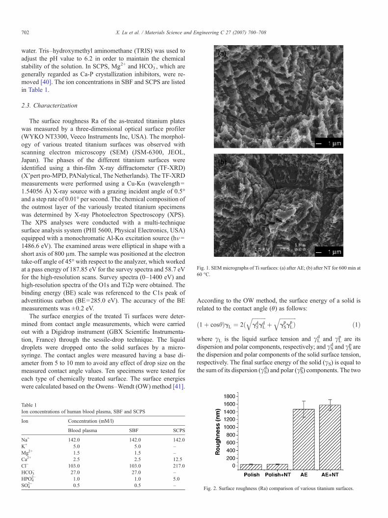
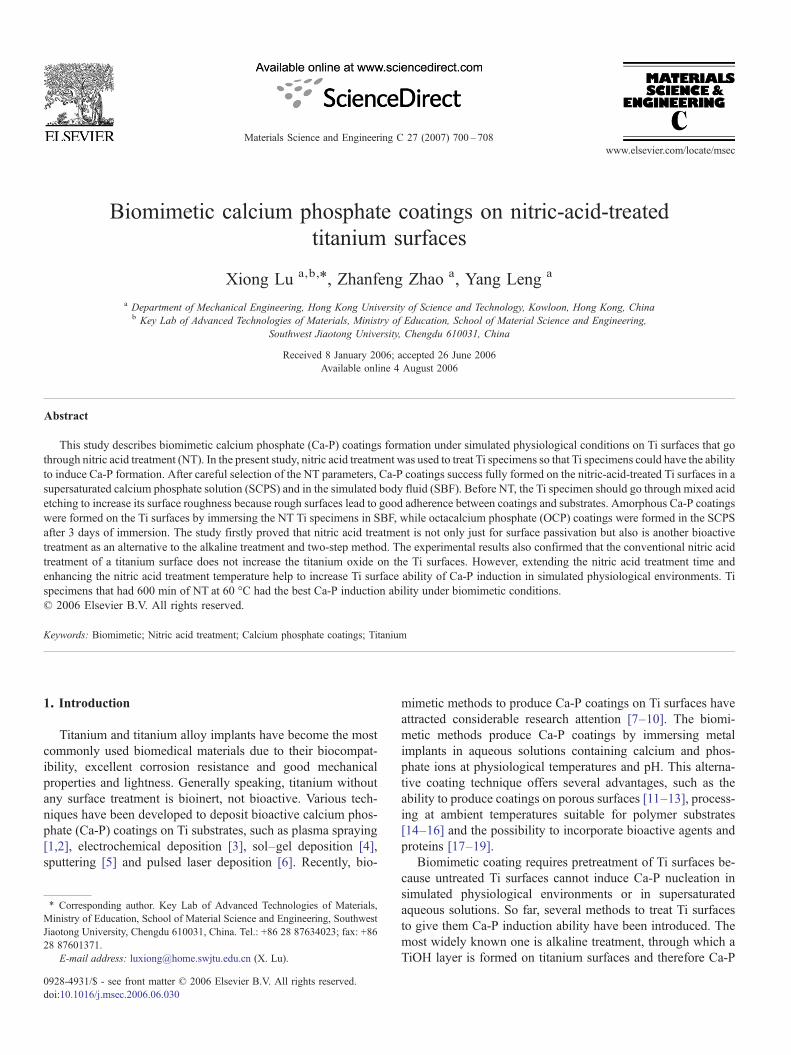
Fig. 1. SEMmicrographs of Ti surfaces: (a) after AE; (b) after NT for 600 min at60 °C.
702 X. Lu et al. / Materials Science and Engineering C 27 (2007) 700–708
water. Tris–hydroxymethyl aminomethane (TRIS) was used toadjust the pH value to 6.2 in order to maintain the chemicalstability of the solution. In SCPS, Mg2+ and HCO3
−, which aregenerally regarded as Ca-P crystallization inhibitors, were re-moved [40]. The ion concentrations in SBF and SCPS are listedin Table 1.
2.3. Characterization
The surface roughness Ra of the as-treated titanium plateswas measured by a three-dimensional optical surface profiler(WYKO NT3300, Veeco Instruments Inc, USA). The morphol-ogy of various treated titanium surfaces was observed withscanning electron microscopy (SEM) (JSM-6300, JEOL,Japan). The phases of the different titanium surfaces wereidentified using a thin-film X-ray diffractometer (TF-XRD)(X'pert pro-MPD, PANalytical, The Netherlands). The TF-XRDmeasurements were performed using a Cu-Kα (wavelength=1.54056 Å) X-ray source with a grazing incident angle of 0.5°and a step rate of 0.01° per second. The chemical composition ofthe outmost layer of the variously treated titanium specimenswas determined by X-ray Photoelectron Spectroscopy (XPS).The XPS analyses were conducted with a multi-techniquesurface analysis system (PHI 5600, Physical Electronics, USA)equipped with a monochromatic Al-Kα excitation source (hν=1486.6 eV). The examined areas were elliptical in shape with ashort axis of 800 μm. The sample was positioned at the electrontake-off angle of 45° with respect to the analyzer, which workedat a pass energy of 187.85 eV for the survey spectra and 58.7 eVfor the high-resolution scans. Survey spectra (0–1400 eV) andhigh-resolution spectra of the O1s and Ti2p were obtained. Thebinding energy (BE) scale was referenced to the C1s peak ofadventitious carbon (BE=285.0 eV). The accuracy of the BEmeasurements was ±0.2 eV.
The surface energies of the treated Ti surfaces were deter-mined from contact angle measurements, which were carriedout with a Digidrop instrument (GBX Scientific Instrumenta-tion, France) through the sessile-drop technique. The liquiddroplets were dropped onto the solid surfaces by a micro-syringe. The contact angles were measured having a base di-ameter from 5 to 10 mm to avoid any effect of drop size on themeasured contact angle values. Ten specimens were tested foreach type of chemically treated surface. The surface energieswere calculated based on the Owens–Wendt (OW) method [41].
Table 1Ion concentrations of human blood plasma, SBF and SCPS
Ion Concentration (mM/l)
Blood plasma SBF SCPS
Na+ 142.0 142.0 142.0K+ 5.0 5.0 –Mg2+ 1.5 1.5 –Ca2+ 2.5 2.5 12.5Cl− 103.0 103.0 217.0HCO3
− 27.0 27.0 –HPO4
2− 1.0 1.0 5.0SO4
2− 0.5 0.5 –
According to the OW method, the surface energy of a solid isrelated to the contact angle (θ) as follows:
ð1þ coshÞgL ¼ 2ðffiffiffiffiffiffiffiffiffiffigdSg
dL
qþ
ffiffiffiffiffiffiffiffiffiffiffiffigpSg
pLÞ
qð1Þ
where γL is the liquid surface tension and γLd and γL
p are itsdispersion and polar components, respectively; and γS
d and γSp are
the dispersion and polar components of the solid surface tension,respectively. The final surface energy of the solid (γS) is equal tothe sum of its dispersion (γS
d) and polar (γSp) components. The two
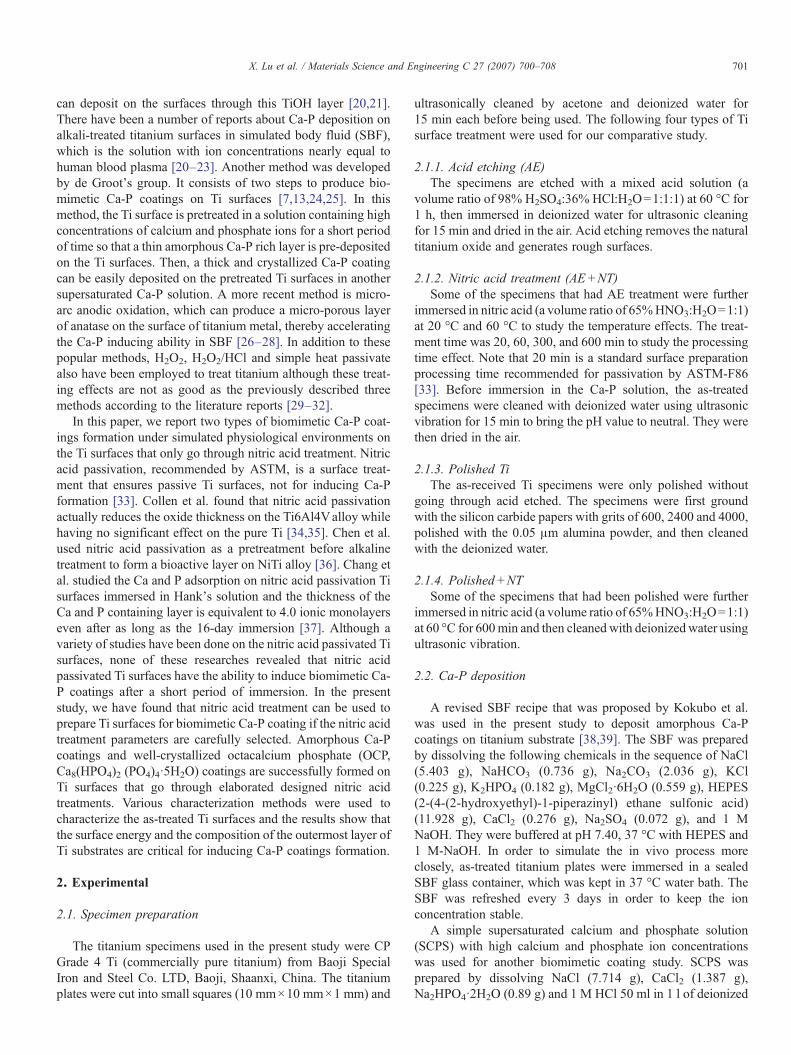
Fig. 2. Surface roughness (Ra) comparison of various titanium surfaces.
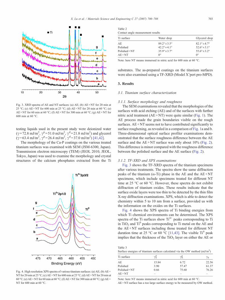
Fig. 3. XRD spectra of AE and NT surfaces: (a) AE; (b) AE+NT for 20 min at25 °C; (c) AE+NT for 600 min at 25 °C; (d) AE+NT for 20 min at 60 °C; (e)AE+NT for 60 min at 60 °C; (f) AE+NT for 300 min at 60 °C; (g) AE+NT for600 min at 60 °C.
Table 2Contact angle measurement results
Ti surface Water drop Glycerol drop
AE 89.2°±3.5° 82.1°±4.7°Polished 42.2°±4.1° 52.6°±3.1°Polished+NT 35.9°±3.7° 55.6°±3.2°AE+NT 0° 0°
Note: here NT means immersed in nitric acid for 600 min at 60 °C.
703X. Lu et al. / Materials Science and Engineering C 27 (2007) 700–708
testing liquids used in the present study were deionized water(γ=72.8 mJ/m2, γp=51.0 mJ/m2, γd=21.8 mJ/m2) and glycerol(γ=63.4 mJ/m2, γp=26.4 mJ/m2, γd=37.0 mJ/m2) [41,42].
The morphology of the Ca-P coatings on the various treatedtitanium surfaces was examined with SEM (JSM-6300, Japan).Transmission electron microscopy (TEM) (JEOL 2010, JEOL,Tokyo, Japan) was used to examine the morphology and crystalstructures of the calcium phosphates extracted from the Ti
Fig. 4. High resolution XPS spectra of various titanium surfaces: (a) AE; (b) AE+NT for 20min at 25 °C; (c)AE+NT for 600min at 25 °C; (d)AE+NT for 20min at60 °C; (e) AE+NT for 60min at 60 °C; (f) AE+NT for 300min at 60 °C; (g) AE+NT for 600 min at 60 °C.
substrates. The as-prepared coatings on the titanium surfaceswere also examined using a TF-XRD (Model X'pert pro-MPD).
3. Results
3.1. Titanium surface characterization
3.1.1. Surface morphology and roughnessThe SEM examinations revealed that the morphologies of the
surfaces with acid etching (AE) and of the surfaces with furthernitric acid treatment (AE+NT) were quite similar (Fig. 1). TheAE process made the grain boundaries visible on the roughsurfaces. AE+NT seems not to have contributed significantly tosurface roughening, as revealed in a comparison of Fig. 1a and b.Three-dimensional optical surface profiler examinations dem-onstrated that the surface roughness difference between the AEsurface and the AE+NT surface was only about 10% (Fig. 2).This difference is minor compared with the roughness differencebetween the polished surface and the AE surface (Fig. 2).
3.1.2. TF-XRD and XPS examinationsFig. 3 shows the TF-XRD spectra of the titanium specimens
after various treatments. The spectra show the same diffractionpeaks of the titanium (α-Ti) phase in the AE and the AE+NTspecimens, which include specimens treated for different NTtime at 25 °C or 60 °C. However, these spectra do not exhibitdiffraction of titanium oxides. These results indicate that thesurface oxide layers were too thin to be detected by the thin filmX-ray diffraction examinations. XPS, which is able to detect thechemistry within 5 to 10 nm from a surface, provided us withthe information on the oxides on the Ti surfaces.
Fig. 4 shows the XPS spectra of Ti binding energies fromwhich Ti chemical environments can be determined. The XPSspectra of the Ti surfaces show Ti4+ peaks corresponding to Tiin TiO2 and Ti0 peaks corresponding to Ti metal on the AE andthe AE+NT surfaces including those treated for different NTduration time at 25 °C or 60 °C [11,43]. The visible Ti0 peakimplies that the thickness of the TiO2 layer on either the AE or
Table 3Surface energies of titanium surfaces calculated via the OW method (mJ/m2)
Ti surface γSd γS
p γS
AE 15.84 6.72 22.56Polished 3.90 57.47 61.37Polished+NT 0.66 75.60 76.26AE+NT – – –
Note: here NT means immersed in nitric acid for 600 min at 60 °C.AE+NT surface has a too large surface energy to be measured by OW method.
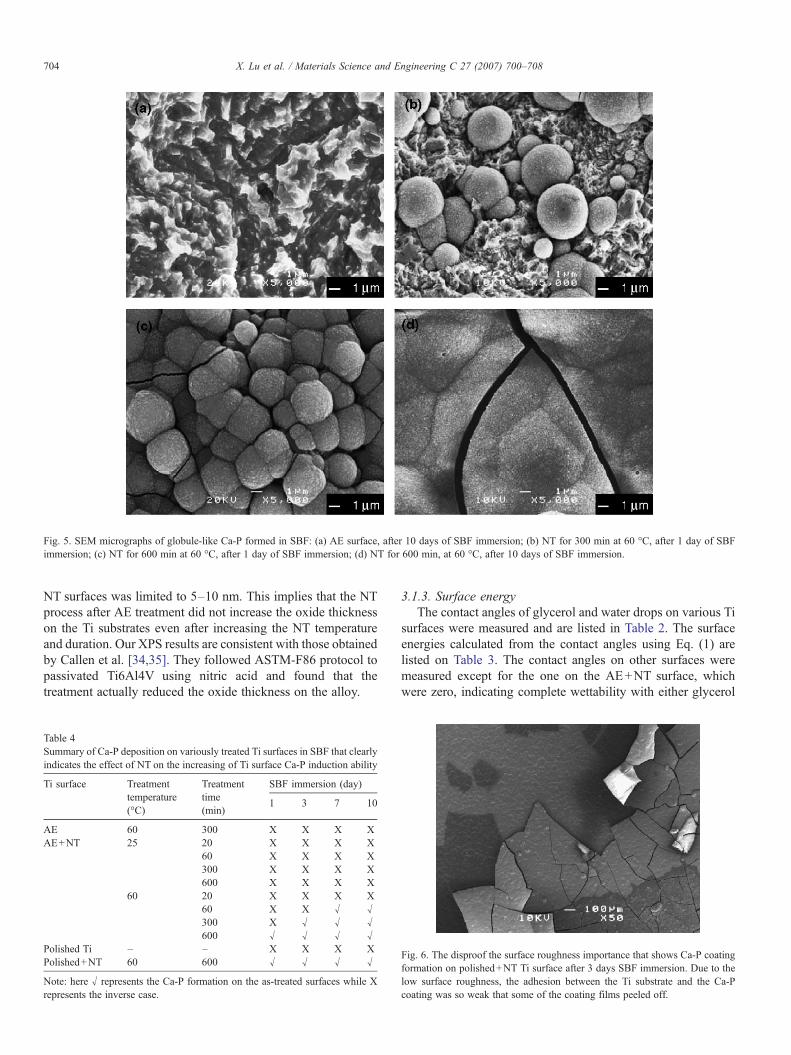
Fig. 5. SEM micrographs of globule-like Ca-P formed in SBF: (a) AE surface, after 10 days of SBF immersion; (b) NT for 300 min at 60 °C, after 1 day of SBFimmersion; (c) NT for 600 min at 60 °C, after 1 day of SBF immersion; (d) NT for 600 min, at 60 °C, after 10 days of SBF immersion.
704 X. Lu et al. / Materials Science and Engineering C 27 (2007) 700–708
NT surfaces was limited to 5–10 nm. This implies that the NTprocess after AE treatment did not increase the oxide thicknesson the Ti substrates even after increasing the NT temperatureand duration. Our XPS results are consistent with those obtainedby Callen et al. [34,35]. They followed ASTM-F86 protocol topassivated Ti6Al4V using nitric acid and found that thetreatment actually reduced the oxide thickness on the alloy.
Table 4Summary of Ca-P deposition on variously treated Ti surfaces in SBF that clearlyindicates the effect of NT on the increasing of Ti surface Ca-P induction ability
Ti surface Treatmenttemperature(°C)
Treatmenttime(min)
SBF immersion (day)
1 3 7 10
AE 60 300 X X X XAE+NT 25 20 X X X X
60 X X X X300 X X X X600 X X X X
60 20 X X X X60 X X √ √300 X √ √ √600 √ √ √ √
Polished Ti – – X X X XPolished+NT 60 600 √ √ √ √
Note: here √ represents the Ca-P formation on the as-treated surfaces while Xrepresents the inverse case.
3.1.3. Surface energyThe contact angles of glycerol and water drops on various Ti
surfaces were measured and are listed in Table 2. The surfaceenergies calculated from the contact angles using Eq. (1) arelisted on Table 3. The contact angles on other surfaces weremeasured except for the one on the AE+NT surface, whichwere zero, indicating complete wettability with either glycerol
Fig. 6. The disproof the surface roughness importance that shows Ca-P coatingformation on polished+NT Ti surface after 3 days SBF immersion. Due to thelow surface roughness, the adhesion between the Ti substrate and the Ca-Pcoating was so weak that some of the coating films peeled off.
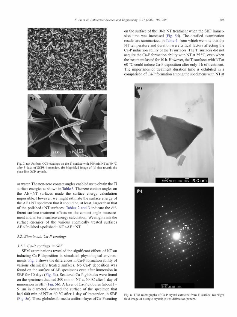
Fig. 7. (a) Uniform OCP coatings on the Ti surface with 300 min NT at 60 °Cafter 3 days of SCPS immersion. (b) Magnified image of (a) that reveals theplate-like OCP crystals.
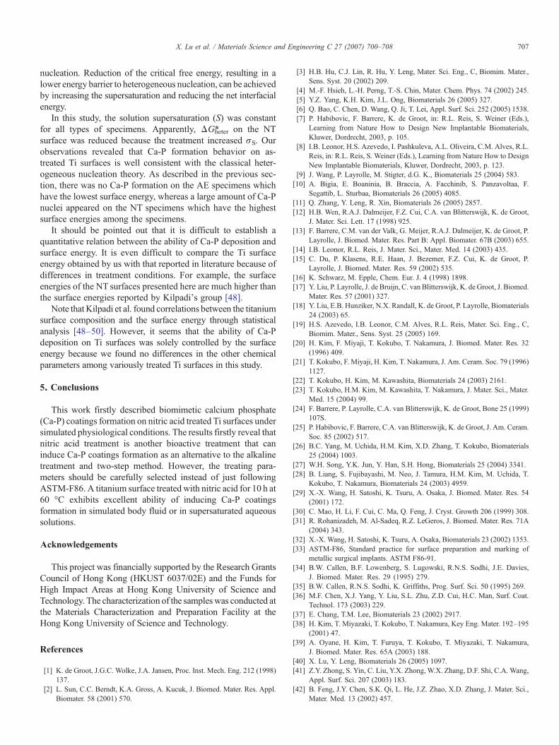
Fig. 8. TEM micrographs of Ca-P crystal extracted from Ti surface: (a) brightfield image of a single crystal; (b) its diffraction pattern.
705X. Lu et al. / Materials Science and Engineering C 27 (2007) 700–708
or water. The non-zero contact angles enabled us to obtain the Tisurface energies as shown in Table 3. The zero contact angles onthe AE+NT surfaces made the surface energy calculationimpossible. However, we might estimate the surface energy ofthe AE+NT specimen that it should be, at least, larger than thatof the polished+NT surfaces. Tables 2 and 3 indicate the dif-ferent surface treatment effects on the contact angle measure-ment and, in turn, surface energy calculation. We might rank thesurface energies of the various chemically treated surfacesAEbPolishedbpolished+NTbAE+NT.
3.2. Biomimetic Ca-P coatings
3.2.1. Ca-P coatings in SBFSEM examinations revealed the significant effects of NT on
inducing Ca-P deposition in simulated physiological environ-ments. Fig. 5 shows the differences in Ca-P formation ability ofvarious chemically treated surfaces. No Ca-P deposition wasfound on the surface of AE specimens even after immersion inSBF for 10 days (Fig. 5a). Scattered Ca-P globules were foundon the specimen that had 300 min of NT at 60 °C after 1 day ofimmersion in SBF (Fig. 5b). A layer of Ca-P globules (about 1–5 μm in diameter) covered the surface of the specimen thathad 600 min of NT at 60 °C after 1 day of immersion in SBF(Fig. 5c). These globules formed a uniform layer of Ca-P coating
on the surface of the 10-h NT treatment when the SBF immer-sion time was increased (Fig. 5d). The detailed examinationresults are summarized in Table 4, from which we note that theNT temperature and duration were critical factors affecting theCa-P induction ability of the Ti surfaces. The Ti surfaces did notacquire the Ca-P formation ability with NT at 25 °C, even whenthe treatment lasted for 10 h. However, the Ti surfaces with NTat60 °C could induce Ca-P deposition after only 1 h of treatment.The importance of treatment duration time is exhibited in acomparison of Ca-P formation among the specimens with NT at
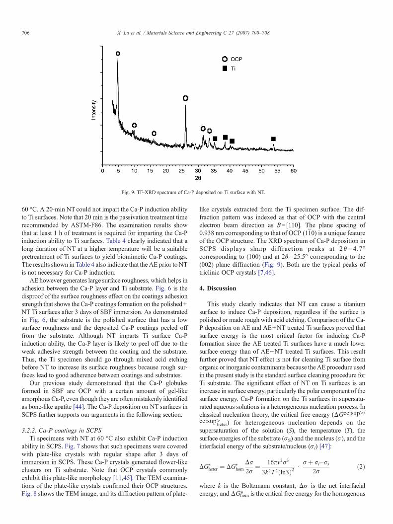
Fig. 9. TF-XRD spectrum of Ca-P deposited on Ti surface with NT.
706 X. Lu et al. / Materials Science and Engineering C 27 (2007) 700–708
60 °C. A 20-min NT could not impart the Ca-P induction abilityto Ti surfaces. Note that 20 min is the passivation treatment timerecommended by ASTM-F86. The examination results showthat at least 1 h of treatment is required for imparting the Ca-Pinduction ability to Ti surfaces. Table 4 clearly indicated that along duration of NT at a higher temperature will be a suitablepretreatment of Ti surfaces to yield biomimetic Ca-P coatings.The results shown in Table 4 also indicate that the AE prior to NTis not necessary for Ca-P induction.
AE however generates large surface roughness, which helps inadhesion between the Ca-P layer and Ti substrate. Fig. 6 is thedisproof of the surface roughness effect on the coatings adhesionstrength that shows the Ca-P coatings formation on the polished+NT Ti surfaces after 3 days of SBF immersion. As demonstratedin Fig. 6, the substrate is the polished surface that has a lowsurface roughness and the deposited Ca-P coatings peeled offfrom the substrate. Although NT imparts Ti surface Ca-Pinduction ability, the Ca-P layer is likely to peel off due to theweak adhesive strength between the coating and the substrate.Thus, the Ti specimen should go through mixed acid etchingbefore NT to increase its surface roughness because rough sur-faces lead to good adherence between coatings and substrates.
Our previous study demonstrated that the Ca-P globulesformed in SBF are OCP with a certain amount of gel-likeamorphous Ca-P, even though they are oftenmistakenly identifiedas bone-like apatite [44]. The Ca-P deposition on NT surfaces inSCPS further supports our arguments in the following section.
3.2.2. Ca-P coatings in SCPSTi specimens with NT at 60 °C also exhibit Ca-P induction
ability in SCPS. Fig. 7 shows that such specimens were coveredwith plate-like crystals with regular shape after 3 days ofimmersion in SCPS. These Ca-P crystals generated flower-likeclusters on Ti substrate. Note that OCP crystals commonlyexhibit this plate-like morphology [11,45]. The TEM examina-tions of the plate-like crystals confirmed their OCP structures.Fig. 8 shows the TEM image, and its diffraction pattern of plate-
like crystals extracted from the Ti specimen surface. The dif-fraction pattern was indexed as that of OCP with the centralelectron beam direction as B=[110]. The plane spacing of0.938 nm corresponding to that of OCP (11̄0) is a unique featureof the OCP structure. The XRD spectrum of Ca-P deposition inSCPS displays sharp diffraction peaks at 2θ = 4.7°corresponding to (100) and at 2θ=25.5° corresponding to the(002) plane diffraction (Fig. 9). Both are the typical peaks oftriclinic OCP crystals [7,46].
4. Discussion
This study clearly indicates that NT can cause a titaniumsurface to induce Ca-P deposition, regardless if the surface ispolished or made rough with acid etching. Comparison of the Ca-P deposition on AE and AE+NT treated Ti surfaces proved thatsurface energy is the most critical factor for inducing Ca-Pformation since the AE treated Ti surfaces have a much lowersurface energy than of AE+NT treated Ti surfaces. This resultfurther proved that NT effect is not for cleaning Ti surface fromorganic or inorganic contaminants because theAEprocedure usedin the present study is the standard surface cleaning procedure forTi substrate. The significant effect of NT on Ti surfaces is anincrease in surface energy, particularly the polar component of thesurface energy. Ca-P formation on the Ti surfaces in supersatu-rated aqueous solutions is a heterogeneous nucleation process. Inclassical nucleation theory, the critical free energy (ΔGce:sup>/ce:sup>
heter) for heterogeneous nucleation depends on thesupersaturation of the solution (S), the temperature (T), thesurface energies of the substrate (σS) and the nucleus (σ), and theinterfacial energy of the substrate/nucleus (σi) [47]:
DG⁎heter ¼ DG⁎
hom
Dr2r
¼ 16pv2r3
3k2T2ðlnSÞ2 drþ ri−rs
2rð2Þ
where k is the Boltzmann constant; Δσ is the net interfacialenergy; andΔG⁎hom is the critical free energy for the homogenous
707X. Lu et al. / Materials Science and Engineering C 27 (2007) 700–708
nucleation. Reduction of the critical free energy, resulting in alower energy barrier to heterogeneous nucleation, can be achievedby increasing the supersaturation and reducing the net interfacialenergy.
In this study, the solution supersaturation (S) was constantfor all types of specimens. Apparently, ΔG⁎heter on the NTsurface was reduced because the treatment increased σS. Ourobservations revealed that Ca-P formation behavior on as-treated Ti surfaces is well consistent with the classical heter-ogeneous nucleation theory. As described in the previous sec-tion, there was no Ca-P formation on the AE specimens whichhave the lowest surface energy, whereas a large amount of Ca-Pnuclei appeared on the NT specimens which have the highestsurface energies among the specimens.
It should be pointed out that it is difficult to establish aquantitative relation between the ability of Ca-P deposition andsurface energy. It is even difficult to compare the Ti surfaceenergy obtained by us with that reported in literature because ofdifferences in treatment conditions. For example, the surfaceenergies of the NT surfaces presented here are much higher thanthe surface energies reported by Kilpadi's group [48].
Note that Kilpadi et al. found correlations between the titaniumsurface composition and the surface energy through statisticalanalysis [48–50]. However, it seems that the ability of Ca-Pdeposition on Ti surfaces was solely controlled by the surfaceenergy because we found no differences in the other chemicalparameters among variously treated Ti surfaces in this study.
5. Conclusions
This work firstly described biomimetic calcium phosphate(Ca-P) coatings formation on nitric acid treated Ti surfaces undersimulated physiological conditions. The results firstly reveal thatnitric acid treatment is another bioactive treatment that caninduce Ca-P coatings formation as an alternative to the alkalinetreatment and two-step method. However, the treating para-meters should be carefully selected instead of just followingASTM-F86. A titanium surface treated with nitric acid for 10 h at60 °C exhibits excellent ability of inducing Ca-P coatingsformation in simulated body fluid or in supersaturated aqueoussolutions.
Acknowledgements
This project was financially supported by the Research GrantsCouncil of Hong Kong (HKUST 6037/02E) and the Funds forHigh Impact Areas at Hong Kong University of Science andTechnology. The characterization of the sampleswas conducted atthe Materials Characterization and Preparation Facility at theHong Kong University of Science and Technology.
References
[1] K. de Groot, J.G.C. Wolke, J.A. Jansen, Proc. Inst. Mech. Eng. 212 (1998)137.
[2] L. Sun, C.C. Berndt, K.A. Gross, A. Kucuk, J. Biomed. Mater. Res. Appl.Biomater. 58 (2001) 570.
[3] H.B. Hu, C.J. Lin, R. Hu, Y. Leng, Mater. Sci. Eng., C, Biomim. Mater.,Sens. Syst. 20 (2002) 209.
[4] M.-F. Hsieh, L.-H. Perng, T.-S. Chin, Mater. Chem. Phys. 74 (2002) 245.[5] Y.Z. Yang, K.H. Kim, J.L. Ong, Biomaterials 26 (2005) 327.[6] Q. Bao, C. Chen, D. Wang, Q. Ji, T. Lei, Appl. Surf. Sci. 252 (2005) 1538.[7] P. Habibovic, F. Barrere, K. de Groot, in: R.L. Reis, S. Weiner (Eds.),
Learning from Nature How to Design New Implantable Biomaterials,Kluwer, Dordrecht, 2003, p. 105.
[8] I.B. Leonor, H.S. Azevedo, I. Pashkuleva, A.L. Oliveira, C.M. Alves, R.L.Reis, in: R.L. Reis, S. Weiner (Eds.), Learning from Nature How to DesignNew Implantable Biomaterials, Kluwer, Dordrecht, 2003, p. 123.
[9] J. Wang, P. Layrolle, M. Stigter, d.G. K., Biomaterials 25 (2004) 583.[10] A. Bigia, E. Boaninia, B. Braccia, A. Facchinib, S. Panzavoltaa, F.
Segattib, L. Sturbaa, Biomaterials 26 (2005) 4085.[11] Q. Zhang, Y. Leng, R. Xin, Biomaterials 26 (2005) 2857.[12] H.B. Wen, R.A.J. Dalmeijer, F.Z. Cui, C.A. van Blitterswijk, K. de Groot,
J. Mater. Sci. Lett. 17 (1998) 925.[13] F. Barrere, C.M. van der Valk, G. Meijer, R.A.J. Dalmeijer, K. de Groot, P.
Layrolle, J. Biomed. Mater. Res. Part B: Appl. Biomater. 67B (2003) 655.[14] I.B. Leonor, R.L. Reis, J. Mater. Sci., Mater. Med. 14 (2003) 435.[15] C. Du, P. Klasens, R.E. Haan, J. Bezemer, F.Z. Cui, K. de Groot, P.
Layrolle, J. Biomed. Mater. Res. 59 (2002) 535.[16] K. Schwarz, M. Epple, Chem. Eur. J. 4 (1998) 1898.[17] Y. Liu, P. Layrolle, J. de Bruijn, C. van Blitterswijk, K. de Groot, J. Biomed.
Mater. Res. 57 (2001) 327.[18] Y. Liu, E.B. Hunziker, N.X. Randall, K. de Groot, P. Layrolle, Biomaterials
24 (2003) 65.[19] H.S. Azevedo, I.B. Leonor, C.M. Alves, R.L. Reis, Mater. Sci. Eng., C,
Biomim. Mater., Sens. Syst. 25 (2005) 169.[20] H. Kim, F. Miyaji, T. Kokubo, T. Nakamura, J. Biomed. Mater. Res. 32
(1996) 409.[21] T. Kokubo, F. Miyaji, H. Kim, T. Nakamura, J. Am. Ceram. Soc. 79 (1996)
1127.[22] T. Kokubo, H. Kim, M. Kawashita, Biomaterials 24 (2003) 2161.[23] T. Kokubo, H.M. Kim, M. Kawashita, T. Nakamura, J. Mater. Sci., Mater.
Med. 15 (2004) 99.[24] F. Barrere, P. Layrolle, C.A. van Blitterswijk, K. de Groot, Bone 25 (1999)
107S.[25] P. Habibovic, F. Barrere, C.A. van Blitterswijk, K. de Groot, J. Am. Ceram.
Soc. 85 (2002) 517.[26] B.C. Yang, M. Uchida, H.M. Kim, X.D. Zhang, T. Kokubo, Biomaterials
25 (2004) 1003.[27] W.H. Song, Y.K. Jun, Y. Han, S.H. Hong, Biomaterials 25 (2004) 3341.[28] B. Liang, S. Fujibayashi, M. Neo, J. Tamura, H.M. Kim, M. Uchida, T.
Kokubo, T. Nakamura, Biomaterials 24 (2003) 4959.[29] X.-X. Wang, H. Satoshi, K. Tsuru, A. Osaka, J. Biomed. Mater. Res. 54
(2001) 172.[30] C. Mao, H. Li, F. Cui, C. Ma, Q. Feng, J. Cryst. Growth 206 (1999) 308.[31] R. Rohanizadeh, M. Al-Sadeq, R.Z. LeGeros, J. Biomed. Mater. Res. 71A
(2004) 343.[32] X.-X. Wang, H. Satoshi, K. Tsuru, A. Osaka, Biomaterials 23 (2002) 1353.[33] ASTM-F86, Standard practice for surface preparation and marking of
metallic surgical implants. ASTM F86-91.[34] B.W. Callen, B.F. Lowenberg, S. Lugowski, R.N.S. Sodhi, J.E. Davies,
J. Biomed. Mater. Res. 29 (1995) 279.[35] B.W. Callen, R.N.S. Sodhi, K. Griffiths, Prog. Surf. Sci. 50 (1995) 269.[36] M.F. Chen, X.J. Yang, Y. Liu, S.L. Zhu, Z.D. Cui, H.C. Man, Surf. Coat.
Technol. 173 (2003) 229.[37] E. Chang, T.M. Lee, Biomaterials 23 (2002) 2917.[38] H. Kim, T. Miyazaki, T. Kokubo, T. Nakamura, Key Eng. Mater. 192–195
(2001) 47.[39] A. Oyane, H. Kim, T. Furuya, T. Kokubo, T. Miyazaki, T. Nakamura,
J. Biomed. Mater. Res. 65A (2003) 188.[40] X. Lu, Y. Leng, Biomaterials 26 (2005) 1097.[41] Z.Y. Zhong, S. Yin, C. Liu, Y.X. Zhong,W.X. Zhang, D.F. Shi, C.A.Wang,
Appl. Surf. Sci. 207 (2003) 183.[42] B. Feng, J.Y. Chen, S.K. Qi, L. He, J.Z. Zhao, X.D. Zhang, J. Mater. Sci.,
Mater. Med. 13 (2002) 457.

708 X. Lu et al. / Materials Science and Engineering C 27 (2007) 700–708
[43] J. Voros, M. Wieland, L. Ruiz-Taylor, M. Textor, D. Brunette, in: D.Brunette, et al., (Eds.), Titanium in Medicine, Springer, Berlin, 2001, p.87.
[44] X. Lu, Y. Leng, Biomaterials 25 (2004) 1779.[45] X. Lu, Z.F. Zhao, Y. Leng, J. Cryst. Growth 284 (2005) 506.[46] J.C. Elliott, Structure and Chemistry of the Apatites and Other Calcium
Orthophosphates, Elsevier, Amsterdam, 1994.
[47] I.V. Markov, Crystal Growth for Beginners: Fundamentals of Nucleation,Crystal Growth, and Epitaxy, World Scientific, Singapore, 1995.
[48] D.V. Kilpadi, J.E. Lemons, J. Biomed. Mater. Res. 28 (1994) 1419.[49] D.V. Kilpadi, G.N. Raikar, J. Liu, J.E. Lemons, Y. Vohra, J.C. Gregory,
J. Biomed. Mater. Res. 40 (1998) 646.[50] D.V. Kilpadi, J.J. Weimer, J.E. Lemons, Colloids Surf., A 135 (1998) 89.