biomimetic and biocompatible chitosan- carbon … · physiochemically characterized as bone graft...
TRANSCRIPT

18TH INTERNATIONAL CONFERENCE ON COMPOSITE MATERIALS
AbstractIn recent years, significant development has
been given to chitosan composites for orthopedic applications. In this study, we have used low and high molecular weight of chitosan with 0.25%, 0.5% and 1.0% weight of f-Multiwalled Carbon Nanotube (f-MWCNT) were fabricated as a scaffold by freezing and lyophilization method and physiochemically characterized as bone graft substitutes. A combination of Fourier Transform Infrared Spectroscopy, X-Ray diffraction analysis, Thermal Gravimetric Analysis, Scanning Electron Microscopy and Optical Microscopy results indicated that the f-MWCNT was uniformly dispersed in chitosan matrix and there was a chemical interaction between chitosan and f-MWCNT. The porosity, water uptake and retention ability and of scaffolds were increased with an increase the amount of f-MWCNT. Composite scaffold materials have greater cell proliferation, protein content, alkaline phosphatase, mineralization as compared to chitosan scaffold. Moreover, addition of hydroxyapatite in chitosan/f-MWCNT lead to improved the biological function at in vitro level. Herewith, we are suggesting that chitosan/f-MWCNT scaffolds are promising biomaterials for bone tissue engineering. IntroductionThe repair and replacement of injured or defect bone is a critical problem in orthopaedic treatment throughout worldwide. In recent years, significant development has been made in organ replacement, surgical reconstruction, and the use of artificial prostheses to treat the loss or failure of an organ or tissue [1-2]. Autograft and allograft are considered as ideal procedures for bone grafting. However, both grafting procedures have their own disadvantages like inadequate supply of bone to fill the gap and diseases transmissible. Due to limited supply of natural bone for grafting, the need for synthetic bone substitutes which posses same physiochemical and biological properties as natural bone is ever increasing. In view of bone is mainly composed of
organic and inorganic portion such as collagen and nano hydroxyapatite. In the recent years, natural polymer has been attracted by researchers due to their biocompatibility and biodegradability in nature. Chitosan is a copolymer consisting of β-(1→4)-2-acetamido-d-glucose and β-(1→4)-2-amino-d-glucose unit linkage [3-4]. Over the past two decade, considerable attention has been given to chitosan materials and their applications in the field of bone tissue engineering. Porous materials have a highly significant role in the bone implantation process. Degradable polymeric implants eliminate the need for a second operation and can prevent some of the problems associated with stress shielding during post-healing, and can also be used simultaneously to deliver therapeutic drugs to treat infections or growth factors to accelerate new bone growth [5]. From the synthesis of carbon nanotube, has unique high mechanical properties compared to any other material and also consider development in biomaterials areas [6]. The amount of carbon nanotube in the polymeric matrix is important such as 50%w/w, 10%w/w, and 0-7%w/w. Researchers have been proven that biocompatible carbon nanotube composite scaffold comprised of 1-4 % w/w. In addition, osteoblast cell growths have been observed on the carbon nanotube composite scaffolds are superior to normal polymeric scaffold [7]. Apatite formation has been found in the surface of chitosan and carbon nanotube membrane [8-9]. In the present study, we are attempting to mimic the natural function of bone with two materials chitosan and carbon nanotube. Thus, we have prepared a scaffold as system with chitosan and f-MWCNT. A freeze dried method has been used for the scaffold preparation. Chitosan scaffold and carbon nanotube/chitosan scaffold materials have been prepared with various amounts of chitosan and f-MWCNT. Then the scaffold was analysed with biodegradability, water uptake and intake retention ability and physiochemical characterization.Materials and methodMedium molecular weight (100-250 KDa) and high
BIOMIMETIC AND BIOCOMPATIBLE CHITOSAN-CARBON NANOTUBE COMPOSITE SCAFFOLDS FOR
BONE TISSUE ENGINEERING J. Venkatesan 1 , S.K. Kim1, 2*
1 Department of Chemistry, Pukyong National University, Busan 608-737, Republic of Korea2 Marine Bioprocess Research Center, Pukyong National University, Busan 608-737, Republic of
Korea* Corresponding author ([email protected]; [email protected])
Keywords: Chitosan; f-multiwalled carbon nanotube; scaffold; bone tissue engineering

BIOMIMETIC AND BIOCOMPATIBLE CHITOSAN-CARBON NANOTUBE COMPOSITE SCAFFOLDS FOR BONE TISSUE ENGINEERING
molecular weight (500 KDa) chitosan samples were received from Kitto chemicals and their degree of deacetylation was 70% and 90 % respectively; Multiwalled carbon nanotubes (Outer diameter <8 nm, length 10-30µm) were purchased from Cheap Tubes.com, USA. Human osteosarcoma (MG-63) cell line was obtained from American Type Culture Collection (Manassas, VA, USA). Dulbecco’s Modified Eagle’s Medium (DMEM) was obtained from Gibco BRL, Life Technology (USA). MTT(3-(4,5-dimethyl-2-yl)-2, 5-diphenyltetrazolium bromide) was purchased from molecular probes (Eugene, OR, USA). All other reagents used in this experiment were of analytical grade. Functionalization and purification of carbon nanotubes was performed with acid treatment.Preparation of chitosan and chitosan–multiwalled carbon nanotube scaffoldsChitosan scaffolds were made-up by freezing and lyophilization method. Chitosan solution was dissolved by dissolving chitosan in 2% (v/v) acetic acid solution. To synthesize composite scaffolds with different fractions of f-MWCNT (0, 0.25%, 0.5% and 1%) was ultrasonically dispersed in deionized water for 2 h. Subsequently, the dispersed solutions were added drop by drop to the chitosan solution, while the solution was being agitated. Next, the chitosan/f-MWCNT dispersion was vigorously mixed using a mechanical stirrer for 24 h to obtain a homogeneous mixture. The obtained pure chitosan solution and Chitosan/f-MWCNT dispersion was transferred to polystyrene petri dishes (60 x 15mm) 20 g of each solution, frozen at -80 °C for 5 h and lyophilized in a freeze dryer. The acetate in resulting chitosan scaffold was neutralized by immersing them in 10% NaOH followed by washing with water until neutralized and again lyophilized. LM – Low molecular weight chitosan, LMCNT-25 (Low molecular weight chitosan/0.025g of f-MWCNT), LMCNT-50 (Low molecular weight chitosan/0.050g of f-MWCNT), LMCNT-100 (Low molecular weight chitosan/0.100g of f-MWCNT), HM-High molecular weight chitosan, HMCNT-25 (High molecular weight chitosan/0.025g of f-MWCNT), HMCNT-50 (High molecular weight chitosan/0.050g of f-MWCNT), HMCNT-100 (High molecular weight chitosan/0.100g of f-MWCNT) (Table 1). Characterization techniquesThermal gravimetric analysis was achieved by the use of Pyris 7 TGA analyzers, Perkin Elmer Inc., USA with scan range from 50 to 900 °C at constant
heating rate of 10 °C min-1 with continuous nitrogen flow. The stretching frequencies of samples were examined by Fourier Transform Infrared Spectroscopy, Perkin Elmer (USA) and spectrum GX spectrometer within the range of 400 to 4000 cm−1. The phase and crystallinity were evaluated using X-ray diffractometer (PHILIPS X’Pert-MPD diffractometer, Netherland) and Cu-Kα radiation 1.5405 Å over a range of 5 to 80° angle, step size 0.02, scan speed 4°/min with 40 kV voltage and 30 mA current. Morphology of the scaffolds was obtained by scanning electron microscopy (SEM JSM-6700F, JEOL, Japan).Results and discussionIn this study, we have used chitosan as a raw material which is biocompatible and biodegradable. f-MWCNT was used to increase the function of chitosan but pristine CNT is reported to be toxic to cells, therefore functionalization of CNT is important for reduction of toxicity [10]. Addition of COOH, dispersion increases in the aqueous phase thereby making it suitable for scaffold preparation by possible interactions with other cationic molecules. Table.1. Weight composition ratio of different type of chitosan/f-MWCNT composite scaffold
Gross Examination of the scaffoldsIn the low and high molecular weight chitosan, by increase the addition of carbon nanotube in the polymer matrix will increases the flexibility of the scaffold. The visual examination of lyophilized raw chitosan and their composite scaffolds showed that they are stiff and inelastic are shown in Fig. 1, it was observed that the chitosan and their scaffolds swelled rapidly. The chemical basis may be the intermolecular hydrogen bonding interaction between the carboxylic groups present in the f-MWCNT and the NH2 group of chitosan. Low and high molecular weight scaffolds are pure white in colour, where as chitosan/f-MWCNT scaffolds were obtained in was deeply black in color when increase
2
Scaffolds type
Chitosan (g)
f-MWCNT(g)
Water (ml)
LM 5 --- 1000LM25 5 0.025 1000LM50 5 0.050 1000LM100 5 0.100 1000HM 5 --- 1000HM25 5 0.025 1000HM50 5 0.050 1000HM100 5 0.100 1000
BIOMIMETIC AND BIOCOMPATIBLE CHITOSAN-CARBON NANOTUBE COMPOSITE SCAFFOLDS FOR BONE TISSUE ENGINEERING
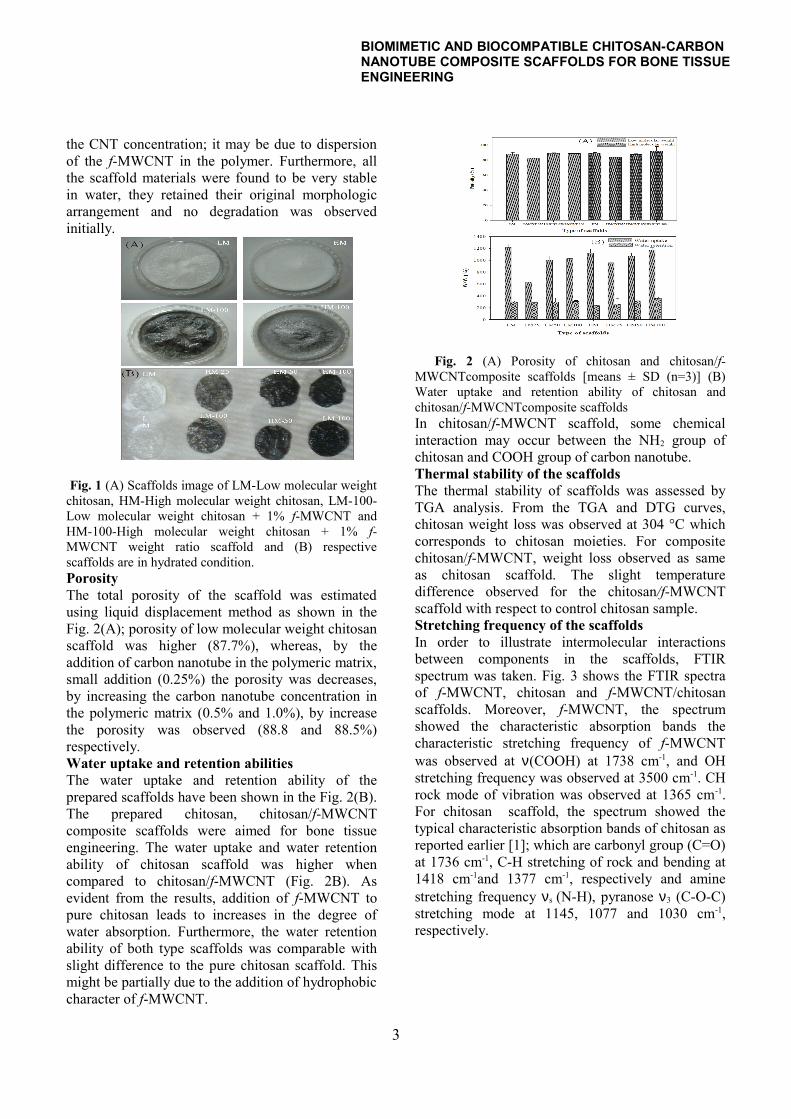
the CNT concentration; it may be due to dispersion of the f-MWCNT in the polymer. Furthermore, all the scaffold materials were found to be very stable in water, they retained their original morphologic arrangement and no degradation was observed initially.
Fig. 1 (A) Scaffolds image of LM-Low molecular weight chitosan, HM-High molecular weight chitosan, LM-100-Low molecular weight chitosan + 1% f-MWCNT and HM-100-High molecular weight chitosan + 1% f-MWCNT weight ratio scaffold and (B) respective scaffolds are in hydrated condition.Porosity The total porosity of the scaffold was estimated using liquid displacement method as shown in the Fig. 2(A); porosity of low molecular weight chitosan scaffold was higher (87.7%), whereas, by the addition of carbon nanotube in the polymeric matrix, small addition (0.25%) the porosity was decreases, by increasing the carbon nanotube concentration in the polymeric matrix (0.5% and 1.0%), by increase the porosity was observed (88.8 and 88.5%) respectively.Water uptake and retention abilitiesThe water uptake and retention ability of the prepared scaffolds have been shown in the Fig. 2(B). The prepared chitosan, chitosan/f-MWCNT composite scaffolds were aimed for bone tissue engineering. The water uptake and water retention ability of chitosan scaffold was higher when compared to chitosan/f-MWCNT (Fig. 2B). As evident from the results, addition of f-MWCNT to pure chitosan leads to increases in the degree of water absorption. Furthermore, the water retention ability of both type scaffolds was comparable with slight difference to the pure chitosan scaffold. This might be partially due to the addition of hydrophobic character of f-MWCNT.
Fig. 2 (A) Porosity of chitosan and chitosan/f-MWCNTcomposite scaffolds [means ± SD (n=3)] (B) Water uptake and retention ability of chitosan and chitosan/f-MWCNTcomposite scaffoldsIn chitosan/f-MWCNT scaffold, some chemical interaction may occur between the NH2 group of chitosan and COOH group of carbon nanotube. Thermal stability of the scaffoldsThe thermal stability of scaffolds was assessed by TGA analysis. From the TGA and DTG curves, chitosan weight loss was observed at 304 °C which corresponds to chitosan moieties. For composite chitosan/f-MWCNT, weight loss observed as same as chitosan scaffold. The slight temperature difference observed for the chitosan/f-MWCNT scaffold with respect to control chitosan sample. Stretching frequency of the scaffolds In order to illustrate intermolecular interactions between components in the scaffolds, FTIR spectrum was taken. Fig. 3 shows the FTIR spectra of f-MWCNT, chitosan and f-MWCNT/chitosan scaffolds. Moreover, f-MWCNT, the spectrum showed the characteristic absorption bands the characteristic stretching frequency of f-MWCNT was observed at ν(COOH) at 1738 cm-1, and OH stretching frequency was observed at 3500 cm-1. CH rock mode of vibration was observed at 1365 cm-1. For chitosan scaffold, the spectrum showed the typical characteristic absorption bands of chitosan as reported earlier [1]; which are carbonyl group (C=O) at 1736 cm-1, C-H stretching of rock and bending at 1418 cm-1and 1377 cm-1, respectively and amine stretching frequency νs (N-H), pyranose ν3 (C-O-C) stretching mode at 1145, 1077 and 1030 cm-1, respectively.
3
BIOMIMETIC AND BIOCOMPATIBLE CHITOSAN-CARBON NANOTUBE COMPOSITE SCAFFOLDS FOR BONE TISSUE ENGINEERING
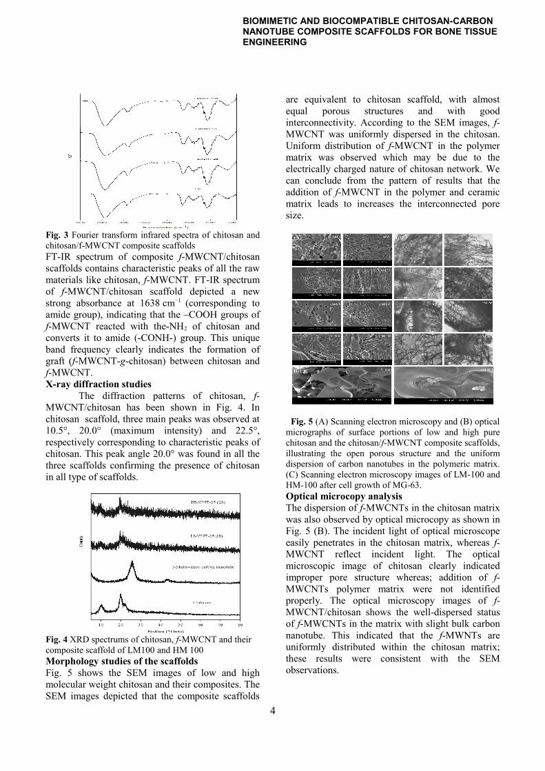
Fig. 3 Fourier transform infrared spectra of chitosan and chitosan/f-MWCNT composite scaffoldsFT-IR spectrum of composite f-MWCNT/chitosan scaffolds contains characteristic peaks of all the raw materials like chitosan, f-MWCNT. FT-IR spectrum of f-MWCNT/chitosan scaffold depicted a new strong absorbance at 1638 cm−1 (corresponding to amide group), indicating that the –COOH groups of f-MWCNT reacted with the-NH2 of chitosan and converts it to amide (-CONH-) group. This unique band frequency clearly indicates the formation of graft (f-MWCNT-g-chitosan) between chitosan and f-MWCNT.X-ray diffraction studies
The diffraction patterns of chitosan, f-MWCNT/chitosan has been shown in Fig. 4. In chitosan scaffold, three main peaks was observed at 10.5°, 20.0° (maximum intensity) and 22.5°, respectively corresponding to characteristic peaks of chitosan. This peak angle 20.0° was found in all the three scaffolds confirming the presence of chitosan in all type of scaffolds.
Fig. 4 XRD spectrums of chitosan, f-MWCNT and their composite scaffold of LM100 and HM 100Morphology studies of the scaffoldsFig. 5 shows the SEM images of low and high molecular weight chitosan and their composites. The SEM images depicted that the composite scaffolds
are equivalent to chitosan scaffold, with almost equal porous structures and with good interconnectivity. According to the SEM images, f-MWCNT was uniformly dispersed in the chitosan. Uniform distribution of f-MWCNT in the polymer matrix was observed which may be due to the electrically charged nature of chitosan network. We can conclude from the pattern of results that the addition of f-MWCNT in the polymer and ceramic matrix leads to increases the interconnected pore size.
Fig. 5 (A) Scanning electron microscopy and (B) optical micrographs of surface portions of low and high pure chitosan and the chitosan/f-MWCNT composite scaffolds, illustrating the open porous structure and the uniform dispersion of carbon nanotubes in the polymeric matrix. (C) Scanning electron microscopy images of LM-100 and HM-100 after cell growth of MG-63.Optical microcopy analysisThe dispersion of f-MWCNTs in the chitosan matrix was also observed by optical microcopy as shown in Fig. 5 (B). The incident light of optical microscope easily penetrates in the chitosan matrix, whereas f-MWCNT reflect incident light. The optical microscopic image of chitosan clearly indicated improper pore structure whereas; addition of f-MWCNTs polymer matrix were not identified properly. The optical microscopy images of f-MWCNT/chitosan shows the well-dispersed status of f-MWCNTs in the matrix with slight bulk carbon nanotube. This indicated that the f-MWNTs are uniformly distributed within the chitosan matrix; these results were consistent with the SEM observations.
4
BIOMIMETIC AND BIOCOMPATIBLE CHITOSAN-CARBON NANOTUBE COMPOSITE SCAFFOLDS FOR BONE TISSUE ENGINEERING
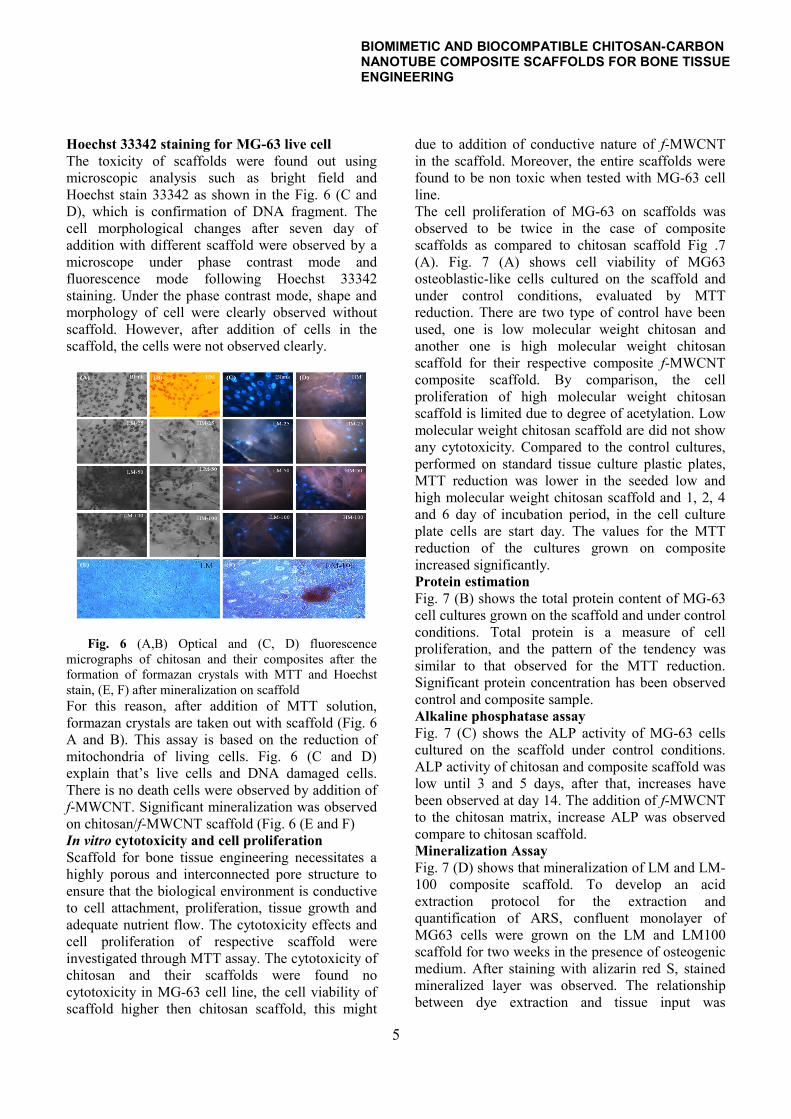
Hoechst 33342 staining for MG-63 live cellThe toxicity of scaffolds were found out using microscopic analysis such as bright field and Hoechst stain 33342 as shown in the Fig. 6 (C and D), which is confirmation of DNA fragment. The cell morphological changes after seven day of addition with different scaffold were observed by a microscope under phase contrast mode and fluorescence mode following Hoechst 33342 staining. Under the phase contrast mode, shape and morphology of cell were clearly observed without scaffold. However, after addition of cells in the scaffold, the cells were not observed clearly.
Fig. 6 (A,B) Optical and (C, D) fluorescence micrographs of chitosan and their composites after the formation of formazan crystals with MTT and Hoechst stain, (E, F) after mineralization on scaffoldFor this reason, after addition of MTT solution, formazan crystals are taken out with scaffold (Fig. 6 A and B). This assay is based on the reduction of mitochondria of living cells. Fig. 6 (C and D) explain that’s live cells and DNA damaged cells. There is no death cells were observed by addition of f-MWCNT. Significant mineralization was observed on chitosan/f-MWCNT scaffold (Fig. 6 (E and F)In vitro cytotoxicity and cell proliferation Scaffold for bone tissue engineering necessitates a highly porous and interconnected pore structure to ensure that the biological environment is conductive to cell attachment, proliferation, tissue growth and adequate nutrient flow. The cytotoxicity effects and cell proliferation of respective scaffold were investigated through MTT assay. The cytotoxicity of chitosan and their scaffolds were found no cytotoxicity in MG-63 cell line, the cell viability of scaffold higher then chitosan scaffold, this might
due to addition of conductive nature of f-MWCNT in the scaffold. Moreover, the entire scaffolds were found to be non toxic when tested with MG-63 cell line. The cell proliferation of MG-63 on scaffolds was observed to be twice in the case of composite scaffolds as compared to chitosan scaffold Fig .7 (A). Fig. 7 (A) shows cell viability of MG63 osteoblastic-like cells cultured on the scaffold and under control conditions, evaluated by MTT reduction. There are two type of control have been used, one is low molecular weight chitosan and another one is high molecular weight chitosan scaffold for their respective composite f-MWCNT composite scaffold. By comparison, the cell proliferation of high molecular weight chitosan scaffold is limited due to degree of acetylation. Low molecular weight chitosan scaffold are did not show any cytotoxicity. Compared to the control cultures, performed on standard tissue culture plastic plates, MTT reduction was lower in the seeded low and high molecular weight chitosan scaffold and 1, 2, 4 and 6 day of incubation period, in the cell culture plate cells are start day. The values for the MTT reduction of the cultures grown on composite increased significantly.Protein estimationFig. 7 (B) shows the total protein content of MG-63 cell cultures grown on the scaffold and under control conditions. Total protein is a measure of cell proliferation, and the pattern of the tendency was similar to that observed for the MTT reduction. Significant protein concentration has been observed control and composite sample.Alkaline phosphatase assayFig. 7 (C) shows the ALP activity of MG-63 cells cultured on the scaffold under control conditions. ALP activity of chitosan and composite scaffold was low until 3 and 5 days, after that, increases have been observed at day 14. The addition of f-MWCNT to the chitosan matrix, increase ALP was observed compare to chitosan scaffold. Mineralization AssayFig. 7 (D) shows that mineralization of LM and LM-100 composite scaffold. To develop an acid extraction protocol for the extraction and quantification of ARS, confluent monolayer of MG63 cells were grown on the LM and LM100 scaffold for two weeks in the presence of osteogenic medium. After staining with alizarin red S, stained mineralized layer was observed. The relationship between dye extraction and tissue input was
5

BIOMIMETIC AND BIOCOMPATIBLE CHITOSAN-CARBON NANOTUBE COMPOSITE SCAFFOLDS FOR BONE TISSUE ENGINEERING
investigated by recovering the monolayer and extracting the dye from various amounts of tissue. The mineralizing cell was recovered by 10% of cetylpyridium chloride solution to form blue colour. Quantify at 555 nm. The quantification of mineralized cell is tricky, because of chitosan solution.
Fig. 7 (A) Cell proliferation of MG-63 osteoblast like cells on chitosan and their composite scaffolds as the function of time. Cell density was significantly higher on the composite scaffold after day 4 onwards (n=4); (B) Protein estimation of chitosan and the composites on different days; (C) Alkaline phosphatase activity of osteoblast like MG-63cells grown on chitosan and their composite scaffold [cells without scaffold was taken as control]; and (D) The degree of mineralization was estimated by alizarin red–S stain, cells were treated with different type of chitosan and their composite scaffolds. The present biological results also revealed that chitosan-carbon nanotube composite are non toxic and improved properties for various biomedical application particularly bone tissue engineering. Using carbon nanotubes for optical, magnetic resonance would provide better tissue formation. Here, carbon nanotubes play a structural reinforcement with chitosan as well as imparting novel properties cell growth. Potential cytotoxic effects associated with carbon nanotubes may be mitigated by chemically functionalizing the surface and combination with natural polymer. Overall, carbon nanotubes may play an integral role as unique biomaterial for creating and monitoring engineered tissue. ConclusionWe have developed a novel chitosan/f-MWCNT composite scaffold by freeze drying method to mimic the function of extracellular matrix of bone. Based on Physiochemical and biological properties of composite scaffold has, interconnected porosity,
controlled in vitro degradation and cell proliferation, alkaline phosphatase activity, increase the protein concentration and mineralization. We conclude that f-MWCNT/chitosan scaffold is a novel composite scaffold that will have great potential applications in the field of bone tissue engineering.
References
[1] W. W. Thein-Han, and R. D. K. Misra. "Biomimetic chitosan-nanohydroxyapatite composite scaffolds for bone tissue engineering." Acta Biomaterialia, Vol.5 No.4, pp 1182-1197, 2009.
[2] J. Venkatesan, Z.-J. Qian, B. Ryu, N. Ashok Kumar, and S.-K. Kim. "Preparation and characterization of carbon nanotube-grafted-chitosan - Natural hydroxyapatite composite for bone tissue engineering." Carbohydrate Polymers, Vol.83 No.2, pp 569-577, 2011.
[3] J. Je, and S. Kim. "Water-soluble chitosan derivatives as a BACE1 inhibitor." Bioorganic & medicinal chemistry, Vol.13 No.23, pp 6551-6555, 2005.
[4] Y. Jeon, F. Shahidi, and S. Kim. "Preparation of chitin and chitosan oligomers and their applications in physiological functional foods." Food Reviews International, Vol.16 No.2, pp 159-176, 2000.
[5] Q. Hu, B. Li, M. Wang, and J. Shen. "Preparation and characterization of biodegradable chitosan/hydroxyapatite nanocomposite rods via in situ hybridization: a potential material as internal fixation of bone fracture." Biomaterials, Vol.25 No.5, pp 779-785, 2004.
[6] S. Iijima. "Helical microtubules of graphitic carbon." nature, Vol.354 No.6348, pp 56-58, 1991.
[7] C. Laurencin, S. Nukavarapu, and S. Kumbar. (2009). Carbon nanotube composite scaffolds for bone tissue engineering. Google Patents.
[8] J. Yang, Z. Yao, C. Tang, B. W. Darvell, H. Zhang, L. Pan, J. Liu, and Z. Chen. "Growth of apatite on chitosan-multiwall carbon nanotube composite membranes." Applied Surface Science, Vol.255 No.20, pp 8551-8555, 2009.
[9] L. Carson, C. Kelly-Brown, M. Stewart, A. Oki, G. Regisford, Z. Luo, and V. I. Bakhmutov. "Synthesis and characterization of chitosan-carbon nanotube composites." Materials letters, Vol.63 No.6-7, pp 617-620, 2009.
[10] A. Abarrategi, M. C. Gutiérrez, C. Moreno-Vicente, M. J. Hortigüela, V. Ramos, J. L. López-Lacomba, M. L. Ferrer, and F. del Monte. "Multiwall carbon nanotube scaffolds for tissue engineering purposes." Biomaterials, Vol.29 No.1, pp 94-102, 2008.
6