biomechanics of interlocking 4 flexible … mukherjee.pdf · kongress fur chirurgie". he...
TRANSCRIPT

1
MANAGEMENT OF
MIDDLE AND LOWER THIRD FRACTURE
TIBIA
BY ENDER’S NAILING
Dr. ABHIJEET MUKHERJI
P.G. HOSPITAL
1933, WRIGHT TOWN
JABLAPUR (M.P.)
INTRODUCTION
The present study was undertaken using ENDERS NAILS for fracture
lower and middle third of Tibia. This nail is stronger than V. Nails, cheaper
than other interlock nails, easily available does not need any special
instrumentation and can be done with simple technique.
AIMS AND OBJECTIVES

2
1. To assess the results of dynamic interlocking nailing by ENDERS
NAIL in unstable and compound fractures of tibia.
2. To review available literature on the treatment of fracture shaft of
tibia with modified interlocking nail and elastic nails.
3. To discuss merits and demerits of procedure.
REVIEW OF LITERATURE
A number of treatment of fractures, particularly for those of leg have been
devised from time to time. The common treatments are :
1. Conservative methods :
Closed reduction and plaster immobilization
Cast bracing
Pins & plaster.
2. Operative - Open reduction & internal fixation :
Encirclage wiring
Screw fixation
Plate and screw fixation.

3
Intramedullary nailing.
3. External Fixation :
External Fixators.
The conservative treatment is still the treatment of choice for stable
type of fracture.
The principle of medullary fixation in fractures of long bones was
evolved during the present era. It's earliest use appears to have been in 1897
by Nicolaysen, in 1906 by Delbet, and in 1913 by Lambotte. Delbet
practised the internal fixation of the fractures of the femoral neck by screws
introduced under X-ray control.
Lambotte's work was mainly concerned with the fixation of small
bone fragments with thin nails. He also practised with the axial method of
Oastosynthesis of Clavicle.
In America in the year 1939, Rush L.V. and Rush H.L. Presented a
entirely new technique of longitudinal fixation of the fragments by means of
a Steinmann Pin, In their method, the pin projected from the skin. This
prosthesis was temporary and after few weeks it was extracted.
In England, Lambrinudi C in 1940 presented as a discovery the
intramedullary use of kirschner wires. He lets the wire protrude through the
skin and removes it between the fourth and fifth weeks.

4
It was Kuntscher of Germany who showed convincingly the
Osteosynthesis could be secured along the medullary cavity of the long
bones. His first paper was presented in March 1940 to the " Deutsche
Kongress Fur Chirurgie". He described longitudinal fixation by means of
metallic material occupying the full length of the medullary space. The
material was strong, a rod, as large size as the bone canal permitted. The
material was introduced by a extra-articular route, as far from the fracture
site as possible, and without exposure of the fracture. The material was
completely burried. The end of the prosthesis which extruded beyond the
bone was covered by the soft parts and the skin. The reduction has been
attained by external manoeuvres before the intramedullary fixation. After
consolidation of the bone the prosthesis was extracted. Thanks to the
adaptability of the material and to the technical skill, which Kuntscher
displayed at the outset, one of the most difficult problems of Osteo-synthesis
was solved.
Merianos, Pazaridis, Serenes, Orfanidis & Smyrnis (1982) reported 31
fractures of tibial shaft treated with closed ender nailing and early weight
bearing. Twenty five of these fractures were closed and six open. All
fractures united between 6 and 16 weeks, No case of deep infection was
encountered. Comminuted fractures of the proximal and distal and were
prone to angular deformities and needed cast protection post operatively.
Nail re-insertion was done in two patients. Delay of the operation renders
closed reduction very difficult. The authors mentioned that this method is
advantageous as Normal knee and ankle movements were possible during
healing and Rapid restoration of bone continuity was possible.

5
THE LOCKING NAIL
In 1968 Kuntscher described the application of nail which was attached to
the femoral shaft with screw proximally and distally he called this technique
" Detensionsnagelung" and gave the name of nail as " Detensor". In 1970
Klemm and Schellman with permission from Kuntscher developed the nail
further initially bolts without thread were used but as these become loose
screw with terminal self tapping threads were used.
In 1972-1977 Grosse and Kempf perfected the osteosynthesis system. In
1977 Grosse and Lafforging designed a target device for distal locking
which could be attached to the image intensifier's C arm enabling distal
locking to be carried out under X-ray control. All these techniques
contributed materially to the routine application of " Locking Nail"
according to a specific protocol.
Vecsei and Heinz (1990) studied the results of the interlocing nail for long
comminuted and compound fracture of femur and tibia, and described
interlocking nail as an optimal method. The cases treated were 208 fractures
of femur and 158 fractures of the tibia. There was a low complication rater
as it was done by a closed method. In the femur the nail fractured in 8 cases
(3.9%), infection is seven cases (3.5%) and in three cases pseudoarthrosis.
Following tibial interlocking authors found deviation of the axis by greater
than degrees in eight cases (5%), fracture of nail occurred in one case
(0.6%), infection in six cases (3.8%) and one case of pseudoarthrosis (0.6%).
6
Reynders, Schonken and Hoogmartens (1990) made a simple selfbuilt
target device which allows easy placement of the distal transverse screws.
Reudi T. (1990) stated that in Gustilo I and II degree of open fractures
of the tibia, the unreamed locked nail may perhaps become the preferred
implant.
Kempf, Grosse, Taglang, Bernhard and Moin (1991) conducted a
study with 837 cases of fresh fractures of the femur (385) and the tibia (397)
treated by locked intramedullary nailing. In two third of the cases the
procedure was static and in one-third it was dynamic. Full weight bearing
was allowed in a mean period of 60 days. Of the femur (371 closed and 65
open fractures) authors noted 8 infection out of which 2 deep infections
(0.45%), 5 non-union, 25 moderate mal-unions in varus and 18 in valgus. Of
the tibia (267 closed and 132 open fractures) authors had observed 15 deep
infections (3.7%) out of which 9 (2.2%) after open fractures, 15 non-union
(3.7%), 22 compartment syndromes, 6 malunion in varus and 54 malunions
in valgus greater than 5 degrees out oof which 15 greater than 10 degrees.
Court-Brown, Keating, McQueen (1991) treated 459 tibial fractures
by primary reamed nailing with the Grosse-Kampf locking nail. Of these
391, were closed or Gustilo type I in severity, 26 were type II, 18 were type
IIIa and 24 were type IIIb. All the closed fracture were treated by the nailing
technique described by Court-Brown (1991) with antibiotic cover by a three
dose regime cefuroxime 1.5g was given at the induction of anaesthesia and

7
750 mg 6 and 12 hours after operation. The average infection rate for nailing
of closed and type I open fractures was 4.1%.
The incidence of infection rate in type II and type III was 3.8% and 9.5%
respectively.
S. Weller (1993) reviewed internal fixation of fractures by intramedullary
nailing and gave indications, contra-indications and mentioned about the
advantages and dis-advantages of intramedullary nailing.
INDICATIONS
Good Indications : Extended Indications :
Transverse Fractures Transitional fractures
Short oblique fractures Segmental fractures
Delayed Union Multifragmentary fractures
Pseudoarthrosis
The author concluded that internal fixation of the long bones (femur, and
tibia) by intramedullary nailing is an extremely efficient and elegant
therapeutic technique.
Riemer B.L., Miranda M.A., Butterfield S.L., Burke C.J. (1995) did
retrospective study comparing dynamic and static undreamed nailing in 88
cases which included closed, Grade I and II Gusited open fractures. They
found that tibia treated with dynamic nails united in an average of 20 weeks
with 3 reparations, tibia treated with static locked nails united in and average
of 30 weeks with 21 re-operations. They concluded that static locking mode
appeared to delay union.

8
BIOMECHANICS OF INTERLOCKING & FLEXIBLE NAILING
All intramedullary nails, regardless of their types act as flexible
internal splints which provide stability for the fracture fragments from
within it is load sharing device in which stress shielding is minimal due to
the fact that it is situated close to the neutral axis of the bone where strain is
minimal. The nail allows considerable load from the muscle action and
weight bearing to be transmitted across the fracture site. The strain induced
is now considered the most important factor in the later stage of fracture
callus remodeling.
A. basics understanding of the bio-mechanical principles of
intramedullary nailing is important to understand the bio-mechanics of
interlocking nails.
In all operative treatment of fractures, there is a race between bony
union and implant failure. Mechanical failure of nail may occur in one
of the two ways.
1. plastic deformation.
2 Fatigue failure.
if a nail is overloaded it will deform after the yield point is palled.
This is called as plastic deformation. Fatigue failure may result after cyclical
loading of the nail as such load is concentrated where there is a hole through
the nail or at any point where the nail is damaged.
9
Bending strength:
The bending strength of an intramedullary nail is proportional to the cube of
the diameter of a cylinder of constant wall thickness.
Rigidity :
The rigidity of an intramedullary nail is inversely proportional to cube
of the working length. For an oblique or transverse fracture stabilized by a
well fitting, reamed intramedullary nail, the working length is short and the
rigidity of the construct high. Construct high. Conversely in a comminuted
fracture fixed with an interlocking nail, the working length is long and
rigidity of the construct is low.
Torsional Rigidity :
The torsional rigidity of an intramedullary nail increases with the fourth
power of its radius. Therefore if the radius of a cylindrical nail is doubled,
the torsional rigidity of nail increases by a factor of 16.
Working length :
Working length is defined as the length of a nail spanning the fracture
site from its distal point of fixation in the proximal fragment to its proximal
point of fixation in the distal fragment. A less technical definition states that
it is the distance between the two points on either side of the fracture where
the bone firmly grips the metal. Thus working length is the unsupported

10
portion of the nail between the two major bone fragments and reflects the
length of nail carrying the majority of the load across the fracture site.
The bending stiffness of a nail is inversely proportional to the square
of its working length, while the torsional stiffness is inversely proportional
to its working length. Shorter working length means stronger fixation.
two techniques which modify the working length are medullary reaming and
interlocking. Medullary reaming prepares a uniform canal and improves nail
bone fixation towards the fracture, thus reducing the working length.
Interlocking screws also modify the working length in torsion by fixing the
nail to the bone at specific points. The torsional stability is substantially
improved by this technique and is directly related to the distance between
the two fixation points. Weight bearing with an interlocked bail further
improves the nail bone contact as the bail bends under axial load reducing
the working length and adding to the overall stiffness of the fixation
Cross section of the nail :
Cross section of the nail determines it's bending and torsional strength. A
circular nail has an area and polar moment of Inertia proportional to its
diameter. Similarly a square cross-sectional nail has an area and polar
moment of inertia proportional to its edge length. Complicated cross
sectional shapes need calculations to assess their moments of inertia. In
simple terms the further the material is distributed from the principal axis
the greater the moments of inertia.
Shape plays an important role in the mechanics of the nail bone interface. A
nail with sharp corners or fluted edges resists torsional forces to a greater
degree than a smooth walled nail.

11
Slotted and Non-slotted nails :
A Slotted nail is flexible. This flexibility is observed more in torsion
than in anteroposterior bending. A flexible nail also tolerates variation in the
point of insertion.
The non-slotted nail bone construct is more stable than the partially
slotted nail bone fixation in torsion and thus prevents shear stress generation
at the fracture site. Absence of shear stress improves fracture healing. A
non-slotted nail, being less flexible, must be introduced risk of bone
splitting.
Comparison of slotted and non-slotted nails
parameters Slotted nail Non-slotted nail
Diameter Larger Smaller
Wall thickness Thicker Thinner
insertion point Variation tolerated Precision mandatory
Stability Less stable More stable
Shear stress More less
Stiffness in torsion Flexible Rigid 40x (slotted nail)
Distal targeting
device
Cannot be used Can be used

12
The standard non-locked nail is useful in the treatment of simple and
minimally comminuted mid shaft fractures of femur and tibia. In unstable
and comminuted fractures interlocking nailing is treatment of choice.
Static locking and bridging fixation :
Screw insertion at the two ends of a nail interlock it with the proximal
and the distal fracture fragments. This technique prevents sliding of these
fragments along the nail and is called static locking. interlocking controls
both the bone length and the rotation of the fragments but mainly improves
the rotational stability of the nail-bone construct. Static locking achieves
bridging fixation. In the presence of severe comminution it is undesirable to
open the fracture site and handle individual fragments to achieve alignment
as further devascularization occur and the risk of infection increases. In
bridging fixation the implant extends across the zone of soft tissue injury
and fracture but is fixed to the major bone fragments proximal and distal to
the injury site. Although the fracture appears to be held in distraction by
bridging fixation, a favorable environment for periosteal callus formation
exists and healing rather than non union is the rule. This occurs because the
tissue remain viable and the fixation permits limited motion. Static locking
is used in fixation of segmental, comminuted long oblique or spiral fractures
and in stabilization of fracture with bone loss or in pseudoarthrosis.
Dynamic Locking :
When the screws are inserted only at one end of the nail, the fixation
is called dynamic locking. Dynamic locking is effective only when the
13
contact area between the two major fragments is at least 50% of the cortical
circumference. Dynamic fixation will fail in the presence of unstable bone
contact between the main fragments.
In an interlocking nail the working length in bending and torsion is
reduced with axial loading, as the nail bends and abuts against the cortex
near the fracture, improving the nail-bone contact.
The holes for locking screws in the intramedullary nail are stress
risers, also the perimeter of the hole may be inadvertently nicked during
screw insertion. An increased possibility of fatigue failure of nail through
these holes exists because of these factors. The stresses in an interlocked nail
increase in a distally located fracture because in this situation the nail is
loaded like a cantilever beam. When the fracture site is within 5 cm of the
most proximal of the distal locking screws, the peak stresses around that
hole may exceed the stress level above which fatigue fracture of the nail may
occur. Such a fixation should be additionally protected.
Dynamization :
It is no longer a standard practice to dynamize weaken the
interlocking nail assembly by removing the locking screws. if healing is

14
progressing normally than there is no need to dynamize. if consolidation is
continuing well the removal of the distal screw will not improve the quality
of the callus.
Dynamization is indicated when there is risk of development of non-
union as in established pseudoarthrosis. The screws are then removed from
the longer fragment, maintaining adequate control of shorter fragment.
Premature removal of locking screw may cause shortening instability and
non-union.
Dynamization does potentially increase the fatigue life of the nail by
decreasing its load bearing however if adequate cortical stability or bone
regeneration has not occurred before bynamization, shortening will result.
MATERIAL AND METHODS
Twenty five fractures of shaft of tibia were treated and followed at P.G.
HOSPITAL, JABALPUR using ENDERS NAILING system between Jan..
2012 to July 2012. Mode of trauma included both high energy and low
energy trauma. 20 of 25 cases were injured in road traffic accidents and
other had sustained injury due to direct blow, fall of weight over leg. The
age of patient ranged between 18 to 45 years. All cases were male.
Criteria for selection of cases :

15
Closed but unstable fresh fractures of middle third or distal third tibia.
Fresh open fractures (Gustilo's grade I)
INSTRUMENTS
The basic instrumentation set is very simple and economical.
It comprises of :
1. Set of awls for perforating the cortex for making an entry for the nail.
2. An introducer to impact as well as change the direction of nail.
3. Punch for final impaction of nail end.
4. Extractor for nail removal.
5. Nail Bender.
6. Nail Straightner.
7. Drill
8. Screw driver and screw Box for locking screws into nail eye.
9. Measuring scale.

16

17
IMPLANTS
The Endres nails are flexible elastic nails made of 316 L stainless steel
with a bevel at one end and an eyelet of the introducing end. The nails come
in diameters of 3 mm 3.5 mm and 4 mm. The eyelets of the 3.5 mm and 4
mm nails are large enough to allow locking of this end of the nail with a 3.5
mm cortical screw, when the nail is desired to be used as a locked nail.
The nails are generally pre contoured to allow easy introduction and
they accommodate according to the medullary canal once hammered inside.
For narrow medullary canal, two 3 mm or 3.5 mm nails are used in
scissoring fashion. (one from medial and the other from lateral said)
For wide medullary canals, 4 mm nails are used and more than 2 nails can
also be used (stack nailing).

18
PRE OPERATIVE MANAGEMENT
Preoperative management was done till the patient was investigated for
surgical intervention.
In closed fractures of tibia above knee plaster of paris slab was applied, limb
was elevated, active toe movements, analgesics, anti-inflammatory drugs
was started.
In case of open fractures anti-tetanus globulin, tetanus toxoid, broad
spectrum antibiotics were started and debridement if needed was done at the
earliest followed by application of above knee plaster of paris posterior
splint.
Measurement of nail size :
Correct length of nail is measured form opposite leg, length is taken from tip
of tibial tuberosity to tip of medial malleolus substract 2.5 cm, or from tip
of tibial tuberosity to ankle joint line, substract 1 cm from it.
OPERATIVE MANAGEMENT.
POSITION OF PATENT–SUPINE ON C-ARM COMPETABLE TABLE.
ANAESHTHIESIA – SPINAL MOSTLY OR EPIDURAL
OPERAVTIVE STEPS -
a) For middle third fracture
Both nails are introduced prograde (from proximal to distal). A hole is
made in the medial tibial condyle 2.5 cm distal to the articular surface.

19
Entry point is confirmed with IITV. The awl is advanced into the
proximal medullary canal. The nail is then introduced into the
proximal tibia, reduction checked under IITV and then the nail is
introduced across the fracture into the distal fragment. At this sage the
reduction may not be perfect but it gets corrected by rotating the nail
at times, and at other times by introducing the second nail.
The second nail is similarly introduced from the lateral aspect. After
final introduction, if the nails are of 3.5 or 4 mm, the eye of the nail is
locked with a 3.5mm screw to prevent proximal migration of the nail
when weight bearing is started. This allows early dynamization with
the advantage of flexible nailing.
The distal ends of the nails must be at least 2.5 to 3 mm short of the
ankle joint to allow sliding compression effect. The distal ends of the
nail must also abut the opposite cortices to given rotational stability to
the fracture.
b) For lower third fractures.
The process of nailing is the same only the nails are introduced from
distal to proximal and are locked only at the medial malleolus.
Proximally the nails are left short of the knee joint by 2.5 to 5cms.
Excessive length of the nail may cause distraction of the fracture site.

20
c) For comminuted fractures or difficult reduction rarely open reduction
is done through a mini open (1” to 2”) exposure of the fracture and
negative suction drain is put before closure.
POST OPERATIVE MANAGEMENT :
Antibiotics, analgesics and anti inflammatory drugs are given. Limb is
elevated, active toe movements, Quadriceps exercises, active knee
movements, active ankle movements are started as soon as pain permits.
Drain is pulled out after 48 hours, check dressings are done on 3rd
post
operative and 7th post operative days. Stitches are removed on 10
th day.
Touch down weight bearing is started on 3rd
– 5th post operative day if no
contraindication such as associated injuries or per-operative complication
are present. All patients are given a PTB type of Brace, removable at rest
and at night.
Follow up :
All patient were advised to attends orthopaedic out patients department on a
fixed date at regular intervals. On each occasion status of union, functional
evaluation and complication, if any were assessed in the following manner.
1. Patients were asked about any subjective complaints like pain,
swelling, range of movements, loss of function, weight bearing etc.
2. Movements of knee, ankle and foot were checked.
3. Standard anteroposterior and lateral X-ray were taken to assess the
radiological union.

21
4. Any complication like infection, implant failure in any form were
checked.
RESULTS WERE ASSESSED AS FOLLOWS :
a. Clinically
No tenderness at fracture site.
No abnormal movements at fracture site.
b. Roentgenographically :
Obliteration of fracture line.
Evidence of bridging callus.
Restoraion of trabecular pattern.
S.No. Functional Excellent Good Fair Poor
1 Malalignment of tibia
(degrees) Valgus or
Varus Antecurvatum or
Internal Rotation
recurvatum
External Rotation
5
5
5
10
5
10
10
15
10
15
15
20
>10
>15
>15
>20
2 Shortening of the tibia
(cm)
1 2 3 >3
3 Rang of movement of the
knee
Degrees)
Flexion
Extension deficit
120
5
120
10
90
15
<90
>15
4 Pain or Swelling None
Minor
Sporadic Significant Severe
5 Range of Ankle Motion
(degrees)
Dorsiflexion
Planter Flexion
20
30
20
30
10
20
<10
<20
6 Foot Motion as Fraction
of Normal
Range of Motion
5/6
2/3
1/3
<1/3

22

23
OBSERVATIONS :
The present study consisted of 25 diaphyseal fractures of tibia who were
treated at Department of ortholpedics and Traumatology, P.G. Hospital,
Jabalpur by ENDERS NAILING TIBIA. system. These cases were managed
from Jan. 2012 to July 2012. The observations made in this study are
presented only here :
AGE INCIDENCE
TABLE NO.1 : AGE DISTRIBUTION
S.NO. Age Group No. of Cases Percentage
1 16-25 7 28
2 26-35 14 56
3 36-45 4 16
Total 25 100
The age of patient varied from 18 years- 40 years. The average age
was 29 years. Majority of cases were young adults in their active period of
life. All the cases incidentally were male probably because males are more
involved in out door activities.
MODE OF INJURY
TABLE NO.2
S.NO. Mode of injury No. of
Cases
Percentage
1 R.T.A. 20 80
2 Assault by hard and blunt object 4 16
3 Fall of weight over limb 25 4
Total 25 100

24
Road traffic accident accounted for 80%of the cases. In 4 cases mode
of injury was assault by hard and blunt object over the limb. Rest one case
sustained injury by fall weight over the limb (mine collapse)
In 20 cases (80%)fracture of tibia was caused by high energy trauma
and in 4 cases (16%) low energy trauma.
TABLE NO.3
SIDE INJURED
Side of Injured leg No. of Cases Percentage
Right 16 64
Left 09 36
Total 25 100
In majority of cases right tibia was fractured.
CLINICAL TYPE OF FRACTURE
TABLE NO.4
Open Vs Closed
injuries
No. of Cases Percentage
Closed 13 52
open
Grade I Gustilo
12 48
Total 25 100
Both closed and open fractures were included in present series and they were
approximately equal in number. Closed being 52% (13 cases) and open
grade I Gustilo 48% (12 cases).

25
RADIOLOGICAL TYPES OF FRACTURE
TABLE NO.5
S.NO. Type of
fracture
Closed % open % Total %
1 Oblique 8 57.14 6 42.85 14 56
2 Transverse 4 50 4 50 8 32
3 Comminuted 1 33.3 2 66.66 3 12
Total 13 52 12 48 25 100
Oblique, transverse and comminuted fractures were included in this series.
Most of them were oblique 56% followed by transverse fracture which were
32% of these 14 cases of oblique fracture 57.14% were closed and 42.85%
open. Similarly of 32% of transverse fractures 50% were Closed and 50%
open.
LAVEL OF FRACTURE
TABLE NO.6
S.NO. Level of Fracture No. of Cases Percentage
1 lower third/Middle third
Junction
11 44
2 Middle third 9 36
3 Lower third 5 20
Total 25 100
Lower tibial diaphyseal fracture lower middle third junction and lower third
which are known to be more prone to delayed union constituted Majority of
cases. Lower third and middle third junction fractures were 44% and lower
26
third fracture 20% which together accounted for 64% of cases and middle
third fractures constituted rest 36% of cases.
TABLE NO.7
CAUSE OF INJURY AND FURTHER CLASSIFICATION OF
FRACTURES
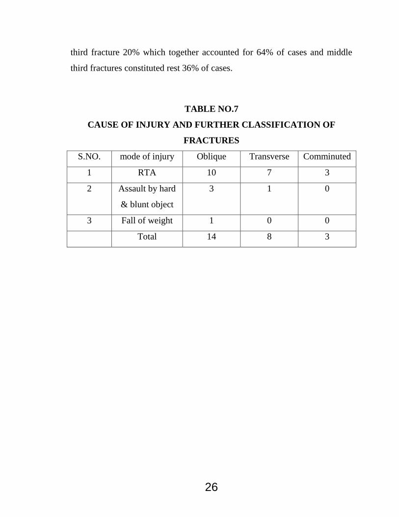
S.NO. mode of injury Oblique Transverse Comminuted
1 RTA 10 7 3
2 Assault by hard
& blunt object
3 1 0
3 Fall of weight 1 0 0
Total 14 8 3

27
TABLE NO.8
ASSOCIATED INJURIES
S.NO. Associated
Fracture
No. of Cases Contralateral/
lpsilateral
%
Fracture shaft
femur
2 lpsilateal 25 2
2 Fracture both
bone leg Upper
third
2 Contralateral 25
3 Fracture medial
Malleolus
1 lpsilateral 12.5
4 Fracture 2nd
&3rd MT
1 lpsilateal 12.5
5 Fracture both
bone forearm
1 lpsilateral 12.5
6 Fracture clavicle 1 lpsilateral 12.5
Out of 25 cases of our series 6 cases had associated lower limb
injuries because of which weight bearing after operation had to be delayed in
5 cases which included 2 cases of fracture shaft femur on ipsilateral side and
2 cases of fracture both bone seg upper 3rd on contralateral side, one case
had fracture medial malleolus of the same side.

28
TABLE NO.9
PERIOD BETWEEN INJURY AND HOSPITALIZATION
S.NO. Duration
Days
Closed % open % Total %
1 0-1 9 52.94 8 47.05 17 68
2 2-3 3 60 2 40 5 20
3 4-5 1 33.33 2 66.66 3 12
Total 13 52 12 48 25 100
Fresh cases were included in the series as is seen in chart out of 25 cases 17
cases (68%) were admitted within 24 hours of injury.5 cases (20%) were
admitted within 3 days of injury and rest (12%) in 4-5 days of injury
TABLE NO.10
INJURY OPERATION INTERWAL
S.NO. Injury operation
Interval days
No. of Cases Percentage
1 3-5 13 52
2 6-8 10 40
3 9-12 2 8
Total 25 100
Early operation was attempted in this series and the cases had the cases had
to wait till routine investigations and pre anaesthetic fitness.52%of cases

29
were operated within 5 days of injury of which 40% were operated within 8
days of injury, one case on 10th day and one on 12th day after injury.
TABLE NO.11
INTRA OPERATIVE COMPLICATIONS
S.NO. Complications No.of Cases Percentage
1 Sphintening of cortex
around window
4 80
2 Broken drill bit 1 20
Total 5 100
In this series there were 6 cases of intraoperative complication of
which 4 cases 66.66% had splintering of anteromedial cortex at the base of
window,1 case 16.67% of splintering of cortex at fracture site was their as
tip of nail was stuck up at posterior cortex of nail There was one case of
broken drill bit as bit was on the nail border rather than being in hole.

30
TABLE NO.12
PARTIAL WEIGHT BEARING WITH/WITHOUT CAST WAS
STARTED EARLY
S.No. Partial weight
bearing started in
post operative
days
No.of
Cases
Total Percentage
with cast With cast
1 5th-10th *5 8 13 52
2 11th-15th 1 1 2 8
3 16th-20th 1 4 5 20
4 21st and above 0 5 5 20
Total 7 18 25 100
* PTB cast was applied after removal of stitches.
Partial weight bearing was promoted during early post operative
period cases were allowed partial weight bearing with the help of standard
walking frame or crutches. 13cases 52% out of 25 case started partial weight
bearing within 10 days of operation of operation of which 5 were given a
PTB cast after stitches removal. These 5 cases were allowed touch down
weight bearing initially with the help of crutches before removal of stitches
without PTB cast. These five cases either had communition at fracture site or
the D nail used was of 8 mm. In 7 cases weight bearing was started after 2
weeks which was mainly done because of superficial infection either at
fracture of at window site,2 cases of these were given PTB after superficial
infection had healed as 8 mm nail was used in these 2 cases. 5 cases had
associated injuries of the lower limbs and these weight bearing was started
after 6 week of operation. No cast was given in these, as patients were

31
confined to bed due to other associated injuries of the lower limb. In these
cases physiotherapy was promoted as soon as pain permitted.
TABLE NO. 13
RELATION BETWEEN ONSET OF PARTIAL WEIGHT BEARING
AND UNION TIME
S.no Partial
weight
bearing
started post
operatively
No.of
Cases
12-14 weeks 15-17
weeks
18-20
weeks
N0. % No. % No. %
1 5th-10th 13 10 76.92 3 23.08 - -
2 11th-15th 2 1 50 1 50 - -
3 16th-20th
day
5 1 20 1 20 2 40
4 21days
&above
5 2 40 2 40 1 20
Total 25 14 7 3
Partial weight bearing was started within 10 days of operation in 13 cases
52% out of these 13 cases 10 cases 76.92% united within 14 weeks and rest
3 cases 23.08% in 17 weeks. In 2 cases the partial weight bearing was
started in around 15 days of operation, one case united within 14 weeks, and
one in 17 weeks, showing that union percentage within 14 weeks fell down
from 76.92% to 50%. In five cases weight bearing was delayed till around
20 days out of these one cases (20%) only united within 14 weeks and one
case of these five cases united within 17 weeks, 2 cases had union after 18
weeks, whereas one case went into non union. In other five cases weight

32
bearing was delayed till around 6 weeks because of associated lower limb
injuries. In these five 40% had union in 14 weeks, 40% in 17 weeks and
20% in 20 weeks. The case in which non-union occurred was one of the
cases which had deep seated infection finally landing into osteomyelitis. In
this case of non union nail had to be removed and canal was reamed. Thus
we found that as we delayed the initial partial weight bearing in patients
union percentage within 14 weeks gradually fell down. Union time raises
between 12-20 weeks averaged being 14.08 weeks.
Table No. 14
USE OF DRAIN AND RELATION TO INFECTIONS
S.NO. Drain No of
Cases
Early
Superficial
infection
Late
deep
infection
No. % No. % No. %
1 Not
used
7 28 4 57.1 2 28.57
2 Used 18 72 3 16.67 - -
Total 25 100 7 2
When procedure was started negative suction drain was not used in 7
cases (28%) and out of these 7 cases, there was superficial infection in 4
cases (57.1%) and of these 4, finally deep infection and osteomyelitis
occurred in 2(28.57%). Later negative suction was used at site of
anteromedial window in 18 cases of which superficial infection occurred in
3 (16.67%) and deep infection in none. Thus use of negative suction drain at
window site decreases the number of infection.

33
TABLE NO. 15
SHOWING TIME TAKEN FOR UNION
S.NO. Union time in
weeks
No. of Cases Percentage
1 12-14 14 56%
2 15-17 7 28
3 18-20 3 12
Union time ranged from 13 weeks to 20 weeks in 24 cases out of 25
cases included in this series. 14 cases (56%) of these united in 12-14 weeks.
Union time on an average was 14.08 weeks. One case had non- union.
34
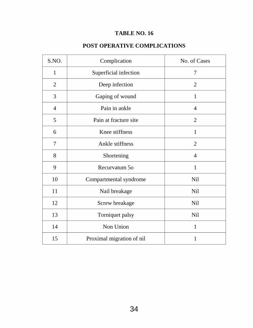
TABLE NO. 16
POST OPERATIVE COMPLICATIONS
S.NO. Complication No. of Cases
1 Superficial infection 7
2 Deep infection 2
3 Gaping of wound 1
4 Pain in ankle 4
5 Pain at fracture site 2
6 Knee stiffness 1
7 Ankle stiffness 2
8 Shortening 4
9 Recurvatum 5o 1
10 Compartmental syndrome Nil
11 Nail breakage Nil
12 Screw breakage Nil
13 Torniquet palsy Nil
14 Non Union 1
15 Proximal migration of nil 1

35
TABLE NO. 17
RESULTS
Results No. of Cases Percentage
Excellent 17 68
Good 6 24
Fair 1 4
Poor 1 4
Total 25 100

36
DISCUSSION
POST OPERATIVE HOSPITAL STAY :
Post operative hospital say varied from 5-10 days, average being 7 days.
Pintore Maffuli et al. (1992) in their series the average hospital say was 9
days regardless of the fracture.
We routinely keep the patient till removal of sutures because most of the
patients were from distant places were adequate health facilities were not
available.
Closed Vs open nailing :
In our series of 25 cases closed nailing could be done in 13 cases (52%) and
open nailing had to be resorted in 12 cases (48%) it was observed that
where injury operation interval was around 1 week or more opening up
fracture site for nailing was done more commonly as compare to those
which were operated within 5 days of injury.
In Arne Ekeland, Thoresen et al. (1988) series of 45 cases 41 were closed
nailing and only 4 were open nailing.
The significant difference between our series and Thoresen et al. series is
due to the fact that we used manual traction and manipulation without X-ray
control where as Thoresen et al. used calcaneal traction, fracture table and
radiological control for their procedure.

37
PARTIAL WEIGHT BEARING :
Partial weight bearing with or without cast was promoted early. In 80% it
was started within 21 days of operation 52% (13) cases it was started within
10 days of operation. In 7 cases (28%) weight bearing was delayed for
around 2 weeks mainly because of superficial infection and rest 5 cases
(20%) weight bearing was delayed till 6 weeks because of associated injuries
in lower limb. In 7 cases out of 25 weight bearing was supported with PTB
cast mainly because there was either communition at fracture site or the nail
used was of 8 mm.
In Arne Ekeland, Thoresen et al. (1992) in their series median time for full
weight bearing was 30 days. In Michael Alms series (1962) average period
of weight bearing was just under 19 days.
TIME TAKEN FOR UNION :
Clinically union was assessed on the basis of absence of tenderness and
abnormal movements at fracture site. Radiological union was mainly judged
on basis of presence of bridging callus, other radiological criteria used were
absence of visibility of fracture line or continuation or trabecular pattern. In
our series of 25 cases 14 cases (56%) united within 14 weeks, 7 cases (28%)
within 17 weeks and 3 cases (12%) took more than 18 weeks to unite. One
case (4%) went into non union, this case was Gustilo grade I compound,
open rainlin had to be done in this case. Partial weight had to be delayed for
2 weeks as patient had superficial infection and finally osteomyelitis,

38
probably all these factors in combination resuled in non union in this case.
Overall union time varied form 12 to 20 weeks averaged being 14.08 weeks.
Arne Ekeland, Thoresen et al. (1988) in their series the median time for
roentgenographic fracture healing was 16 weeks (8-40 weeks). Pintore
Maffuli et al. (1992) in their series consolidation was achieved
approximately in 3 months.
The median time for fracture healing in our present study is comparable to
that of Arne, Thoresen et al. (1988) and Pintore Maffuli et at. (1992) series.
POST OPERATIVE COMPLICATIONS :
In our series there were 7 cases of superficial infection, 2 cases of
deep infection, 1 case of gaping of wound, 4 cases of pain in ankle, 2 cases
of ankle stiffness, 2 cases of pain at fracture site, 1 case of recurvtum (10
degree), 4 cases of shortening, one case of non union, and no case of
compartment sysndrome, tourniquet palsy. There was one case of proximal
migration of nail.
RESULTS :
In our present study all the 25 cases were followed for a period of 4 to
14 months (the average being 6.96 months). The average age of those
injured was 29 years. There was a predominance of young adults. All cases
incidentally were male. Predominant aetiology was road traffic accident
(80%). There were 52% closed and 48% Grade I Gustilo fractures. 56%
were oblique, 32% transverse and 12% comminuted fractures. Cases
selected were mainly of lower and middle third shaft tibia.
39
Preoperative antibiotics was administered in all patients 6 hours
before surgery, intraoperatively before the inflation of tourniquet. In this
series 13 cases were operated within 5 days of injury, 10 cases were 6 to 8
days of injury and 2 cases in 10 days. Partial weight bearing was started with
P.T.B. cast in 7 cases and without cast in 18 cases, 52% of cases started
partial weight bearing within 10 days of operation. Knee and ankle
movements were started as soon as pain permitted. Average hospital stay
was 21 day, 60% cases united within 14 weeks with an overall average
union period being 14.08 weeks. Final functional results were assessed on
the basis of criteria given by Thoresen. Superficial infection in 9 cases, deep
infection in 10 cases, revurvatum of 10 degree in 1 case, shortening in 4
cases, pain in ankle in 4 cases, pain at fracture site in 1 case of non union,
one case of proximal migrations of nail were the post operative complication
seen.
Based on Thoresen’s criteria results were 68% excellent 24% good
and 4% fair and 4% poor.
Arne Ekeland, Thoresen et al. (1988) treated 45 tibial shaft fracture
with Gross Kempf nailing. The Final evaluation was made using Thoresen’s
criteria. The results were 64.44% excellent, 28.88 good, 4.44 fair and 2.22%
poor.
Riquelme Rodriguez et al. (1992) treated 50 tibial shaft fractures with
Grosse Kempf interlocking nail. The final evaluation were made as per
Thorensen’s criteria the results were 92% excellent, 4% good and 4% poor.

40
The results of our present study are comparable with he results of the
studies conducted by Arne Ekeland, Thoresen et al (1988).
In this series of ours we did not use negative suction drain in initial 7
cases and we found a direct relation of it with superficial infection. After we
started using negative suction drain infection rate went down to almost nil.
Therefore we recommend use of negative suction drain at window site.

41
SUMMARY AND CONSLUSION
The study of MANAGEMENT OF LOWER AND MIDDLE THIRD TIBIA
FRACTURE BY ENDERS INTERLOCKING NAIL was carried out in he
Department of Orthopaedics and Traumatology, P.G. Hospital, Jabalpur
consisting of 25 cases of fracture shaft of Tibia.
In the light of results obtained it can be summarized and concluded that :
1. It is an effective modality which allows early ambulation and
weight bearing, and decreased dependency.
2. Infection rate is low provided negative suction drain is used ate
window site.
3. It reduces hospital stay of patients and later patient can return early
to work, thus minimizes psychological trauma and financial burden
to the patient.
4. It given good results in terms of union and functional recovery.
5. It is economical.
6. It is done by a simple technique can be mastered easily and does
not require costly instrumentation of IITV thus can be done in
remote areas.
Fracture of tibia arises controversies regarding any standardized
method of treatment, but with encouraging and convincing results
here is a better future for fracture of tibia leading to good solution
to controversial problem of fractures of tibia by ENDERS
interlocking Nailing System.