biomathematical pattern of emg signal propagation in ...€¦ · malgorzata domino, bartosz...
TRANSCRIPT

RESEARCH ARTICLE
Biomathematical pattern of EMG signal
propagation in smooth muscle of the non-
pregnant porcine uterus
Malgorzata Domino, Bartosz Pawlinski, Zdzislaw Gajewski*
Department of Large Animal Diseases with Clinic, Veterinary Research Centre and Center for Biomedical
Research, Faculty of Veterinary Medicine, Warsaw University of Life Sciences (WULS – SGGW), Warsaw,
Poland
Abstract
Uterine contractions are generated by myometrial smooth muscle cells (SMCs) that com-
prise most of the myometrial layer of the uterine wall. Aberrant uterine motility (i.e., hypo- or
hyper-contractility or asynchronous contractions) has been implicated in the pathogenesis
of infertility due to the failure of implantation, endometriosis and abnormal estrous cycles.
The mechanism whereby the non-pregnant uterus initiates spontaneous contractions
remains poorly understood. The aim of the present study was to employ linear synchroniza-
tion measures for analyzing the pattern of EMG signal propagation (direction and speed) in
smooth muscles of the non-pregnant porcine uterus in vivo using telemetry recording sys-
tem. It has been revealed that the EMG signal conduction in the uterine wall of the non-preg-
nant sow does not occur at random but it rather exhibits specific directions and speed. All
detectable EMG signals moved along the uterine horn in both cervico-tubal and tubo-cervi-
cal directions. The signal migration speed could be divided into the three main types or cate-
gories: i. slow basic migration rhythm (SBMR); ii. rapid basic migration rhythm (RBMR); and
iii. rapid accessory migration rhythm (RAMR). In conclusion, the EMG signal propagation in
smooth muscles of the porcine uterus in vivo can be assessed using a linear synchroniza-
tion model. Physiological pattern of the uterine contractile activity determined in this study
provides a basis for future investigations of normal and pathologicall myogenic function of
the uterus.
Introduction
Uterine contractions are generated by myometrial smooth muscle cells (SMCs) comprising
most of the myometrial layer of the uterine wall. Synchronous contractions of SMCs are
responsible for normal gamete transport in the female reproductive tract and contribute to the
expulsion of uterine debris [1]. The occurrence of uterine contractions must be strictly con-
trolled and coordinated to sustain these reproductive functions [1, 2]. Abnormal patterns of
uterine motility such as hypoactive, hyperactive or asynchronous contractions have been
PLOS ONE | DOI:10.1371/journal.pone.0173452 March 10, 2017 1 / 14
a1111111111
a1111111111
a1111111111
a1111111111
a1111111111
OPENACCESS
Citation: Domino M, Pawlinski B, Gajewski Z
(2017) Biomathematical pattern of EMG signal
propagation in smooth muscle of the non-pregnant
porcine uterus. PLoS ONE 12(3): e0173452.
doi:10.1371/journal.pone.0173452
Editor: Roger C. Young, UNITED STATES
Received: July 26, 2016
Accepted: February 22, 2017
Published: March 10, 2017
Copyright: © 2017 Domino et al. This is an open
access article distributed under the terms of the
Creative Commons Attribution License, which
permits unrestricted use, distribution, and
reproduction in any medium, provided the original
author and source are credited.
Data Availability Statement: All relevant data are
within the paper.
Funding: This work was supported by the State
Committee for Research Investigation grant NN308
077439, Narodowe Centrum Nauki, https://www.
ncn.gov.pl/. This work was conducted in the
Veterinary Research Centre WULS (WCB) and the
Center for Biomedical Research (CBB) supported
by EFRR RPO WM 2007-2013. The funders had no
role in study design, data collection and analysis,
decision to publish, or preparation of the
manuscript.
implicated in the pathogenesis of infertility, implantation failure, endometriosis and abnormal
estrous cycles [3]. However, the specific mechanism by which the non-pregnant uterus auton-
omously initiates myometrial contractions remains poorly described. In spite of their clinical
significance, contractile properties of the uterus during the entire estrous cycle have not been
examined [4]. A better understanding of the complex myometrial activity would help to
develop effective therapies for an array of reproductive disorders associated with the flawed
rhythmicity of uterine contractions.
A single impulse can initiate a myometrial contraction but multiple, coordinated impulses
are needed for forceful and sustained contractions [5]. Moreover, individual impulses within
the myometrial tissue may differ in speed and direction, and such intrinsic variations in con-
duction velocities are essential for sustaining forceful and coordinated muscular contractions
[6, 7]. Uterine SMCs themselves trigger cellular processes resulting in uterine contractions.
The typical sequence of these interrelated processes is referred to as the excitation-contraction
(EC) coupling [8] and entails three stages: Ca2+ entry into the cell; Ca2+ expulsion from the
cell; and SMC contraction. Calcium influx occurs through the voltage-dependent calcium
channels in response to depolarization of the cell membrane and results in a transient increase
in intracellular concentrations of Ca2+ ions [1, 2]. The voltage- and time-dependent changes in
the membrane ionic permeability initiate an action potential (AP) or myoelectrical activity [5].
Cell membrane stimulated by external impulses responds with a change in the transmembrane
electrical potential, which leads to opening of membrane channels to let ions enter or leave the
cell [8]. Calcium ions constitute the main component of depolarizing current during the prop-
agation of AP as well as the most important factor determining contractile force of myometrial
myocytes [1]. Elevated level of intracellular Ca2+ ions allows for binding of Ca2+ by calmodu-
lin, which ultimately results in activation of the light-chain myosin kinase and phosphoryla-
tion. Subsequently, the cross-bridges between actin and myosin filaments are formed and
SMCs contract [1, 2]. There is a paucity of studies on the generation of EC processes by the
uterine SMCs in situ. Such investigations are confined to the determination of intrauterine
pressure and uterine electrical activity during simulated uterine contractions [8, 9]. For exam-
ple, Cochran and Gao [9] designed the electromechanical model to simulate intrauterine pres-
sure and to assess uterine contractions during parturition. Sharifimajd et al. [8] developed an
electro-chemo-mechanical model to replicate excitation, activation, and contraction of uterine
smooth muscle cells. In both cases, uterine contractions were induced “from cellular level to
the organ level” due to electrophysiological properties of the pacemaker cells and ensuing
propagation of electrical discharges via gap junctions within the myometrium. Most of earlier
studies on uterine activity deal primarily with the electrophysiology of the SMC membrane [4,
10]. In all in vitro investigations, at the level of single myometrial myocyte, equations describ-
ing various ionic membrane currents were established from their respective activation curves.
Parameters were determined using the voltage-clamp technique and measurements performed
on isolated cells [11]. This particular approach is excellent for describing contribution of cellu-
lar phenomena in muscular contractions but provides inadequate information on contractile
activity of the tissue or organ [8, 12]. Alternatively, in vivo models of uterine contractility uti-
lize the “top-down approach” in which function in the entire organ or tissue following activa-
tion of SMCs is monitored.
Changes in electrical activity of smooth muscle fibers during contraction or relaxation can
be detected on surface of skin (surface EMG) or directly within the myometrium (needle
EMG) as an electromyography (EMG) signal. The EMG is the sum of bioelectrical signals or
electric currents produced by differences in bioelectrical potentials along a specialized tissue
and is associated with electrochemical events occurring during the propagation of action
potential [13]. Two distinctive forms of action potential have been recorded in myometrium of
Biomathematical pattern of EMG signal in smooth muscle of the non-pregnant porcine uterus
PLOS ONE | DOI:10.1371/journal.pone.0173452 March 10, 2017 2 / 14
Competing interests: The authors have declared
that no competing interests exist.

various species, namely a single action potentials and multiple action potentials. The single
action potential, called the "single spike", consists of rapid depolarization followed by immedi-
ate repolarization of cell membranes. These spikes often occur in sequels called "bursts", num-
ber and frequency of spikes within burst predetermine strength and rapidness of smooth
muscle contraction. Multiple spontaneous action potentials (bursts) seem to arise simulta-
neously in different regions of the uterine wall [1, 12]. The frequency and duration of the
bursts as well as the frequency of spikes within a burst shows considerable individual variations
and vary with the stage of parturition [7]. The propagation of electrical activity in the myome-
trium mainly depends on arrangement of myometrial bundle fibers [6], population of gap
junctions formed between adjacent cells during each contraction [14, 15], and waves of cal-
cium transport [16]. In contrast to skeletal muscle, in which the propagation of action poten-
tials that progresses in muscle fibers is highly predictable, the direction and speed of electrical
activation in myometrial cells remain to be elucidated [17]. Most of previously published stud-
ies were devoted to the analysis of entire bursts or single spikes in pregnant uterus [8, 18, 19,
20], and only few studies dealt with non-pregnant state [4, 21]. Hence, the main goal of the
present study was to determine the biomathematical pattern describing EMG signal propaga-
tion in the non-gravid porcine uterus in vivo.
Materials and methods
Data acquisition
The experiment has been conducted on 10 mature Polish Landrace sows (n = 10). The experi-
ment has been conducted according to applicable national and international ethical guidelines
and all efforts were made to minimize animals suffering. The protocol was approved by the III
Local Ethical Committee on Animal Testing in Warsaw (Permit Number: 71/2009, from
19.11.2009) on behalf of the National Ethical Committees on Animal Testing. Sows had been
adapted to the animal facilities for 7 days before studies. During the entire experiment animals
were housed in metabolic cages, fed and watered ad libitum. In order to maintain animal wel-
fare surgery was carried out under general anesthesia and the telemetry EMG recording
method was applied. Telemetry method [22] allows to reduce the number of animals used in
the experiment and provide long-term registration without stress connected with immobiliza-
tion. Spontaneous uterine activity in non-pregnant state during diestrus was recorded by the
combination of three electrodes connected to 3-channel transmitter used in large animals. The
telemetry transmitter TL10M3-D70-EEE (DSI, St. Paul, Minnesota, USA) was surgically posi-
tioned between abdominal muscles and three silver bipolar needle electrodes were sutured
onto different topographic regions of uterus: right uterine horn (RUH—channel 1), corpus
uteri (CU—channel 2) and the left uterine horn (LUH—channel 3) surfaces (Fig 1). The dis-
tance between electrodes (channel 1 to channel 2 and channel 2 to channel 3) was constant
and fixed at 17 cm. Animals were premedicated with an intramuscular injection of azaperone
(Stresnil, 3 [mg/kg b.wt.], i.m., Janssen Pharmaceutica) and then catheter was inserted into the
auricular vein. Surgery was carried out under general anesthesia, consisting of combined
administration of medetomidyne (Cepetor, 1 [mg/kg b.wt.], i.v., CP- Pharma Handelsges),
butorphanol (Butomidor, 0,2 [mg/kgb.wt.], i.v., Ricgter Pharma AG), ketamine (Bioketan, 3
[mg/kg b.wt.], i.v., Vetoquinol Biowet) and propofol (Propofol, 2–4 [mg/kg b.wt.], i.v., Pfizer).
Then pigs recovered from surgery analgesic—meloxicam (Metacam 0.4 [mg/kg b.wt.], i.m.,
Boehringer Ingelheim) and anti-microbial—cefquinom (Cobactan, 2.0 [mg/kg b.wt.], i.m.,
Intervet) had been administered for 5 days. Obtained analog signal was digitalized and sent by
radio waves to the telemetric receiver (DL10 analog output (DSI)). The signal was acquired
with a 3-channel transmitter (PowerLab (ADInstruments, Melbourne, Australia) and
Biomathematical pattern of EMG signal in smooth muscle of the non-pregnant porcine uterus
PLOS ONE | DOI:10.1371/journal.pone.0173452 March 10, 2017 3 / 14

analyzed. Sampling frequency was 100 Hz. Pigs were euthanized at the end of the experiment
by—Sodium Pentobarbital (Morbital 100.0 or [mg/kg b.wt.], i.v., Biowet Pulawy).
EMG data processing
The EMG signals were digitally filtered with a band-pass filter [5–50 Hz], then power line
interference was lowered from the EMG recordings with a notch filter. Mean and linear trends
were removed [23].
Uterine contractions were defined as series of electrical potentials with amplitude above
5 μV and a duration of more than 3 s, separated from each of next series by a time period not
less than 5 s. Any new electrical activity after this period (5 s) was interpreted as a subsequent
contraction (8). Each contraction was described using spike and burst parameters. The spike
represented single action potentials while the burst comprised multiple action potentials.
Mean amplitude [mV], mean RMS (root mean square) [mV], duration of electrical activity [s],
duration of pauses [s], and number of spikes forming a burst were analyzed relative to bursts
and spikes, respectively.
The EMG signal spectral content was analyzed in addition to studying changes in its time
domain features. The spectrum analysis gave amplitudes of each of pure tones of different fre-
quencies and phases, summed in single EMG signal. The amplitude spectrum of investigated
signals was defined as the distribution of amplitudes over different frequencies. By means of
Fourier analysis (FFT—Fast Fourier transform), dominant frequency (DF) [Hz] (the frequency
at which most signal energy was transmitted) was assessed for each data series. We used the
Hamming window only for the Fourier analysis [21, 24].
EMG channel synchronization
The uterus is indubitably a complex system in which billions of cells comprising myometrium
interact in a complex manner. Our understanding of the co-ordination of uterine contractions
is incomplete. In physiological research, multivariate data sets containing two or more simul-
taneously recorded time series are usually examined to establish signals similarity [24]. We
used similarity measures based on the concept of time series data synchronization. In order to
describe similarities between EMG signals, we used two-dimensional functions analysis.
Degree of synchronization between three simultaneously recorded data series (channels 1(x),
Fig 1. Diagram of the measurement system. Electrodes were arranged in the porcine reproductive tract (A) and the EMG signals (B)
were sampled from: channel 1, right uterine horn, RUH; channel 2, corpus uteri, CU; and channel 3, left uterine horn, LUH.
doi:10.1371/journal.pone.0173452.g001
Biomathematical pattern of EMG signal in smooth muscle of the non-pregnant porcine uterus
PLOS ONE | DOI:10.1371/journal.pone.0173452 March 10, 2017 4 / 14

2(y), 3(z)) was estimated for two signal pairs (xy and yz) using linear measures: the cross-cor-
relation function (ƒx,y(l), ƒy,z(l)) and the cross-coherence function (Cxy(ƒ), Cyz(ƒ)). The coher-
ence function in frequency domain was equivalent to the correlation function in time domain.
Cxy was estimated after Fourier analysis (FFT) [21].
Cross-correlation analysis. Cross-correlation function gives the correlation degree
between the two signals—amplitude data series (pair xy—channel 1 and channel 2 / pair yz—
channel 2 and channel 3) [25]. The cross-correlation function of two discrete data series x(n)
and y(n) is a statistical quantity defined as follows:
fx; yðlÞ ¼covðx; yÞSdxx Sdy
ð1Þ
Signals x(n), y(n) and z(n) are collected from the right uterine horn, corpus uteri and left
uterine horn electrodes, respectively.
After normalization, the value of cross-correlation parameters will be between -1 and 1.
The result is 1 if x(n) = y(n) or y(n) = z(n) and l = 0; ƒx,y(l) = 0 if x,y are statistically indepen-
dent and it is -1 if x(n) = —y(n) or y(n) = —z(n) and l = 0. Synchronization is high if |ƒx,y(l)| =
1 and absent if ƒx,y(l) = 0.
Cross-coherence analysis. The cross-coherence function gives the coherence degree
between two signals—dominant frequencies data series (pair xy—channel 1 and channel 2 /
pair yz—channel 2 and channel 3) [25]. The cross-coherence function of two discrete data
series x(n) and y(n) is a statistical quantity defined as follows:
Cxyðf Þ ¼jPxyðf Þj
2
Pxxðf Þ x Pyyðf Þð2Þ
It is real-valued, positive and normalized to vary between 0 (no coherence) and 1 (complete
coherence). In the same way, high synchronization corresponds to a value of 1, whereas a
value of 0 indicates lack of synchronization.
Description of the experimental model. Values for the cross-correlation coefficient |r|
and cross-coherence coefficient |R| were estimated separately for two pairs of discrete time
series x(n) and y(n) as well as y(n) and z(n). Synchronization between the right uterine horn
(x(n)) and corpus uteri (y(n)) as well as the corpus uteri (y(n)) and left uterine horn (z(n)) was
observed. The threshold at which the values of |r| and |R| were considered significant was
determined empirically, and was always over > 0.50 with significant strength from middle to
very high. The burst pairs with an empirical value of |R| over threshold were considered as syn-
chronized. Results of linear synchronization measures application to pairs of EMG signals
demonstrated that the cross-coherence function is effective in determining bursts similarity.
Similarity measures based on the concept of synchronization allow detection of bursts propa-
gation in different topographic regions. They were used to "identify" the frequency fingerprint
of the spatial burst and determined when the burst reached the other location during
propagation.
Locations of the first and second electrodes (on the x/y or y/z axis) were used to measure
highly synchronized signals, and the direction of bursts propagation was evaluated. The sec-
ond signal location (on the x/y or y/z axis) in the time function pointed towards cervico-tubal
and tubo-cervical directions.
The speed of bursts propagation was evaluated based on the time elapsed for highly syn-
chronized signals to move from the first electrode to the second one (measuring points on the
x/y or y/z axis).
Biomathematical pattern of EMG signal in smooth muscle of the non-pregnant porcine uterus
PLOS ONE | DOI:10.1371/journal.pone.0173452 March 10, 2017 5 / 14
Statistical analysis. To assess statistical differences among three channel data series, a
one-way ANOVA and the Kruskal-Wallis test (the level of statistical significance was set to
P<0.05) were performed. To evaluate the percentage of signal propagation (direction and
speed) in relation to the total number of synchronizations examined, non-parametric, two-
tailed Mann-Whitney test was applied (P<0.01). The independence of speed was tested using
a non-parametric Kruskal-Wallis test with Dunn’s multiple comparisons test (P<0.05) after-
wards, while the homogeneity of speed populations was defined using a one-way ANOVA test
followed by Tukey’s multiple comparisons test (P<0.05) using GraphPad Prism 6 (GraphPad
Software Inc., San Diego, CA, USA).
Results
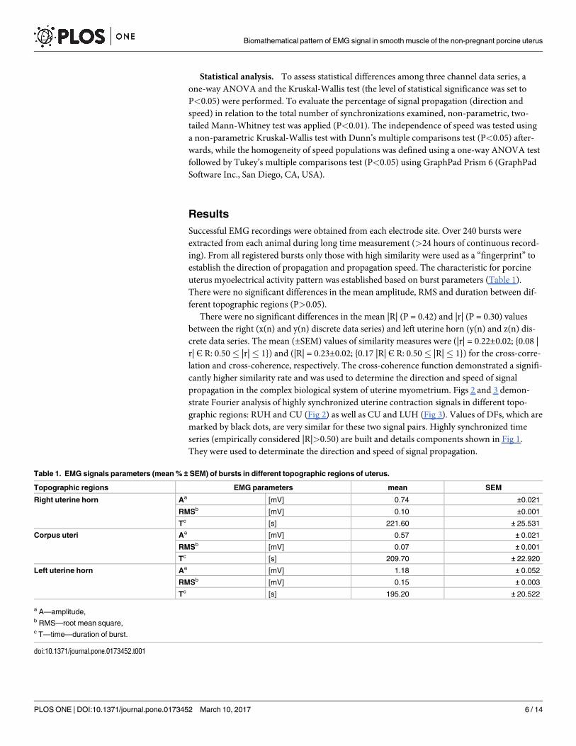
Successful EMG recordings were obtained from each electrode site. Over 240 bursts were
extracted from each animal during long time measurement (>24 hours of continuous record-
ing). From all registered bursts only those with high similarity were used as a “fingerprint” to
establish the direction of propagation and propagation speed. The characteristic for porcine
uterus myoelectrical activity pattern was established based on burst parameters (Table 1).
There were no significant differences in the mean amplitude, RMS and duration between dif-
ferent topographic regions (P>0.05).
There were no significant differences in the mean |R| (P = 0.42) and |r| (P = 0.30) values
between the right (x(n) and y(n) discrete data series) and left uterine horn (y(n) and z(n) dis-
crete data series. The mean (±SEM) values of similarity measures were (|r| = 0.22±0.02; {0.08 |
r| Є R: 0.50� |r|� 1}) and (|R| = 0.23±0.02; {0.17 |R| Є R: 0.50� |R|� 1}) for the cross-corre-
lation and cross-coherence, respectively. The cross-coherence function demonstrated a signifi-
cantly higher similarity rate and was used to determine the direction and speed of signal
propagation in the complex biological system of uterine myometrium. Figs 2 and 3 demon-
strate Fourier analysis of highly synchronized uterine contraction signals in different topo-
graphic regions: RUH and CU (Fig 2) as well as CU and LUH (Fig 3). Values of DFs, which are
marked by black dots, are very similar for these two signal pairs. Highly synchronized time
series (empirically considered |R|>0.50) are built and details components shown in Fig 1.
They were used to determinate the direction and speed of signal propagation.
Table 1. EMG signals parameters (mean % ± SEM) of bursts in different topographic regions of uterus.
Topographic regions EMG parameters mean SEM
Right uterine horn Aa [mV] 0.74 ±0.021
RMSb [mV] 0.10 ±0.001
Tc [s] 221.60 ± 25.531
Corpus uteri Aa [mV] 0.57 ± 0.021
RMSb [mV] 0.07 ± 0,001
Tc [s] 209.70 ± 22.920
Left uterine horn Aa [mV] 1.18 ± 0.052
RMSb [mV] 0.15 ± 0.003
Tc [s] 195.20 ± 20.522
a A—amplitude,b RMS—root mean square,c T—time—duration of burst.
doi:10.1371/journal.pone.0173452.t001
Biomathematical pattern of EMG signal in smooth muscle of the non-pregnant porcine uterus
PLOS ONE | DOI:10.1371/journal.pone.0173452 March 10, 2017 6 / 14

The EMG signals were propagated along the uterine horn in both cervico-tubal (from CU
to UH) and tubo-cervical (from UH to CU) directions. There were no significant differences
(P = 0.20) between the percentages (mean % ± the standard error of mean (SEM)) of bursts
propagated in both directions, from CU to UH (45.30% ± 3.27) and from UH to CU (4.73% ±3.25) in relation to the total number of highly synchronized uterine contraction signals. There
were also no significant differences (P = 0.20 and P = 0.60, respectively) between bursts propa-
gation in the right and left uterine horns (Table 2).
EMG signals were propagated along the uterine horn with three significantly different
(P<0.0001), independent speeds: SBMR (slow basic migration rhythm), RBMR (rapid basic
migration rhythm) and RAMR (rapid accessory migration rhythm). Bursts propagation speed
was determined experimentally and tested for homogeneity inside the population for: SBMR,
RBMR and RAMR. All propagation speeds were considered to be homogeneous (P>0.05).
The SBMR, RBMR and RAMR values (mean ± SEM) as well as the percentage (mean %) of
bursts propagation speed in relation to total number of highly synchronized uterine contrac-
tion signals are presented in Table 3.
Fig 2. Fourier analysis of highly synchronized EMG signals. EMG signals from the right uterine horn (A, B) and corpus uteri (C, D) in
time (A, C) and frequency (B, D) domains.
doi:10.1371/journal.pone.0173452.g002
Biomathematical pattern of EMG signal in smooth muscle of the non-pregnant porcine uterus
PLOS ONE | DOI:10.1371/journal.pone.0173452 March 10, 2017 7 / 14

Fig 3. Fourier analysis of highly synchronized EMG signals. EMG signals from the corpus uteri (A, B) and left uterine horn (C, D) in
time (A, C) and frequency (B, D) domains.
doi:10.1371/journal.pone.0173452.g003
Table 2. The percentage (mean % ± SEM) of bursts propagation directions along the uterus in relation
to the total number of highly synchronized uterine contraction signals.
The direction Tubo-cervical Cervico-tubal
Right uterine horn RUH-CU a CU-RUH b
mean [%] 24.5 20.8
SEM ±2.27 ±3.75
Left uterine horn LUH-CU c CU-LUH d
mean [%] 30.2 24.5
SEM ±1.07 ±0.53
P 0.20 0.60
a RUH-CU—tubo-cervical direction in the right uterine horn,b CU-RUH—cervico-tubal direction in the right uterine horn,c LUH-CU—tubo-cervical direction in the left uterine horn,d CU-LUH—cervico-tubal direction in the left uterine horn. Mann-Whitney test (P<0.01)
doi:10.1371/journal.pone.0173452.t002
Biomathematical pattern of EMG signal in smooth muscle of the non-pregnant porcine uterus
PLOS ONE | DOI:10.1371/journal.pone.0173452 March 10, 2017 8 / 14

Discussion
Changes in the number of spikes affected the amplitude measures in time domain without
altering the frequency domain and the DF patterns. This could be an explanation for signifi-
cantly higher similarity rates in the frequency vs. time domain for burst similarity parameters.
Variation in number of spikes can be caused by the progressive and reverse recruitment phe-
nomenon where the muscle area affected by spike within burst enlarges progressively [20].
Moreover, contractions demonstrate different propagation parameters, so within the same
burst, analysis of single spike can lead to more extensive information compared to the whole
burst analysis [7, 20]. Consequently, the combination of simultaneous single spike and burst
analyses used in this study significantly reduce inherent variability in the EMG sensitivity in
the porcine myometrium.
In a previous study [21] we demonstrated the usefulness of the cross-coherence function
in synchronization between uterine horn and corpus uteri for multiple action potentials in
short time measurement (<10 hour of continuous recording). In this study the linear syn-
chronization measured in frequency domain was used to determine direction and speed of
signal propagation in long time measurement (>24 hours of continuous recording). We
stated that determination of detail of signal propagation in short time measurement is unreli-
able. Recent studies have suggested that both frequency and synchrony of the uterine electri-
cal activity could be potential indicators of predicting uterine contraction [26]. We stated
that frequency and synchrony are also suitable for determining direction and speed of those
contractions in the non-pregnant uterus. Nevertheless both of these studies [21, 26] and the
current one considered cross-coefficients over > 0.50 based on Fourier and synchronization
analysis of experimental data. Therefore we stated that similarity measures based on the con-
cept of synchronization analysis can be implemented with fast algorithms and that this
method is suitable for real-time applications. The analysis method may be generalized to ani-
mal and human data obtained during labor, in pregnancy and non-pregnant state.
The high accuracy of identifying the frequency fingerprint of the spatial burst allowed to
determine the signal migration based on three measuring points. Analysis methods, used in
the experiment, eased the limits on the ability to interpret data obtained from a limited num-
ber of probes in each horn. Langendijk et al. [27] successfully described spontaneous uterine
activity around estrus in sows based on open-end catheter technique and one measuring
point. Bower and Zerobin et al. [28, 29] established porcine uterine activity including direction
of contractility propagation and used for this purpose two measuring points. Gajewski et al.
[22, 30] efficiently completed the myoactivity findings based on three measuring points but
didn’t carry out biomatematical signal analysis. The myometrial activity detecting methods
Table 3. The values (mean ± SEM) and the percentage (mean %) of bursts propagation speed along
the uterus in relation to the total number of highly synchronized uterine contraction signals.
The speed SBMR a RBMR b RAMR c
mean [mm/min] 1.25 2.47 7.07
SEM ±0.04 ±0.08 ±0.60
mean [%] 46.01 26.30 27.30
a SBMR—slow basic migration rhythm,b RBMR—rapid basic migration rhythm,c RAMR—rapid accessory migration rhythm. For independence: Kruskal-Wallis test with Dunn’s multiple
comparisons test (P<0.05). For homogeneity: one-way ANOVA test with Tukey’s multiple comparisons test
(P<0.05).
doi:10.1371/journal.pone.0173452.t003
Biomathematical pattern of EMG signal in smooth muscle of the non-pregnant porcine uterus
PLOS ONE | DOI:10.1371/journal.pone.0173452 March 10, 2017 9 / 14

result in the limited number of probes. All authors recorded myometrial activity directly
within the myometrium (EMG) [22, 28, 29, 30, 31] or used intrauterine pressure (IUP) [27].
An alternative method for monitoring the uterine activity based on surface EMG—EHG (elec-
trohysterogram) allowed to application from 3 [32] to 16 [33] and 64 [20] active electrodes
located on abdomen skin. The needle EMG picked up the bioelectrical signal directly associ-
ated with the muscular activity of myometrium, while EHG collected signal abundant in noise
generated between cells and electrodes. Therefore, the electrical activity recorded directly from
uterus may provide essential information about uterine activity and permit the prediction of
the EHG associated with the contraction. However different dedicated methods for parameter
estimation have been applied, from animal in vitro experiments (invasive EMG) and human
clinical studies (EHG). Limitations during invasive in vivo experiments result from hardware
and equipment deficiencies. Implantable telemetry systems, designed for monitoring and col-
lecting data from large animals, have disadvantage such as only 3-channel transmitter, limited
source of energy and no possibility to implant more than one transmitter in one animal
[30, 31].
The uterine activity in non-pregnant sows was recorded via uterine intraluminal pressure
measurements (non-surgical technique) [27]. Oviductal and myometrial activity during the
periovulatory period in pigs was described using a long-term electromyography combined
with the telemetry recording system (invasive method) [22, 30, 31]. Both of those approaches
failed to determine the specific direction of waves of uterine contractions. Others have sug-
gested that uterine contractions occur along uterine horns [28, 29] in two opposite directions,
tubo-cervical and cervico-tubal, although the tubo-cervical direction appeared to be predom-
inant. In those studies, the myometrial activity was recorded at two different sites in the tips
of both uterine horns [28, 29], and it was suggested that the bulk of myometrial activity origi-
nated in those regions and was conveyed along uterine horns in tubo-cervical direction [27].
Both ends of the uterine horn (tubal and the cervical) are tentatively anatomical regions with
the strong pacemaker activity [22]. Based on our present observations, EMG signals preced-
ing myometrial contractions in the pig’s uterus are propagated relatively uniformly in both
cervico-tubal and tubo-cervical directions. This is in agreement with the results of myoelec-
trical burst analyses in women where both upward and multidirectional propagation patterns
have been reported [33, 34]. This “mixed” propagation pattern may serve to facilitate sperm
transport along the uterine horns and/or re-distribution of semen throughout the two horns
in the case of unilateral deposition of semen [35]. While the cervico-tubal direction of uterine
contractions promotes the distribution of semen over the two horns, the tubo-cervical con-
tractions are important for expelling seminal plasma after mating. Therefore, uniform bidi-
rectional contractions of uterus may play a key role in fertilization process; the direction in
which uterine contractions are propagated may ultimately influence the rate of sperm trans-
port and conception [36].
Based on the tubular structure of porcine uterus two directions were analyzed: cervico-
tubal and tubo-cervical. Extending the discussion of histology structure of the uterus where
billions of cells comprising myometrium interact in a complex manner in longitudinal and cir-
cular muscle layers the oblong, the spiral path as well as a special path via pacemaker cells net-
work directions have to be considerate. Those complex pathways of signal may be reflected in
speed of signal propagation: the slowest signal propagation via spiral path in circular muscle
layer, faster via oblong path in longitudinal muscle layer and the fastest via ICLC network.
Having obtained frequency fingerprint identification of high similar bursts we measured
the time lag between signals and, the distance between sensors, and calculated the speed. The
speed at which an action potential propagates along a muscle fiber or tissue is referred to as
propagation (or conduction) speed [17, 37]. Recent studies described the speed of action
Biomathematical pattern of EMG signal in smooth muscle of the non-pregnant porcine uterus
PLOS ONE | DOI:10.1371/journal.pone.0173452 March 10, 2017 10 / 14
potential propagation in the muscular layer of isolated uterine strips in animals [18, 36, 38]
and whole uterus in women [20]. Once again the analysis method may be generalized to pig,
guinea pig, rabbit, cat and human data obtained during in vivo and in vitro experiments.
As is evident from the analysis of over 240 bursts extracted from each animal, in many occa-
sions the EMG signal started between the sensors. In this case we observed no synchronization
between time series data. The unsynchronized signals were not taken under consideration dur-
ing speed calculation. We suggested the analysis of propagation speed, based on assumption
that the signal spreads out linearly and thus it can be detected at different locations after a cer-
tain delay [6, 19, 20]. However, there is a great deal of evidence that the linear propagation of
single electrical spikes occurs in the uterus and that speed of their propagation can also be
measured [18, 19, 20, 29]. In this study, we employed the concept of simultaneous single spike
and burst linear analysis in the frequency domain. We suggested to divide the propagation
speed uterus into one of three distinctive categories: slow and rapid basic migration rhythm
(SBMR, RBMR) and rapid accessory migration rhythm (RAMR).
In circular and longitudinal muscles of the uterus, the single spike is the result of the depo-
larization phase. The single spike is able to initiate a short contraction, but multiple, higher-
frequency, coordinated spikes forming bursts are necessary to trigger forceful and sustained
contractions [14]. We speculate, that in the porcine non-pregnant uterus the bungles are prop-
agated across fibers in circular muscles of uterus and along the fibers in longitudinal muscles.
We suspect these two propagation pathways to be connected with the two estimated basic
migration rhythms: SBMR in circular and RBMR in longitudinal muscles. The conduction of
electrical activity in the uterus relies on the cell-to-cell coupling by gap junctions composed of
connexin proteins [5]. The grouping of connexins provides channels of low electrical resis-
tance between cells, and thereby furnishes pathways for efficient conduction of action poten-
tials [14]. Circular muscles (in inner layer of the myometrium) contain more gap junctions
than longitudinal muscles (the outer layer of the myometrium) per length unit. In the myome-
trium bioelectrical signal propagation occurs more rapidly in the longitudinal direction then
in the circumferential one [17].
Moreover, the presence of interstitial Cajal-like Cells (ICLC) located among SMCs was
demonstrated in the non-pregnant human [39] and porcine [40] myometrium. ICLC have
been shown to form a network integrating SMCs, nerves and blood vessels. ICLC possess puta-
tive bioelectrical properties based on their morphological features such as numerous gap junc-
tions and presence of calcium handling units typical of pacemaker cells [41]. It was suggested
that the c-kit activity of ICLC is involved in the spontaneous rhythmic contractions of uterine
myocytes. The contraction frequency in longitudinal layer of uterine strips from human can
be reduced or inhibited with imatinib (the c-kit/CD117 tyrosine-kinase inhibitor) in a dose-
dependent manner [42]. It is attractive to speculate that cellular networks containing ICLC
may be responsible for the third accessory migration rhythm detected in the present study, the
RAMR. Rapid bursts could be propagated by the ICLC system including longest cellular pro-
cesses (except for neurons) that form intricate networks between smooth muscle cells.
Conclusions
In conclusion, the EMG signal propagation in smooth uterine muscles could be determined
using linear synchronization measures. We described for the first time the successful cross-
correlation function the EMG signal propagation in a long term in vivo experiment in the
non-gravid porcine uterus. The pattern of the EMG signal propagation is not random but it
occurs in an orderly, bidirectional fashion and at distinctive speed. The spontaneous potentials
are propagated equally in both directions between both horns and the uterus. Since other
Biomathematical pattern of EMG signal in smooth muscle of the non-pregnant porcine uterus
PLOS ONE | DOI:10.1371/journal.pone.0173452 March 10, 2017 11 / 14

studies have shown that the presence of intercellular contacts appears to be controlled by
changing estrogen and progesterone levels, further information is needed to elucidate the
coordination of contractility during different stages of the estrous cycle. Nevertheless, the anal-
ysis of electrical signal propagation in the uterus offers a unique opportunity to understand the
mechanisms underlying uterine contractility both in animals and human beings.
Acknowledgments
This work was conducted in the Veterinary Research Centre WULS (WCB) and the Center for
Biomedical Research (CBB) supported by EFRR RPO WM 2007–2013.
Author Contributions
Conceptualization: ZG.
Data curation: MD BP.
Formal analysis: MD.
Funding acquisition: ZG.
Investigation: BP MD ZG.
Methodology: ZG.
Project administration: ZG.
Resources: ZG.
Software: BP MD.
Supervision: ZG.
Validation: MD.
Visualization: MD.
Writing – original draft: MD.
Writing – review & editing: MD ZG.
References1. Bursztyn L, Eytan O, Jaffa AJ, Elad D. Mathematical model of excitation-contraction in a uterine smooth
muscle cell. Am J Physiol Cell Physiol 2007; 292: 1816–1829.
2. Wray S, Jones K, Kupittayanant S, Li Y, Matthew A, Monir-Bishty E, et al. Calcium signaling and uterine
contractility. J Soc Gynecol Investig 2003; 10: 252–264. PMID: 12853086
3. Aguilar HN, Mitchell BF. Physiological pathways and molecular mechanisms regulating uterine contrac-
tility. Hum Reprod Update 2010; 16: 725–744. doi: 10.1093/humupd/dmq016 PMID: 20551073
4. Dodds KN, Staikopoulos V, Beckett EAH. Uterine Contractility in the Nonpregnant Mouse: Changes
During the Estrous Cycle and Effects of Chloride Channel Blockade. Biology of Reproduction 2015; 92
(6): 141, 1–13. doi: 10.1095/biolreprod.115.129809 PMID: 25926436
5. Garfield RE, Maul H, Maner W, Fittkow C, Olson G, Shi L, et al. Uterine Electromyography and Light-
Induced Fluorescence in the Management of Term and Preterm Labor. J Soc Gynecol Investig 2002; 9:
265–275. PMID: 12383910
6. Rabotti C, Mischi M. Propagation of electrical activity in uterine muscle during pregnancy: a review.
Acta Physiol 2015; 213: 406–416.
7. Rabotti C, Mischi M, Oei G, Bergmans JWM. Noninvasive Estimation of the Electrohysterographic
Action-Potential Conduction Velocity Conf Proc IEEE Eng Med Biol Soc, 2010; 57(9): 2178–2186.
Biomathematical pattern of EMG signal in smooth muscle of the non-pregnant porcine uterus
PLOS ONE | DOI:10.1371/journal.pone.0173452 March 10, 2017 12 / 14

8. Sharifimajd B, Thore CJ, Stålhand J. Simulating uterine contraction by using an electro-chemo-mechan-
ical model Biomech Model Mechanobiol 2016; 15: 497–510. doi: 10.1007/s10237-015-0703-z PMID:
26162461
9. Cochran AL, Gao Y. A model and simulation of uterine contractions. Math Mech Solids 2013; 20: 540–
564.
10. Tong WC, Choi CY, Karche S, Holden AV, Zhang H, Taggart MJ. A computational model of the ionic
currents, ca2+ dynamics and action potentials underlying contraction of isolated uterine smooth muscle.
PloS One 2011; 6(4): e18,685.
11. Maggio CD, Jennings SR, Robichaux JL, Stapor PC, Hyman JM. A Modified Hai—Murphy Model of
Uterine Smooth Muscle Contraction. Bull Math Biol 2012; 74: 143–158. doi: 10.1007/s11538-011-
9681-1 PMID: 21882077
12. Young RC, Barendse P. Linking Myometrial Physiology to Intrauterine Pressure; How Tissue-Level
Contractions Create Uterine Contractions of Labor. PLOS Computational Biology 2014; 10: 1–15.
13. Alkan A, Gunay M. Identification of EMG signals using discriminant analysis and SVM classifier. Expert
Systems with Applications 2012; 39: 44–7.
14. Garfield RE, Maner WL. Physiology and electrical activity of uterine contractions. Seminars in Cell &
Developmental Biology 2007; 18: 289–295.
15. Garfield RE, Sims S, Daniel EE. Gap junctions: Their presence and necessity in myometrium during
parturition. Science 1977; 198: 958–960. PMID: 929182
16. Young RC. Mechanotransduction in rat myometrium: coordination of contractions of electrically and
chemically isolated tissues. Reprod Sci 2011; 18: 64. doi: 10.1177/1933719110379637 PMID:
20713968
17. Devedeux D, Marque C, Mansour S, Germain G, Duchene J. Uterine electromyography: a critical
review. Am J Obstet Gynecol 1993; 169: 1636–1653. PMID: 8267082
18. Lammers WJ, Arafat K, El-Kays A, El-Sharkawy TY. Spatial and temporal variations in local spike prop-
agation in the myometrium of the 17-day pregnant rat. Am J Physiol 1994; 267: c1210–c1223. PMID:
7977684
19. Lammers WJEP, Mirghani H, Stephen B, Dhanasekaran S, Wahab A, Al Sultan MAH, et al. Patterns of
electrical propagation in the intact pregnant guinea pig uterus. Am J Physiol Regul Integr Comp Physiol
2011; 294: R919–R928.
20. Rabotti C, Mischi M. Two-dimensional estimation of the electrohysterographic conduction velocity. Conf
Proc IEEE Eng Med Biol Soc 2010: 4262–4265. doi: 10.1109/IEMBS.2010.5627172 PMID: 21096643
21. Domino M, Pawlinski B, Gajewski Z. The linear synchronization measures of uterine EMG signals: Evi-
dence of synchronized action potentials during propagation, Theriogenology 2016:
22. Gajewski Z, Woliński J, Korczynski W, Ziecik A, Babelewska M, Zabielski R. Application of telemetry to
long term electromyography recordings of the reproductive tract in the pig. Newsletter 5th ESDAR,
Viena, 2001; 1:36–41,
23. Muthuswamy J, Thakor NV. Spectral analysis methods for neurological signals. J Neurosci Methodes
1998; 83: 1–14.
24. Oczeretko E, Kitlas A, Borowska M, Swiatecka J, Laudanski T. Visualization of synchronization mea-
sures in two simultaneously recorded signals. Ann N Y Acad Sci 2007; 101: 49–60.
25. Pereda E, Quiroga RQ, Bhattacharya J. Nonlinear multivariate analysis of neurophysiological signal.
Prog Neurobiol 2005; 77: 1–37. doi: 10.1016/j.pneurobio.2005.10.003 PMID: 16289760
26. Govindan RB, Siegel E, Mckelvey S, Murphy P, Lowery CL, Eswaran H Tracking the changes in syn-
chrony of the electrophysiological activity as the uterus approaches labor using magnetomyographic
technique. Reprod Sci. 2015; 22(5): 595–601. doi: 10.1177/1933719114556484 PMID: 25352329
27. Langendijk P, Bouwman EG, Soede NM, Taverne MAM, Kemp B. Myometrial activity around estrus in
sows: spontaneous activity and effects of estrogens, cloprostenol, seminal plasma and clenbuterol.
Theriogenology 2002; 57: 1563–1577. PMID: 12054214
28. Bower RE. Factors affecting myometrial activity in the pig. PhD Thesis, University of Minnesota, Minne-
sota, USA: 1974: 117.
29. Zerobin K, Sporri H. Motility of the bovine and porcine uterus and fallopian tube. Adv. Vet. Sci. 1972;
16: 303–354.
30. Gajewski Z, Blitek M, Klos J, Pawlinski B, Ziecik A. EMG activity of oviduct and uterus in relation to pre-
ovulatory LH surge in the pig. Proceedings of the 6th Congress of ESDAR; Dublin 2003; 1: 4.
31. Gajewski Z, Blitek M, Klos J, Gromadzka-Hliwa K, Pawlinski B, Andrzejczak A, et al. Oviductal and uter-
ine myometrial activity during periovulatory period in the pig. Reprod. Dom. Anim. 2004; 1: 41–47.
Biomathematical pattern of EMG signal in smooth muscle of the non-pregnant porcine uterus
PLOS ONE | DOI:10.1371/journal.pone.0173452 March 10, 2017 13 / 14

32. Lucovnik M, Maner WL, Chambliss LR, Blumrick R, Balducci J, Novak-Antolic Z, et al. Noninvasive
uterine electromyography for prediction of preterm delivery. Am J Obstet Gynecol 2011; 204: 228.e1–
228.e10.
33. Mikkelsen E, Johansen P, Fuglsang-Frederiksen A, Uldbjerg N. Electrohysterography of labor contrac-
tions: propagation velocity and direction. Acta Obstet Gynecol Scand 2013; 92: 1070–1078. doi: 10.
1111/aogs.12190 PMID: 23730731
34. Lange L, Vaeggemose A, Kidmose P, Mikkelsen E, Uldbjerg N, Johansen P. Velocity and directionality
of the electrohysterographic signal propagation. 2014; PLoS ONE 9, e86775. doi: 10.1371/journal.
pone.0086775 PMID: 24466235
35. Langendijk P, Soede NM, Kemp B. Uterine activity, sperm transport, and the role of boar stimuli around
insemination in sows. Theriogenology 2005; 63: 500–513. doi: 10.1016/j.theriogenology.2004.09.027
PMID: 15626413
36. Bozler E. The action potentials of visceral smooth muscle. Am J Physiol 1938; 124: 502–510.
37. Miller SM, Garfield RE, Daniel EE. Improved propagation in myometrium associated with gap junctions
during parturition. Am J Physiol 1989; 256: 130–141.
38. Ciontea SM, Radu E, Regalia T, Ceafalan L, Cretoiu D, Gherghiceanu M, et al. C-kit immunopositive
interstitial cells (Cajal-type) in human myometrium. J. Cell. Mol. Med. 2005; 9: 407–420. PMID:
15963260
39. Hassan M, Terrien J, Muszynski C, Alexandersson A, Marque C, Karlsson B. Better pregnancy monitor-
ing using nonlinear correlation analysis of external uterine electromyography. IEEE Trans Biomed Eng
2013; 60: 1160–1166. doi: 10.1109/TBME.2012.2229279 PMID: 23192483
40. Kilianczyk R, Domino M, Gajewski Z, Zabielski R, Godlewski M, Pawlinski B. Localization of the Intersti-
tial Cajal-like Cells in the porcine reproductive tract. Reprod Dom Anim. 2015; 50: 43–44.
41. Cretoiu D, Ciontea SM, Popescu LM, Ceafalan L, Ardeleanu C. Interstitial Cajal-like cells (ICLC) as ste-
roid hormone sensors in human myometrium: immunocytochemical approach. J Cell Mol Med. 2006;
10: 789–795. doi: 10.1111/j.1582-4934.2006.tb00438.x PMID: 16989738
42. Popescu LM, Ciontea SM, Cretoiu D. Interstitial Cajal-like cells in human uterus and fallopian tube. Ann
N Y Acad Sci. 2007; 1101: 139–165. doi: 10.1196/annals.1389.022 PMID: 17360808
Biomathematical pattern of EMG signal in smooth muscle of the non-pregnant porcine uterus
PLOS ONE | DOI:10.1371/journal.pone.0173452 March 10, 2017 14 / 14