biomarkers of cancer immunotherapy · – buddy guy’s legends be there!!! title: slide 1 author:...
TRANSCRIPT
Biomarkers of cancer immunotherapy
Jason Luke, MD, FACPAssistant Professor of Medicine
Disclosures• Consultancy:
– 7 Hills, Actym, Amgen, Array, AstraZeneca, BeneVir, Bristol-Myers Squibb, Castle, CheckMate, Compugen, EMD Serono, Gilead, Janssen, Merck, NewLink, Nimbus, Novartis, Palleon, RefleXion, Syndax, Tempest, WntRx
• Research Support:
– AbbVie, Array, Boston Biomedical, Bristol-Myers Squibb, Celldex, CheckMate, Corvus, Delcath, Five Prime, Genentech, Immunocore, Incyte, MedImmune, Macrogenics, Novartis, Pharmacyclics, Palleon, Merck, Tesaro, Xencor
• Travel:
– Amgen, Array, AstraZeneca, BeneVir, Bristol-Myers Squibb, Castle, CheckMate, EMD Serono, Gilead, Janssen, Merck, NewLink, Novartis, RefleXion
Molecular predictors for IO | 2
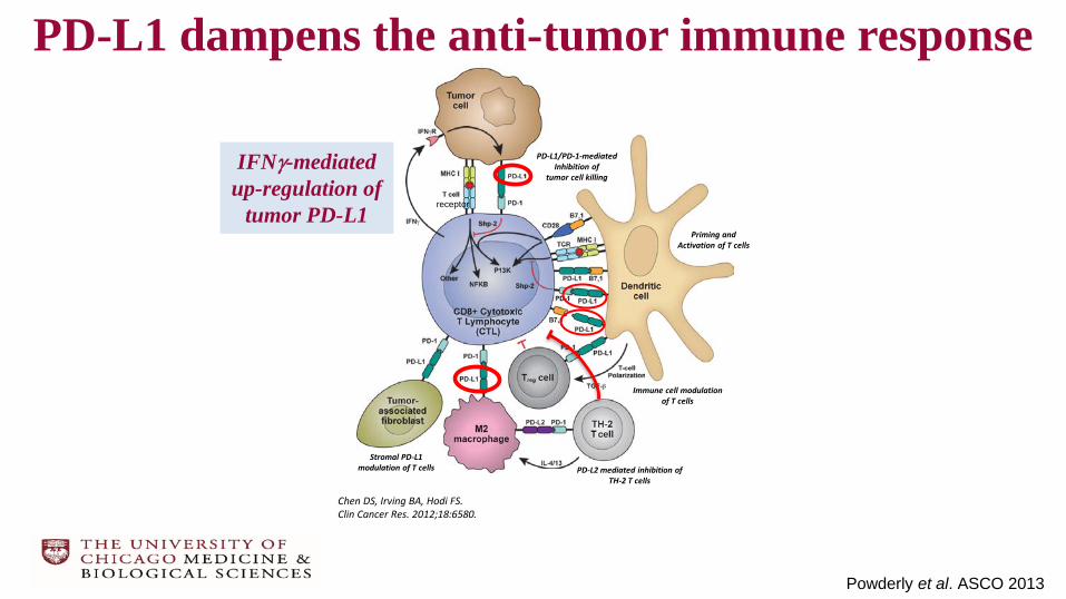
PD-L1 dampens the anti-tumor immune response
Stromal PD-L1 modulation of T cells
Immune cell modulation of T cells
PD-L1/PD-1-mediated Inhibition of
tumor cell killingIFNγ-mediated
up-regulation of tumor PD-L1
Priming and Activation of T cells
PD-L2 mediated inhibition of TH-2 T cells
receptor
Chen DS, Irving BA, Hodi FS. Clin Cancer Res. 2012;18:6580.
Powderly et al. ASCO 2013

Expression of PDL1 is heterogeneous and varies by biopsy location and antibody stain
McLaughlin et al. JAMA Oncol. 2016
E1L3N SP142
Neg
ativ
ePo
sitiv
e
1 mm
H&E
Immunofluorescence shows that stroma and epithelial staining are
often concordant and adjacent.
Green = cytokeratinBlue = nuclei
Red = PD-L1 (SP142)

Given limitations surrounding tumor sampling on biopsy, are there genomic techniques that may assess the tumor
microenvironment more robustly?
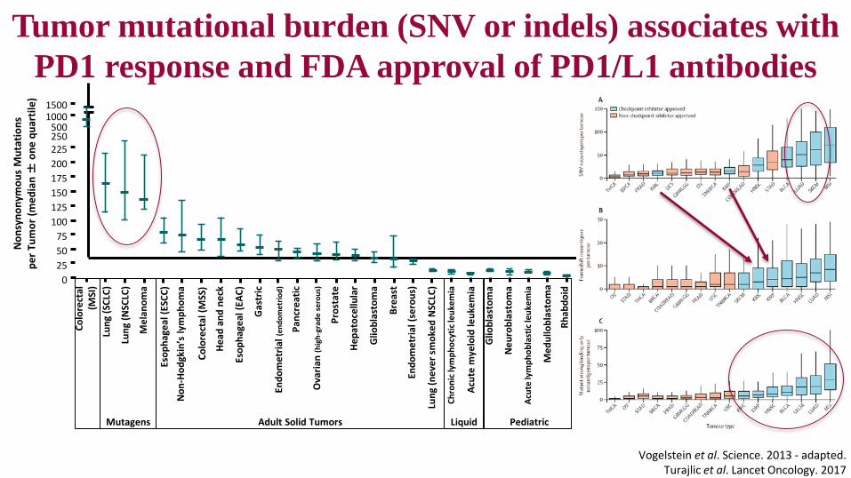
PediatricLiquid
Vogelstein et al. Science. 2013 - adapted.Turajlic et al. Lancet Oncology. 2017
Non
syno
nym
ous
Mut
atio
ns
per T
umor
(med
ian
±on
e qu
artil
e) 15001000
250225
500
200175150125100
755025
0
Colo
rect
al
(MSI
)Lu
ng (S
CLC)
Lung
(NSC
LC)
Mel
anom
a
Esop
hage
al (E
SCC)
Non
-Hod
gkin
’s ly
mph
oma
Colo
rect
al (M
SS)
Head
and
nec
k
Esop
hage
al (E
AC)
Gas
tric
Endo
met
rial (
endo
met
riod)
Panc
reat
ic
Ova
rian
(hig
h-gr
ade
sero
us)
Pros
tate
Hepa
toce
llula
r
Glio
blas
tom
a
Brea
st
Endo
met
rial (
sero
us)
Lung
(nev
er sm
oked
NSC
LC)
Chro
nic l
ymph
ocyt
ic le
ukem
ia
Acut
e m
yelo
id le
ukem
ia
Glio
blas
tom
a
Neu
robl
asto
ma
Acut
e ly
mph
obla
stic
leuk
emia
Med
ullo
blas
tom
a
Rhab
doid
Adult Solid TumorsMutagens
Tumor mutational burden (SNV or indels) associates with PD1 response and FDA approval of PD1/L1 antibodies

PD-1 antibodies sometimes active in tumors with “zero” PD-L1 expression
pembropembro

TMB and PDL1 improves response selection in NSCLC to nivolumab + ipilimumab
Hellmann et al. Cancer Cell 2018

Total exome mutations correlate with mutations via hybrid capture-based next-generation sequencing in commercial assays
Based on in silico analysis filtering on 315 genes in FoundationOne comprehensive genomic profile
(Foundation Medicine, Inc, Cambridge, MA, USA)
100
50
1
FoundationOne Panel (mutations/MB)
Tota
l Exo
me
Mut
atio
ns(m
utat
ions
/MB)
10
501 10 100
Peters et al. AACR. 2017Johnson et al. Cancer Immunol Res. 2016
Mutational load in TCGA skin cutaneous melanoma (SKCM) samples using 315 genes included on the hybrid capture NGS panel is highly correlated
with mutations assessed by WES (Foundation Medicine, Inc, Cambridge, MA, USA)
MelanomaNSCLC
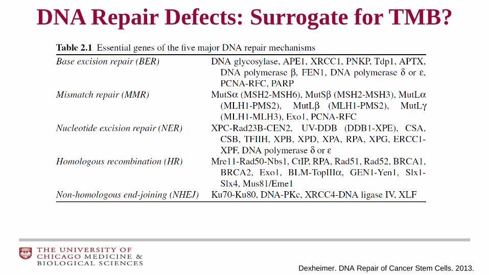
Dexheimer. DNA Repair of Cancer Stem Cells. 2013.
DNA Repair Defects: Surrogate for TMB?
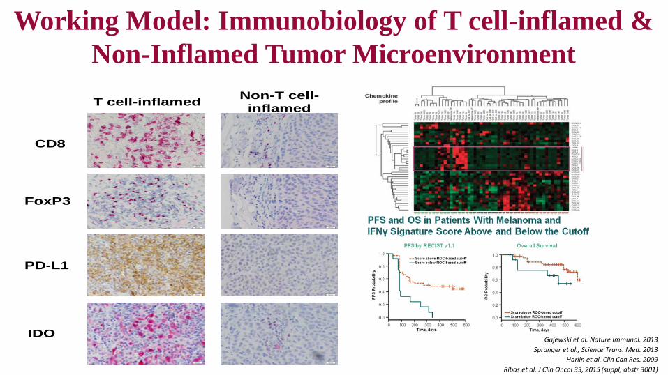
Working Model: Immunobiology of T cell-inflamed &Non-Inflamed Tumor MicroenvironmentT cell-inflamed
CD8
FoxP3
PD-L1
IDO
Non-T cell-inflamed
Gajewski et al. Nature Immunol. 2013 Spranger et al., Science Trans. Med. 2013
Harlin et al. Clin Can Res. 2009Ribas et al. J Clin Oncol 33, 2015 (suppl; abstr 3001)
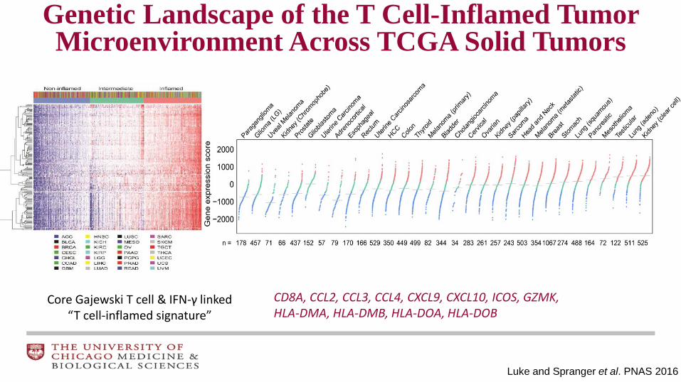
Genetic Landscape of the T Cell-Inflamed Tumor Microenvironment Across TCGA Solid Tumors
Luke and Spranger et al. PNAS 2016
Core Gajewski T cell & IFN-γ linked “T cell-inflamed signature”
CD8A, CCL2, CCL3, CCL4, CXCL9, CXCL10, ICOS, GZMK, HLA-DMA, HLA-DMB, HLA-DOA, HLA-DOB
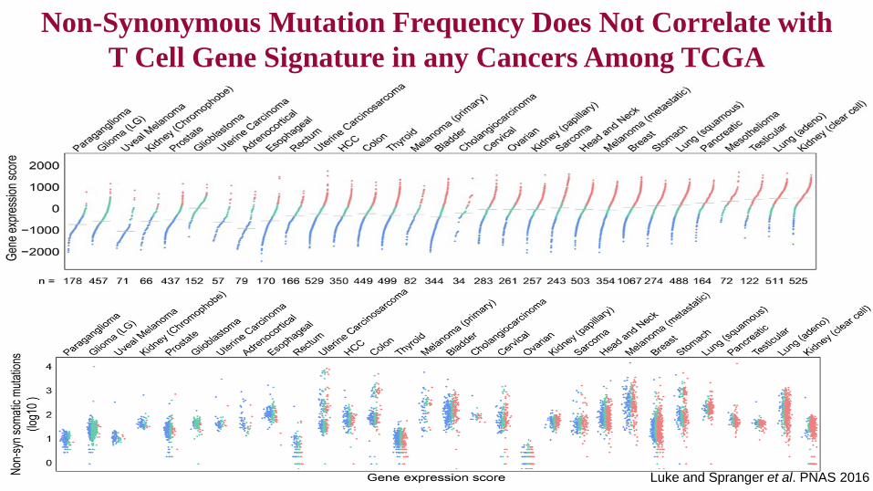
Non-Synonymous Mutation Frequency Does Not Correlate with T Cell Gene Signature in any Cancers Among TCGA
Luke and Spranger et al. PNAS 2016

Seiwert et al. ASCO-SITC. 2016
Relationship of IFN-γ related gene expression & TMB with response to anti–PD1 for HNSCC and gastric cancer in KN012 and KN028.
GEP 18-Gene Score
Merck Interferon-γsignature
CCL5CD27CD274CD276CD8ACMKLR1CXCL9CXCR6HLA.DQA1
HLA.DRB1HLA.EIDO1LAG3NKG7PDCD1LG2 PSMB10STAT1TIGIT
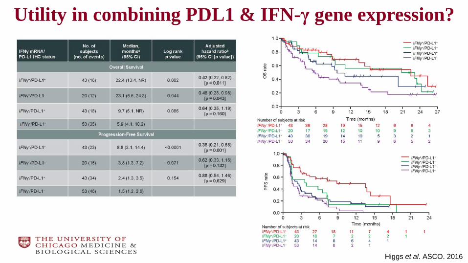
Higgs et al. ASCO. 2016
Utility in combining PDL1 & IFN-γ gene expression?
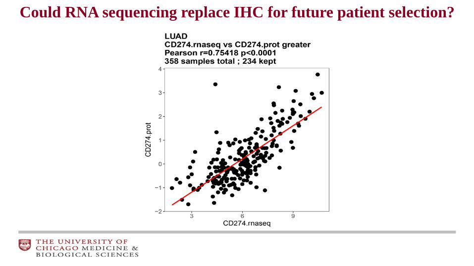
Could RNA sequencing replace IHC for future patient selection?

If tumors can be phenotyped at baseline as T cell-inflamed or non-T cell-inflamed,
is it possible to develop personalized approaches of IO therapy based on this?

Genes Separate Into Those Strongly Correlated and Less Correlated to PDL1
CD163, CCR8, PRDM1, SIGLEC1, CD28, FOXP3, CSF1R, CD33, CD4, TLR7, IL10, HLA−DRA, HLA−DPA1, HLA−DQA1, HLA−DOA, CD69, RSAD2, HLA−DMB, CD3E, CD3D, CCL5, CD247, PDCD1, CD8A, SIRPG, CTLA4, IFNG, TBX21, LAG3, KLRK1, LTA, ICOS, TIGIT, CD3G, ITGAL, CXCL10, CXCL9, CD80, CD86, HAVCR2, ITGB2, CCR1, TNFRSF9, IDO1, STAT1, JAK2, PDCD1LG2, HLA−DPB1, HLA−DRB1, HLA−DMA, HLA−DRB5, HLA−DQB1, KIR2DL4, HLA−DQA2, HLA−DOB, HLA−DQB2, BTLA, LAMP3, CD244, CSF2RA, CD14, IL1B, CD40LG, ITGAX, ITGAM, CD68, ICAM1, MICB, IRF4, STAT4, TNF, CD27, CD72, STAT2, CD40, HLA−B, HLA−C, HLA−A, CXCR3, JAK1, CCR6, LY75, CD79A
IL12B, IRF9, ADAM8, NCR1, TNFSF4, KIR2DL3, KIR2DS4, KIR2DL1, KIR3DL2, KIR3DL1, ISG20, TNFRSF18, IL18, CD93, TMEM173, IL1A, STAT3, FCGR3B, IL6, BATF3, CD70, ENTPD1, TGFB1, CD79B, IL3RA, TNFRSF4, ADORA2A, LAYN, A4GALT, CX3CL1, TNFSF9, IL23A, IL13, CD19, FCER2, TLR9, CLEC4C, CD22, CD160, RORA, BCL6, GATA3, NT5E, IL12A, MME, CCL20, ICOSLG, XBP1, STAT5B, MST1R, TGFB3, TGFB2, IL17A, CD276, STAT6, EDNRB, SMAD3, VEGFA, IFNB1, MICA, KIR3DL3, TYK2, TNFRSF14, ST6GAL1, RORC, CEACAM8, ARG2, KRT20, VTCN1, CD24, IFNK, NCAM1, MAGEH1, IL17F, IL5, HMGB1, IFNA1, IFNA13, IFNW1, IFNA21, ARG1, IL4, NDUFA2
Strongly correlated
Less correlated

Association of PD1 and therapeutically relevant molecules across T cell-inflamed spectrum in melanoma

ADaptiVe Biomarker Trial that InformS Evolution of therapy after nivolumab (ADVISE)
Current selection markers: CD8, PDL1, LAG3, IDO, CSF1R, FoxP3, GITR, NKp46
Future combination options could be included when appropriate combination safety data + potential IHC assays
Biomarker Defined
Treatment Selection
Screen(28 day)
nivo + relatlimab(LAG3)
nivo + BMS-986205(IDO)
nivo + cabiralizumab(CSF1R)
nivo + ipilimumab(FOXP3)
Treat to PD,
Toxicity,or 1 yr
Pre-txbiopsy
NSCLC, MEL, RCC, Gastric,
SCCHN, Urothelial nivo + BMS-986156
(GITR)
nivo + lirilumab(KIR)
nivo + SBRT ClinicalTrials.gov Identifier: NCT03335540BMS CA028-001 / UC IRB17-0731
PI: Jason Luke

Microbiome May Mediate Immunotherapy Efficacy
Zitvogel et al. Nat Rev Microbiol. 2017

PCA analysis sorts significant OTUs by responders and non-responders in metastatic melanoma
63 OTUs identified
N = 42
Matson et al. Science. 2018
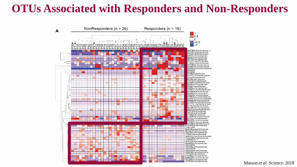
OTUs Associated with Responders and Non-Responders
Matson et al. Science. 2018

Support vector machine model and integration of sequencing methods identifies bacterial species associated
response to anti-PD-1Responder Non-responder
Enterococcus faecium Ruminococcus obeum
Collinsella aerofaciens Roseburia intestinalis
Bifidobacterium adolescentis
Klebsiella pneumoniae
Veillonella parvula
Parabacteroides merdae
Lactobacillus sp
Bifidobacterium longum
Matson et al. Science. 2018

Summation PCR score significantly higher in responders and ratio of beneficial:non-beneficial
OTUs correlates with RECIST
Matson et al. Science. 2018
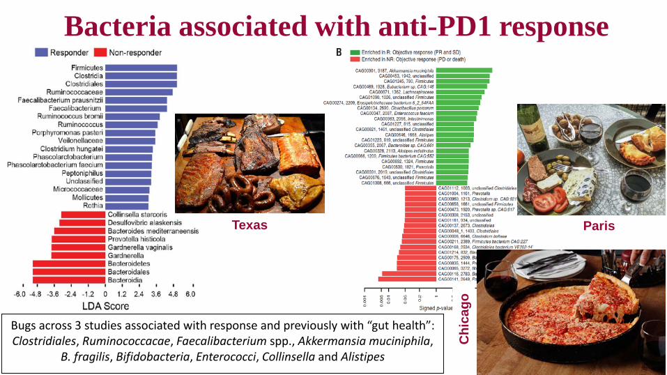
Bacteria associated with anti-PD1 response
Gopalakrishnan et al. Science. 2018Routy et al. Science. 2018
Matson et al. Science. 2018
Texas Paris
Bugs across 3 studies associated with response and previously with “gut health”: Clostridiales, Ruminococcacae, Faecalibacterium spp., Akkermansia muciniphila,
B. fragilis, Bifidobacteria, Enterococci, Collinsella and Alistipes
Chi
cago

Phase II Study Evaluating Gut Microbiota Modulation in Combination with
Pembrolizumab in Advanced Melanoma Merck MISP #53690 - Collaboration with Merck and Evelo
PI: Jason Luke, MD
Bifidobacterium alone Pembrolizumab + BifidoPrimary Endpoint: - RECIST response rateSecondary Endpoints:- PFS, Toxicity- Microbiome Analaysis
Cohort 1: PD1 Ab naïve
Cohort 2: PD1 Ab refractory/resistant

Requirements for an effective anti-tumor immune response The Cancer Immunogram
Blank et al. Science. 2016
29
Conclusions• PD-L1 IHC alone is of limited value as a predictive biomarker
• Tumor mutational burden or IFN-γ may be better but combos even more so
• T cell-inflamed tumor microenvironment may serve as a model for predicting rational combination immunotherapies
• Immunotherapy combination regimens should be targeted toward either T cell-inflamed or non-inflamed tumors
• Future may include patient level immune target identification and incorporation of tumor extrinsic factors such as microbiome
Molecular predictors for IO |

Acknowledgements– Department of Defense Career Development Award (W81XWH-17-1-0265)– National Cancer Institute (P30CA014599-41S4, P30CA014599-43S)– Young Investigator Awards from Cancer Research Foundation and Melanoma Research Alliance– Team Science Award from the Prostate Cancer Foundation– Scientific Research Agreements with Bristol-Myers Squibb, Array, CheckMate and Palleon– Arthur J Schreiner Family Melanoma Research Fund, J. Edward Mahoney Foundation Research Fund, Brush Family Immunotherapy Fund and support from Center for Research Informatics of The University of Chicago Biological Science Division and The Institute for Translational Medicine/CTSA (NIH UL1 RR024999)
@jasonlukemd @UCCancerCenter ASCO Sun June 3rd – Buddy Guy’s Legends BE THERE!!!