biocontrol activity of trichoderma viride...
TRANSCRIPT

Journal of Agricultural Technology 2011, Vol. 7(6): 1589-1602 Available online http://www.ijat-aatsea.com
ISSN 1686-9141
1589
Biocontrol activity of Trichoderma viride and Pseudomonas fluorescens against Phytophthora infestans under greenhouse conditions Ephrem Debebe Zegeye1, Amutha Santhanam2*, Dereje Gorfu3, Mesfin Tessera3 and Bekele Kassa3
1Department of Biology, Ambo University P.O.Box 19, Ambo, Ethiopia 2Department of Biology, Addis Ababa University, Addis Ababa, Ethiopia 3Ethiopian Agriculture Research Organization, Holetta Agricultural Research Center, Holetta, Ethiopia Ephrem Debebe Zegeye, Amutha Santhanam, Dereje Gorfu, Mesfin Tessera, Bekele Kassa (2011). Biocontrol activity of Trichoderma viride and Pseudomonas fluorescens against Phytophthora infestans under greenhouse conditions. Journal of Agricultural Technology 7(6):1589-1602. The biocontrol potential of Trichoderma viride-ES1 and Pseudomonas fluorescens-Bak150 against potato late blight pathogen, Phytophthora infestans, were studied in vitro and under greenhouse conditions. In vitro antagonism test carried out between T. viride and P. infestans showed a radial growth inhibition of the pathogen by 36.7% and a complete overgrowth of T. viride on P. infestans later, whereas P. fluorescens inhibited the radial growth of the pathogen by 88%. Foliar spray method used in the greenhouse trials involved foliar spray of healthy potato plants with suspensions of (i) T. viride only, (ii) P. fluorescens only, (iii) mixed culture of T. viride and P. fluorescens, and (iv) Mancozeb, followed by spraying the pathogen inoculum three days later. The infected leaf area was measured weekly and area under the disease progress curve (AUDPC) was calculated and compared among the treatments. The result showed that T. viride (AUDPC=260) and P. fluorescens (AUDPC=765.1) significantly (P<0.05) reduced the disease compared to the untreated check (AUDPC=1045). T. viride was found to be more efficient than P. fluorescens and mixed culture. No significant difference was observed between the mixed culture and the inoculated/untreated check. This study revealed that the foliar application of T. viride-ES1 has good potential in controlling the late blight disease of potato. Key Words: Biological control, Potato late blight, Phytophthora infestans, Trichoderma viride, Pseudomonas fluorescens
* Corresponding author: Amutha Santhanam; e-mail: [email protected]
Journal of Agricultural Technology 2011, Vol. 7(6): 1589-1602
1590
Introduction
Late blight disease caused by the pathogen Phytophthora infestans, is probably the single most important disease of potatoes and tomatoes worldwide (Son et al., 2008). Worldwide losses due to late blight are estimated to exceed $5 billion annually and thus the pathogen is regarded as a threat to global food security (Latijnhouwers et al., 2004). In the past few decades, the frequency and severity of the disease have increased in many parts of the world including Ethiopia and have been a serious threat to potato production (Bakonyi et al., 2002). Despite the fact that much of the success in controlling the disease has been due to the application of large quantities of chemical fungicides, their extensive use is causing a serious pollution problem in the environment (Ragunathan and Divakar, 1996). Further, the chemical control of late blight is becoming more difficult due to the appearance of new and more aggressive P. infestans strains (Fernandez-Northcote et al., 2000). Thus, an alternative control strategy such as biological control should be sought (Ellis et al., 1999). Biological control of crop disease is receiving increased attention as an environmentally sound alternative to chemical pesticides. Some species of Trichoderma and Pseudomonas are among the major microorganisms that have shown great potential for biological control of several plant pathogens. Trichoderma species have shown biocontrol potential against many plant pathogens including diseases caused by Sclerotinia minor (Jones and Stewart, 1997; Dolatabadi et al., 2011), Botryosphaeria berengeriana f.spp. piricola, Cladosporium herbarum (Barbosa et al., 2001), Dioscorea spp. (Okigbo and Ikediugwu, 2000) and Pythium ultimum (Naseby et al., 2000). Besides, Trichoderma species have also shown efficacy against diseases caused by Rhizoctonia solani, Pythium aphanidermatium, Fusarium oxysporum, Fusarium culmorum, Gaeumannomyces graminis var.tritici, Sclerotium rolfsii, Phytophthora cactorum, Botrytis cinerea and by Alternaria spp. (Kucuk and Kivanc, 2003). Similarly, specific strains of Pseudomonas species have shown efficacy in controlling a number of fungal diseases, including Pythium root and seed rot of many crops (Mazzola, 1998; Ellis et al., 1999), Fusarium wilt in cotton and tomato (Gamliel and Katan, 1992), Verticillium wilt of potato (Leben et al., 1987), Rhizoctonia stem and root rot of peanut (Savithiry and Gnanamanickam,1987), banana wilt caused by Fusarium oxysporum (Sivamani and Gnanamanickam, 1988), bean disease caused by Sclerotium rolfsii (Gamliel and Katan, 1992), Rhizoctonia solani root infection in tomato (Siddiqui and Shaukat, 2002) and late blight of tomato (Tran et al., 2007). However, significant studies on biological control of late blight of potato are scarce and hence the main aim of this study was to evaluate the efficacy of
Journal of Agricultural Technology 2011, Vol. 7(6): 1589-1602 Available online http://www.ijat-aatsea.com
ISSN 1686-9141
1591
local isolates, Trichoderma viride-ES1 and Pseudomonas fluorescens-Bak150, against late blight of potato in vitro and under greenhouse conditions.
Materials and methods
Media used for the culture cultivation and bioassay of the strains were King’s B broth for P. fluorescens (20 g l-1 proteose peptone No.3, 1.5 g l-1 K2HPO4, 1.5 g l-1 MgSO4. 7H2O and 15 ml glycerol) and for agar plates 15 g l-
1 agar was added. For T. viride, Potato dextrose agar (PDA, Oxoid ) was used. Rye agar (60 g l-1 rye extract, 20 g l-1 sucrose, 0.05 g l-1 β- sitosterol and 15 g l-1 agar) was employed for antagonistic tests. To isolate P. infestans, vegetable juice (V8) (100 ml V8 juice, 1.5 g l-1 CaCO3, 0.05 g l-1 β-sitosterol and 15 g l-1 agar) supplemented with dimethylsulfoxide mix of 20 mg l-1 Griseofulvin, 19 mg l-1 Nystatin, 10 mg l-1 Benlate (50 % w/v, 5 mg l-1 Methoxypurine, 30 mg l-1 Rifamycin, 5 mg l-1 Nalidixic acid, 40 mg l-1 8-azaguanine and 30 mg l-1 Neomycin was used. Microorganisms
T. viride-ES1 was isolated and maintained in PDA slant and P. fluorescens-Bak150 was isolated from a potato field in Bako district, Ethiopia, and maintained in King’s B agar slants. P. infestans was isolated from infected potato leaves at Holetta Agricultural Research Center (HARC) potato field, Ethiopia.
In vitro antagonistic tests Antagonistic test between T. viride and P. infestans
Dual culture method was employed to analyze whether T. viride inhibits the growth of P. infestans as described in Sivakumar et al. (2000). Briefly, 1 cm diameter mycelial plug of P. infestans (9 days old) was placed on one side of a petri-dish (9 cm diameter) containing rye agar and pre-incubated at 18C for 2 days to initiate growth. Later, 1 cm diameter disc of T. viride (9 days old) was placed 6 cm away from the pathogen on the dual plates; whereas sterile PDA disc was placed in the control plates. The assay was done in triplicates and the radial growth of the pathogen was measured 4 days after incubation at 18C. The percent radial mycelial growth inhibition (I) was calculated as I= [(C–T)/C] X100 (Sivakumar et al., 2000); where C is radial growth measurement of the pathogen in the control plates and T is radial growth of the pathogen in the dual plates.

Journal of Agricultural Technology 2011, Vol. 7(6): 1589-1602
1592
Antagonistic test between P. fluorescens and P. infestans
The antagonism test was carried out on rye agar as described in Georgakopoulos et al. (2002). A 20 μl of an overnight culture of P. fluorescens grown in King’s B broth was spotted at the center of a petri-dish and pre-incubated for 2 days at 18C followed by placing 1 cm disc of P. infestans (9 days old) at either side of the bacterial culture. In the control plates, 20 μl of sterile King’s B broth was spotted at the center. This test was done in triplicates. Radial growth inhibition of P. infestans was assessed ten days later by measuring the radial growth of the pathogen in the dual and control plates.
Antagonist compatibility test
In vitro compatibility test between P. fluorescens-Bak150 and T. viride-ES1 was conducted in order to determine whether they can be used in combination. Dual culture plate method described by Siddiqui and Shaukat (2003) was employed. Accordingly, an overnight culture of P. fluorescens which was grown in King’s B broth was streaked on one side of a petri-dish (9 cm diameter) containing King’s B agar (KBA). The other side of the petri-dish was inoculated with 1 cm disc of T. viride (9 days old). The plates were then incubated at 25C and zone of inhibition (if any) was measured. The test was performed in triplicates.
Observation of mycoparasitism by T. viride
Slide culture method described in Sivakumar et al. (2000) was employed to determine whether T. viride-ES1 parasitizes P. infestans. Clean slides were placed on a Z shaped glass rod in 9 cm petri-dishes and sterilized. Small amount of molten rye agar was then poured evenly on the slides. A few drops of sterile water were added to the petri-dish to prevent drying. Mycelial disc of P. infestans was inoculated on one side of the slide and pre-incubated at 18o C. T. viride was inoculated two days later at a distance of 2.5 cm from P. infestans. The presence or absence of coiling and other hyphal interactions were studied under compound microscope after four days of incubation at 18C.
Greenhouse Trials Experimental design
Chambers were made in greenhouse using transparent 0.3 mm thick polythene sheet. The temperature in the chambers was adjusted to range
Journal of Agricultural Technology 2011, Vol. 7(6): 1589-1602 Available online http://www.ijat-aatsea.com
ISSN 1686-9141
1593
between 14 and 24C while conducting the experiment. The relative humidity of the chamber was maintained at above 90% by humidifier. Foliar spray method was employed to evaluate the efficiency of spraying the antagonists in controlling airborne inocula of the pathogen. The treatments were (i) T. viride only, (ii) P. fluorescens only (iii) mixed culture of T. viride and P. fluorescens, and (iv) Mancozeb (a chemical fungicide included as a standard check). Negative and positive controls were included. Nine replicates were used for each treatment and the pots were arranged in a completely randomized block design method.
Soil, pots and fertilizers
Three kilograms of clay soil (pH=5.7) was added to each of surface sterilized plastic pots of 5 L volume (diameter and depth of 20 cm each) after being autoclaved at 121C for 30 min. Appropriate doses of fertilizers (i.e., 0.6 g di-ammonium phosphate and 0.5 g urea) were added to each pot.
Selection and preparation of potato seed tubers
The potato cultivar Awash (CIP-378501.3) was used in the experiment. Both the foliage and the tubers are known to be highly susceptible to P. infestans strains in Holetta region, Ethiopia. The seed tubers were harvested from HARC and had been stored for 6 months. Disease free and uniform tubers (6-8 cm long) were selected and surface sterilized with 3.5% sodium hypochlorite for one minute and rinsed with sterile distilled water three times.
Preparations of microbial suspensions for foliar spray
A 250 ml suspension of T. viride spores was prepared from 9 days old culture plates which were grown on PDA at 25C. The plates were rinsed with sterile distilled water and the mycelia were carefully scraped off the agar with a bent glass rod. The suspension was then filtered through four layered gauze bandage to separate the spores from the mycelia. The concentration was adjusted to 3.7×10⁸ spores/ml (Dubos, 1987) with the help of haemocytometer. A 250 ml of P. fluorescens cell suspension was prepared by inoculating the strain into King’s B broth followed by shaking for 48 hr (150 rpm) at room temperature. The bacterial suspension was roughly adjusted optically at ca.1x109 cfu/ml (O.D600= 1) (Mulya et al., 1996). A mixed culture of T. viride and P. fluorescens was prepared by mixing 125 ml of each of T. viride (3.7×10⁸

Journal of Agricultural Technology 2011, Vol. 7(6): 1589-1602
1594
spores/ml) and P. fluorescens (1x10⁹ cfu/ml). Mancozeb (Dithane M-45) at a concentration of 5 g l-1 was employed.
P. infestans was grown on V8 agar for 9 days in dark at 18C. Sporangia
were harvested from the plates by rinsing the sporangial/mycelial mat with sterile distilled water and scraping the mat using bent glass rod. The suspension was filtered through four folds of sterile gauze bandage to separate the sporangia from the mycelia. The concentration of the sporangia was adjusted to 1x10³ sporangia per ml with the help of haemocytometer. Foliar spraying of antagonists and the pathogen
Potato tubers were planted in pots containing the sterilized soil. After the seedlings reached the rapid expansion phase (18 days after emergence), the control agents were sprayed to run-off onto the plants. For the negative and the positive controls, the plants were sprayed with sterile distilled water. Three days following the application of the control agents, 300 ml suspension (1x10³sporangia/ml) of P. infestans was sprayed to all plants of the different treatments except to those of the positive controls. Prior to spray of the pathogen, the plants were exposed to a high relative humidity (R.H=100%) to moisten their leaves. Following spray of the pathogen, the individual plants were covered with polythene bags to prevent cross infection among adjacent plants.
Data collection and Statistical analysis
The individual plants were rated visually on weekly intervals for percentage of leaf area with symptoms of late blight over the disease progress period based on the assessment key of James, (1971). The average amount of disease developed over the disease progress period was expressed as the area under the disease progress curve (AUDPC) and estimated using the midpoint rule described in Campbell and Madden (1990).
The ratio of infected to total leaves at each leaf position on the main stem
of the replicates were also calculated at the 14th day after the foliar inoculation of the pathogen (Kirk et al., 1999). The first leaf above the soil on the main stem was counted as leaf one. Statistical analysis was conducted using the general linear models procedures of the SPSS (version 12). Analysis of variance of differences in the treatments and least significance tests were carried out. Significance was evaluated at P<0.05 for the tests.
Journal of Agricultural Technology 2011, Vol. 7(6): 1589-1602 Available online http://www.ijat-aatsea.com
ISSN 1686-9141
1595
Results
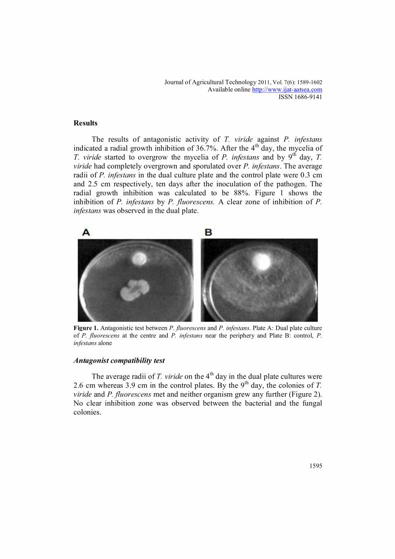
The results of antagonistic activity of T. viride against P. infestans indicated a radial growth inhibition of 36.7%. After the 4th day, the mycelia of T. viride started to overgrow the mycelia of P. infestans and by 9th day, T. viride had completely overgrown and sporulated over P. infestans. The average radii of P. infestans in the dual culture plate and the control plate were 0.3 cm and 2.5 cm respectively, ten days after the inoculation of the pathogen. The radial growth inhibition was calculated to be 88%. Figure 1 shows the inhibition of P. infestans by P. fluorescens. A clear zone of inhibition of P. infestans was observed in the dual plate.
Figure 1. Antagonistic test between P. fluorescens and P. infestans. Plate A: Dual plate culture of P. fluorescens at the centre and P. infestans near the periphery and Plate B: control, P. infestans alone
Antagonist compatibility test
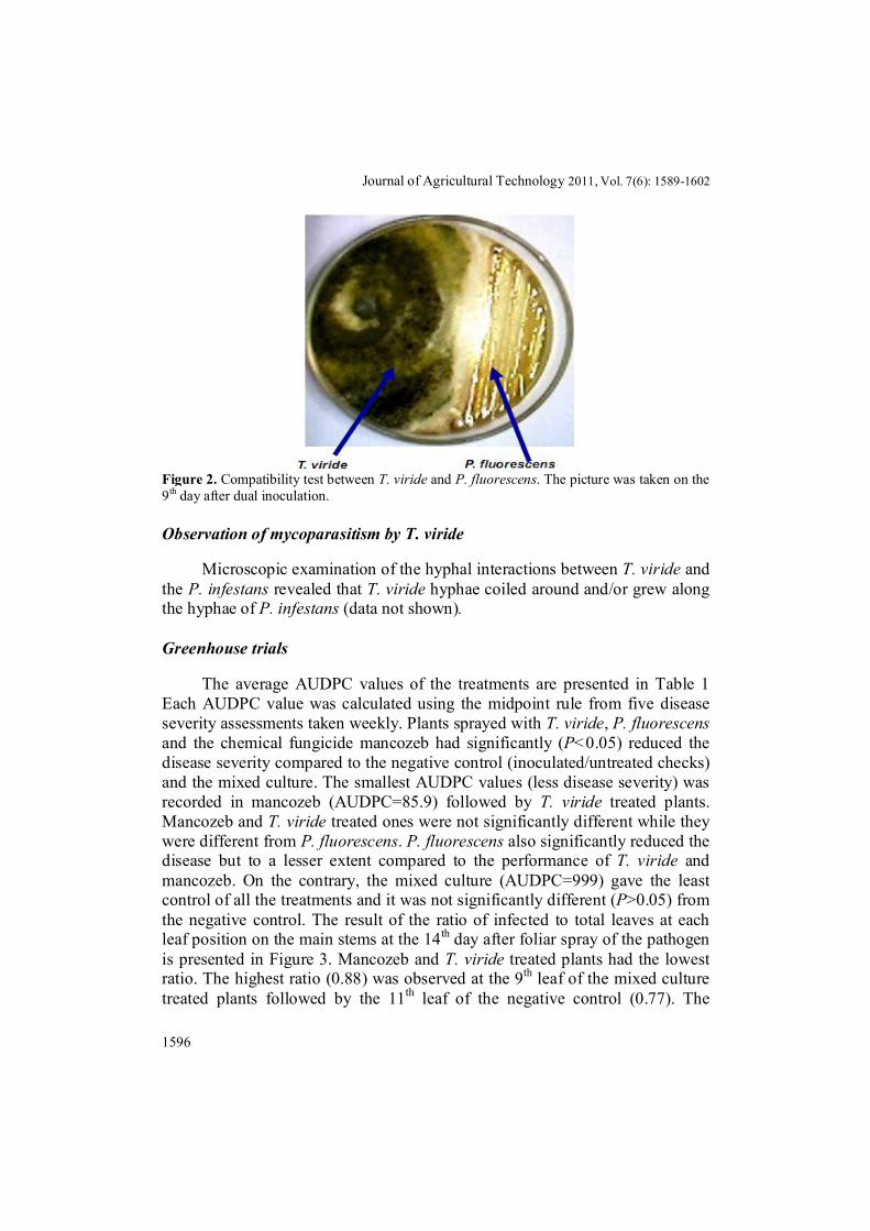
The average radii of T. viride on the 4th day in the dual plate cultures were 2.6 cm whereas 3.9 cm in the control plates. By the 9th day, the colonies of T. viride and P. fluorescens met and neither organism grew any further (Figure 2). No clear inhibition zone was observed between the bacterial and the fungal colonies.
Journal of Agricultural Technology 2011, Vol. 7(6): 1589-1602
1596
Figure 2. Compatibility test between T. viride and P. fluorescens. The picture was taken on the 9th day after dual inoculation.
Observation of mycoparasitism by T. viride
Microscopic examination of the hyphal interactions between T. viride and the P. infestans revealed that T. viride hyphae coiled around and/or grew along the hyphae of P. infestans (data not shown).
Greenhouse trials
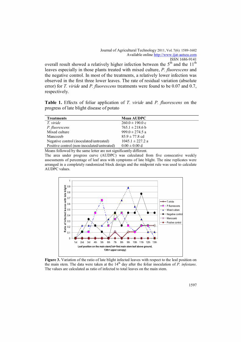
The average AUDPC values of the treatments are presented in Table 1 Each AUDPC value was calculated using the midpoint rule from five disease severity assessments taken weekly. Plants sprayed with T. viride, P. fluorescens and the chemical fungicide mancozeb had significantly (P<0.05) reduced the disease severity compared to the negative control (inoculated/untreated checks) and the mixed culture. The smallest AUDPC values (less disease severity) was recorded in mancozeb (AUDPC=85.9) followed by T. viride treated plants. Mancozeb and T. viride treated ones were not significantly different while they were different from P. fluorescens. P. fluorescens also significantly reduced the disease but to a lesser extent compared to the performance of T. viride and mancozeb. On the contrary, the mixed culture (AUDPC=999) gave the least control of all the treatments and it was not significantly different (P>0.05) from the negative control. The result of the ratio of infected to total leaves at each leaf position on the main stems at the 14th day after foliar spray of the pathogen is presented in Figure 3. Mancozeb and T. viride treated plants had the lowest ratio. The highest ratio (0.88) was observed at the 9th leaf of the mixed culture treated plants followed by the 11th leaf of the negative control (0.77). The
Journal of Agricultural Technology 2011, Vol. 7(6): 1589-1602 Available online http://www.ijat-aatsea.com
ISSN 1686-9141
1597
overall result showed a relatively higher infection between the 5th and the 11th leaves especially in those plants treated with mixed culture, P. fluorescens and the negative control. In most of the treatments, a relatively lower infection was observed in the first three lower leaves. The rate of residual variation (absolute error) for T. viride and P. fluorescens treatments were found to be 0.07 and 0.7, respectively.
Table 1. Effects of foliar application of T. viride and P. fluorescens on the progress of late blight disease of potato
Treatments Mean AUDPC T. viride 260.0 ± 190.0 c P. fluorescens 765.1 ± 218.6 b Mixed culture 999.0 ± 274.5 a Mancozeb 85.9 ± 77.8 cd Negative control (inoculated/untreated) 1045.1 ± 227.2 a Positive control (non-inoculated/untreated) 0.00 ± 0.00 d
Means followed by the same letter are not significantly different. The area under progress curve (AUDPC) was calculated from five consecutive weekly assessments of percentage of leaf area with symptoms of late blight. The nine replicates were arranged in a completely randomized block design and the midpoint rule was used to calculate AUDPC values.
0
0.1
0.2
0.3
0.4
0.5
0.6
0.7
0.8
0.9
1
1st 2nd 3rd 4th 5th 6th 7th 8th 9th 10th 11th 12th 13thLeaf position on the main stem(1st= first main stem leaf above ground,
13th= upper canopy)
Rat
io o
f inf
ecte
d le
aves
with
late
blig
ht
T.virideP.fluorescensMixed cultureNegative controlMancozebPositve control
Figure 3. Variation of the ratio of late blight infected leaves with respect to the leaf position on the main stem. The data were taken at the 14th day after the foliar inoculation of P. infestans. The values are calculated as ratio of infected to total leaves on the main stem.

Journal of Agricultural Technology 2011, Vol. 7(6): 1589-1602
1598
Discussion
Late blight, caused by the oomycete pathogen Phytophthora infestans, is probably the single most important disease of potatoes worldwide. It is destructive wherever potatoes are grown without the use of fungicides, except in hot, dry, and irrigated areas (Thurston and Schultz, 1981). Biological control is a good alternative for sustainable agriculture to overcome the problems of public concern associated with pesticides and pathogens resistant to chemical pesticides and to become eco-friendly (Akhtar and Siddiqui, 2008). In this study T. viride has been found to retard the radial growth of P. infestans. This antagonistic mode of action of Trichoderma could be attributed to the production of antibiotics and fungal cell wall degrading enzymes (Chutrakul et al., 2008; Sharma et al., 2009). A similar inhibitory action of Trichoderma strains (TH1, N47 and T12) against a related oomycete pathogen, Pythium ultimum, was reported earlier (Naseby et al., 2000). The observed mycoparasitic action of Trichoderma in this study suggests that it has good potential in controlling P. infestans. Similar mycoparasitic action of Trichoderma strains were also reported against related pathogens, Phytophthora cinnamomi (Pugeg and Ian, 2006) and Phytophthora capsici (Ezziyyani et al., 2007). Similarly, the pathogen was also strongly (88%) inhibited by P. fluorescens. This is corroborated by the study of Torres-Rubio et al. (2000) in which P. fluorescens inhibited P. infestans to an extent of 74%. The clear inhibition zone that was observed in the dual plate is suggestive of production of antagonistic metabolites by P. fluorescens. In the compatibility test between T. viride and P. fluorescens, the colonies of the fungus and the bacterium met on the 9th day and no inhibition zone was formed between the two. In addition, no overgrowth of either organism occurred. The absence of inhibition zone may indicate that their antagonistic metabolites were not inhibitory to each other. This observation was the basis for testing a combination of the two antagonists as “mixed culture” in the greenhouse trial. Similar in vitro compatibility was reported between Pseudomonas aeruginosa and Pochonia chlamydosporia (Siddiqui and Shaukat, 2003) and between Trichoderma harzianum and Streptomyces rochei (Ezziyyani et al., 2007). The result obtained from the foliar spray method under greenhouse indicated that both of the individual antagonists significantly reduced the severity of foliar phase of late blight infection. The smallest area of leaf infection was observed in mancozeb treated plants and the highest in the untreated checks (negative control). Even though mancozeb treated plants showed the least infection, the difference with that of T. viride treated plants was not statistically significant. This indicated that the performance of T. viride was comparable to that of the chemical fungicide. It is possible that more than one antagonistic mechanism
Journal of Agricultural Technology 2011, Vol. 7(6): 1589-1602 Available online http://www.ijat-aatsea.com
ISSN 1686-9141
1599
could have been involved in the reduction of the disease. However, it needs to be further examined as the relative importance of the mechanisms is dependent on the particular isolate, the target organism and also the ambient environmental conditions (Tronsmo, 1996). The rationale behind the use of mixed culture is that multiple strains allow the deployment of several different biocontrol mechanisms simultaneously. Besides, effective control of the target pathogen over diverse set of environmental conditions could be expected if strains with same ecological requirements are included in the inoculant (Mazzola, 1998). The performance of the mixed culture, however, was the least and it was not significantly different from the inoculated/untreated check. Perhaps competition for iron between the two antagonists could be one possible reason; as such incompatibility was reported between Trichoderma hamatum and Pseudomonas spp in iron deficient soil (Hubbard et al., 1983). However, evidence for the role of siderophores in competition for iron in the phyllosphere is needed if this argument is to be true. The agar test showed compatibility as neither inhibition zone nor overgrowth between the antagonists was observed. Hence, this implies that in vitro compatibility does not always indicate in vivo compatibility. Therefore, at this point it would be worth mentioning that sometimes mixtures of bioagents do not ensure improved biological control (Mazzola, 1998). In the greenhouse trial, the control agents were applied three days ahead of the pathogen. The reason is antagonists should occupy the site earlier than the pathogen’s arrival (Baker and Cook, 1974). In a study by Kexiang et al. (2002), Trichoderma species were found to give better control of Botryosphaeria berengeriana when inoculated three days in advance than when the two are co-inoculated. The ratio of infected leaves on the main stem was significantly lower in those treated with mancozeb and T. viride than the rest of treatments. This reflected the strong performance of T. viride. The reason for a lower infection that was observed in the lower leaves of most of the treatment is because potato leaves at the base of the stem are less susceptible to late blight infection than leaves lying closer to the flower (Carnegie and Colhoun, 1982). This result is in agreement with Kirk et al. (1999).
Germination of fungal spores on the leaf surface is a critical stage in the
development of the host-pathogen interface, and one in which the pathogen is often vulnerable (Campbell, 1989). The motile zoospores of P. infestans have no cell wall and hence are probably extremely vulnerable to adverse conditions, and are also the main infective propagules (Erwin and Ribeiro, 1996). Thus, zoospores can be a potential target in biocontrol of P. infestans. In conclusion, T. viride-ES1 showed good potential in controlling potato late blight under greenhouse conditions. In addition to that, the T. viride used in this study was

Journal of Agricultural Technology 2011, Vol. 7(6): 1589-1602
1600
isolated from local climatic conditions and effectively proved that it has potential to be used as a biocontrol agent in the future. Further research is needed to determine its field performance. In general, as any other biocontrol systems, biological control of late blight should be regarded as one facet of the integrated control program rather than a method to be used alone.
Acknowledgements
The authors are grateful to Holetta Agriculture Research Center (HARC) for providing Laboratory and Greenhouse facilities. The technical assistance of Tiruwork and Atsede is greatly acknowledged. This work was supported by Graduate studies research fund of Addis Ababa University. References Akhtar, M.S and Siddiqui, Z.A. (2008). Arbuscular mycorrhizal fungi as potential
bioprotectants against plant pathogens. Mycorrhizae: Sustainable Agriculture and Forestry, 61–97.
Baker, K.F and Cook, R.J. (1974). Biological control of plant pathogens. W.H. Freeman and Co., San Francisco, 443 pp.
Bakonyi, J., Heremans, B and Jamart, G. (2002). Characterization of Phytophthora infestans isolates collected from potato in Flanders, Belgium. Journal of Phytopathology 150: 512-516.
Barbosa, M.A.G., Rehn, K.G., Menezes, M and Mariano, R.L. (2001). Antagonism of Trichoderma species on Cladosporium herbarium and their enzymatic characterization. Brazilian Journal of Microbiology 32: 98-104.
Campbell, C.L and Madden, L.V. (1990). Introduction to plant disease epidemiology. John Wiley and Sons, New York, 532 pp.
Campbell, R. (1989). Biological control of microbial plant pathogens. Cambridge University press, Cambridge, 232 pp.
Carnegie, S.F and Colhoun, J. (1982). Susceptibility of potato leaves to P. infestans in relation to plant age and leaf position. Phytopathology 104: 157-167.
Chutrakul, C., Alcocer, M., Bailey, K and Peberdy, J.F. (2008). The production and characterization of Trichotoxin peptaibols by Trichoderma asperellum. Chemistry and Biodiversity 5: 1694- 1706.
Dolatabadi, K.H., Goltapeh, E.M., Varma, A and Rohani, N. (2011). In vitro evaluation of arbuscular mycorrhizal-like fungi and Trichoderma species against soil borne pathogen. Journal of Agricultural Technology 7(1): 73-84.
Dubos, B. (1987). Fungal antagonism in aerial agrobiocenoses. In: Innovative approaches to plant disease control, ed. by I. Chet, pp.107-135. John Wiley and Sons, New York.
Ellis, R.J., TimmsWilson, T.M., Beringer, J.E., Rhodes, D., Renwick, A., Stevenson, L and Bailey, M.J. (1999). Ecological basis for biocontrol of damping-off diseases by Pseudomonas fluorescens 54/96. Journal of Applied Microbiology 87: 454-463.
Erwin, D.C and Ribeiro, O.K. (1996). Phytophthora diseases worldwide. American Phytopathological Society, Minnesota, 562 pp.
Journal of Agricultural Technology 2011, Vol. 7(6): 1589-1602 Available online http://www.ijat-aatsea.com
ISSN 1686-9141
1601
Ezziyyani, M., Requena, M.E., Egea-Gilabert, C and Candela, M.E. (2007). Biological control of Phytophthora root rot of pepper using Trichoderma harzianum and Streptomyces rochei in combination. Journal of Phytopathology 155: 342-349.
Fernandez-Northcote, E.N., Navia, O and Gandarillas, A. (2000). Basis of strategies for chemical control of potato late blight developed by PROINPA in Bolivia. Fitopatologia 35: 137-149.
Gamliel, A and Katan, J. (1992). Suppression of major and minor pathogens by fluorescent pseudomonads in solarized and non-solarized soils. Phytopathology 83: 68-75.
Georgakopoulos, D.G., Fiddaman, P., Leifert, C and Malathrakis, N.E. (2002). Biological control of cucumber and sugar beet damping–off caused by Pythium ultimum with bacterial and fungal antagonists. Journal of Applied Microbiology 92:1078-1084.
Hubbard, J.P., Harman, G.E and Hadar, Y. (1983). Effect of soilborne Pseudomonas spp. on the biological control agent, Trichoderma hamatum, on pea seeds. Phytopathology 73: 655-659.
James, C. (1971). A manual assessment keys for plant diseases. Canada Department of Agriculture. Publication No. 1458. APS Press, Canada.
Jones, E.E and Stewart, A. (1997). Biological control of Sclerotinia minor in lettuce using Trichoderma species. Proc. New Zealand Plant protection conf. 50:154-158.
Kexiang, G., Xiaoguang, L., Yonghong, L. Tianbo, Z. and Shuliang, W. (2002). Potential of Trichoderma harzianum and T. atroviride to control Botryosphaeria berengriana f.sp. piricola, the cause of Apple ring rot. Journal of Phytopathology 150: 271-276.
Kirk, W.W., Niemira, B.A., Stein, J.M and Hammerschmidt, R. (1999). Late blight (Phytophthora infestans (Mont.) De Bary) development from potato seed pieces treated with fungicides. Pesticide Science 55: 1151-1158.
Kucuk, C and Kivanc, M. (2003). Isolation of Trichoderma spp. and determination of their antifungal, biochemical and physiological features. Turkish Journal of Biology 27: 247-253.
Latijnhouwers, M., Ligterink, W., Vleeshouwers, V.G., VanWest, P and Govers, F. (2004). A Gα subunit controls zoospore mobility and virulence in the potato late blight pathogen Phytophthora infestans. Molecular Microbiology 51: 925-936.
Leben, S.D., Wadi, J.A and Easton, G.D. (1987). Effects of Pseudomonas fluorescens on potato plant growth and control of Verticillium dahliae. Phytopathology 77: 1592-1595.
Mazzola, H. (1998). The potential of natural and genetically engineered fluorescent Pseudomonas spp. as biological control agents. In: Microbial interaction in agriculture and forestry, ed. by N. S. S. Rao and Y.R. Dommergues, pp.193-217. Science publishers, Plymouth.
Mulya, K., Wataneabe, M., Goto, M., Takikawa, Y and Tsuyumu, S. (1996). Suppression of bacterial wilt disease of tomato by root dipping with P. fluorescens pfg32. Annual Phyto pathological Society of Japan 62: 134-140.
Naseby, D.C., Pascual, J.A and Lynch, J.M. (2000). Effect of biocontrol strains of Trichoderma on plant growth, Pythium ultimum populations, soil microbial communities and soil enzyme activities. Journal of Applied Microbiology 88: 161-169.
Okigbo, R.N and Ikediugwu, F.E.O. (2000). Studies on biological control of post-harvest rot in yams (Discorea spp.) using Trichoderma viride. Journal of Phytopathology 148: 351-355.
Pugeg, I.N.A and Ian, D.G. (2006). Mycoparasitic and antagonistic inhibition on Phytophthora cinnamomi rands by microbial agents isolated from manure composts. Plant Pathology 5: 291- 298.
Journal of Agricultural Technology 2011, Vol. 7(6): 1589-1602
1602
Ragunathan, V and Divakar, B.J. (1996). Integrated pest management strategies. In: Molecular biology of the biological control of pests and disease of plants, ed. by M. Gunasekaran and D. J. Weber, pp.191-194. CRC Press, Florida.
Savithiry, S and Gnanamanickam, S.S. (1987). Bacterization of peanut with Pseudomonas fluorescens for biological control of Rhizoctonia solani and for enhanced yield. Plant and Soil 102: 11-15.
Sharma, K., Kumar, M and Misra, R.M. (2009). Morphological, biochemical and molecular characterization of Trichoderma harzianum isolates for their efficacy as biocontrol agents. Journal of Phytopathology 157: 51-56.
Siddiqui, I.A and Shaukat, S.S. (2002). Resistance against the damping-off fungus Rhizoctonia solani systemically induced by the plant-growth-promoting rhizobacteria Pseudomonas aeruginosa (IE-6S ) and P. fluorescens (CHA0). Journal of Phytopathology 150: 500-506.
Siddiqui, I.A and Shaukat, S.S. (2003). Combination of Pseudomonas aeruginosa and Pochonia chlamydosporia for control of root-infecting fungi in tomato. Journal of Phytopathology 151: 215-222.
Sivakumar, D., Wijeratnam, W.R.S., Wijesundera, R.L.C., Marikar, F.M.T and Abeyesekere, M. (2000). Antagonistic effect of Trichoderma harzianum on post-harvest pathogens of Rambutan (Naphelium lappaceum). Phytoparasitica 28: 240-247.
Sivamani, E and Gnanamanickam, S.S. (1988). Biological control of Fusarium oxysporum f.sp. cubense in banana by inoculation with Pseudomonas fluorescens. Plant and soil 107: 3-9.
Son, S.W., Kim, H.Y., Choi, G.J., Lim, H.K., Jang, K.S., Lee, S.O., Sung, N.D and Kim, J.C. (2008). Bikaverin and fusaric acid from Fusarium oxysporum show antioomycete activity against P. infestans. Journal of Applied Microbiology 104: 692-698.
Thurston, H.D. and Schultz, O. (1981). Late blight. In: Compendium of potato diseases, pp. 40-42, (Hooker, ed). American Phytological Society, U.S.A.
Torres-Rubio, M.G., Valencia-Plata, S.A., Bernal-Sastillo, J and Martinez-Nieto, P. (2000). Isolation of Enterobacteria, Azotobacter spp. and Pseudomonas spp., producers of Indole-3-acetic acid and siderophores, from Colombian rice rhizosphere. Revista Latinoamericana de Microbiologia 42: 171-176.
Tran, H., Ficke, A., Asiimwe, T., Hofte, M and Raaijmakers, J.M. (2007). Role of cyclic lipopeptide massetolide A in biological control of Phytophthora infestans and in colonization of tomato plants by Pseudomonas fluorescens. New Phytologist 175:731-742.
Tronsmo, A. (1996). Trichoderma harzianum in biological control of fungal diseases. In: Principles and practice of managing soil borne plant pathogens, ed. by R. Hall, pp. 213-236. The American Phytopathological society, St. Paul, Minnesota.
(Received 6 June 2011; accepted 1 October 2011)