biochemical properties and acid composition …jcm.asm.org/content/31/1/26.full.pdf · been...
TRANSCRIPT

Vol. 31, No. 1JOURNAL OF CLINICAL MICROBIOLOGY, Jan. 1993, p. 26-300095-1137/93/010026-05$02.00/0Copyright X 1993, American Society for Microbiology
Biochemical Properties and Fatty Acid Composition ofMycobacterium haemophilum: Study of 16
Isolates from Australian PatientsF. PORTAELS,1* D. J. DAWSON,2 L. LARSSON,3 AND L. RIGOUTS1
Laboratory ofMycobacteriology, Department ofMicrobiology, Institute of Tropical Medicine, Nationalestraat155, 2000 AntwerT, Belgium; Laboratory ofMicrobiology and Pathology, Queensland Health, Brisbane,
Australia ; and Department ofMedical Microbiology, University ofLund, Lund, Sweden3
Received 25 June 1992/Accepted 6 October 1992
The biochemical properties and fatty acid compositions of 16 strains of Mycobacterium haemophilum fromAustralian patients were studied. The strains proved to be indistinguishable from each other but could readilybe differentiated from other slowly growing mycobacteria with similar cultural features. Mycolic acid analysesrevealed the presence of a-, methoxy-, and ketomycolates. The fatty acid composition supports the validity ofthe fact that M. haemophilum is a distinct species. The fatty acid composition was consistent among the 16strains, but it was unusual in that there was some resemblance to the fatty acid composition ofM. kprae. Thewide range ofpHs (5.4 to 7.4) that supported growth ofM. haemophilum on artificial medium is in keeping withsuggestions that M. haemophilum exists in an environmental habitat.
Mycobacterium haemophilum was first described in 1978as a fastidious organism that caused skin lesions in an Israelipatient receiving immunosuppressive therapy for Hodgkin'sdisease (20). It is now regarded as a relatively common
pathogen of immunosuppressed patients, typically followingorgan transplantation and in association with human immu-nodeficiency virus infection (1, 13). The organism has alsobeen implicated in lymph node infections in apparently
TABLE 1. Characteristics of M. haemophilum and other nonpigmented slowly growing mycobacteriaa
Characteristic M. haemophilum Cluster 4b M. gast,ib.c M. shimoidei M. aviumbC M. malmoenseb c M. ulceransc
Colony morphologyd SmK/R SmK/R Rf SmS/SmT SmS/SmT R
Growth on L6wenstein-Jensen - + + + + + +medium
Growth at 37°C - + + + + + F
Growth in presence ofIsoniazid (10pg/ml) + F - - + _ MHydroxylamine (250 p,g/ml) + + + - + M Fp-Nitrobenzoate (500 p,g/ml) - + - + + +
Optimal pH rangee 5.4-7*4 5.4.-65 5.4-6.5 5.4-6.9 5.4-7.4
Enzymatic propertiesUrease - - + - - M FTween hydrolysis (10 days) - - + + - +Acid phosphatase + M + +
Mycolate typesfa-Mycolates + + + + + +a'-Mycolates - + - +Methoxymycolates + + - - - +Ketomycolates + + + + + +w-Carboxymycolates - + +a +, >85% of strains positive; M, 50 to 85% of strains positive; F, 15 to 49% of strains positive; -, <15% of strains positive; blank spaces, not tested. SmK/R,
smooth M. kansasii/rough; Rf, rough; SmS/SmT, smooth scotochromogenic/smooth transparent.b Data from Wayne et al. (25).c Data from Jenkins et al. (11).d For colony morphology descriptions, see Jenkins et al. (11).' Data from Portaels and Pattyn (18).f Terminology of Dobson et al. (10).
* Corresponding author.
26
on July 16, 2018 by guesthttp://jcm
.asm.org/
Dow
nloaded from

CHARACTERIZATION OF M. HAEMOPHILUM 27
plate I
a,
a b c ~~~~~de22 . F X T sF :f- wt ~~~~~~~~plateI1,<, +^
a b c d e f g h i j k l
FIG. 1. Results of one-dimensional thin-layer chromatography analysis after double development with petroleum ether-acetone (95/5;
vol/vol) (plate I) and a single development with dichloromethane (plate II). Lanes a, M. avium; lanes b, M. gordonae; lanes c, M. simiae;
lanes d through h, M. haemophilum; lanes i, M. shimoidei; lanes j, M. avium; lanes k, M. simiae; lanes 1, M. marinum. A, a-mycolates;A', a'-mycolates; B, methoxymycolates; C, ketomycolates; D, wax ester mycolates; E, eicosanol; F, nonhydroxylated fatty acid methylesters.
healthy children (8, 22) and in a nonimmunosuppressed65-year-old woman (15).M. haemophilum is a nonpigmented mycobacterium
which grows slowly at temperatures of about 30°C. Becauseof its low enzymatic activity, some investigators (26) havesuggested that this organism be classified as an inert subspe-cies of M. avium, like M. paratuberculosis and M. leprae-munium. It is unusual in that it requires medium enrichedwith either blood products, such as hemin, or iron-contain-
ing compounds, such as ferric ammonium citrate (FAC), forgrowth. This fact has undoubtedly caused it to be over-looked in many laboratories. Also, unlike most of the othermycobacterial pathogens, its characteristics have not beenwell studied. The availability of a collection of 16 isolates ofM. haemophilum from Australian patients provided us withthe opportunity to further investigate its biochemical prop-erties and fatty acid composition. This report presents ourfindings.
I
.i.
:t,i
VOL. 31, 1993
':
on July 16, 2018 by guesthttp://jcm
.asm.org/
Dow
nloaded from
28 PORTAELS ET AL.
MATERIALS AND METHODS
The 16 strains ofM. haemophilum were grown from eitherskin lesions or lymph nodes from Australians (QueenslandHealth, Brisbane, Queensland, Australia) by using Lowen-stein-Jensen medium supplemented with FAC. The strainswere isolated from nine patients who were immunosup-pressed because of renal transplantation, long-term steroiduse, or AIDS and six patients who were considered other-wise healthy. Identification was established on the basis ofthe organisms' (i) requirements for FAC, (ii) preference for30°C, (iii) unreactivities in conventional biochemical tests,and (iv) agglutination by a type-specific antiserum (9).Strains POW (ATCC 33207) and RM (ATCC 33208) formedpart of a previous study by Dawson and Jennis (9), in whichit was shown that the strains are indistinguishable from theprototype strain (ATCC 29548).
Cultural, biochemical, and physiological properties, in-cluding growth at a range of pHs, were determined asdescribed previously (11, 18). Lipid analyses were per-formed on freeze-dried bacteria harvested from subculturesin Dubos broth medium supplemented with Dubos oleic-albumin complex (100 ml/liter) and 1.5% FAC that wereincubated at 30°C for 3 months.The mycolic acid compositions were analyzed by one-
dimensional thin-layer chromatography after heating thefreeze-dried material in alkaline methanolic solution andthen esterifying the liberated acids with iodomethane (5, 10).M. avium, M. gordonae, M. marinum, M. shimoidei, and M.simiae (one strain each) were used as reference strains.The fatty acids were studied by gas chromatography (GC)
after heating the freeze-dried material in methanolic hydro-gen chloride and extracting the fatty acid methyl esters (12).The extracts were introduced and separated on a narrow-bore fused-silica capillary column by using a nonpolar sta-tionary phase. Peak identification was performed by GC-mass spectrometry analysis in the electron impact mode(24).
RESULTS
The 16 strains that were previously identified as M.haemophilum had identical cultural, physiological, and bio-chemical characteristics. Results for M. haemophilum aregiven in Table 1, along with those for other slowly growingnonpigmented mycobacteria which share several propertieswith M. haemophilum, e.g., low catalase activity, negativeniacin production, negative nitrate reduction, resistance to 1,ug of thiophen-2-carboxylic acid hydrazide per ml, andsusceptibility to 5% sodium chloride. The optimal pH rangefor growth of the M. haemophilum strains was 5.4 to 7.4.
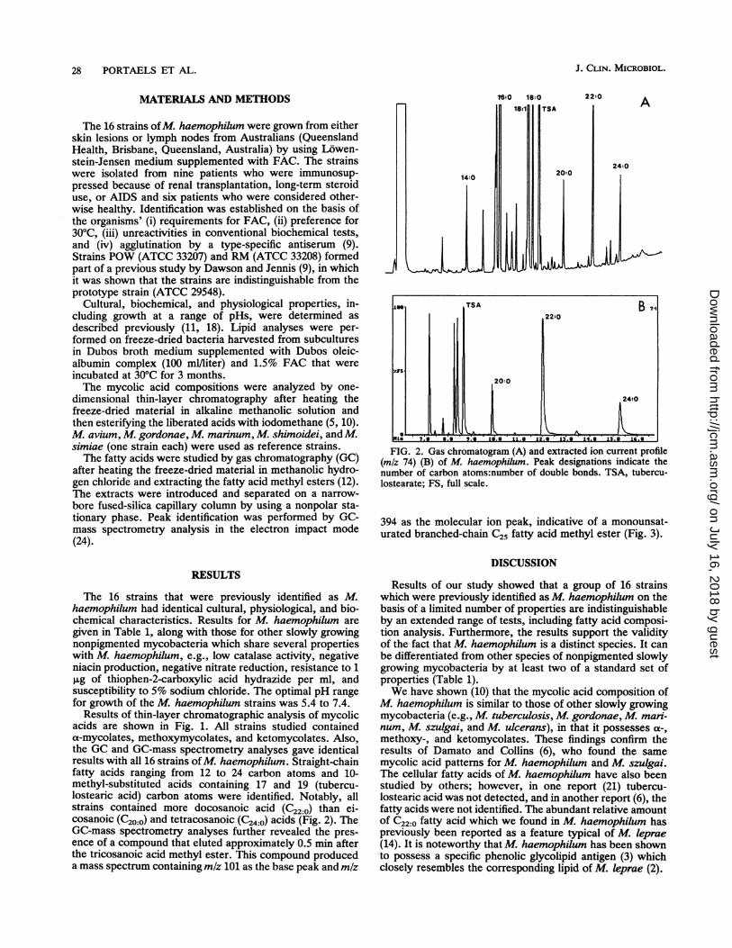
Results of thin-layer chromatographic analysis of mycolicacids are shown in Fig. 1. All strains studied containeda-mycolates, methoxymycolates, and ketomycolates. Also,the GC and GC-mass spectrometry analyses gave identicalresults with all 16 strains ofM. haemophilum. Straight-chainfatty acids ranging from 12 to 24 carbon atoms and 10-methyl-substituted acids containing 17 and 19 (tubercu-lostearic acid) carbon atoms were identified. Notably, allstrains contained more docosanoic acid (C22:0) than ei-cosanoic (C20:0) and tetracosanoic (C24:0) acids (Fig. 2). TheGC-mass spectrometry analyses further revealed the pres-ence of a compound that eluted approximately 0.5 min afterthe tricosanoic acid methyl ester. This compound produceda mass spectrum containing m/z 101 as the base peak and mlz
I<14:0
18:0 18:0181
22:0
TSA A
24:0
FIG. 2. Gas chromatogram (A) and extracted ion current profile(m/z 74) (B) of M. haemophilum. Peak designations indicate thenumber of carbon atoms:number of double bonds. TSA, tubercu-lostearate; FS, full scale.
394 as the molecular ion peak, indicative of a monounsat-urated branched-chain C25 fatty acid methyl ester (Fig. 3).
DISCUSSION
Results of our study showed that a group of 16 strainswhich were previously identified as M. haemophilum on thebasis of a limited number of properties are indistinguishableby an extended range of tests, including fatty acid composi-tion analysis. Furthermore, the results support the validityof the fact that M. haemophilum is a distinct species. It canbe differentiated from other species of nonpigmented slowlygrowing mycobacteria by at least two of a standard set ofproperties (Table 1).We have shown (10) that the mycolic acid composition of
M. haemophilum is similar to those of other slowly growingmycobacteria (e.g., M. tuberculosis, M. gordonae, M. man-num, M. szulgai, and M. ulcerans), in that it possesses et-,methoxy-, and ketomycolates. These findings confirm theresults of Damato and Collins (6), who found the samemycolic acid patterns for M. haemophilum and M. szulgai.The cellular fatty acids of M. haemophilum have also beenstudied by others; however, in one report (21) tubercu-lostearic acid was not detected, and in another report (6), thefatty acids were not identified. The abundant relative amountof C22:0 fatty acid which we found in M. haemophilum haspreviously been reported as a feature typical of M. leprae(14). It is noteworthy that M. haemophilum has been shownto possess a specific phenolic glycolipid antigen (3) whichclosely resembles the corresponding lipid of M. leprae (2).
II
j
I
u
J. CLIN. MICROBIOL.
II
s^n20:0
on July 16, 2018 by guesthttp://jcm
.asm.org/
Dow
nloaded from

VOL.31,1993~~~~~CHARACTERIZATIONOF M. HAEMOPHILUM 29
inin 12.6U 12.8U 13.90 13.29 13.49 13C0 13.80 14.901291470 40d 14.fi
199.
29 .99
xFS_ 251.99 397.99
~~266.099AL .11,d .LL. l.1
.-
299
35 .99
.I -L 3 :19r99499
294.99251.99 3~.9
199 159~ 299 259,
399 359o 4- 4--
5049 ---5-59--- 609FIG. 3. Extracted ion current profile of a selected time interval in analysis of M. haemwphilum indicative of methyl esters of
branched-chain (m/z 101) and straight-chain (m/z 74) fatty acids (A); mass spectrum of the branched-chain fatty acid methyl ester with a
retention time of 13.94 min (B). FS, full scale.
Other mycobacterial taxa which have unusual growth
requirements in vitro include M. lepraemurium (17), M.
paratuberculosis (19), "wood-pigeon mycobacteria" (19),now called M. avium subsp. "silvaticum" (23), and some
armadillo-derived mycobacteria (16). WVhile these taxa have
been regarded as probably distinct species, recent nucleic
acid analyses suggest that M. lepraemurium, M. paratuber-
culosis, and M. avium subsp. "silvaticum" are variants of
M. avium (25). The demonstrated phenetic divergences are
likely due to adaption to different hosts through almost
absolute parasitism. While we await results of genetic stud-
ies such as analysis of 16S rRNA from M. haemophilum, it
seems reasonable to speculate that it is a distinct species.
Any further evidence of its relatedness to M. leprae will be
of great interest.
It is likely that M. haemophilum is an ubiquitous organism
(1, 4) of environmental origin that causes infections in only a
small proportion of exposed humans. Its occurrence in
children's lymph nodes and surface wounds (1, 7) supports
this contention. In the present study, we showed that M.
haemophilum grows over a wide pH range, as do most
saprophytic mycobacteria (18). It may show a preference for
iron-rich natural environments. No thorough search of the
environment for M. haemophilum has yet been carried out.
The present investigation has increased the available data
on one of the more recently recognized mycobacterial patho-
gens. M. haemophilum is apparently a potential pathogen of
global distribution, and it is likely that further cases of M.
haemophilum infection will be identified as laboratories
become more aware of the need to use appropriate media.
Consideration ofM. haemophilum must be given in any case
of surface lesions containing noncultivable mycobacteria.Once isolated, recognition is not difficult. A requirement for
FAC (or blood, hemin, etc.) and a preference for tempera-
tures of about 300C are adequate for a presumptive identifi-
cation of M. haemwphilum. Better data regarding its drug
IS.A 84941
101
ZFS-
E~~~~~~~~~~~~~~~~~~~~~~~~~~~~~~~~~~~~~~~~199~~~~~~~~~~~~~~~I
B Ls..s199-
ZFS -
43.99
142.99169.99
IiLlW''
259 399 359
0/Z. 5,9
a . .. -, . 1- . .- -1 -1 .-- I -4 t 'r Im . Ift -- . - -- . - -- . - -- . - - - . - -. I . .
V M .-V '! 4"! gamin, IN - - . 1.1. W., M. Sw I-M6 .S 1, I MI. 0. -1 1 Our! 0
Falk 0 JIMAIIII
VOL. 31, 1993
" .1
on July 16, 2018 by guesthttp://jcm
.asm.org/
Dow
nloaded from

30 PORTAELS ET AL.
susceptibility, genetic relatedness to other species, andhabitat are still required.
REFERENCES1. Becherer, P., and R. L. Hopfer. 1992. Infection with Mycobac-
terium haemophilum. Clin. Infect. Dis. 14:793.2. Besra, G. S., M. McNeil, D. E. Minnikin, F. Portaels, M. Ridell,
and P. J. Brennan. 1991. Structural elucidation and antigenicityof a novel phenolic glycolipid antigen from Mycobacteriumhaemophilum. Biochemistry 30:7772-7777.
3. Besra, G. S., D. E. Minnikin, L. Rigouts, F. Portaels, and M.Ridell. 1990. A characteristic phenolic glycolipid antigen fromMycobacterium haemophilum. Lett. Appl. Microbiol. 11:202-204.
4. Branger, B., A. Gouby, R. Oules, J. P. Balducchi, G. Mourad, J.Fourcade, C. Mion, F. Duntz, and M. Ramuz. 1985. Mycobac-terium haemophilum and Mycobacterium xenopi associatedinfection in a renal transplant patient. Clin. Nephrol. 23:46-49.
5. Daffe, M., M. A. Laneelle, C. Asselineau, V. Uvy-Frebault, andH. David. 1983. Interet taxonomique des acides gras des myco-bacteries: proposition d'une methode d'analyse. Ann. Micro-biol. (Inst. Pasteur). 134:241-256.
6. Damato, J. J., and M. T. Collins. 1984. Radiometric studies withgas-liquid and thin-layer chromatography for rapid demonstra-tion of hemin dependence and characterization of Mycobacte-rium haemophilum. J. Clin. Microbiol. 20:515-518.
7. Dawson, D. J. Unpublished data.8. Dawson, D. J., Z. M. Blacklock, and D. W. Kane. 1981.
Mycobacterium haemophilum causing lymphadenitis in an oth-erwise healthy child. Med. J. Aust. 2:289-290.
9. Dawson, D. J., and F. Jennis. 1980. Mycobacteria with a growthrequirement for ferric ammonium citrate, identified as Myco-bacterium haemophilum. J. Clin. Microbiol. 11:190-192.
10. Dobson, G., D. E. Minnikin, S. M. Minnikin, J. H. Parlett, M.Goodfellow, M. Ridell, and M. Magnusson. 1985. Systematicanalysis of complex mycobacterial lipids, p. 237-265. In M.Goodfellow and D. E. Minnikin (ed.), Chemical methods inbacterial systematics. Academic Press, Inc. (London), Ltd.,London.
11. Jenkins, P. A., S. R. Pattyn, and F. Portaels. 1982. Diagnosticbacteriology, p. 441-469. In C. Ratledge and J. L. Stanford(ed.), The biology of the mycobacteria. Academic Press, Inc.(London), Ltd., London.
12. Jimenez, J., and L. Larsson. 1986. Heating cells in acid metha-nol for 30 min without freeze-drying provides adequate yields offatty acids and alcohols for gas chromatographic characteriza-tion of mycobacteria. J. Clin. Microbiol. 24:844-845.
13. Kristjansson, M., V. M. Bieluch, and P. D. Byeff. 1991. Myco-bacterium haemophilum infection in immunocompromised pa-
tients: case report and review of the literature. Rev. Infect. Dis.13:906-910.
14. Kusaka, T., and S. Izumi. 1983. Gas chromatography of consti-
tutive fatty acids in Mycobacterium leprae. Microbiol. Immu-nol. 27:409-414.
15. McBride, M. E., A. H. Rudolph, J. A. Tschen, P. Cernoch, J.Davis, B. A. Brown, and R. J. Wallace. 1991. Diagnostic andtherapeutic considerations for cutaneous Mycobacterium hae-mophilum infections. Arch. Dermatol. 127:276-277.
16. Portaels, F., C. Asselineau, I. Baess, M. Daffe, G. Dobson, P.Draper, D. Gregory, R. M. Hall, T. Imaeda, P. A. Jenkins, M. A.Lan6elle, L. Larsson, M. Magnusson, D. E. Minnikin, S. R.Pattyn, G. Wieten, and P. R. Wheeler. 1986. A cooperativetaxonomic study of mycobacteria isolated from armadillos in-fected with Mycobactenum leprae. J. Gen. Microbiol. 132:2693-2707.
17. Portaels, F., and S. R. Pattyn. 1981. Parameters influencing thein vitro growth of Mycobacterium lepraemunium. Int. J. Lepr.49:194-197.
18. Portaels, F., and S. R. Pattyn. 1982. Growth of mycobacteria inrelation to the pH of the medium. Ann. Microbiol. (Inst.Pasteur). 133:213-221.
19. Saxegaard, F., and I. Baess. 1988. Relationship between Myco-bactenium avium, Mycobacterium paratuberculosis and 'woodpigeon mycobacteria.' APMIS 96:37-42.
20. Sompolinsky, D., A. Lagziel, D. Naveh, and T. Yankilevitz. 1978.Mycobacterium haemophilum sp. nov., a new pathogen ofhumans. Int. J. Syst. Bacteriol. 28:67-75.
21. Sompolinsky, D., A. Lagziel, and I. Rosenberg. 1979. Furtherstudies of a new pathogenic mycobacterium (M. haemophilumsp. nov.). Can. J. Microbiol. 25:217-225.
22. Thibert, L., F. Lebel, and B. Martineau. 1990. Two cases ofMycobactenium haemophilum infection in Canada. J. Clin.Microbiol. 28:621-623.
23. Thorel, M. F., M. Krichevsky, and V. V. Levy-Frebault. 1990.Numerical taxonomy of mycobactin-dependent mycobacteria,emended description ofM. avium, and description of M. aviumsubsp. avium, supsp. nov., M. avium subsp. paratuberculosissubsp. nov., and M. avium subsp. silvaticum subsp. nov. Int. J.Syst. Bacteriol. 40:254-260.
24. Valero-Guillen, P., F. Martin-Luengo, L. Larsson, J. Jimenez, I.Juhlin, and F. Portaels. 1988. Fatty and mycolic acids ofMycobacterium malmoense. J. Clin. Microbiol. 26:153-154.
25. Wayne, L. G., R. C. Good, M. I. Krichevsky, Z. Blacklock, H. L.David, D. J. Dawson, W. Gross, J. Hawkins, V. Levy-Frebault,C. McManus, F. Portaels, S. Rusch-Gerdes, K. H. Schroder,V. A. Silcox, M. Tsukamura, L. Van den Breen, and M. A.Yakrus. 1991. Fourth report of the cooperative, open-endedstudy of slowly growing mycobacteria by the InternationalWorking Group on Mycobacterial Taxonomy. Int. J. Syst.Bacteriol. 41:463-472.
26. Wayne, L. G., and H. A. Sramek. 1992. Agents of newlyrecognized or infrequently encountered mycobacterial diseases.Clin. Microbiol. Rev. 5:1-25.
J. CLIN. MICROBIOL.
on July 16, 2018 by guesthttp://jcm
.asm.org/
Dow
nloaded from