bioadhesive buccal discs of fluvastatin sodium by
TRANSCRIPT
134 Az. J. Pharm Sci. Vol. 51, March, 2015.
BIOADHESIVE BUCCAL DISCS OF FLUVASTATIN SODIUM
BY
Nada Abdulla Assaedi1, Nagia N. Afifi
2, Gehanne A. Awad
1
FROM
1Department of Pharmaceutics and Industrial Pharmacy, Faculty of Pharmacy, Ain
Shams University, Cairo, Egypt; 2Department of Pharmaceutical Technology, Faculty
of Pharmacy and Biotechnology, German University in Cairo, Cairo, Egypt
ABSTRACT
Fluvastatin sodium (FVS) is a cholesterol lowering agent (HMG-CoA reductase
inhibitor) which undergoes extensive hepatic first pass metabolism causing an absolute
bioavailability of ~30%. The aim of this work was to formulate a buccoadhesive disc of
FVS to be applied to the buccal mucosa, releasing the drug in a unidirectional manner,
in order to improve the bioavailability of the drug and lower the dose-dependent side
effects. The bioadhesive discs were prepared by direct compression method using
several polymers such as: guar gum, sodium alginate, sodium carboxymethyl cellulose,
carbopol 934P, and hydroxypropylmethyl cellulose. Impermeable ethyl cellulose was
applied as the backing layer. Different permeation enhancers such as bile salts,
surfactants, fatty acids, chitosan, dimethyl sulfoxide, and polyethylene glycol 6000
(PEG 6000) were tested to improve the permeability of buccal mucosal membranes. The
optimized formulation contained FVS, guar gum, PEG 6000, and sodium deoxycholate
(permeation enhancer, 4%). It showed a drug release of 95.4% in 80 min, drug
permeation through chicken pouch membrane (flux (Jss) = 3.74 mg cm-2
h-1
), ex vivo
bioadhesion strength of 2.543 g, along with satisfactory bioadhesion time of 4.87 h.
Physicochemical characteristics of the buccal discs such as drug content uniformity,
disc thickness, disc hardness, surface pH, and swelling index were also evaluated.
Keywords: Fluvastatin sodium, bioadhesive buccal discs, PEG 6000, sodium
deoxycholate.

Az. J. Pharm Sci. Vol. 51, March, 2015. 135
Introduction
Oral drug administration is the most suitable and widely acceptable route for the
delivery of most therapeutically active agents. However, many drugs are subjected to
presystemic clearance in the liver, which often leads to a lack of correlation between
membrane permeability, absorption and bioavailability (Harris and Robinson, 1992).
In recent years, delivery of therapeutic agents through various transmucosal routes has
received significant attention. Among these, the buccal route provides a number of
advantages such as well vascularization, relatively large surface area of absorption, ease
of accessibility, simple delivery devices, feasibility of controlled drug delivery,
avoidance of gastrointestinal degradation and hepatic first pass metabolism due to direct
access of the drug into the systemic circulation through the internal jugular vein
(Neelagiri et al., 2013).
The most important determinant of buccal delivery is the degree of permeability
of the mucosa since the therapeutic efficacy of a drug mainly depends on its ability to
penetrate the tissue fast enough to provide the required effective plasma concentrations
(De Caro et al., 2009). Permeation enhancers may be utilized to overcome the
permeability barrier in which they act by increasing the retention time of drug
around the buccal mucosa, interaction with the buccal mucosal protein and intercellular
lipid, and/or enhancing the drug partitioning across the buccal mucosa (Meher et al.,
2012). In addition, drug absorption through the oral mucosal membranes requires that
the drug dissolves sufficiently in a very small volume of saliva, which may represent an
obstacle for poorly soluble drugs (Turunen et al., 2011).
Fluvastatin sodium (FVS) is an antilipemic agent which competitively inhibits
hydroxymethylglutaryl-coenzyme A (HMG-CoA) reductase. FVS belongs to a class of
medications called statins and is widely used to reduce plasma cholesterol levels and
prevent cardiovascular disease. However, it undergoes extensive hepatic first pass
metabolism causing an absolute bioavailability of ~30% (Sweetman, 2005). Thereby, a
new approach to increase its bioavailability will be of prime benefit. With its low oral
bioavailability, short half-life of 2-3 hours, and suitable molecular size of 433.45 g/mol,
FVS is considered a good candidate for buccal route administration which has the
benefit of escaping first pass effect as the drug passes directly into the bloodstream
resulting in a reduction of dose and dose-dependent adverse events.
Materials and methods
Materials
FVS was kindly gifted by Biocon Ltd, Bangalore, India. Guar gum (GG),
sodium alginate (SALG), sodium carboxymethyl cellulose, high viscosity (SCMC),
hydroxypropylmethyl cellulose, 4,000 cp (HPMC), sodium cholate (SC), sodium
deoxycholate (SDC), cetrimide, and dimethyl sulfoxide (DMSO) were purchased from
Sigma Chemicals, USA. Chitosan was obtained from Acros Organics, USA. Carbopol
934P (CP) was obtained from Goodrich Chemical Co., USA. 2-pyrrolidone was
obtained from Aldrich, USA. Polyethylene glycol 6000 (PEG), microcrystalline
cellulose (MCC), ethyl cellulose (EC), sodium lauryl sulfate (SLS), tween 80, oleic
acid, magnesium stearate (MgSt), sodium chloride (NaCl), potassium dihydrogen
phosphate (KH2PO4), and disodium hydrogen phosphate (Na2HPO4) anhydrous were
purchased from El-Nasr Pharmaceutical Chemical Co., Cairo, Egypt.

136 Az. J. Pharm Sci. Vol. 51, March, 2015.
Preparation of bioadhesive buccal discs
Disc compositions are shown in Tables (1-2). The ingredients were accurately
weighed and mixed in a glass mortar and pestle for 10 minutes. Magnesium stearate was
added as lubricant and mixed again for 2 minutes. The discs were prepared manually by
direct compression method using a flat faced 12 mm punch. First, the powder mix was
precompressed then the backing layer of ethyl cellulose was added and compressed at
maximum force. The peripheral sides of the discs were coated with EC in ethanol
solution (10% w/v) by brushing and were left to dry in room temperature. The design of
the buccal disc is shown in Figure 1. The main function of the backing layer is to
provide unidirectional drug flow to the buccal mucosa and prevent the drug from being
dissolved in saliva and hence swallowed (Neelagiri et al., 2013). MCC and PEG 6000
were used as release enhancers.
Release study
The release rate from buccal discs was studied using USP type II (paddle)
dissolution test apparatus (Hanson SR8 plus dissolution tester, Germany). Buccal discs
were fixed to a glass slide sitting at the bottom of the dissolution flask using an instant
adhesive (cyanoacrylate) so that the core layer was facing the dissolution medium. The
space between the paddle and the buccal disc was 2.5 cm. The dissolution medium
comprised 500 mL phosphate buffer pH 6.8 to simulate buccal environment and
maintain sink conditions. The release study was performed at 37±0.5°C with a rotation
speed of 50 rpm. Samples of 5 mL were withdrawn, replaced with fresh medium at time
intervals: 15, 30, 60, 90, 120, 150, 180, 210, and 240 min, and analyzed using UV
spectrophotometer (Shimadzu UV visible 1601 PC, Kyoto, Japan) at 303 nm.
Experiments were performed in triplicate (n = 3).
In order to determine the drug release mechanism from the prepared discs, the
release data (up to 60% release) of the optimized formulation was fitted to the
Korsmeyer-Peppas equation (Peppas, 1985):
Mt/M∞= ktn, Eq. (1)
where Mt/M∞ is the fractional release of the drug, „t‟ denotes the release time,
„k‟ represents a constant, incorporating structural and geometrical characteristics of the
drug/polymer system, and „n‟ is the diffusional exponent and characterizes the type of
release mechanism during the dissolution process. For the case of cylindrical discs, n ≤
0.45 corresponds to a Fickian (case I) diffusion, 0.45 < n < 0.89 to an anomalous (non-
Fickian) transport (where release is controlled by a combination of diffusion and
polymer relaxation), n = 0.89 to a zero order (case II) transport (where the drug release
rate is independent of time and involves polymer relaxation), and n > 0.89 to a super
case II transport (Harland et al., 1988).
Permeation study
Chicken pouch mucosa, an easily available biological membrane having a non-
keratinized uniform surface morphology similar to humans (Maswadeh et al., 2010),

Az. J. Pharm Sci. Vol. 51, March, 2015. 137
was chosen as a model membrane for this study. Chicken heads were obtained
immediately post sacrifice from a local slaughterhouse and transported to the laboratory.
The excised mucosa was immersed in isotonic saline at 60°C for 1 min and the
epithelium was then peeled away from the connective tissue by heat separation method
(Kulkarni et al., 2010). Resulting membranes of thickness ~180-200 µm were briefly
dipped in distilled water and frozen until use in a period of 3 weeks.
Permeation study was performed using a modified USP type II (paddle) dissolution test
apparatus (Hanson SR8 plus dissolution tester, Germany). Figure 2 displays the method
where the buccal membrane was stretched over an open end of a glass tube (13 mm
diameter, opened from both ends) and made water tight by rubber band forming the
donor chamber. The tube was then immersed in 500 mL phosphate buffer saline pH 7.4
contained in the dissolution flask so that the membrane was just below the surface of the
recipient solution (Mohamed et al., 2011). The temperature was maintained at
37±0.5°C and paddle speed was set at 100 rpm. The buccal membrane was allowed to
stabilize for 1 h before applying the buccoadhesive disc inside the tube. Donor
compartment was filled with 1 mL phosphate buffer pH 6.8. Samples of 5 mL were
withdrawn and replaced with fresh medium at time intervals: 0.5, 1, 1.5, 2, 2.5, 3, 3.5, 4,
5, and 6 h.
The formula yielding the highest permeation was selected for further study with
permeation enhancers. Ten permeation enhancers were used in different concentrations:
bile salts (sodium cholate and sodium deoxycholate), surfactants (tween 80, sodium
lauryl sulfate, and cetrimide), fatty acid (oleic acid), chitosan, 2-pyrrolidone, dimethyl
sulfoxide, and polyethylene glycol 6000 (Dodla and Velmurugan, 2013). The
experiments were performed 4 times (n = 4).
The cumulative amount of permeated drug (mg) was plotted versus time (h) and
steady-state flux was measured from the slope of the linear portion of the plot using the
following equation:
Flux =Jss = (dQ/dt)/A, Eq. (2)
where Jss is the steady-state flux; dQ/dt is the permeation rate; A is the active
diffusion area (1.33 cm2). The permeability coefficient P was calculated as follows (De
Caro et al., 2008):
P = Jss/Cd, Eq. (3)
where P is the permeability coefficient and Cd is the donor drug concentration.
2.5. Bioadhesion study (ex vivo)
2.5.1. Bioadhesion strength
Several techniques have been reported in literature for the measurement of bioadhesive
strength. In the present study, bioadhesive strength was measured using the modified
physical balance method. The method utilized chicken pouch membrane as the model

138 Az. J. Pharm Sci. Vol. 51, March, 2015.
mucosal membrane. A piece of mucosa was fixed to the lower stainless steel support
with a cyanoacrylate adhesive and moistened with phosphate buffer pH 6.8. The disc
was attached to the upper clamp of the apparatus using cyanoacrylate. The lower
support was slowly raised so that the disc touched the mucosa. Then both pans were
balanced by adding an appropriate weight to the left-hand pan. Previously weighed
empty beaker was placed on the right hand pan. A preload of 50 g was placed on the
clamp for 1 min to establish the adhesive bond, then water (equivalent to weight) was
slowly added to the beaker at a constant rate until the disc detached from the mucosal
surface (Velmurugan and Srinivas, 2013; Sudarshan et al., 2014). The weight
required to detach the disc from the mucosal surface gave the measure of bioadhesive
strength. The experiment was performed on 6 discs of each formulation using a different
chicken pouch membrane each time. From the bioadhesive strength (g), force of
adhesion was calculated.
Force of adhesion (N) = (Bioadhesive strength (g) /1,000) x 9.81 Eq. (4)
Bioadhesion time
The ex vivo bioadhesion time was studied by the application of buccoadhesive
discs on freshly obtained chicken buccal mucosa. Mucosal membrane was fixed on the
internal side of a beaker using cyanoacrylate. Discs were pasted onto the membrane by
applying a light force for 30 s. The beaker was filled with 200 mL phosphate buffer (pH
6.8) and kept at 37±1°C. After 2 minutes, a 50 rpm stirring was applied to simulate the
buccal cavity environment. Time for disc to detach or completely dissolve was recorded
as the bioadhesion time (Sudarshan et al., 2014). The experiment was performed in
triplicate.
Evaluation of physicochemical characteristics
Physicochemical characteristics of the optimized formula were evaluated
including the swelling index, drug content uniformity, disc thickness, disc hardness, and
surface pH.
Swelling Index: Three buccal discs were individually weighed (W1) and placed
separately in petri dishes with 5 mL of phosphate buffer of pH 6.8. At the time interval
of 15, 30, 60, 90, and 120 min, disc was removed from the petri dish and carefully
blotted using filter paper (Bhanja et al., 2013). The swollen disc was then reweighed
(W2) and the percentage hydration was calculated using the following formula:
Percentage hydration = [(W2-W1)/ W1] * 100 Eq. (5)
Drug content uniformity was evaluated by dissolving one buccal disc in 250 mL
phosphate buffer (pH 6.8). The solution was then passed through a whatmann filter
paper and analyzed spectrophotometrically at the predetermined λmax of FVS after
sufficient dilution with phosphate buffer of pH 6.8 (Kassem et al., 2014). The test was
done in triplicate and the mean drug content was deduced ± SD.

Az. J. Pharm Sci. Vol. 51, March, 2015. 139
Disc thickness of three buccoadhesive discs was measured using a micrometer
(Tri-Circle, Shanghai, China) and recorded.
2.6.4. Disc hardness was measured using a hardness tester (Erweka, Germany) for six
buccoadhesive discs.
Surface pH was found by placing one disc in a petri dish in contact with 1 mL of
phosphate buffer (pH 6.8) for 2 hours at room temperature. The pH was identified by
bringing the electrode into contact with the disc surface and allowing equilibration for 1
minute (Viswanadhan et al., 2012). The experiment was repeated three times.
RESULTS AND DISCUSSION
Release study
The maximum duration for buccal drug delivery is usually limited to
approximately 4–6 h, since meal intake and/or drinking may require dosage form
removal (Neelagiri et al., 2013). Therefore, a formulation with an appropriate release
profile of at least 80% drug release over a 3 h period was desired for the purpose of this
study. The obtained release curves of F1-F25 are shown in Figure 3.
Discs containing single polymers (F1-F4) showed a slow drug release of less
than 50% in 4 h. On the other hand, discs containing HPMC (F5) released 90% of the
drug in 2 h but they were excluded due to rapid erosion. Therefore, a blend of HPMC
and each of the other polymers was formed in a 1:1 ratio (F6-F9) as a way to improve
the release and minimize erosion. CMC-HPMC and SALG-HPMC blends showed
improved drug release over the use of CMC or SALG alone, whereas in the GG and CP
formulas, the addition of HPMC did not improve drug release.
MCC and PEG 6000 were added in 15-35% w/w as release enhancers to
previous formulas F1-F4 and F6-F9. PEG 6000 was reported to increase porosity of the
matrix and produce channels, which in turn facilitate the dissolution medium to
penetrate the matrix and dissolve the drug more rapidly (Hassan et al., 2009). MCC
allows water to enter the disc matrix by means of capillary pores and exhibits very good
disintegrant property (Bala et al., 2012). GG (in F10 and F18) was the most influenced
polymer by the addition of MCC or PEG where it released > 90% in 3 h compared to
only 11.3% using GG alone (F1). When release enhancers were added to HPMC
polymer blends, these formulas showed the highest drug release rates. However, F14
and F22 were excluded due to excessive swelling which caused the discs to come off
the glass slide. Therefore, the selected formulations which showed ≥ 80% drug release
in 3 h were: F10, F11, F15, F16, F18, F23, and F24. They were tested for drug
permeation through chicken pouch mucosa.
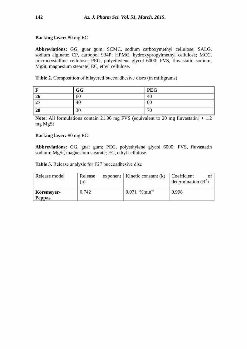
Drug release of formula F27 is shown in Figure 4. F27 was picked as the
optimized formulation as will be discussed in section “3.2. Permeation Study”. F26 and
F28 were therefore not tested for drug release since all three formulations (F26-F28)
were expected to have higher drug release than F18 (70 GG, 30 PEG) due to possessing
higher PEG concentrations. The formulation F27 showed a drug release of 95.4% in 80

140 Az. J. Pharm Sci. Vol. 51, March, 2015.
min. The values of T50%, T70% and T90% were found to be 13.8, 24.2, and 49 min
respectively. Polymeric matrices release the drug via a combination of mechanisms.
Korsmeyer-Peppas release exponent (n) was found to be 0.742, indicating anomalous
(non-Fickian) release kinetics, where different processes such as diffusion, swelling,
and erosion simultaneously occurred. The obtained value of k (kinetic constant), n
(diffusional exponent) and R2 (correlation coefficient) of the in vitro release data are
presented in Table 3.
Permeation study
Initial permeation results of the selected 7 formulations were in the range of 2.9-
6.6% drug permeation in 4 h. The formula with the highest permeation, F18 (70 GG, 30
PEG), was chosen for further evaluation with permeation enhancers.
Upon using different permeation enhancers with formula F18 (Table 4), sodium
deoxycholate (SDC) was found to be the most effective enhancer. Addition of SDC
results in the extraction of mucosal lipids from the intercellular spaces, via
micellization, which enhances the diffusivity of the drug through the paracellular route.
At higher concentrations, SDC perturbs the lipid membranes of the epithelial cells,
possibly facilitating transcelluar transport as well (Ganem-Quintanar et al., 1997;
Shanker et al., 2009). It was suggested that SDC can also cause the uncoiling and
extension of the protein helices, which leads to opening of the polar pathways for
diffusion (Nicolazzo et al., 2005).
Although the drug permeation has improved but it was still considered slow
(11.8% in 4 h using 4% SDC). This was probably due to the inability to release the drug
in the small volume of liquid available. Therefore, F18 formulation had been changed
concerning the ratio of guar gum to PEG 6000. New formulations: F26, F27, and F28
were prepared with decreasing GG and increasing PEG concentrations (Chinta et al.,
2014). PEG 6000 has also been used as a permeation enhancer for buccal delivery of
Simvastatin (Goud and Samanthula, 2011) and Atorvastatin calcium (John et al.,
2010). SDC (4%) was added to the new formulas and permeation was tested again.
Results are shown in Table 5. Formulations F27 and F28 demonstrated
acceptable drug permeation of 75.6% and 81.2% in 4 h respectively. Table 6 lists a
comparison of permeation properties of the two formulations such as permeation flux
(J) and permeability coefficient. Permeation curve of the chosen formula F27 is
displayed in Figure 5.
Bioadhesion study
Formulas F27 and F28 were evaluated for their bioadhesion properties. F27
showed a higher bioadhesion strength (2.534 g) than F28 (1.668 g). Increase in polymer
concentration (40mg GG in F27 compared to 30mg GG in F28) resulted in increased
force of adhesion. When the concentration of polymer is low, the number of chains
penetrating glycoprotein chains per unit volume of mucus is low resulting in weaker
interaction (Salamat-Miller et al., 2005).
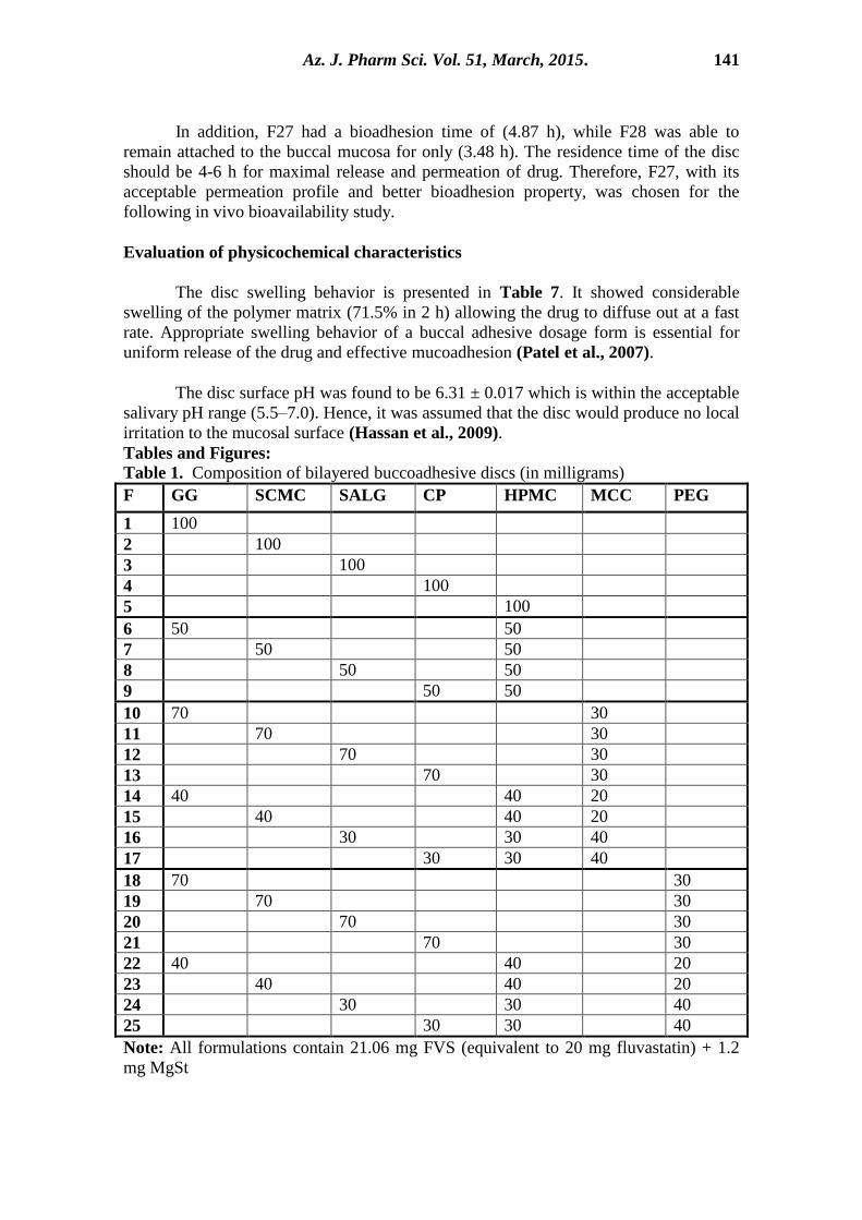
Az. J. Pharm Sci. Vol. 51, March, 2015. 141
In addition, F27 had a bioadhesion time of (4.87 h), while F28 was able to
remain attached to the buccal mucosa for only (3.48 h). The residence time of the disc
should be 4-6 h for maximal release and permeation of drug. Therefore, F27, with its
acceptable permeation profile and better bioadhesion property, was chosen for the
following in vivo bioavailability study.
Evaluation of physicochemical characteristics
The disc swelling behavior is presented in Table 7. It showed considerable
swelling of the polymer matrix (71.5% in 2 h) allowing the drug to diffuse out at a fast
rate. Appropriate swelling behavior of a buccal adhesive dosage form is essential for
uniform release of the drug and effective mucoadhesion (Patel et al., 2007).
The disc surface pH was found to be 6.31 ± 0.017 which is within the acceptable
salivary pH range (5.5–7.0). Hence, it was assumed that the disc would produce no local
irritation to the mucosal surface (Hassan et al., 2009).
Tables and Figures:
Table 1. Composition of bilayered buccoadhesive discs (in milligrams)
F GG SCMC SALG CP HPMC MCC PEG
1 100
2 100
3 100
4 100
5 100
6 50 50
7 50 50
8 50 50
9 50 50
10 70 30
11 70 30
12 70 30
13 70 30
14 40 40 20
15 40 40 20
16 30 30 40
17 30 30 40
18 70 30
19 70 30
20 70 30
21 70 30
22 40 40 20
23 40 40 20
24 30 30 40
25 30 30 40
Note: All formulations contain 21.06 mg FVS (equivalent to 20 mg fluvastatin) + 1.2
mg MgSt
142 Az. J. Pharm Sci. Vol. 51, March, 2015.
Backing layer: 80 mg EC
Abbreviations: GG, guar gum; SCMC, sodium carboxymethyl cellulose; SALG,
sodium alginate; CP, carbopol 934P; HPMC, hydroxypropylmethyl cellulose; MCC,
microcrystalline cellulose; PEG, polyethylene glycol 6000; FVS, fluvastatin sodium;
MgSt, magnesium stearate; EC, ethyl cellulose.
Table 2. Composition of bilayered buccoadhesive discs (in milligrams)
F GG PEG
26 60 40
27 40 60
28 30 70
Note: All formulations contain 21.06 mg FVS (equivalent to 20 mg fluvastatin) + 1.2
mg MgSt
Backing layer: 80 mg EC
Abbreviations: GG, guar gum; PEG, polyethylene glycol 6000; FVS, fluvastatin
sodium; MgSt, magnesium stearate; EC, ethyl cellulose.
Table 3. Release analysis for F27 buccoadhesive disc
Release model Release exponent
(n)
Kinetic constant (k) Coefficient of
determination (R2)
Korsmeyer-
Peppas
0.742 0.071 %min-n
0.998

Az. J. Pharm Sci. Vol. 51, March, 2015. 143
Table 4. Permeation of F18 using permeation enhancers
Permeation enhancer
Amount
permeated
in 4 h
(mg)
Drug
percent
permeated
in 4 h
No enhancer 1.32 6.6%
Sodium cholate 2% 1.33 6.7%
4% 2.12 10.5%
Sodium deoxycholate 2% 1.66 8.3%
4% 2.35 11.8%
Tween 80 4% 1.42 7.1%
8% 1.50 7.5%
Sodium lauryl sulfate 1% 1.61 8.1%
2% 2.04 10.2%
Cetrimide 2% 1.33 6.7%
4% 1.32 6.6%
Chitosan 4% 1.56 7.8%
8% 1.62 8.1%
Oleic acid 4% 1.18 5.9%
2-Pyrrolidone 4% 1.30 6.5%
Dimethyl sulfoxide 4% 1.27 6.4%
Table 5. Permeation of modified formulas in addition to 4% sodium deoxycholate
GG (mg) PEG 6000 (mg) Amount permeated
in 4 h (mg)
Drug percent
permeated in 4 h F18 70 30 2.35 11.8%
F26 60 40 3.88 19.4%
F27 40 60 15.12 75.6%
F28 30 70 16.24 81.2%
Abbreviations: GG, guar gum; PEG 6000, polyethylene glycol 6000

144 Az. J. Pharm Sci. Vol. 51, March, 2015.
Table 6. Permeation and bioadhesion parameters of fluvastatin sodium discs
Table 7. Swelling behavior of F27 buccoadhesive discs
Time (min) Percentage hydration (%)
15 38.3
30 46.9
60 57.2
90 65.4
120 71.5
Table 8. Physicochemical characteristics of F27 buccoadhesive disc
Drug content (%) 98.6 ± 1.46
Thickness (mm) 1.94 ± 0.042
Hardness (N) 58.67 ± 5.033
Surface pH 6.31 ± 0.017
Parameters F27 F28
Permeation
study
Amount of drug permeated in 4 h
(mg) 15.12 ± 0.862 16.24 ± 0.784
Flux (J) (mg h-1
cm-2
) 3.743 4.068
Permeability coefficient (cm h-1
) 0.187 0.203
Bioadhesion
study
Bioadhesion strength (g) 2.534 ± 0.784 1.668 ± 0.697
Bioadhesion force (N) 0.025 ± 0.008 0.016 ± 0.007
Bioadhesion time (h) 4.87 ± 0.51 3.48 ± 0.39

Az. J. Pharm Sci. Vol. 51, March, 2015. 145
Figure 1. Bioadhesive buccal discs design
Figure 2. Permeation apparatus

146 Az. J. Pharm Sci. Vol. 51, March, 2015.
Figure 3. Release curves of F1-F25 bioadhesive buccal discs

Az. J. Pharm Sci. Vol. 51, March, 2015. 147
Figure 4. Release curve of formula F27
Figure 5. Permeation curve of optimized formula F27
Conclusion
The results of the study indicated that buccoadhesive discs of fluvastatin sodium
could be successfully prepared by direct compression method using guar gum as the
mucoadhesive polymer, PEG 6000 as the release enhancer, sodium deoxycholate as the
permeation enhancer, and ethyl cellulose as the backing layer. It exhibited well drug
release, bioadhesion property, and drug permeation in 4 h. The mechanism of drug
release was found to be non-Fickian diffusion. Therefore, there is a good potential of the
prepared buccoadhesive discs for systemic delivery with added advantages of
circumventing the hepatic first pass metabolism and substantial dose reduction. Further

148 Az. J. Pharm Sci. Vol. 51, March, 2015.
in vivo study is required to attain the relative bioavailability of this optimized buccal
disc formulation in comparison to a peroral product in the market.
Acknowledgment
The authors are thankful to Biocon Ltd, Bangalore, India, for providing a generous gift
sample of fluvastatin sodium.
REFERENCES
Bala, R., Khanna, S., Pawar, P., (2012):. Polymers in fast disintegrating tablets - A
review. Asian J. Pharm. Clin. Res. 5, 8-14.
Barilla, D., Pratapa, P., Hubert, M., Gumbhir-Shah, K., (2004):. Steady-state
pharmacokinetics of fluvastatin in healthy subjects following a new extended
release fluvastatin tablet, Lescol® XL. Biopharm. Drug Dispos. 25, 51-59.
DOI: 10.1002/bdd.378.
Bhanja, S., Shafeeque, C., Sudhakar, M., (2013):. Mucoadhesive buccal tablets of
glimeperide - formulation and evaluation. Int. J. Pharm. Pharm. Sci. 5, 502-
510.
Chinta, B., Tnvss, S., Santosh, T., Nannapaneni, M., (2014):. Formulation and
evaluation of diclofenac diethylamine gels prepared using hyaluronic acid:
Influence of various permeation enhancers on rat skin penetration. Indo Amer.
J. Pharm. Res. 4, 320-328.
De Caro, V., Giandalia, G., Siragusa, M.G., Paderni, C., Campisi, G., Giannola,
L.I., (2008). Evaluation of galantamine transbuccal absorption by
reconstituted human oral epithelium and porcine tissue as buccal mucosa
models: Part I. Eur. J. Pharm. Biopharm. 70, 869-873.
De Caro, V., Giandalia, G., Siragusa, M.G., Campisi, G., Giannola, L.I., (2009). Galantamine delivery on buccal mucosa: Permeation enhancement and design
of matrix tablets. J. Bioequiv. Availab. 1, 127-134.
Dodla, S., Velmurugan, S., (2013):. Buccal penetration enhancers - An overview.
Asian J. Pharm. Clin. Res. 6, 39-47.
Ganem-Quintanar, A., Kalia, Y.N., Falson-Rieg, F., Buri, P., (1997):. Mechanisms
of oral permeation enhancement. Int. J. Pharm. 156, 127-142.
Goud, B.A., Samanthula, K.S., (2011):. Formulation and evaluation of bioadhesive
buccal tablets of simvastatin. J. App. Pharm. Sci. 1, 68-91.
Harland, R.S., Gazzaniga, A., Sangalli, M.E., Colombo, P., Peppas, N.A., (1988):. Drug/polymer matrix swelling and dissolution. Pharm. Res. 5, 488–494.

Az. J. Pharm Sci. Vol. 51, March, 2015. 149
Harris, D., Robinson, J.R., (1992):. Drug delivery via the mucous membranes of the
oral cavity. J. Pharm. Sci. 81, 1–10.
Hassan, N., Khar, R.K., Ali, M., Ali, J., (2009):. Development and evaluation of
buccal bioadhesive tablet of an anti-emetic agent ondansetron. AAPS Pharm.
Sci. Tech. 10, 1085-1092. DOI: 10.1208/s12249-009-9304-4.
John, A.S., Sathesh, B.P.R., Divakar, G., Jangid, M.K., Purohit, K.K., (2010):. Development and evaluation of buccoadhesive drug delivery system for
atorvastatin calcium. J. Curr. Pharm. Res. 1, 31-38.
Kassem, M.A., Elmeshad, A.N., Fares, A.R., (2014):. Enhanced bioavailability of
buspirone hydrochloride via cup and core buccal tablets: formulation and in
vitro/in vivo evaluation. Int. J. Pharm. 463, 68-90. DOI:
10.1016/j.ijpharm.2014.01.003.
Kulkarni, U., Mahalingam, R., Pather, I., Li, X., Jasti, B., (2010):. Porcine buccal
mucosa as in vitro model: Effect of biological and experimental variables. J.
Pharm. Sci. 99, 1265-1277. DOI: 10.1002/jps.21907.
Maswadeh, H.M., Kanaan, R.A., Aljarbou, A.N., Al-Hanbali, O.A, (2010):. Effect
of different biological membranes on in vitro bioadhesion property. Drug
Invent. Today 2, 155-159.
Meher, J.G., Yadav, N.P., Yadav, K.S., Rai, V., (2012):. Buccal drug delivery system
and penetration enhancer: A review. The Pharm. Rev. 8, 113-117.
Mohamed, M., Haider, M., Ali, M., (2011):. Buccal mucoadhesive films containing
antihypertensive drug: In vitro/in vivo evaluation. J. Chem. Pharm. Res. 3,
665-686.
Neelagiri, R., Reddy, M.S., Rao, N.G.R., (2013):. Buccal patch as drug delivery
system: An overview. Int. J. Curr. Pharm. Res. 5, 40-47.
Nicolazzo, J., Reed, B., Finnin, B., (2005):. Buccal penetration enhancers - How do
they really work? J. Control. Release 105, 1-15.
DOI:10.1016/j.jconrel.2005.01.024.
Patel, V.M., Prajapati, B.G., Patel, M.M., (2007):. Formulation, evaluation, and
comparison of bilayered and multilayered mucoadhesive buccal devices of
propranolol hydrochloride. AAPS Pharm. Sci. Tech. 8, E147–E154.
Peppas, N.A., (1985):. Analysis of fickian and non-fickian drug release from polymers.
Pharm. Acta Helv. 60, 110-111.
Salamat-Miller, N., Chittchang, M., Johnston, T.P., (2005):. The use of
mucoadhesive polymers in buccal drug delivery. Adv. Drug Deliv. Rev. 57,
1666-1691.
150 Az. J. Pharm Sci. Vol. 51, March, 2015.
Shanker, G., Kumar, C.K., Gonugunta, C.S.R., Kumar, B.V., Veerareddy, P.R.,
(2009):. Formulation and evaluation of bioadhesive buccal drug delivery of
tizanidine hydrochloride tablets. AAPS Pharm. Sci. Tech. 10, 530-539. DOI:
10.1208/s12249-009-9241-2.
Sudarshan, S., Savaliya, D., Shah, S.D., Bothara, S.B., (2014):. Development and in
vivo bioavailability evaluation of sumatriptan succinate buccal tabets. Int. J.
Pharm. Sci. Nanotech. 7, 2477-2486.
Sweetman, S.C., editor, (2005):. Martindale: The Complete Drug Reference. 34th ed.
London: The Pharmaceutical Press.
Turunen, E., Mannila, J., Laitinen, R., Riikonen, J., Lehto, V.P., Jarvinen, T.,
Ketolainen, J., Jarvinen, K., Jarho, P., (2011):. Fast-dissolving sublingual
solid dispersion and cyclodextrin complex increase the absorption of
perphenazine in rabbits. J. Pharm. Pharmacol. 63, 19-25. DOI: 10.1111/j.2042-
7158.2010.01173.x.
Velmurugan, S., Srinivas, P., (2013):. Formulation and in vitro evaluation of losartan
potassium mucoadhesive buccal tables. Asian J. Pharm. Clin. Res. 6, 125-130.
Viswanadhan, P.V., Padole, A., Abraham, A., Mathew, S.T., (2012):. Buccal tablets
of lisinopril by direct compression method for buccal drug delivery. Int. R. J.
Pharmaceuticals 2, 30-38.

Az. J. Pharm Sci. Vol. 51, March, 2015. 151
الملخص العربي
أسطوانات فمية لاصقة لعقار صوديوم الفلوفاستاتين
للسادة الذكاترة
ذ عجذالله انصبعذ1ففبجخ جت ع،
2، جب عجذانسع عض1
نـــــــــــــــــــــــــــم
1 جبيعخ ع شس، يذا انعجبسخ، انقبشح، يصش انصذنخ انصبعخ، كهخ انصذنخ، قسى انصذلابد
2 قسى ركنجب انصذنخ، كهخ انصذنخ، انجبيعخ الأنبخ ف انقبشح، انزجع انخبيس، انقبشح، يصش
الأىط انيى الأنى نذى يشرفىع نكى سجخ انكنسزشل فى انىذوصدو انفهفبسزبر عقبس قهم
صبغخ أسطابد فخ لاصقخ نهصقب ثبنغشىب رذف انذساسخ انيبنخ إن %(.33يب قهم ي إربحز انيخ )~
نعىشف أ انف ثيث أ طهق انعقبس ثطشقخ أحبدىخ اترجىب يى انىذسح انذيىخ يزفبدىب انجىبص اناى. ا
اتزب انيى يى الأشىكبل انصىذنخ انفىخ زسىى ثزفىبد الأىط انيى الأنى. عى فى انزقىع أ رزيسى
قىذ . قهم ي الأعشاض انجبجىخ انشرجطىخ ثبنجشعىخقذ ك إقبص جشعخ انذا يب قذ اتربحخ انيخ نهذا
صى انغىاس، صىدو ثهىشاد لاصىقخ ي ىمنجبشىش ثسسىزخذاو عىذح رى رياىش ىز الأسىطابد ثاسىطخ انكىجس ا
كىىب رىىى .434 انكبسثىىبثل الأنجىىذ، صىىدو انكبسثكسىى ي ىىم سىىههص، انذسكسىى ثشثىىم ي ىىم سىىههص،
ل كطجقخ عبصنىخ. قىذ أجشىذ عىذح رجىبسة نزيسى فبرىخ انىذا يى ىاغش انفز نهب إسزخذاو إ م انسهنص
غشىىب انىىذجبف انفىى عهىى عىىذد يخزهىىا يىى ييسىىبد انفبرىىخ ي ىىم أيىىات انصىىفشا ، يخفاىىبد انزىىرش انسىىطي،
. جىذ أ انزشكجىخ ان بنىخ 6333 ه جهكلإ انجن ي م سهفكسذ، ، ثبئ الأحبض الأيخ، انشزصا
صىدو انذكسى 6333انجن إ ه جهكىل رهك انيزخ عه صدو انفهفبسزبر ص انغاس
دققىىخ أ دسجىىخ 03% يىى انىىذا فىى 45.4%(. حىىث أرىىش ىىزا انشكىىت إطىىا 4د )ييسىى انفبرىىخ، كىىلا
أ قىىح انهصىىق انيىى كبىىذ يجى/سى/سىىبعخ(. 4..3رىىذفق )انفبرىىخ يىى ىىال غشىىب انىىذجبف انفىى ثهغىىذ
إ زجىبس ان جىبد انعجىم سبعخ عه غشب انذجبف انف. كب نى ظش .4.0ل قذس جى يع صي نصق يقج 2.543
فىى ييزىىبد انىىذا يعىىخ شىىس أ رغىىشاد 3% سطثىىخ سىىجخ نىىذح 5.يئىىخ 43ريىىذ دسجىىخ حىىشاسح
.اتطا انفبرخ انهصق اني
، صىىدو 6333انجىىن إ هىى جهكىىل لاصىىقخ، صىىدو انفهفبسىىزبر، أسىىطابد فىىخ الكلمااات المفتاةيااة
.دانذكس كلا