bioadhesive drug delivery systems -...
TRANSCRIPT

PHARMAQUEST
BIOADHESIVE DRUG DELIVERY SYSTEMS LIST OF CONTENTS:-
1 INTRODUCTION
2 MECHANISM OF BIOADHESION
3 THEORIES OF BIOADHESION
4 FACTORS IMPORTANT TO MUCOADHESION
5 BIOADHESIVE POLYMERS
6 CLASSIFICATION OF BDDS
7 BUCCAL BDDS
8 GI BDDS
9 INTRA – PERIODONTAL POCKET BDDS
10 NASAL BDDS
11 OCULAR BDDS
12 EVALUATION OF BDDS

PHARMAQUEST
INTRODUCTION
The term bioadhesion commonly defined as adhesion between two materials
where at least one of the materials is of biological origin. In the case of bioadhesive drug delivery system, bioadhesion often refers to the adhesion between the excipients and biological tissue.
When adhesion is restricted to mucous layer lining of the mucosal surface layer known
as Mucoadhesion. For the purpose of drug delivery, the term bioadhesion is defined as the ability of the
drug carrier system or the material to adhere to a biological tissue for extended period of time, leads to an increased drug concentration gradient at the absorption site and
therefore improved bioavailability of systemically delivered drugs. In addition, bioadhesive dosage forms have been used to target local disorders at the
mucosal surface (e.g. mouth ulcer) to reduce the overall required and minimize side
effect that may be caused by systemic administration of drugs. Now, due to bioadhesion, the immobilization of drug carrying particles at the mucosal
surface would result in,
A prolonged residence time at a site of absorption or action A localization of the drug delivery system (DDS) at a given target site.
Increase in the drug concentration gradient due to the intestine contact of the particles with mucosal surface.
Possible by pass of first pass effect Avoidance of presystemic elimination within GIT.
Depending on the particular drug, a better enzymatic flora for drug absorption. Inclusion of penetration enhancers such as sodium glycocholate, sodium
taurocholate and protease inhibitors in dosage form results in better absorption of peptides and proteins.
MECHANISM OF BIOADHESION The process involved in the formation of bioadhesive bonds has been described in three steps – Wetting and swelling of polymer to permit intimate contact with biological tissue. Interpenetration of bioadhesive polymer chain and entanglement of polymer and
mucin chains. Formation of weak chemical bonds between entangled chain.

PHARMAQUEST
BIOLOGICAL MEMBRANE
Membranes of internal tracts of the body are covered with a thick gel like structure
known as mucin and mucin is synthesized by goblet cells and special exocrine glands with mucous cell acini.
This bioadhesive mucin consists of highly hydrated, cross -linked, linear, flexible and random coil glycoprotein molecules with net negative charge.
The cell surface membrane also possesses a net negative charge due to the presence of charged groups. Thus the binding of mucin to cell surfaces, which is a result of interaction between the two surfaces with same net charge, indicates that adhesive forces dominate the electrostatic repulsive forces between the two surfaces.

PHARMAQUEST
Composition and characteristic of mucous Mucins are synthesized by the goblet cells and special exocrine glands Mucin is of glycoprotein family, having mol.wt.1-40 dalton Mucin network is negative because of Presence of sialic acid which has pKa of 2.6 Presence of charged groups.
Two basic steps have been identified for mucoadhesion.
(1) Contact stage :- An intimate contact is formed between the mucoadhesive
and mucous membrane.
(2) Consolidation stage :-
It has been proposed that if strong or prolonged adhesion is required, with larger formulations exposed to stresses such as blinking or mouth movements, then a second “ consolidation “ stage is required. The mucoadhesive, the mucosa, and the interfacial region, consisting of mucous.
Adhesive joint failure may occur at weakest components of the joint. The strength of
the adhesive joint will depend on the cohesive nature of the weakest region.

PHARMAQUEST
(Possibilities in mucoadhesion failure) To understand the above problem there are two theories of how this gel strengthening occurs.
(1) Macromolecular interpenetration effect (2) Rheological synergy study :-
The rheological synergy study suggests that as soon as mucus and mucoadhsive
interpenetrate, they are likely to interact and form a surface gel layer that wi ll substantially inhibit any further interpenetration.
The theory proposed that consolidation arises from the ability of dry or partially hydrated mucoadhesive materials to swell and hydrate mucous gel, and it is water movement rather macromolecular interpenetration.
MECHANISM OF HYDROGEL HYDRATION:- Swelling is an affinity consequence of the affinity of polymeric components for
water. Polymers swell because of an imbalance between the chemical potential of
solvent within the polymer and that in the surrounding medium. Thus solvent moves as a result of polymeric “osmotic pressure “until equilibrium is achieved and the internal and external chemical potentials are equivalent.
For low- molecular weight hydrophilic polymers the equilibrium state is a solution; for high molecular weight crossed linked polymers it can be a water swollen gel.
The extent and rate of swelling are affected by the degree of crsslinking and chain length.
If the surrounding medium contains solute, the rate of swelling decreases, particularly if the solute is large and cannot enter the hydrogels network.
THEORIES OF BIOADHESION
(1) Electronic theory: - According to this theory, electron transfer occurs upon
contact of an adhesive polymer with a mucous glycoprotein due to difference in

PHARMAQUEST
their electronic structure. This results in formation of electrical double layer at the
interface.
(2) Adsorption theory: - After an initial contact between two surfaces, the material
adheres because of surface forces acting between the atoms in the two surfaces.
(3) Wetting theory: - Predominantly applicable to liquid bioadhesive systems. The
thermodynamic work of adhesion is a function of surface tension of the surface in contact as well as interfacial tension. The interfacial energy is responsible for the contact between the two surfaces and adhesive strength.
(4) Fracture theory: - It attempts to relate the difficulty of separation of two
surfaces after adhesion.
(5) Diffusion theory: - The polymer chains and mucus mix to a sufficient depth to
create a semipermant adhesive bond.
FACTORS AFFECTING MUCOADHESION
(1) POLYMER RELATED FACTORS :- 1) Molecular weight:-
There is certain molecular weight at which bioadhesion is at a maximum. The interpenetration of polymer molecules is favorable for low molecular weight
polymers, whereas entanglements are favored for high molecular weight polymers. It seems hat the bioadhesive forces increases with the molecular weight of the
bioadhesive polymer up to 100000, and that beyond this level there is not much affect.
2) Concentration of active polymer Bremecker relates that there is an optimum concentration of polymer corresponding
to the best bioadhesion. In highly concentrated systems, the adhesive strength drops significantly. In fact, in
concentrated solutions, the coiled molecules become solvent-poor, and the chains available for interpenetration are not numerous.
This result seems to be of interest only for more or less liquid bioadhesive forms 3) Degree of hydration
Depending on the degree of hydration adhesive properties are different. It is maximum at a certain degree of hydration.
When the degree of hydration is high, adhesiveness is lost probably due to formation of slippery, non-adhesive mucilage in an environment of large amount of water at or near the interface.
4) Charge on polymer
Mucosal surface is negatively charged. So positively charged polymer might facilitate the mucoadhesive process. Perhaps the initial step of mucoadhesion of a positively charged polymer to the biologic surface is through electrostatic attraction, followed by mechanical interlinking of polymer chains, vanderwaal forces, H bonds and other

PHARMAQUEST
forces. Chitosan have bioadhesion due to electrostatic attraction between positively
charged D- glucosamine residue of chitosan and negatively charged sialic acid residues.
5) Flexibility of polymer chain
6) Spatial confirmation
7) Swelling
8) Presence of functional group
Non-invasive delivery of hydrophilic macromolecular drugs such as peptides, nucleic acids & polysaccharides is one of the major challenges in modern pharmaceutical
technologies. Thiomers are thiolated polymers.
Due to immobilization of thiol groups on well established polymers like chitosan & polyacrylic acid their permeation enhancement, enzyme inhibitory & mucoadhesive properties are improved.
The immobilization of thiol groups on microparticles improves mucoadhesive properties.
FOR EXAMPLE:-
Chitosan – Thyoglycolic acid conjugates were synthesized and their characteristic including thiol group content and bioadhesive property evaluated.
Finally concluded that chitosan – Thyoglycolic acid conjugate with a 5.56
% weight exhibited better bioadhesion.
Surface modification of PLGA nanoparticles with Chitosan – 4 – Thiobutylamidine .
Polymer
SH SH
SH
Thiomer
THIOMERS

PHARMAQUEST
PLGA nanoparticles were prepared by emulsion solvent evaporation
method.
Immobilization of thiolated chitosan to the surface of PLGA nanoparticles via amide bonds shows 3.3 fold prolonged residence time on mucosa.
(2) ENVIRONMENT RELATED FACTORS:- (1) pH
pH was found to have a significant effect on mucoadhesion.
pH influences the charge on the surface of both mucus and the polymers. Mucus will have a different charge density depending on the pH because of the difference in the
dissociation of the functional groups on the carbohydrate moiety and amino acids of the polypeptide backbone.
Robinson et al. Observed that the pH of the medium is critical for the degree of hydration of highly cross linked polyacrylic acid polymers, increasing between pH 4 to pH 5, continuing to increase slightly at pH 6- pH 7, and decreasing at more alkaline levels. This behavior was attributed to difference in the charge density at the different pH levels.
(2) Applied strength
To place a solid bioadhesive system, it is necessary to apply a defined strength. The adhesion strength increases with the applied strength or with the duration of its application, up to an optimum level.
(3) Initial contact time
The initial contact time between the mucoadhesives and the mucus layer determines the extent of swelling and the interpenetration of the polymer chains. The mucoadhesive strength increases as the initial contact time increases.
(4) Swelling Interpenetration of chains is easier when polymer chains are disentangled and free
of interactions. When swelling is too great, a decrease in the bioadhesion occurs, such a phenomena must not occur too early, in order to lead to a sufficient time for action of the bioadhesive system.
(3) PHYSIOLOGICAL FACTORS :-
(1) Mucin turnover
The natural turnover of the mucin molecules from the mucus layer is important for at least two reasons-
.

PHARMAQUEST
The mucin turn over is expected to limit the residence time of mucoadhesive dosage form on the mucus layer.
Mucin turnover results in substantial amount of soluble mucin molecules. These mucin molecules interact with mucoadhesive before they have a chance to interact with the mucus layer.
(2) Disease states
The physiological properties of the mucus are known to change during disease conditions such as the common cold, gastric ulcers etc. The exact structural changes taking place in mucus under these conditions are not yet clearly understood.
There are some other factors that influence the chemical or physical characteri stics of mucin or mucoadhesive layer and will have an effect on the extent of interaction and strength of mucoadhesion.
BIOADHESIVE POLYMERS
They are water soluble and water insoluble polymers which are swellable networks jointed by crosslinking agents.
Characteristics of an ideal polymer ….. Degradation products should be non toxic and non absorbable from g.i.t Non irritant to mucous membrane. Form a strong non covalent bond with mucin epithelial cell surfaces.
Should adhere quickly to moist tissue and should possess site specificity. Allow easy incorporation of the drug and offer no hindrance to its release.
Polymer must not decompose on storage or during shelf life of dosage form. Cost effective.
POLYMER BIOADHESIVE PROPERTY
Carboxy methyl cellulose +++
Carbopol 934 +++
Polycarbophil +++
Tragacanth +++
Poly (acrylic acid / divenyl benzene) +++
Sodium alginate +++

PHARMAQUEST
Hydroxy ethyl cellulose +++
Gum karaya ++
Gelatin ++
Guar gum ++
POLYMER BIOADHESIVE PROPERTY
Thermally modified starch +
Pectin +
PVP +
Acacia +
PEG +
Psyllium +
Amberlite – 200 resin +
HPC +
Chitosan +
Hydroxy ethyl methacrylate +
+++ :- Excellent ++:- Fair +:- Poor
Summary of work on Mucoadhesive dosage Forms:- (1). Anti hypertensive, Antianginal, and related drugs:
Drug Route/Purpose Dosage form
Polymer
Captopril Oral, SR Tablet Carbopol 934 Chlorthiazide Orar, SR Beads POlycarbophil
Nifedipine Buccal, SR
Nasal SR
Patch
Gel
Sodium alginate, PEG6000 PEG 6000, carbopol

PHARMAQUEST
Isosorbide dinitrate
Buccal SR Tablet PVP, Polyacrylic acid
Verapamil HCl Buccal SR Tablet HPC-M, arbopol 934
DeltiazemHCl Buccal SR Tablet Carbopol 934, PVP Propranolol Buccal SR
Nasal SR Patch Gel
Sodium CMC -
Nitroglycerine Buccal SR Tablet - Hydralazine Buccal SR Tablet Carbopol 934, CMC
Vasopressin Nasal SR Solution Sodium hyaluronate.
Dopamine Nasal SR Solution HPC
(2) Analgesic and anti-inflammatory drugs
Morphine sulphate
Oral SR Tablet Protein Prosobet L85, HPMC
Buprenorphine Buccal SR Patch Polyisobutylene, Polyisoprene, Carbopol 934
P
Ketorolac tromethamine
Buccal SR Tablet -
Lignocaine HCl Gingival SR Film -
Triamcinolone acetonide
Buccal SR Tablet HPC, Carbopol 934
Prednisolone Buccal SR Ointment Carbopol, white petrolatum
Antipyrine Rectal SR Gel Hydroxy ethyl methacrylate
(3) Anti asthmatic drugs
Salbutamol sulphate
Buccal SR Buccal SR
Film Tablet
- -
Terbutaline
sulphate
Buccal SR Film -
Beclomethasone Dipropionate
Nasal SR Powdre HPC
Di- isoproterenol Oral CR Tablet HPC

PHARMAQUEST
(4) Anti infective drugs Metronidazole Oral SR
Buccal SR
Oral, vaginalSR
Tablet Carbopol 934, HPMC
HPMC, polyacrylic acid
HPC, Carbopol 934 P
Miconazole Buccal SR Tablet Drum dried starch, Polyacrylic acid
Cetyl pyridinium
chloride
Buccal SR Tablet -
Clotrimazole Buccal SR Tablet -
Gentamycin Nasal SR Microsphere Starch
(5) Anti neoplastic drugs
Bleomycin Vaginal SR Disk HPC, Carbopol 934 5- Fluorouracil Vaginal SR Stick HPC, Carbopol 934
Interferon B Nasal SR Powder Avicel, Human serum albumin
(6) Hormonal Drugs
Insulin Oral Tablet HPC, Carbopol934
Insulin Nasal
Gel
Powder
Polyacrylic acid Carbopol 934
Testosterone Buccal Tablet -
Calcitonin Nasal Gel Polyacrylic acid
(7) Ophthalmic drugs Progesterone Occular - -
Pilocarpine Occular SR Gel Hyaluronic acid Tropicamide Occular SR Gel Hyaluronic acid

PHARMAQUEST
Advantage of poly ( dimethyl aminoethyl methacrylate) { PDMAEMA } over chitosan is that it has more selectivity to mucous. While
chitosan binds to both mucous and cellular surfaces. Other advantage is that these polymers are easy to handle. They are readily soluble and can easily be modified to suit application.
Mucosal Permeation Enhancers
23-lauryl ether
Aprotinin Azone
Benzalkonium chloride Cetylpyridinium chloride Cetyltrimethylammonium bromide Cyclodextrin Dextran sulfate Lauric acid

PHARMAQUEST
POTENTIAL SITES FOR BIOADHESIVE DRUG DELIVERY The mucosal layer lines number of regions of the body including the GI tract, urogenital
tract the airways, the ear, nose, eye etc. These represent the potential sites for the attachment of many bioadhesive systems and hence mucoadhesive drug delivery system
include the following-
Buccal Drug Delivery system Sublingual Drug Delivery system Oral Drug Delivery system Nasal Drug Delivery system Ocular Drug Delivery system Vaginal Drug Delivery system Rectal Drug Delivery system
Other classification of bioadhesive dosage form:-
Solid bioadhesive formulations Tablets
Bioadhesive microparticles Bioadhesive inserts
Bioadhesive wafers Lozenges
Semisolid bioadhesive Formulations Gels Films Liquid bioadhesive formulations Suspensions Gel forming liquids
BUCCAL BIOADHESIVE DRUG DELIVERY: -
Oral cavity has rich blood supply and direct access to systemic circulation. The oral route is suitable for drugs which are susceptible to acid hydrolysis in the stomach or
which are extensively metabolized in the liver. In oral cavity, buccal and gingival areas are associated with a smaller flow of saliva as
compared to the sublingual region, thus the duration of adhesion of the delivery system would be longer at these areas than at the sublingual region.

PHARMAQUEST
Buccal absorption of drug
To penetrate the mucosa to a significant degree a drug should have relatively low
molecular weight and exhibit biphasic solubility patterns, that is, be soluble in both the aqueous salivary fluid and lipid membrane barrier to show penetration. High molecular
weight muccopolysachrides such as heaperin and proteins such as insulin are not well absorbed. A significant amount of drug should be un- ionized at salivary pH and the drug
should also not bind strongly to the oral mucosa.
Oral mucosa as site for drug delivery
Within the oral mucosal cavity, delivery of drugs is classified into three categories:
1. Sublingual delivery: - Which is systemic delivery of drugs through the mucosal membranes lining the
floor of the mouth. Sublingual mucosa is relatively permeable due to the thin membrane and large
veins, hence allow rapid absorption and acceptable bioavailability of many drugs. Sublingual dosage forms are of two different designs, those composed of rapidly
disintegrating tablets, and those consisting of soft gelatin capsules filled with liquid drug.
Such systems create a very high drug concentration in the sublingual region before they are systemically absorbed across the mucosa.
2. Buccal delivery: -
Which is drug administration through the mucosal membranes lining the cheeks (buccal mucosa)
Buccal mucosa is significantly less permeable than sublingual mucosa, which makes it more suitable for sustained drug delivery and is generally not able to provide the
rapid absorption and good bioavailabilities seen with sublingual administration.

PHARMAQUEST
The buccal mucosa has an expanse of smooth muscle and relatively immobile
mucosa, which makes it a more desirable region for retentive systems used for oral transmucosal drug delivery.
Thus the buccal mucosa is more fitted for sustained delivery applications, delivery of less permeable molecules, and perhaps peptide drugs.
Get higher patient compliance due to accessibility of the cheek lining and lack of invasive measures.
3. Local delivery: -
Which is drug delivery into the oral cavity.
TYPES OF BUCCAL BIOADHESIVE DOSAGE FORM
BIOADHESIVE BUCCAL TABLETS
Bioadhesive tablets are immobilized drug delivery systems. They can be formulated into monolithic partially coated or multilayered matrices.
Drug can be co-incorporated with an absorption enhancer, if required. Partial coating of a monolithic tablet affords the protection of every face of the tablet, which is not in contact with the mucosa.
In case of bi-layered tablets, drug can be incorporated in the adhesive layer, which comes in contact with the mucosal surface.
Following are the possible designs for buccal bioadhesive drug delivery-

PHARMAQUEST
The limitations of bioadhesive tablets are: -
The small surface of contact with the mucosa.
Their lack of physical flexibility. It is difficult to get high release rates, which is required for some drugs.
The extent and frequency of contact may cause irritation following chronic application on the buccal and sublingual mucosa.
e.g. of buccoadhesive tablets:-
a. Sublingual mucosal delivery of nitroglycerin - Susadrin®
b. Buccal mucosal delivery of prochlorperazine - Buccastem® chewing gum buccal mucosal delivery of Nicotine – Nicorette
BUCCAL PATCHES
Adhesive patches can be designed either for unidirectional release or multidirectional
release.
The adhesive part of the system can be used as drug carrier or as an adhesive for the
retention of a drug loaded non-adhesive layer. The use of as an impermeable backing layer will maximize the drug concentration
gradient and prolong adhesion because the system is protected from saliva.
Backing
Unidirectional release
No backing
Multidirectional release
Backing
Matrix System

PHARMAQUEST
Polyacrylic acid based patches have been used successfully for the delivery of opoid
analgesics Application aids Depending on the therapeutic aim of a buccal patch, it may be necessary to consider a design with an application aid. A good application aid should help a patient handle a thin and small patch in such a way that the patch itself does not have to be held with the f ingers. As it may be difficult to put two fingers holding a patch deep into the mouth to reach an administration site deep into the distal region of the buccal cavity. An example of this is
shown in the figure-
Other buccal drug delivery systems:- Lozenges Act typically within the mouth including the antimicrobials, corticosteroids, local
anaesthetics, antibiotics ant antifungal. Bioadhesive liquids Dry mouth is treated with artificial saliva solution that is retained on mucosal surfaces to
provide lubrication. These solutions contain bioadhesive polymers including sodium carboxymethyl cellulose.
Films
It can reach to the base of the pocket to be treated. Hollow fibers Burnside et al designed a microporous hollow fiber of poysulfone, intended for delivery
of histrelin.
This fiber is intended to be placed in the buccal cavity for oral mucosal drug delivery. The lack of intimate contact of the delivery system with the mucosa may be detrimental
to peptide absorption and the delivery system does not afford protection of the drug
Reservoir system

PHARMAQUEST
from the environment of the oral cavity and may subject the peptide drug to enzymatic
degradation
Oravescent:- It is oral effervescent tablet, which is kept in the buccal cavity.
Chewing gums:-
Chewing gum preparations like nicotine are commonly available in the market.
Buccal spray:-
It is a spray, which is put into the lingual region leading to very quick drug absorption. Therefore, it is called as immediate –immediate release lingual spray.
ADVANTAGES OF BUCCOADHESIVE DRUG DELIVERY SYSTEMS
Good patient compliance. Administration and termination of therapy is easy.
Due to lack of langerhans cells it is tolerant to potential allergens. This route can administer drugs that are unstable in the acidic environment of the
stomach or are destroyed at the enzymatic or alkaline environment of the intestine. Permits localization of the drug to the oral cavity for prolonged period of time.
Offers an excellent route for systemic delivery of drugs having drawbacks of first pass metabolism, convenient for drugs that show poor bioavailability.
Significant dose reduction can be achieved. The presence of saliva ensures relatively large amounts of water for drug dissolution
unlike the rectal and transdermal routes. Offers a passive system for drug absorption and does not require any activation. Consist of non-keratinised epithelium resulting in somewhat more permeable tissue
than the skin.
LIMITATION OF BUCCOADHESIVE DRUG DELIVERY SYSTEM One of the major limitations with buccal drug delivery is the low flux, which results in
low drug bioavailability. Drugs which irritate the mucosa or have bitter or unpleasant taste or an obnoxious
odor or unstable at buccal pH cannot be administered by this route. Only drugs with small dose requirements and drugs that are absorbed by passive
diffusion can be administered by this route. There is a possibility of patient swallowing the dosage form.
Eating and drinking may become restricted. Over hydration may lead to formation of slippery surface. Swelling and hydration of
the bioadhesive polymers may disrupt structural integrity of the formulation.
GASTROINTESTINAL BIO/MUCO ADHESIVE DRUG DELIVERY

PHARMAQUEST
GIT as a target for drug delivery
The target sites for bioadhesion in GIT are- The mucosal tissue.
The mucosal gel layer.
The thickness of the mucin gel layer varies regionally through out the GIT. There is a continuous renewal of the mucosal layer by a turnover process,
which limits the duration of mucoadhesion. The micro particles are attached to the mucosal layer through specific or non-specific
interactions. NON – SPECIFIC BIOADHESION Non-specific bioadhesion with the intestinal membrane occurs through physiochemical
interactions. In the GIT, particles are directly mixed with liquid materials in the stomach, which is
likely to strongly decrease the adhesiveness of such polymers because of the premature hydration of the polymer, which takes place before the contact with mucosal surface.
So the various approaches of GI bioadhesion of colloidal particles are based on the use of non-swellable, hydrophobic polymers.
In this case, adhesion is mainly due to inherent tendency of these small particles to develop intimate contacts with large mucosal surfaces.
Non-specific bioadhesion suffers from two major drawbacks- Only a fraction of the dosage form administered is absorbed while remaining part is
subjected to direct fecal elimination. Due to unspecificity of the interactions, targeting to a specialized area of the mucosa
with unmodified particles is unrealistic.

PHARMAQUEST
SPECIFIC BIOADHESION
Specific adhesion is adhesion directly to surface of the cells of the mucosa and this involves specific ligand receptor interactions between complementary structures.
Ideally, the adhesion takes place when the dosage form reaches the desired site. Different targets within GIT can be identified depending on the pharmaceutical
applications. The targets are, Mucosal glycoprotein, M-cells
Epithelial cells, Payer’s patches or gut-associated lymphoid tissue etc.
Limitation of specific bioadhesion strategy-
Specific bioadhesion strategy is likely to be limited in vivo by the limited capacity of the particles to diffuse through the mucous layer before reaching cell surfaces.
The search of ligands exhibiting a sufficient specificity and lack of toxicity at the same time may be crucial task.
A possible alteration or a blockage of the cell membrane functions and the
immunogenicity of the ligand should be considered
Lectin conjugates (cytoadhesion)
The concept is specifically based on certain materials that can reversibly bind to cell
surfaces in the GIT. This next generation of mucoadhesives functions with greater specificity because
they are based on receptor-ligand-like interactions in which the molecules bind strongly and rapidly directly onto the mucosal cell surface rather than the mucus
itself. One such class of compounds that has these unique requirements is called lectins.
Lectins have been used extensively for oral delivery in recent years because of their inherent property to provide specific binding to biological surfaces bearing sugar
residues located at the surface of epithelial cells and they are resistant to acidic pH and enzymatic degradation.

PHARMAQUEST
The binding of lectins is only possible if corresponding sugar moieties are available
on the mucosal surface. Lectin-based drug delivery systems have applicability in targeting epithelial cells,
intestinal M cells, and enterocytes. Lectins favor binding at neutral pH; it is more likely that they will be suited to small
intestinal applications. Toxicity is an important factor to bear in mind, as some lectins can be toxic at
certain levels.
COLONIC BIOADHESIVE DRUG DELIVERY
Kakoulides et al., synthesized azo-crosslinked poly (acrylic acid) for colonic delivery as
well as for adhesion specificity. They evaluated in vitro degradation and ex vivo bioadhesion of the synthesized polymer.
Azo- networks based on acrylic backbone croslinked with 4,4'- divenyl benzene. The study indicates that there is optimum crosslinking density to allow non-adhesive
particles to reach the colon. In colonic environment, the azo-network degrades to produce a structure capable of
developing subsequent mucoadhesive interaction with colonic mucosa.
Suspensions
Sucralfate suspensions adhere directly to mucosal surfaces within the GIT. This adhesion is not due to bioadhesive polymer but due to the acidification of the insoluble powder leading to the formation of an adhesive paste. Incorporation of a bioadhesive agent, however, has demonstrated enhanced invitro adhesion of sucralfate formulation within the oesophagus.
Bioadhesive liquids
Gastric reflux of acidic materials from the stomach into the oesophagus leads to damage of the oesophagal tissue, bioadhesive liquids that coat the oesophagus after oral
administration may be used to protect this mucosal surface from gastric reflux. These
adhesive liquids that coat the oesophagus may be used to deliver drugs for the treatment of local disorders including motility dysfunction, fungal infections and
oesophagal cancer.
In situ gelling system Rectal insitu gelling and mucoadhesive Meberevine HCl solution for rectal
administration by using poloxamer 407 and poloxamer 188 which are having thermogelling property. Meberevine HCl undergo first pass metabolism. It is used in
the irritable bowl syndrome.

PHARMAQUEST
INTRA-PERIODONTAL POCKET BIOADHESIVE DRUG DELIVERY
FIBERS- Commercially available delivery system (AcitsiteO) is based on a monolithic ethylene vinyl acetate fiber that delivers tetracycline FILMS - It can reach to the base of the pocket to be treated. The physical properties of the
film with its sufficient adhesiveness keeps it sufficiently submerged without any noticeable interference with patients eating and oral hygiene habits.
DEGRADABLE DEVICES- Resorbable hydroxy propyl cellulose based devices for delivery of tetracycline and chloerhexidine as well as ofloxacin have been tested clinically (in vivo retention was seen even after 24 hrs). PERIOCHIP –is a film made up of degradable matrix of crosslinked-hydrolyzed gelatin. It is a subgingival delivery method.
PRIODONTAL BIOADHESIVE GEL: - Made with bioadhesive polymers like CMC, methyl cellulose, PVP, carbopol. This has been formulated for metronidazole.
NASAL BIOADHESIVE DRUG DELIVERY SYSTEMS
The key parameters in case of nasal drug delivery are–
o Dispersion patterns. o Bioadhesion.
The nasal mucosa allows effective absorption of a variety of lipophilic drug and
hydrophilic drugs such as peptides and proteins. The major difficulty in administering these drugs intra-nasally is their low bioavailability
due to enzymatic degradation, mucociliary clearance and poor mucosal membrane permeability. This problems can be overcome by co-administering penetration
enhancers or/and mucoadhesive substance. Chitosans are biodegradable high molecular weight cationic polysaccharide having
mechanism of transport enhancement by transient opening of tight junction in nasal

PHARMAQUEST
membrane and the property of bioadhesion , enhance the nasal absorption in human
volunteers of polypeptides and other polar drugs.
1. Liquid Bioadhesive Technology
A range of studies has been performed with liquid bioadhesive formulations of variable viscosity.
Pennigton et al. has shown that an increase in viscosity of a solution by means of the bioadhesive material hydroxypropylmethyl cellulose results in a prolonged clearance time from the nasal cavity. Concentrations of 0.6, 0.9, and 1.25% HPMC resulted in
clearance half-life of 0.47, 1.7, and 2.2 hrs respectively in human.
2. Self- Gelling Bioadhesive System A problem may be encountered in therapeutic use with application of the bioadhesive
liquid gel system in the nasal cavity, especially if a high concentration of the polymer is used. The formulations are not likely to be readily delivered using a normal nasal spray device but rather will have to be applied with the means of a tube.
To overcome this problem, bioadhesive formulation that gel upon interaction with the nasal mucosa (due to either increase in temperature, increase in ionic strength, or
presence of calcium ions), so- called environmentally responsive polymers have been exploited for nasal drug delivery. For e.g. thermogelling polymer Pluronic F127 is a
poyoxyethylene polyoxypropylene block copolymer that is liquid at a concentration of more than 25% in buffer at 4ºC, whereas room temperature or at higher temperature it
forms a clear viscous gel.
3. Bioadhesive Powder System Nagai and co-workers investigated the use of bioadhesive powder dosage form for the
administration of peptides such as insulin to the nasal cavity. The bioadhesive agents studied in combination with the freeze-dried insulin includes
crystalline cellulose, hydroxy propyl cellulose and Corbopol 934. All formulations tested gave significant decrease in the plasma glucose levels when administered nasally to dog
and rabbit models.
4.Bioadhesive Microsphere System Illum et al first suggested the use of the bioadhesive microspheres. These microspheres swell when they come in contact with the nasal mucosa to form
a gel and control the rate of clearance from the nasal cavity, thereby giving poorly absorbed drugs sufficient time to absorb from the nasal mucosa.
OCULAR BIOADHESIVE DRUG DELIVERY SYSTEMS
1. Hydrogels
Hydrogels-sodium hyaluronate and carbomer are the two hydrogels, providing considerable bioadhesive nature. Artificial tears for the treatment of dry eye (e.g.
Viscotear®, Novartis) are the carbomer solutions that adhere on the surface of the eye providing a lubricated surface
PHARMAQUEST
Carbopol is considered superior for sustained drug delivery in case of ocular drug
delivery because it has similar features to mucin. E.g. negative charge, expanded nature etc.
2. Solid Formulations
Solid ophthalmic delivery devices are thin disks or small cylinders made with appropriate polymeric materials and fitting into the lower or upper conjuctival sac. Some inserts like now classical occusert can release the drug at a slow constant rate for one week. So, mucoadhesive polymers can be profitably used as constituents of inserts to achieve prolonged contact with the conjunctival sac and to alleviate the risk of expulsion from cul-de-sac.
3.Particulate Drug Delivery systems Liposomes, microspheres and nanoparticles – are manufactured with bioadhesive
polymers to show controlled drug release properties.
EVALUATION OF BIOADHESIVE DRUG DELIVERY SYSTEM: -
1. IN VITRO / EX VIVO METHODS a. Methods based on measurement of tensile strength. b. Methods based on measurement of shear strength. OTHER IN VITRO METHODS
c. Adhesion weight method d. Fluorescent probe method e. Flow channel method
f. Falling liquid film method g. Colloidal gold staining method
h. Mechanical spectroscopic method i. Thumb test
j. Viscometric method k. Adhesion number l. Electrical conductance
2. IN VIVO METHODS a. Use of radio isotopes b. Use of gamma scintigraphy
Measurement of residence time / retention time
Measured at site of application. Provides quantitative information on mucoadhesive properties.

PHARMAQUEST
The GI transit time of many mucoadhesives have been examined using radioisotopes
e.g. 51Cr and the time dependent distribution of the radioactivity in the GIT is measured.
As same, redionuclides such as 99mTc, 113mIn or 123I are used and their transit through the GIT is measured by γ scintigraphy.
If we want to test the esophageal bioadhesive retention, then Longitudinal sections of ex vivo porcine oesophageal tissue is used and sections are equilibrated to 37°C in a humidity chamber immediately prior to use. The tissue is washed at a rate of 1ml/min to simulate saliva flow.
1.5 mL of formulation was mixed with ~0.2 MBq Tc99m as a radioactive label and it is spread evenly over the mounted tissue surface and washing initiated. Eluate was collected into tubes at regular intervals up to 30 minutes. The radioactivity in each tube was measured to determine the percentage of the dose washed off at each time point
Measurement of adhesive strength
Three different types of stress, tensile, shear and peel stress are measured. For simulation of actual application conditions, the ideal substrate would be the
tissue to which the mucoadhesive system will be applied and the force required to
In-vitro apparatus used to measure bioadhesive retention to a particular tissue

PHARMAQUEST
separate mucoadhesives from mucosal tissue is measured using modified automatic
surface tensiometer. The results from measuring tensile strength provides information regarding the
effects of charge density, hydrophobicity and experimental conditions such as pH, ionic strength, mucolytic agents and applied pressure on bioadhesion.
The shear stress measures the force that causes mucoadhesive to slide with respect to the mucus layer in a direction parallel to their plane of contact.
The shear mucoadhesive strength is measured by flow channel method where force necessary for the detachment of a particle placed on the mucin gel was determined by passing humid air through the flow cell.
The peel test involves the application of stress over a fine line at the edge rather than over the entire area of contact sites.
Thumb test
Here, the adhesiveness is qualitatively measured by the difficulty of pulling the thumb from the adhesive as a function of the pressure and the contact time.
It provides useful information on mucoadhesive potential.
Adhesion Number
With a mucoadhesive in the form of small particles, the adhesion number can be used as a parameter for Mucoadhesion.
The adhesion number (Na) is, Na = (N/No)*100
Where, No = total no. of applied particles
N = no. of particles attached to the substrate. It is assumed that as the adhesion strength increases, the adhesion number also
increases.
PHARMAQUEST
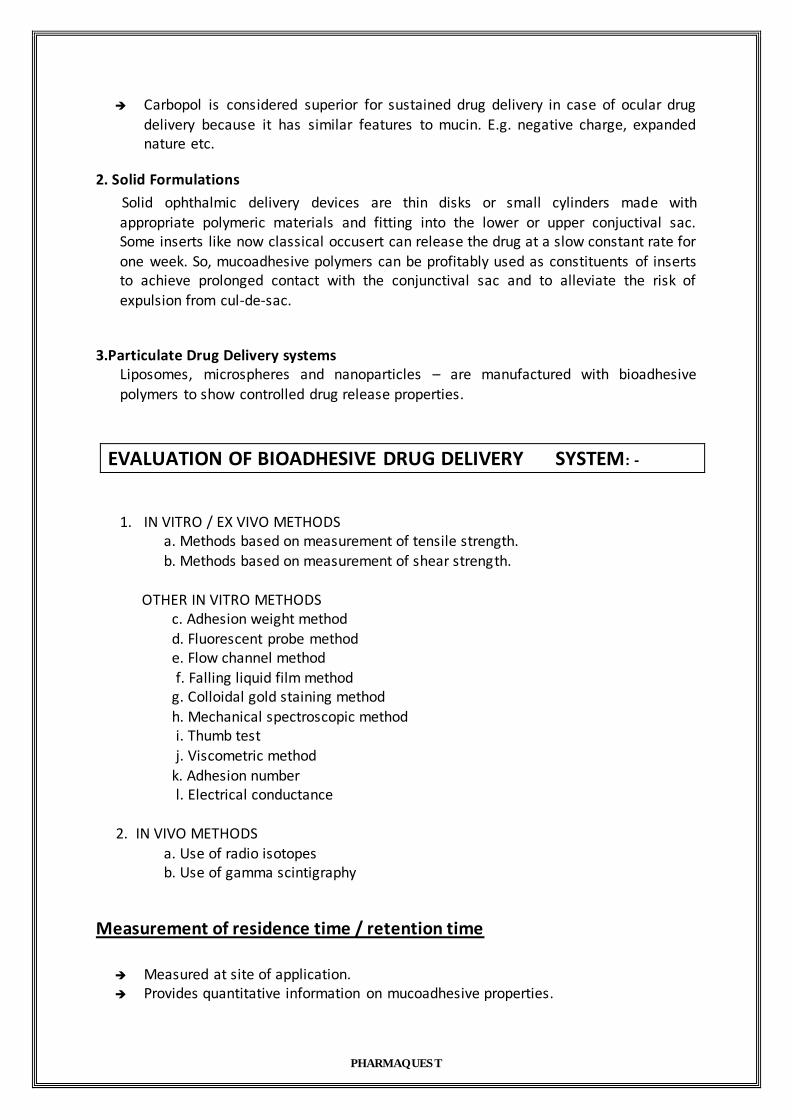
Falling liquid film method
Small intestinal segments from rats were placed at an inclination on a tygon tube.
The adhesion of particles to this surface is measured by passing the particle suspension over the surface and by comparing the fraction of particles adhered to the tissue; the adhesion strength of different polymers can be determined.
Membrane viscosity
The interaction between polymers and cell membranes was examined by labeling the cell membranes with fluorescent probes.
The lipid bilayer and proteins of cell membranes were labeled with pyrene and fluorescein isothiocyanate.
The fluorescence spectrum of pyrene and the fluorescence depolarization of fluorescein isothiocyanate were used to examine the change in membrane viscosity
after interaction with polymer.
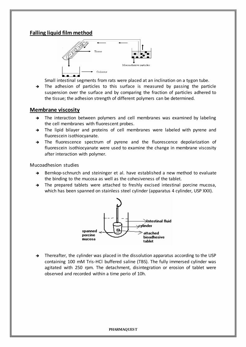
Mucoadhesion studies
Bernkop-schnurch and steininger et al. have established a new method to evaluate the binding to the mucosa as well as the cohesiveness of the tablet.
The prepared tablets were attached to freshly excised intestinal porcine mucosa, which has been spanned on stainless steel cylinder (apparatus 4 cylinder, USP XXII).
Thereafter, the cylinder was placed in the dissolution apparatus according to the USP
containing 100 mM Tris-HCl buffered saline (TBS). The fully immersed cylinder was agitated with 250 rpm. The detachment, disintegration or erosion of tablet were observed and recorded within a time perio of 10h.

PHARMAQUEST
In vivo evaluation methods
In vivo methods used for evaluation methods are based on administration of polymers to a laboratory animal and tracking their transit through the GI system. Administration methods
include forced oral gavage, surgical stomach implantation and infusion through a loop placed in situ in the small intestine. Tracking generally followed with the help of X-ray
studies, radio opaque markers and radioactive elements etc. For e.g. X- ray studies for monitoring GI transit time for bioadhesive tablet made of BaSO4 and radiolabelled
microspheres and nanoparticles is carried out.
Mucoadhesive strength measurement. Here first tissue novel bioadhesive system (NBAS) is placed or adhered to the rabbit or porcine buccal mucosa. Whole assembly paced in the krebs solution . Then NBAS is
clamped. On other side, from the burette liquid is poured and amount of liquid required to detach the NBAS from tissue is measured. An thus bioadhesive strength measured.
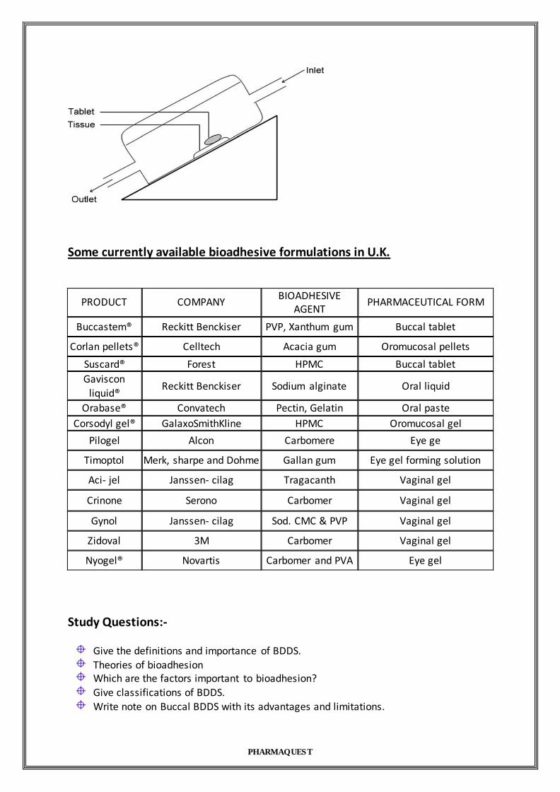
Dissolutin of Buccal tablet:- Mumtaz and Chang model for the dissolution of the buccal tablet as shown in figure. From the inlet dissolution medium is poured and from outlet it is collected. And assayed.
PHARMAQUEST
Some currently available bioadhesive formulations in U.K.
PRODUCT COMPANY BIOADHESIVE
AGENT PHARMACEUTICAL FORM
Buccastem® Reckitt Benckiser PVP, Xanthum gum Buccal tablet
Corlan pellets® Celltech Acacia gum Oromucosal pellets
Suscard® Forest HPMC Buccal tablet
Gaviscon liquid®
Reckitt Benckiser Sodium alginate Oral liquid
Orabase® Convatech Pectin, Gelatin Oral paste
Corsodyl gel® GalaxoSmithKline HPMC Oromucosal gel
Pilogel Alcon Carbomere Eye ge
Timoptol Merk, sharpe and Dohme Gallan gum Eye gel forming solution
Aci- jel Janssen- cilag Tragacanth Vaginal gel
Crinone Serono Carbomer Vaginal gel
Gynol Janssen- cilag Sod. CMC & PVP Vaginal gel
Zidoval 3M Carbomer Vaginal gel
Nyogel® Novartis Carbomer and PVA Eye gel
Study Questions:-
Give the definitions and importance of BDDS.
Theories of bioadhesion Which are the factors important to bioadhesion?
Give classifications of BDDS.
Write note on Buccal BDDS with its advantages and limitations.

PHARMAQUEST
How will you do the evaluation of bioadhesive drug delivery systems?
What is the importance of Transmucosal routes of drug delivery? Suggest potential sites and mechanism of adhesion? Enumerate mucosal permeation enhancers and
experimental methods to evaluate them. Write a note on intraperiodental drug delivery systems. ( March- 2005)
What is the importance of Transmucosal routes of drug delivery? Suggest potential sites and mechanism of adhesion? Enumerate mucosal permeation enhancers and
experimental methods to evaluate them. How you will approach combination drug therapy in transdermal patch? ( University Exam – 2005)
Write a short note on Buccal patch ( Sept-2006)