bileleakfollowinganelectivelaparoscopic cholecystectomy
TRANSCRIPT
From the Case Records ofthe Hospital ofthe UniversityofPennsylvania, Philadelphia,Pennsylvania
was a postoperative bile leak. The patient was treated withbroad spectrum intravenous antibiotics and received nooral medications.
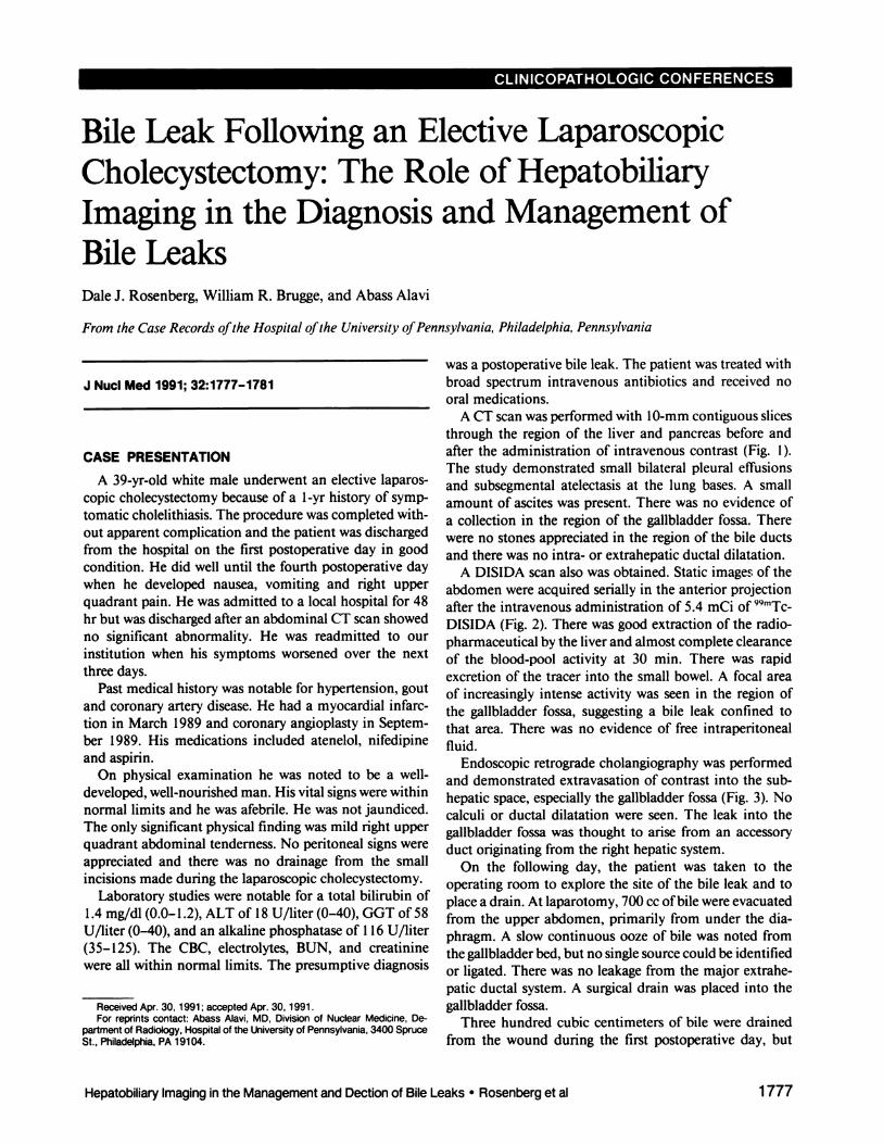
A CT scan was performed with 10-mm contiguous slicesthrough the region of the liver and pancreas before andafter the administration of intravenous contrast (Fig. 1).The study demonstrated small bilateral pleural effusionsand subsegmental atelectasis at the lung bases. A smallamount of ascites was present. There was no evidence ofa collection in the region of the gallbladder fossa. Therewere no stones appreciated in the region of the bile ductsand there was no intra- or extrahepatic ductal dilatation.
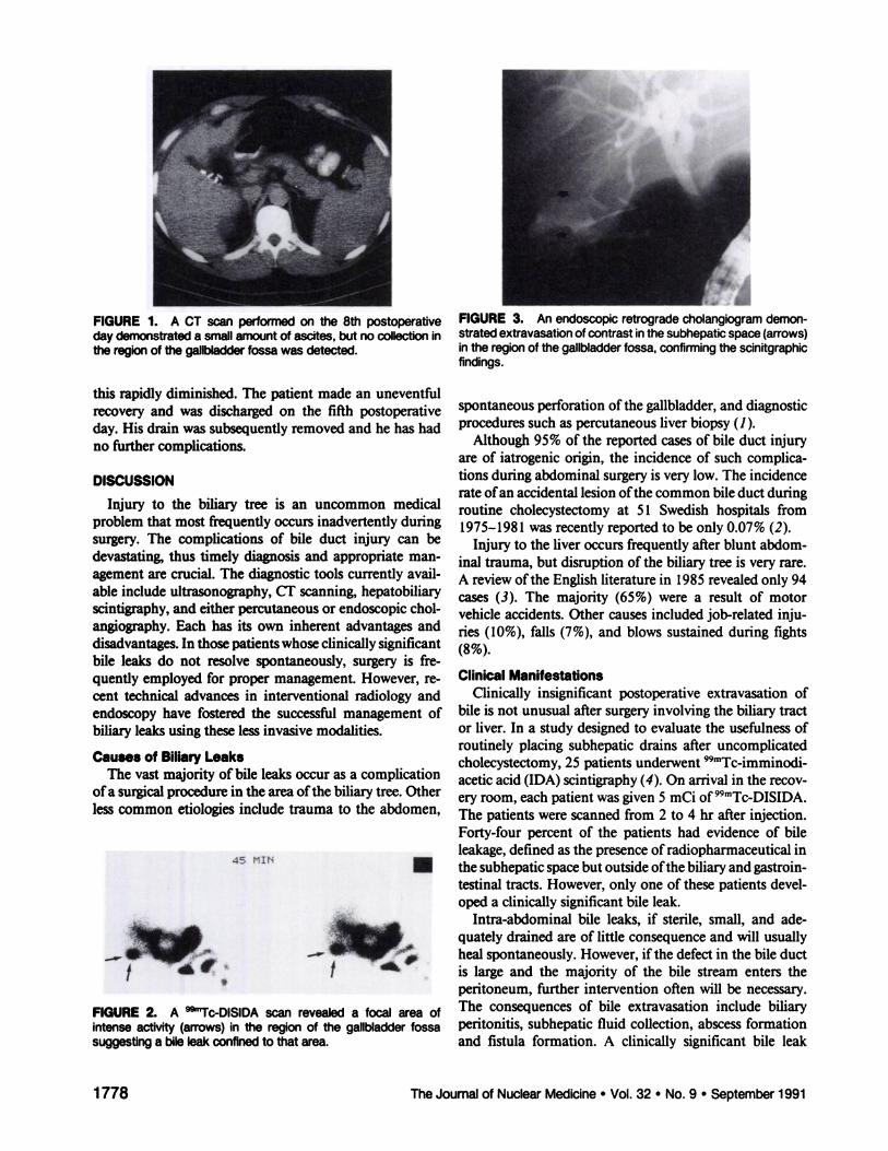
A DISIDA scan also was obtained. Static images of theabdomen wereacquired seriallyin the anterior projectionafter the intravenous administration of 5.4 mCi of 99mTc@DISIDA (Fig. 2). There was good extraction of the radiopharmaceutical by the liver and almost complete clearanceof the blood-pool activity at 30 mm. There was rapidexcretion of the tracer into the small bowel. A focal areaof increasingly intense activity was seen in the region ofthe gallbladder fossa, suggesting a bile leak confined tothat area. There was no evidence of free intraperitonealfluid.
Endoscopic retrograde cholangiography was performedand demonstrated extravasation of contrast into the subhepatic space, especially the gallbladder fossa (Fig. 3). Nocalculi or ductal dilatation were seen. The leak into thegallbladder fossa was thought to arise from an accessoryduct originating from the right hepatic system.
On the following day, the patient was taken to theoperating room to explore the site of the bile leak and toplace a drain. At laparotomy, 700 cc ofbile were evacuatedfrom the upper abdomen, primarily from under the diaphragm. A slow continuous ooze of bile was noted fromthe gallbladder bed, but no single source could be identifiedor ligated. There was no leakage from the major extrahepatic ductal system. A surgical drain was placed into thegallbladder fossa.
Three hundred cubic centimeters of bile were drainedfrom the wound during the first postoperative day, but
J NucI Med 1991; 32:1777—1781
CASE PRESENTATION
A 39-yr-old white male underwent an elective laparoscopic cholecystectorny because of a 1-yr history of symptomatic cholelithiasis. The procedure was completed without apparent complication and the patient was dischargedfrom the hospital on the first postoperative day in goodcondition. He did well until the fourth postoperative daywhen he developed nausea, vomiting and right upperquadrant pain. He was admitted to a local hospital for 48hr but was discharged after an abdominal CT scan showedno significant abnormality. He was readmitted to ourinstitution when his symptoms worsened over the nextthree days.
Past medical history was notable for hypertension, goutand coronary artery disease. He had a myocardial infarction in March 1989 and coronary angioplasty in Septernber 1989. His medications included atenelol, nifedipineand aspirin.
On physical examination he was noted to be a welldeveloped, well-nourished man. His vital signs were withinnormal limits and he was afebrile. He was not jaundiced.The only significant physical finding was mild right upperquadrant abdominal tenderness. No peritoneal signs wereappreciated and there was no drainage from the smallincisions made during the laparoscopic cholecystectomy.
Laboratory studies were notable for a total bilirubin of1.4 mg/dl (0.0—1.2), ALT of 18 U/liter (0—40),GGT of 58
U/liter (0—40),and an alkaline phosphatase of 116 U/liter(35—125). The CBC, electrolytes, BUN, and creatininewere all within normal limits. The presumptive diagnosis
Received Apr. 30, 1991 ; accepted Apr. 30, 1991.For reprints contact: Abass Alavi, MD, Division of Nuclear Medicine, De
partment of Radiology, Hospital of the University of Pennsylvania, 3400 SpruceSt., Philadelphia, PA 19104.
1777HepatobiliaryImaginginthe Managementand Dectionof BileLeaks •Rosenberget al
Bile Leak Following an Elective LaparoscopicCholecystectomy: The Role of HepatobiliaryImaging in the Diagnosis and Management ofBile LeaksDale J. Rosenberg, William R. Brugge, and Abass Alavi
N
FIGURE2. A @“‘Tc-DlSlDAscanrevealeda focalareaofintense actMty (arrows) in the region of the gallbladder fossasuggesting a bile leak confined to that area.
/@
FIGURE1. A CT scanperformedon the 8th postoperativeday demonstrateda smallamountof asates, but no collectioninthe region of the gallbladder fossa was detected.
this rapidly diminished. The patient made an uneventfulrecovery and was discharged on the fifth postoperativeday. His drain was subsequently removed and he has hadno further complications.
DISCUSSION
Injury to the biliary tree is an uncommon medicalproblem that most frequently occurs inadvertently duringsurgery. The complications of bile duct injury can bedevastating, thus timely diagnosis and appropriate management are crucial. The diagnostic tools currently available include ultrasonography, CT scanning, hepatobiiaryscintigraphy, and either percutaneous or endoscopic cholangiography. Each has its own inherent advantages anddisadvantages.In thosepatientswhoseclinicallysignificantbile leaks do not resolve spontaneously, surgery is frequently employed for proper management. However, recent technical advances in interventional radiology andendoscopy have fostered the successful management ofbiliary leaks using these less invasive modalities.
Causes of Biliary LeaksThe vast majority of bile leaks occur as a complication
ofa surgical procedure in the area ofthe biliary tree. Otherless common etiologies include trauma to the abdomen,
45 MIN
FIGURE3. Anendoscopicretrogradecholangiogramdemonstratedextravasationofcontrast in the subhepaticspace(arrows)intheregionofthegallbladderfossa,confirmingthescinitgraphicfindings.
spontaneous perforation of the gallbladder, and diagnosticprocedures such as percutaneous liver biopsy (1).
Although 95% of the reported cases of bile duct injuryare of iatrogenic origin, the incidence of such complications during abdominal surgery is very low. The incidencerate ofan accidental lesion ofthe common bile duct duringroutine cholecystectomy at 51 Swedish hospitals from1975—1981was recently reported to be only 0.07% (2).
Injury to the liver occurs frequently after blunt abdominal trauma, but disruption of the biiary tree is very rare.A review ofthe English literature in 1985 revealed only 94cases (3). The majority (65%) were a result of motorvehicle accidents. Other causes included job-related injuries (10%), falls (7%), and blows sustained during fights(8%).
Clinical ManifestationsClinically insignificant postoperative extravasation of
bile is not unusual after surgery involving the biliary tractor liver. In a study designed to evaluate the usefulness ofroutinely placing subhepatic drains after uncomplicatedcholecystectomy, 25 patients underwent 99mTc@imminodi@acetic acid (IDA) scintigraphy (4). On arrival in the recovery room, each patient was given 5 mCi of [email protected] patients were scanned from 2 to 4 hr after injection.Forty-four percent of the patients had evidence of bileleakage, defined as the presence ofradiopharmaceutical inthe subhepatic space but outside ofthe biliary and gastrointestinal tracts. However, only one of these patients develo_ a clinically significant bile leak.
Intra-abdominal bile leaks, if sterile, small, and adequately drained are of little consequence and will usuallyheal spontaneously. However, ifthe defect in the bile ductis large and the majority of the bile stream enters theperitoneum, further intervention often will be necessary.The consequences of bile extravasation include biliaryperitonitis, subhepatic fluid collection, abscess formationand fistula formation. A clinically significant bile leak
1778 The Journal of Nuclear Medicine•Vol.32 •No. 9 •September1991

should be suspected in any patient who has recently sustamed abdominal trauma or surgery and develops fever,jaundice, abdominal pain or bilious drainage from anincision or drain. The time interval between the onset ofbile leakage and the development of symptoms can rangefrom 24 hr to 1 wk and may be dependent on whether ornot the bile is infected (5).
DiagnosisDocumenting the presence and extent ofa leak can pose
a difficult diagnostic challenge. Real-time ultrasonographyand CT scanning are two readily available and noninvasivetechniques for diagnosing fluid collections within the abdomen (1). The main advantage of ultrasonography is itsoptimal anatomic resolution, particularly for space-occupying lesions deep within the liver. CT scan provides evenhigher resolution and the opportunity to make densitymeasurements. Unfortunately, the differentiation of fluidswith the same density as water (e.g., bile, serous ascites,lymph and urine) is not possible. Therefore, althoughultrasound and CT scans can identify fluid collectionswithin the abdomen, they cannot demonstrate whetherthey contain bile or freely communicate with the biliarytree.
Imaging techniques that use 99mTc@IDAderivatives provide a noninvasive, physiologic means of detecting andevaluating bile drainage and therefore bile leaks. Althoughthe primary role of 99mTC4DA cholescintigraphy has beenthe evaluation of cystic duct patency in patients withsuspected acute cholecystitis, it also demonstrates the physiologic route of bile flow and can diagnose a bile leak andits relation to the collections detected by the other means.
After intravenous injection, IDA analogues are efficiently cleared from the blood stream by the hepatocytesand are excreted unchanged into the biliary system andultimately the intestines. High photon flux allows excellentimages of the common bile duct, gallbladder and smallintestine to be obtained (5).
Technetium-99m-IDA scintigraphy has been shown tobe an important tool for detecting extravasation of bile.There are many case reports in the literature documentingits usefulness in the assessment of damage to the biliarytree after trauma or surgery (3—8).Technetium-99m-IDAscintigraphy has many advantages (6):
•It is noninvasive and physiologic and therefore causesno changes in the existing condition in the biliarysystem.
•It can be performed in the face of hyperbilirubinemia.•It also allows for a smaller concentration ofthe tracer
to be detected than the concentration ofiodine neededfor visualization by conventional radiographic techniques.
Unfortunately, there have been no studies to establish thesensitivity and specificity of the test in diagnosing bileleaks and no large scale comparisons of scintigraphy with
radiographic studies such as percutaneous transhepaticcholangiography (PTC) or endoscopic retrograde cholangiopancreatography (ERCP) have been performed.
Technetium-99m-IDA scintigraphy has been shown tobe particularly useful in diagnosing bile leaks that occurduring abdominal trauma. Small bile duct injury, oftendeep within the liver parenchyma, frequently occurs whenthe liver is damaged. Intrahepatic bile duct injury andbiloma formation may not be recognized for days or weeksuntil fever or bilious drainage occurs. Zeeman has performed Tc-IDA scintigraphyon 21 patients suspectedofhaving hepatobiliary trauma—6 due to penetrating traumaand 15 due to blunt trauma (8). All of the patients withpenetrating trauma underwent emergency exploratory laparotomy, while the 12 patients with blunt trauma eventually had surgery. Although no biliary injuries were identifled at laparotomy in these patients, eight of them hadparenchymal defects on postoperative IDA scintigraphy.In five of these cases, radioactive bile eventually was seenwithin the defect, indicating an intrahepatic biliary leak.The authors suggest that the routine preoperative use ofTc-IDA scintigraphy in patients who have sustained abdominal trauma will expedite the diagnosis of occult intrahepatic bile duct leaks.
Several authors have made recommendations to improve the sensitivity of Tc-IDA scintigraphy in the diagnosis of bile leaks. Weissman has noted that it is essentialto obtain delayed views when looking for bile leaks. In sixof nine patients she had evaluated, the bile leak was onlyapparent in those films obtained after 1 hr (6). Lette hasnoted that it can be difficult to differentiate right upperquadrant pathology, including bile leaks, from radiopharmaceutical in the small intestine (7). He has recommendedstanding views to overcome this problem. Other techniques to differentiate true pathology from enteric radiotracer activity include supplementary oblique views andthe use of water ingestion to dilute the tracer.
Although Tc-IDA is a sensitive way to detect the presence of a bile leak, it has limited anatomic resolution.Injection of radiographic contrast material directly intothe biliary tract using PTC or ERCP is the best way toestablish the exact site of extravasation.
ManagementThere are three therapeutic options available for those
patients with bile leaks who do not respond to conservativetherapy or develop fistulous tracts: (1) surgery; (2) percutaneous transhepatic biliary drainage (PTBD); and (3)endoscopic sphincterotomy and stent placement.
Clearly, those bile duct injuries detected intraoperativelyshould be repaired surgically. Bile leaks discovered postoperativelycan also be repaired during a second surgicalprocedure, but this is often difficult because of inflammation. The incidence of postoperative bile duct stricture orthe need for further intervention is high after reparativebiliary surgery. Sixty-nine percent of the patients in
1779Hepatobiliary Imaging in the Management and Dection of Bile Leaks •Rosenberg et al

Andren-Sanberg's review required additional surgeryeither from a postoperative biiary complication or becausethey developed a biliary stricture. The best SUrgicalresultsare obtained when the repair is made by creating a Rouxen-Y choledocho- or hepaticoenterostomy (2,9).
The morbidity associated with the surgical repair of bileduct leaks has been an impetus in recent years for thedevelopment of alternative techniques to treat these injuries. Kaufman reported a series of 12 patients with bileleaks or fistulas who were managed with PTBD (10). Insevenpatients, the leak stopped with drainagealone; fivepatients required additional surgery. Although PTBD hasbeen used successfully to control bile leaks, it has a numberof disadvantages. The catheter can be uncomfortable andinconvenient for the patient. It may also serve as an avenuefor bacteria to enter the biliary tract. Continuous biliarydrainage may also produce fluid and electrolyte disturbances, particularly in elderly patients. In addition, PTC isoften difficult in patients with nondilated ducts, a frequentproblem encountered in patients with biiary leaks.
The most recent advance in the management of biliaryleaks and fistulas has been the introduction of endoscopicsphincterotomy and the insertion ofstents and nasobiiarydrainage catheters. Iatrogemc bile duct injuries are mostcommonly located in the extrahepatic biliary tree and arewell suited for endoscopic management. In 1986, Smithand Sauerbruch each reported the successful treatment ofpostoperative biliary fistulas with endoscopic biliary drainage (11,12). Leakage from various locations, including thecommon hepatic duct, cystic duct stump, common bileduct and T-tube sites, were successfully managed with onlyendoscopic techniques.
Although it was originally believed that stents inducedhealing by bridging the hole at the site of extravasation,recent reports suggest that these maneuvers help by decreasing the pressure gradient across the ampulla andrelieving obstruction ifit is present (13). The largest seriesofpatientswithendoscopicallytreatedbiliaryfistulaswasreported by Ponchon in 1989 (13). His team treated 24patients with persistent biliary fistulas, most as a result ofprevious surgery. Eleven ofthe patients had distal bile ductobstruction. Twelve of the patients were managed bysphincterotomy alone and nine were cured by this method.Success was achieved in 7 ofthe remaining 12 patients, allof whom were managed with either endoprostheses ornasobiliary drainage. The only complication noted wasbleedingfrom one sphincterotomy.
Ponchon noted four factors that influence the outcomeof endoscopic treatment: (1) the type of distal biiaryobstruction; (2) the size of the bile duct injury; (3) thelocation of the injury; and (4) the presence of associatedlesions. In those patients where sphincterotomy alone wassufficient to reduce the pressure gradient between thecommon bile duct and the duodenum, a successful outcome could frequently be achieved without the use of anendoprosthesis or external drainage. Those patients with
duct injuries larger than 0.5 cm did not heal. Intrahepaticlesions responded less well than extrahepatic lesions. Finally, a poor outcome was noted in patients with hepaticabscesses unless they were diagnosed and adequatelydrained.
Goldin has reported the successful endoscopic treatmentof two patients with peripheral intrahepatic bile leakswithout bridging the site of extravasation (14). Sphincterotomy and stent placement through the papilla sufficientlydecreased the pressure within the biliary tree to allow thesite of disruption to heal.
Laparoscopic CholecystectomyAs previously stated, injury to the biliary tree is an
uncommon problem. With incidence rates of less than0. 1% after routine biliary surgery, only a few cases peryear would be expected, even at large university centers.However, with the rapid emergence of laparoscopic cholecystectomy, bile duct injury may become a more common problem.
Over the past two years laparoscopic cholecystectomyhas emerged as an alternative to traditional open cholecystectomy. It offers the advantage of a shorter postoperative hospital stay, less postoperative pain, a shorter convalescent period, and a more acceptable cosmetic result(15). Although preliminary data suggests that laparoscopic
cholecystectomy is a safe alternative to conventional cholecystectomy, the true incidence of complications has notyet been determined. To date, seven series (a total of 758patients) have appeared in the literature (15—21).In thesereports, complication rates have ranged from 0% to 5%.
Four patients have required conversion to an opencholecystectomy because of bile duct injury during thelaparoscopic procedure. Peters reported one patient withan inflamed gallbladder who suffered an intraoperativetear of the common bile duct (16). Phillips noted onepatient with unsuspected subacute cholecystitis who suffered a bile duct injury that was sutured during laparotomy(1 7). Salky noted a laceration of the common bile duct ina patient in whom the cyclic duct originated from the righthepatic duct (18). The fourth case was a common hepaticduct injury reported by Zucker (19).
Bile leaks have also been the major postoperative complication described after laparoscopic cholecystectomy. Inthe reported series, seven patients currently have beennoted to havepostoperativebile leaks:two from the cysticduct, two from the hepatic duct, two from small biliaryradicals entering the gallbladder fossa, and one from thegallbladder bed (15,16,20). Five of these patients requiredadditional surgery. The two patients with leakage fromsmall biliary radicals were both successfully treated withendoscopically placed stents. Kozarek and Traverso havealso reported a case in which a cystic duct leak afterlaparoscopic cholecystectomy was successfully managedwith endoscopic stent placement (22).
In addition to the patient presented in this article, we
I780 The Journal of Nuclear Medicine•Vol.32 •No. 9 •September 1991

have seen two other patients at this institution with bileleaks after laparoscopic cholecystectomy. One leak onginated from the cystic dump stump and the other probablyfrom an accessory bile duct. We have also seen fourpatients who underwent laparoscopic cholecystectomy atother medical centers who developed biliary obstructionafter sustaining bile duct injuries (23).
During the 758 laparoscopic cholecystectomies reportedin the literature to date, six patients have sustained injuriesto the hepatic or common bile ducts, an incidence rate of0.8%. This figure is higherthan thosethat have beenreported after traditional open cholecystectomy (2). Additional data must be accumulated to determine if thisincrease in bile duct injury simply reflects inexperiencewith this new technique or an inherent risk of removingthe gallbladder using the laparoscope.
CONCLUSION
Injury to the biliary tree is an uncommon medicalproblem that is most likely to occur during surgery orother types of.trauma. The extravasation of a small quantity of bile occurs commonly after biliary surgery and is oflittle clinical significance. However, patients who developfever, jaundice, abdominal pain, or profuse bilious drainage should be suspected of having a significant bile leak.
Ultrasonography and CT scanning are useful for detecting the presence of fluid collections in these patients, butthey cannot demonstrate if the collection communicateswith the biliary tree. Technetium-99m-IDA scintigraphyis a noninvasive and physiologic means of evaluatingpatients suspected of having bile leaks. It will documentcommunication between a fluid collection and the biliarytree and will also establish the primary route of bile flow(i.e., through the leak or into the small intestine). Thosepatients with preferential flow into the small intestine willgenerally respond well to conservative management, including drainage of the fluid collection. Those patientswith preferential flow through the bile leak frequently havebiliary obstruction and require more invasive therapy.Injection of radiographic contrast directly into the biliarytree via either PTC or ERCP is the best means of localizingthe anatomic site of the leak and diagnosing possibleunderlying biliary obstruction.
Those leaks detected intraoperatively are best treated bysurgical repair. Postoperative leaks may also be repairedsurgically, but the incidence of postoperative complications and stricture formation is significant. PTBD can beused to manage bile leaks but is fraught with drawbackssuch as discomfort, inconvenience and infection. Endoscopic sphincterotomy and biliary endoprostheses haverecently been employed in the successful management of
both intra- and extrahepatic bile leaks and fistulas. Although the short-term results of these nonsurgical techniques are encouraging, the incidence of long-term complications such a biliary stricture has not yet been determined.
REFERENCES
1. Lorenz R, Beyer D, Peters P. Detection of intraperitoneal bile accumulations: significance of ultrasound, CT and cholescintigraphy. GastrointesiRadio! l984;9:2l3—2l7.
2. Andren-Sandberg A, Johansson S, Bengmark S. Accidental lesions ofcommon bile duct at cholecystectomy. Ann Surg l983;20l:452—455.
3. Michelassi F, Ranson J. Bile duct disruption by blunt trauma. J Traumal985;25:454—457.
4. Gilsdorf JR. Phillips M, McLeod MK, et al. Radionuclide evaluation ofbile leakage and the use of subhepatic drains after cholecystectomy. Am JSurg 1986; 151:259—264.
5. Weissmann HS, Byun KJ, Freeman LM. Role ofTc-99m-IDA scintigraphyin the evaluation of hepatobiliary trauma. Semin Nuc! Med 1983;l3:l99—222.
6. Weissmann HS, Gliedman ML, Wilk PJ, et al. Evaluation of the postoperative patient with @mTc@IDAcholescintigraphy. Semin Nuc! Med1982; 12:27—40.
7. Lette J, Morn M, Heyen F, Paquet A, Levasseur A. Standing views todifferentiate gallbladder or bile leak from duodenal activity on cholescintigrams. C!in Nuc! Med 1990; 15:231—236.
8. Zeeman RK, Lee CH, Stahl R, Viscomi GN, Baker C, Cahow CE, et al.Strategy for the use ofbiliary scintigraphy in non-iatrogenic biliary trauma.Radio!ogy1984;151:771—777.
9. Saber K, El-Manialawi M. Repair of bile duct injuries. Wor!d J Surg1984;8:82—89.
10. Kaufman SI, Kadir 5, Mitchell SE, Chang R, Kinnison ML, Cameron JL,et al. Percutaneous transhepatic biliary drainage for bile leaks and fistulas.AiR l985;l44:l055—l058.
11. Smith AC, Schapiro RH, Kelsey PB, Warshaw AL. Successful treatmentof nonhealing biliary-cutaneous fistulas with biliary stents. Gasiroentero!ogy l986;90:764—769.
12. Sauerbruch T, Weinzierl M, Holl J, Pratschke E. Treatment of postoperative bile fistulas by internal endoscopic biliary drainage. Gasiroentero!ogy1986;90: 1998—2003.
13. Ponchon T, Gallez J, Valette P, Chavaillon A, Bory R. Endoscopic treatment ofbiliary fistulas. Gastrointest Endoscop l989;35:490—498.
14. Goldin E, Katz E, Wengrower D, ci al. Treatment of fistulas of the biliarytract by endoscopic insertion of endoprostheses. Surg Gyneco! Obstet1990; 170:418—423.
15. Gadacz TR, Talamini MA, Lillemoe KD, Yeo CJ. Laparoscopic cholecystectomy. Surg C!in North Am l990;70: 1249—1262.
16. Peters JH, Ellison EC, Innes JT, et al. Safety and efficacy of laparoscopiccholecystectomy. Ann Surg l991;213:3—l2.
17. Phillips EH, Berci G, Carroll B, Daykjhovsky L, Sackier J, Paz-Partlow M.The importance of intraoperative cholangiography during laparoscopiccholecystectomy. Am Surg 1990;56:792—795.
18. Salky BA, Bauer ii, Kreel I, Gelernt IM, Gorfine SR. Laparoscopiccholecystectomy: an initial report. Gastrointesi Endoscop 1991;37: 1—4.
19. Zucker KA, Bailey RW, Gadacz TR, Imbembo AL. Laparoscopic guidedcholecystectomy. Am J Surg 1991; 161:36—44.
20. Dubois F. Coelioscopic cholecystectomy. Endoskopie Heute 1990;3:30—32.
21. Reddick EJ, Olsen DO. Laparoscopic laser cholecystectomy. A comparisonwith mini-lap cholecystectomy. Surg Endoscop l989;3: 131—133.
22. Kosarek RA, Traverso LW. Endoscopic stent placement for cystic ductleak after laparoscopic cholecystectomy. Gastroiniesi Endoscop 1991;37:71—73.
23. Rosenberg DJ, Brugge WR. Complications after laparoscopic cholecystectomy. Am J Gasiroentero! 1991: in press.
1781HepatobiliaryImaginginthe Managementand Dectionof BileLeaks •Rosenberget al