transcylindrical cholecystectomy for the treatment of ... · transcylindrical cholecystectomy for...
TRANSCRIPT

1
Transcylindrical Cholecystectomy for the Treatment of Cholelithiasis and Its Complications: Cholecystectomy
Under Local Anesthesia
E. Javier Grau Talens, Julio Horacio Cattáneo, Rafael Giraldo Rubio and Pablo Gustavo Mangione Castro
Siberia-Serena Hospital, Talarrubias (Badajoz) Extremadura University
Spain
1. Introduction
Cholecystectomy is the primary treatment of cholelithiasis. But the prevention of the formation and the dissolution of the stones were popular in the 80's . The clinical use of the chenodeoxycholic and after the ursodeoxycholic acid emerged in the 70's, when proved that this acids reduced biliary cholesterol saturation in bile. Important aspects were significant but reversible hepatotoxicity in 3%, diarrhea in 8%, abandonment of treatment in 15% and a similar proportion of abdominal pain. Probably, more important was the increase in total serum cholesterol and low density lipoprotein during treatment with chenodesoxycholic acid. In general, ursodeoxycholic acid appears to have fewer side effects, works faster and causes less liver damage. In patients with small cholesterol stones and floating radiolucent treated with ursodeoxycholic acid, for 6-12 months, partial or complete dissolution can be expected in 40-55% of cases. The direct dissolution of cholesterol gallstones using methyl tert-butyl ether (MBTE) requires the insertion of a percutaneous transhepatic catheter in the gallbladder. The MBTE (5-10 mL) should be infused in a manner that involves the calculi but does not flow into the common bile duct and duodenum. In 4-16 hours the stones are dissolved. The patient should stay overnight in the hospital. Side effects include pain and nausea; haemolysis and duodenitis are serious consequences of the spilling of the solvent into the duodenum . Transabdominal mechanical lithotripsy is another treatment modality, which leads to fragmentation of the stones in selected cases in almost 100% of patients. All of these treatments have in common the recurrence of stones (from 45% to 70% at 5 or 7 years of follow-up), due to persistence of a place for the precipitation of cholesterol crystals (gallbladder) and bile prone to precipitate (lithogenic bile). A report by Gilliland and Traverso in 1990 settled any doubts about the alternatives in the treatment of cholelithiasis (Gilliland & Traverso, 1990) These authors reviewed outcomes of 671 cholecystectomy patients during the years 1982-1987 and found no mortality and 2.2% of complications. They conclude that open cholecystectomy is a definitive treatment for symptomatic cholelithiasis with minimal risk to the patient and a high degree of cure of the symptoms.
www.intechopen.com

Updated Topics in Minimally Invasive Abdominal Surgery
4
The first truly major surgery on the biliary tract was performed in 1867 in Indiana (USA). John S. Bobbs, professor of surgery at the Medical College of Indiana, operates a tumor in the right upper quadrant in a 30 year old woman, at home and under general anesthesia, resulting in the diagnosis of gallbladder hydrops which was evacuated and drained. It was the first cholecystostomy performed in the history. Fifteen years later, in 1882, Carl J. Langenbush of Berlin performed the first cholecystectomy by lithiasis, after exercising cholecystectomy in cadavers for several years. However, as more than a century later would happen with the laparoscopy and in the same Germany, Langenbush's communication in the German Congress of Surgery of three cases of cholecystectomy that evolved successfully, was received with apathy and without due consideration that the time reserved.
1.2 Development of mini-lap (small-incision) cholecystectomy Minilaparotomy was used for several decades for the diagnosis of obstructive jaundice. Through a small incision is valued, in addition to the aetiology, the operability by palpation of the gallbladder and hilum liver and usually the diagnosis included a cholecistocholangiography. In 1982 F. Dubois and B. Berthelot (Dubois & Berthelot, 1982) published the first paper on the minilaparotomy for operations on the bile duct, performing the procedure in 1500 patients, including alongside cholecystectomy, some cases of choledochotomy, sphincterotomy and choledochoduodenostomy. All these interventions were carried out with a transverse or oblique skin incision 3 to 6 cm in length, but the duration of surgery, the authors say, was "twice that of a normal operation". Intervention was carried out with the help of an autostatic (if no more than one assistant was available), a vaginal valve for retraction of the liver, a malleable valve for retraction of the hepatic flexure of the colon and the positioning of two packs for the separation of the colon and stomach, referenced with a tape. The description of the intervention with this procedure and its duration arise a suspicion of some difficulty with exposure of the structures and the easement of the procedure. However, the authors describe: a minimization of cosmetic damage, solidness of the wall closure an a reduction of pain and postoperative ileus. Moss, in 1983, published the first cases of cholecystectomy with stay less than 24 hours, and in 1986, 100 cases. Later, he operates 160 patients by midline laparotomy, with an incision that "barely allows the surgeon's hand”, which were discharged the day after surgery without receiving narcotics, tolerating food intake between 8 and 18 hours and only 3 readmissions. The author concluded in 1996 that the benefits of laparoscopy may be more related to the enthusiasm and expectations for the new technology that in the technique by itself (Moss, 1996). In 1985, Morton (Morton, 1985) performs a cost containment study of cholecystectomy with intraoperative cholangiography in 96 patients through an incision of 4 to 5 cm with a mean operating time of 45 minutes. The average stay was 2.5 days and analgesic requirements were lower than in the classical subcostal incision. The period of sick leave decreased significantly. Goco and Chambers in 1988 (Goco & Chambers, 1988) studied the impact of mini-cholecystectomy in the management of health expenditure, considering the reduction of hospital costs compared to traditional cholecystectomy. The authors conclude, by analyzing 450 interventions, that a 4-cm incision produces an average stay of 1.22 days and that the savings stay was 4.78 days per case. Rating the daily cost at $ 200 USA in 1988, it is easy to
www.intechopen.com

Transcylindrical Cholecystectomy for the Treatment of Cholelithiasis and Its Complications: Cholecystectomy Under Local Anesthesia
5
see the savings produced by minilaparotomy, especially if applied to the 600,000 cholecystectomies performed annually in the United States. Despite these and further studies, minilaparotomy was never popular. For example, out of seven standard textbooks: Norton al al., (Harris, 2008). Sabiston (Arendt & Pitt, 2004), Schwartz (Schwatz 1989), Doherty (Doherty, 2010), Maingot (Karam & Roslyn, 1997), Marlow & Sherlock (Dawnson, 1985). Morris & Malt (Britton & Bickerstaff, 1994), only the latter describes the technique of cholecystectomy by minilaparotomy.
1.3 Laparoscopic cholecystectomy – Eric Mühe
Laparoscopy has not only caused a revolution in the treatment of cholelithiasis, but that has changed an old surgical proverb: "a large incision, a great surgeon." It seems reasonable to assert that "a smaller incision, less abdominal wall trauma and better aesthetic results." The era of minimally invasive surgery began and laparoscopy has been extended to almost all abdominal surgical operation and almost any procedure has been performed by laparoscopic approach, including resections and all types of gastrointestinal suture. Interestingly, laparoscopic cholecystectomy was not well received by the German Surgical Society when E. Mühe reported the first operation in 1986. On September 12, 1985, Mühe selected with great care the first patient to perform the first laparoscopic cholecystectomy, almost five years after the first laparoscopic appendectomy by Semm. Like him, Mühe performed the pneumoperitoneum with the Veress needle, inserted the trocar and introduced his own "galloscope" through the umbilicus. Two hours later he concluded successfully the first laparoscopic cholecystectomy (Litynski, 1996). His presentation at the congress was not published and only a summary appeared in Langenbecks Archiv für Chirurgie 1986 (Mühe, 1986). However, with subsequent amendments Mühe concluded that inserting the laparoscope (galloscope) as close as possible to the gallbladder the "cumbersome" pneumoperitoneum could be avoided. After several cholecystectomies without gas, trying to simplify and adapt the technique to be used by most surgeons, he realized that the optical instrument was not necessary, "with or without galloscope, the magic surgical approach could be the same". Soon operated through sheath of the galloscope without the optical instrument with the advantages of minimal incision: - The abdominal musclulature is not cut - Little postoperative pain that disappears in two or three days - short Immobilization(even elderly patients need to be in bed only the day of operation) - Short hospital stay (4-5 days) - Quick return to work (50-75% earlier than with traditional surgery) This outlined the bases of minimally invasive surgery. Sadly, Mühe didn´t publish the evolution of his technique for cholecystectomy in any international journal and we haven´t hat notice of it until 1996 with the Litynski´s. book. Many reasons can be considered to explain the success of laparoscopic cholecystectomy: 1. It is obvious that the ports, about 1 cm, scattered in the upper abdomen and a umbilical
opening for the introduction of optic, produce a minimal aesthetic disorder. 2. The trauma to the abdominal wall caused by an incision about 15 cm is large and has a
well-known impact on the respiratory physiology, a greater possibility of formation of adhesions, hernias and, above all, pain.
3. The acceptance by the patient has been quick, because it was publicized with all of the above advantages. The charisma of laparoscopic technology is undeniable, its elegance, too.
www.intechopen.com

Updated Topics in Minimally Invasive Abdominal Surgery
6
4. The commercial pressure has been relentless. Technological research has been overturned in the design and implementation of increasingly sophisticated and safer instruments. Sponsoring of the learning of the technique to the interested surgeons was a strategic objective.
5. Finally, the health financier had an opportunity to reduce hospital stays. Given all the above mentioned facts it is obvious that the introduction of the technique is an undeniable fact and that, at present, nobody doubt that laparoscopy is the technique of choice for cholecystectomy. However, the advantages of laparoscopic cholecystectomy have been put in evidence, deliberately, with the open cholecystectomy with a generous wound of about 15 cm. But what if the comparison is made against a technique that uses an incision of 5 cm or smaller? It is possible that the above mentioned advantages were less obvious and that the assessment had to be made over other aspects than aesthetics, postoperative pain, parietal trauma, hospital stay, re-employment, etc., entering the field of cost, security and benefits to the patient.
2. Laparoscopic vs. small-incision cholecystectomy
A review in 1993 (Olsen, 1993) concluded that there are no good studies comparing conventional cholecystectomy by minilaparotomy or by laparoscopy. However, it was apparent that the small incision was better than the big one and that the length of the incision appears to be associated with hospital stay and return to the workplace. The ultimate goal is to achieve a safe surgery with the maximum benefit for the patient, and the keys are: knowledge of anatomy, good surgical view and a proper exposure. This last key to safety, exposure, is a limiting factor for minilaparotomy, which leads the question of how small an incision can still provide a exposure to perform the cholecystectomy safely. For Olsen, the answer is the laparoscopy, which allows for smaller incision, but it is noteworthy that the sum of the incisones made for the insertion of four trocars is about 4 cm and two-dimensional view. An incision of this size can provide adequate exposure for cholecystectomy under direct three-dimensional vision. An overview of the Cochrane Hepato-Biliary Group reviews in January 2010 (Keus et al.,
2010) revealed the evidence to date of the revisions that assess the effect of differents
techniques of cholecystectomy: open, small-incision, or laparoscopic. A total of 5246 patients
in 56 randomized trials are included. Total complications of laparoscopic cholecystectomy
and small-incision were similar (17%), hospital stay and convalescence were not
significantly different, small-incision cholecystectomy operative time was shortest (16.4
minutes) and is less costly. In our study of 1998 (Grau-Talens et al., 1998) small-incision
cholecystectomy was $ 1003 U.S less costly than laparoscopic. The effects of anesthesia and
surgery on lung function have been well studied (Lindell & Hendenstierna 1976). There is a
reduction in FVC (Forced Vital Capacity) and FEV1 (Forced Expiratory Volume in one
second) to 75% of baseline for a separate incision without cutting the muscles, while
reducing down to 40-55% in the subcostal incisions and midline laparotomy. An incision
that spares the muscular section can prevent postoperative pulmonary complications. The
restrictive pattern of lung dysfunction in postoperative abdominal surgery is influenced by
several factors and is not well understood. The size, location and direction of the incision are
responsible for the alteration of mechanical ventilation, by themselves and the pain. Kind of
anesthetic agent and diaphragmatic dysfunction are also involved (Craig 1981).
www.intechopen.com

Transcylindrical Cholecystectomy for the Treatment of Cholelithiasis and Its Complications: Cholecystectomy Under Local Anesthesia
7
In some studies, laparoscopic cholecystectomy has shown lower spirometric reductions when compared to open cholecystectomy (Frazee et al., 1991) and to mini-lap (McMahon et al., 1994) although the latter with incisions between 5 and 10 cm. Presumably, a reduction in the length of the incision could be rewarded by a smaller reduction in the impairment of lung physiology, ie, an incision of 4.5 cm, uniform to all layers of the abdominal wall could improve postoperative spirometric results as happened in our study (Grau-Talens et al.,1998) wich shows that the reduction of spirometrics values were similar in laparoscopic and small-incision cholecystectomy, ie over 20% of preoperative value for FVC and 25% for FEV1. The results obtained by keus et al. are similar to ours (Keus et al 2008).
3. Treatment options in biliary lithiasis complications
3.1 Acute cholecystitis Early cholecystectomy is the best treatement for acute cholecystitis. Laparoscopic
cholecystectomy was a relative contraindication in acute cholecystitis, but now is the
preferred aproach for most patients. The first articles appear in the early 90´s (Cooperman,
1990) (Yamashita et al., 2007). However, in our experience, cholecystectomy in this way was
not easy: the difficulties are related to the inflammatory process, with greater difficulty for
dissection and recognition of structures, the possibility of further contamination of the
cavity (not the surgical wound), the need for instruments to 10 mm in diameter, greater
difficulty in haemostasis and, of course, a greater proportion of conversions (35%) and
duration of the intervention.
With these preliminary considerations we began to operate the acute cholecystitis by early
transcylindrical cholecystectomy (within 72 hours or more of admission), thinking that the
abdominal wall injury should not be higher than laparoscopy, even using the cylinder of 5
cm in diameter, that manipulation of the gallbladder (gripping, aspiration, recovery of
stones etc.) could be done in a simpler way than by laparoscopy, and that contamination of
the surgical wound could be avoided by the protective and insulating effect of the cylinder.
We have only found an article of acute cholecystitis treated by minilaparotomy in the
context of a randomized study comparing minilaparotomy with conventional laparotomy
(Assalia et al., 1997). The authors show figures contrasting results in a very favourable way,
not only with traditional laparotomy, but with the laparoscopic approach. In this article the
average time (+ /-SD) of the intervention was 69.1 (+ / - 17.0) minutes and mean hospital
stay was 3.1 days.
3.2 Choledocholithiasis The choledochotomy was first performed in 1884 by Kummel and in 1889 by Thornton and Abbe, who made the first ideal suture of the choledochotomy. In the late nineteenth and early twentieth century the common bile duct exploration was guided by the subjective clinical impression of the surgeon, until the introduction of intraoperative cholangiography by Mirizzi in 1937. In the Massachusetts General Hospital (Bartlett & Waddell, 1958) were reviewed 1000 choledochotomy for suspected choledocholithiasis with a mortality of 1.8% (three times higher than simple cholecystectomy) and 16% global choledocholithiasis. In the presence of previous pancreatitis, stones were found at choledochotomy in 12% of the patients; in the presence of jaundice or a reliable history of jaundice, 35%; in the previous situation more palpable stone in 99%; with bile duct larger than 1 cm diameter, 58%;
www.intechopen.com
Updated Topics in Minimally Invasive Abdominal Surgery
8
jaundice and only cystic dilated (greater than 4 mm), 50%; when occurred only jaundice and small stones (<0.5 cm) in 34%. In patients without jaundice, the presence of stones in the choledochotomy was as follows: If calculation palpable, 89%; if dilated common bile duct, 53%; if the cystic duct dilated, 29%; and in the presence of small stones, 16%. With the arrival of cholangiography the negative common bile duct exploration decreased from 50% to 6%, the incidence of retained stones also fell from 25% to 11%. Moreover, although it was not popular until the 70, the introduction of rigid choledochoscope in 1941 by McIver reduced the incidence of retained stones. A big progress in the treatment of retained stones was the introduction of endoscopic sphincterotomy in 1974 by german and japanese authors (Classen &Demling, 1974) with a success rate of 95%, 15% morbidity and mortality from 0.2 to 1.5% ( Escorrou et al., 1984), relativized the problem of retained stones and its treatment and compared favourably with surgical sphincterotomy, whose mortality was 2.9 to 4.4%. With the introduction of laparoscopic cholecystectomy, surgery for gallstones changed and preoperative endoscopic retrograde cholangiography became the rule in the care of patients suspected of gallstones in the bile duct to avoid open choledochotomy. In experienced centres, the success rate of ERCP in the extraction of common duct stones is 90% but 1% overall mortality and complication rate of 6% to 10% (Fink, 1993). The risk of mortality and morbidity should be added to the subsequent laparoscopic cholecystectomy. If we accept a risk of death of 0.3% and 5% complication rate for laparoscopic cholecystectomy, the overall mortality of the sum of the two procedures can be 1.3% and morbidity of 11 to 15% (Tomkin, 1997). Other notable aspects of this sequence of treatments (first ERCP and posterior cholecystectomy) are: the cost and the negative ERCP, ie, discriminating which patients have choledocholithiasis preoperatively. A study by Koo and Traverso (Koo &Traverso, 1996) revealed that the history is the best predictor of choledocholithiasis, but was only able to predict 45% of cases, surpassing the biochemistry of liver function and ultrasound. For this reason, preoperative ERCP is rewarded with the discovery of choledocholithiasis in no more than 50% of cases, which are obviously exposed to morbidity and mortality, and raise the cost of surgical practice. In another recent study, ERCP was performed only if the patient had any of the following criteria: dilatation of the bile duct by ultrasound, gallstone pancreatitis or abnormalities of liver function tests (Katz et al., 2004). ERCP was performed in 41 patients and stones were found in 22 (53.7%). The authors conclude that dilatation of the bile duct along with liver function abnormalities are the most useful, with a yield of 82% correct in detecting choledocholithiasis. In the last decade has improved radiological assessment of patients with suspected common bile duct stones. Transabdominal ultrasounds are not very sensitive in detecting common bile duct stones, but if ultrasounds are negative and liver function is normal, the chances of choledocholithiasis are minimal. Magnetic resonance cholangiopancreatograpy and endoscopic ultrasonography have high sensitivity and specificity (grater than 90%) and are the best options as preoperative assessment (Werbesey & Birkett 2008). There are different diagnostic and therapeutic options to address the common bile duct, but not an algorithm that can be considered the standard criterion. The management of this disease depends on the experience and the possibilities of available technology of each working group. The therapeutic approaches are: - Preoperative ERCP and later laparoscopic cholecystectomy - Laparoscopic surgery and rendezvous
www.intechopen.com

Transcylindrical Cholecystectomy for the Treatment of Cholelithiasis and Its Complications: Cholecystectomy Under Local Anesthesia
9
- Laparoscopic cholecystectomy and, if necessary, laparoscopic common bile duct exploration
- Transcylindrical cholecystectomy and, if necessary, transcylindrical common bile duct exploration
- Conventional open surgery - Laparoscopic cholecystectomy more postoperative ERCP The first option does not seem reasonable for the reasons already discussed, the last remains reserved for the failures of laparoscopic choledochotomy and retained stones. Conventional open surgery may remain as an option, but at the much higher wall trauma, the greater number of stays, the worse aesthetic outcome and greater disability after surgery. A randomized study demonstrated a greater benefit for the treatment of choledocholithiasis with laparoscopic common bile duct exploration than with postoperative ERCP (Rhodes et al., 1998). Laparoscopic exploration of common bile duct has been developed in the 90's, almost simultaneously with laparoscopic cholecystectomy, and is performed through the cystic duct or choledochotomy. Laparoscopic choledochotomy is technically demanding, is a difficult procedure that requires a great deal of laparoscopic skill (Kroh & Chand, 2008). In this sense, a simple technique, as is the transcylindrical approach, can have a place in the common bile duct exploration. In our Hospital this is the algorithm for suspected choledocholithiasis:
4. Transcylindrical cholecystectomy
In 1992, we started laparoscopic cholecystectomy in the Hospital Verge del Toro (Mahon, Menorca, Spain) after a training period at another hospital. The technique quickly settled in the hospital, in a time of full discussion of the validity of this approach and the need for prior training. We conducted a series of 11 laparoscopic cholecystectomy, until the absence of capnography and other circumstances prevented continuation of the procedure The laparoscopic view of Calot's triangle, with the camera close enough to the structures, as it’s
www.intechopen.com

Updated Topics in Minimally Invasive Abdominal Surgery
10
set to perform the dissection, does not focus more than a few square centimetres area, which is where the dissections and sections between clips of the cystic duct and cystic artery are performed. It crossed our minds that this limited field, but sufficient for the laparoscopic dissection, could be constructed in a straightforward manner, without camera, with a cylindrical or tubular separator that prevented the interposition of intraperitoneal mobile structures between the surgeon's eyes and structures hepatocystic triangle. Of course, the dissection should be performed through the cylinder with material that could be used in laparoscopic or open surgery. With these premises we entrust the construction of the first steel cylinder, 5 cm in diameter and 10 in length, with a polypropylene plunger, like a piston, which protruded from the distal end, with the purpose of helping to introduce and reject the intraperitoneal mobile structures, which could interpose and hinder the hepatocystic triangle. The first time we use it (August 1993) we were rewarded with the success of an intervention without mishap. With the cylinder of 5 cm in diameter were obtained an incision 6-7 cm in length, which could be reduced by a smaller diameter cylinder, therefore, we inquired the construction of another cylinder, 3.8 cm in diameter and with the same length. The choice of length is based on measurements made in emergency surgery, from skin to the triangle hepatocystic. Cholecystectomy with the new cylinder was still easy, but with an incision 4.5 cm length uniform in all the layers of the abdominal wall, aesthetics and a smooth postoperative period where they drew more attention to nausea and vomits than pain. Hepatocystic triangle dissection and recognition of the structures left us less uncertainty than in the laparoscopic approach, we could ensure the identity of the structures and fingertip exploration of the consistency of the organs. We considered it a safe, as it allowed the steps of the classical open cholecystectomy. We decided to call the technique transcylindrical cholecystectomy. The first communication in a conference dates back to 1994 when we presented a video communication with the first 20 cases in "The X Surgical Day of District Hospitals” (Tarragona, May 6, 1994). That same year it was admitted to the "XX National Congress of Surgery of the Surgical Spanish Association" Madrid, November, 1994 (Grau-Talens et al., 1994). The review of the literature on minilaparotomy cholecystectomy and the method used by
the authors showed no results of a technique similar to ours, although other types of
separators or optical instruments have been developed (O´Dwyer et al.,1990), (O´Kelly et al.,
1991) (Rozsos et al.,2003) (Russell & Shankar, 1987) (Shumacher & Kohaus 1994). Rozsos et
al., 1997 distinguish between: microlaparotomy, where the incision is less than 4 cm in
length, modern minilaparotomy, where it comes to 4-6 cm incision and classical
minilaparotomy, with 6-8 cm.
The first operation of transcylindrical cholecystectomy under local anesthesia and sedation dates to 1996, in a patient with low body mass index and followed by other cases performed sporadically. The experience accumulated over 15 years and 387 interventions (Grau-Talens & Giner, 2010) showed us the safety and applicability of transcylindrical cholecystectomy and was applied to realization of the technique in outpatient surgery in the Hospital Siberia-Serena (Talarrubias, Badajoz, Spain), where we offer the transcylindrical cholecystectomy under local anesthesia and sedation to all patients with almost no exceptions (Grau-Talens et al., 2010). Patients greatly appreciate the possibility of not being entirely deprived of consciousness and not to be connected to a respirator during cholecystectomy perhaps resulting in a reduction of preoperative anxiety and stress.
www.intechopen.com

Transcylindrical Cholecystectomy for the Treatment of Cholelithiasis and Its Complications: Cholecystectomy Under Local Anesthesia
11
4.1 Selection of patients for transcyndrical cholecystectomy in hospitalization and day-case surgery under local anesthesia plus sedation Between 1993 and 2008 the patients with symptomatic cholelithiasis, recovering from mild/moderate acute biliary pancreatitis or acute cholecystitis were treated by transcylindrical cholecystectomy. Since 1996 we treat choledocholithiasis in this way. Informed consent was requested for each patient explaining both the novelty of the transcylindrical cholecystectomy and its rationality, like a minilaparotomy, for aesthetic and functional benefits of a small incision, in order to prevent biliary colic and complications of lithiasis (acute cholecystitis, pancreatitis or recurrent pancreatitis and gallbladder cancer), with emphasis on uncertainty about other symptoms such as headache, dyspepsia, bitter taste, abdominal pain not related to gallstones and food intolerance. All possible and reasonable complications are listed in the informed consent of the Asociación Española de Cirujanos (Spanish Association of Surgeons), the patient read and sign before being included in the surgical waiting list. With the exception of a randomized study period for comparison with laparoscopic cholecystectomy this series of cases should be considered consecutive. In this way, 387 patients have been included in the study. Although the 3.8 cm cylinder has been used in most cases, the 5 cm cylinder was used, primarily, in the following situations: diagnosis of acute cholecystitis, strong suspicion of choledocholithiasis (medical history jaundice, common bile duct dilatation greater than 12 mm) and when doubt exists in the identification of structures of the hepatocystic triangle with the cylinder of 3.8 cm. This was used in light of the diagnosis of biliary colic, regardless of the ultrasound findings (normal gallbladder or sclerotic) and in patients recovering from acute pancreatitis. Intraoperative cholangiography was performed selectively. From 2008 to the present day we exercise our practice in the Hospital Siberia-Serena (Badajoz, Spain), a public community hospital with short-stay and ambulatory surgical facilities. All of our patients are referred to us for elective surgery. The surgical emergencies are translated to de District General Hospital in the area, nevertheless, we accept hospitalized patients with complications of biliary lithiasis and are operated as a as soon as possible. Include patients with cholelithiasis, acute cholecystitis, acute pancreatitis before discharge and choledocholithiasis. We have 4 beds for patients who require hospitalization for short stay surgery. Patients scheduled for day-case surgery must meet the general criteria of suitable personal and familiar environment and distance from the centre of not more than 45 minutes, together with the ASA I-III. The selection of patients who would undergo transcylindrical cholecystectomy under local
anesthesia plus sedation was done under the following assumptions: 1. Acceptance by the patient to undergo the procedure under local anaesthesia, and the
possibility that it will be converted to general anaesthesia if necessary 2. Assessment by the surgeon that the patient meets the general requirements to be
involved in ambulatory surgery 3. The assessment by the anaesthesiologist in charge of the case, the degree of patient
anxiety, which might conspire with the necessary cooperation of the latter in the case of sedation and local anaesthesia, in addition to the usual pre-anaesthetic evaluation.
4.2 The cylinders Initially we have designed and constructed a stainless steel cylinder with a polypropylene perforated plunger, like a piston, which protrudes from one end. It is 10 cm long and 3.8 cm
www.intechopen.com

Updated Topics in Minimally Invasive Abdominal Surgery
12
in diameter providing a surgical field area of 11.33 cm2, and another which is 10 cm long but 5 cm in diameter providing to surgical field area of 19.62 cm2 . These sizes have been based on the distance between the wall and the hepatocystic triangle, measured in open surgery, and the minimally area necessary for the identification and dissection of its structures. We currently use a transparent methacrylate plunger that there exercises an effect of magnifying glass and once introduced into the abdomen allows visualization of the surgical field before unplugging (figure 1).
Fig. 1. Cylinders used in Transcylindrical cholecystectomy
The cylinder commonly used is made of stainless steel, though we occasionally use a
cylinder totally made in methacrylate to facilitate intraoperative cholangiography. The size
of cylinders is always 10.0 cm long and either 3.8 or 5 cm in diameter. But we have cylinders
12 and 14 cm in length, rarely needed in the bigest patients or abnormal liver depth under
www.intechopen.com

Transcylindrical Cholecystectomy for the Treatment of Cholelithiasis and Its Complications: Cholecystectomy Under Local Anesthesia
13
the ribs in the subdiaphragmatic space. In young people, the use of a cylinder of 2.8 cm in
diameter produces an almost imperceptible scar (Figure 2, 3).
Fig. 2. Methacrylate cylinder, 2,8 cm in diameter
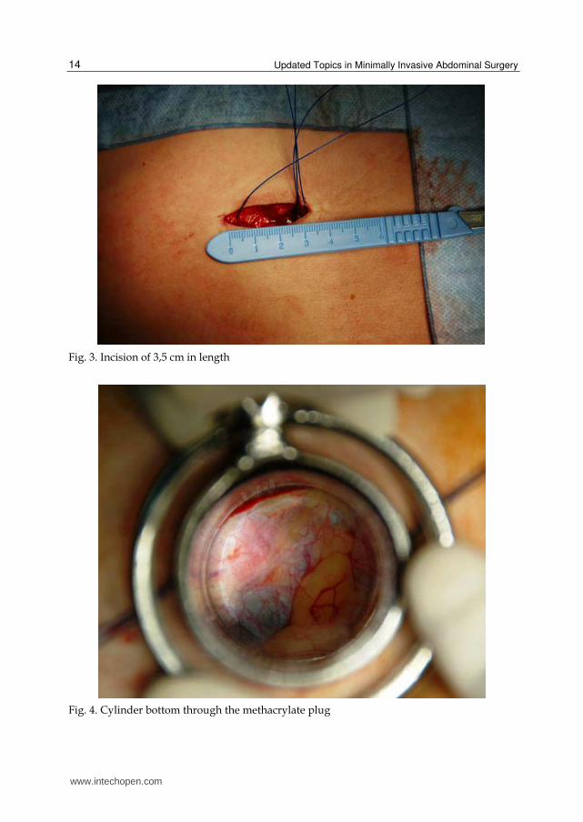
4.3 Technique and equipment To introduce the 3.8-cm cylinder one makes a right transversal-epigastric incision of 4.5 cm two fingerbreath lateral to the midline, approximately at the seventh or eighth costochondral cartilages level. One then proceeds with a longitudinal incision of the rectus sheath, splitting the muscle and cutting the posterior leaf and peritoneum. This is an uniform 4.5 cm section of all the abdominal wall layers. A suture of polypropylene (No. 1) is then passed through the whole thickness of the wall (not including the skin) on both side of the incision, which helps to guide the introduction of the cylinder. We make sure that there is nothing adhering and check the normality of neighbouring organs by two finger exploration. Once it is past the surface of the skin it is softly slided and enters without difficulty to its full extent towards the hepatocystic triangle. While we are inserting the cylinder we are seeing the intraperitoneal structures through the transparent plunger, especially the white appearance of the anteromedial aspect of the gallbladder and Hartmann's pouch and we can see, with a little pressure , the cystic duct and common bile duct (Figures 4, 5). Any gallbladder adherence to the hepatic flexure of the colon or omentum can be freed.
www.intechopen.com
Updated Topics in Minimally Invasive Abdominal Surgery
14
Fig. 3. Incision of 3,5 cm in length
Fig. 4. Cylinder bottom through the methacrylate plug
www.intechopen.com

Transcylindrical Cholecystectomy for the Treatment of Cholelithiasis and Its Complications: Cholecystectomy Under Local Anesthesia
15
Before reaching a working position of the cylinder, this is gently moved inside of the abdomen. The blunt shape of the plunger end, slightly protruding from the intra-abdominal side of the cylinder, facilitates this movement. The plunger can be withdrawn and reintroduced as many times as necessary to identify anatomical structures. Lamp lights usually suffice to illuminate the operative field, but a cold light may be of help occasionally.
Fig. 5. View of the hepatocystic triangle through the plug
Part of the gallbladder with its infundubulum is visible at the bottom of the cylinder, as well as the omentum, duodenal bulb or colon. The infundibulum or Hartmann Pouch is grasped with tissue Foerster forceps and is drawn anterior and laterally and a medium swab inside the cylinder is used to displace the organs that impede the sight of the angle between the gallbladder and the hepatoduodenal ligament. Afterwards the hepatocystic triangle is dissected using conventional material (Figure 6). The peritoneum is incised on the hepatocystic triangle, close to the gallbladder neck, and the fat is carefully dissected away on the free edge of the angle between the infundibulum and the hepatoduodenal ligament using gauze pledget held in an other Foerster forceps, until the cystic duct (Figure 7) and common bile duct are clearly defined (no always this later). Afterwards, we check that the cystic duct follows clearly from the gallbladder neck. If the cystic duct lymph node and cystic artery are not yet visible, the dissection is done gently upwards to discover the cystic artery, which will be followed up to its entrance into the
www.intechopen.com

Updated Topics in Minimally Invasive Abdominal Surgery
16
gallbladder (a right angle dissector is required). The cystic artery can be sectioned between two distal clips and a proximal one.
Fig. 6. Calot’s triangle (as shown by the arrow) after extracting the plug
At this time, surgeon and assistant must agree on the identity of the visible anatomic structures and make sure that there are no more tubular structures above the cystic duct, other than the cystic artery. Accessory extrahepatic ducts and ductus subvesicularis have to be taken into account, as well as the double cystic artery or any abnormal situation or origin. Once the cystic duct has been identified, a silk ligature is passed around it and prepared for cholangiography (performed selectively) and sectioned with two distal clips. To finish the dissection of the hepatocystic triangle we retract the infundibulum or corpus with the help of a pledget gauze, as much as we can, from its bed in the liver, keeping the dissection close to the gallbladder wall (to avoid structures of the hilum). Separation of the gallbladder from the hepatic bed follows in a retrograde fashion using electrocautery. Perhaps, this is de more laborious part of the procedure because we needs to change the point of traction to free the corpus and fundus that are attached to the liver in a somewhat posterior position. The puncture and emptying of the gallbladder helps freeing it and, finally, we extract it from the interior of the cylinder. We check out the hepatocystic zone and the gallbladder bed by means of the reintroduction of the cylinder and check for oozing and bile spill from the gallbladder bed. Bleeding can be
www.intechopen.com

Transcylindrical Cholecystectomy for the Treatment of Cholelithiasis and Its Complications: Cholecystectomy Under Local Anesthesia
17
restrained by gentle pressure of a moist gauze pad through the cylinder or electrocautery. The subhepatic space is irrigated with saline solution through the cylinder and after closing the posterior wall (polydioxanone sulphate) the wound is irrigated again.
Fig. 7. Cystic duct with right angle dissector
4.4 Transcylindrical cholecystectomy under local anaesthesia plus sedation All patients were fitted to the following protocol: 1. In the preparation area an intravenous cannula was placed, vital signs were monitored,
and was given 50 mg ranitidine and metoclopramide 10 mg intravenously. 2. Once in the operating room after the patient monitor ECG, pulse oxymetry (SpO2), BIS
(bispectral index) and noninvasive blood pressure we proceeded to the supply of oxygen with nasal cannula with the end tidal CO2 (ETCO2), Midazolam 0.05 mg/kg/ev and initiation of infusion of remifentanil in doses of 0.05 mcg/kg/min to 0.1 mcg/kg/ min.
The objective was to obtain a sedation 2-3 on the Ramsay scale and/or a BIS value of 70 to 85 before the application of local anesthesia. For anesthesia of the abdominal wall surgical area was used 300-500 mg of mepivacaine 1% was used. The infiltration began in the line previously marked for incision, which is located in the epigastrium about 4 cm to the right of the midline and 3 cm from the costal margin. Follows the infiltration of the muscular plane and transverse oblique, lateral to the incision site with the intention of blocking the intercostal nerves VII-IX in the lateral costal margin. Finally we infiltrate the rectus muscle
www.intechopen.com

Updated Topics in Minimally Invasive Abdominal Surgery
18
of abdomen in the epigastric region right under the incision line (Figure 8). Once the cylinder has been introduced the triangle of Calot is infiltrate with 2-4 cc of 2% mepivacaine (Figure 9). At the end of surgery and subcutaneous muscle planes were infiltrated with 10-20 ml of bupivacaine 0.25%. Before leaving the operating room the patients receives: paracetamol 1g/ev, dexamethasone 8mg/ev, ondansetron 4mg/ev and ketorolac 1mg/kg, although the latter was avoided in patients 70 years or older.
Fig. 8. Local anesthesia on intercostals nerves IX-VII and incision planes
Fig. 9. Infiltration with mepivacaine 2% of the hepatocystic triangle
www.intechopen.com

Transcylindrical Cholecystectomy for the Treatment of Cholelithiasis and Its Complications: Cholecystectomy Under Local Anesthesia
19
All patients were assessed for pain after the procedure and were discharged when they met the criteria (pain control, oral tolerance, no bleeding, nausea or vomiting, etc.), and follow analgesia regime alternating paracetamol 1g/6 h and metamizole 1g/6h orally at home. At 24 hours, through a telephone call, we assessed the pain at rest and with movement (scale of Andersen). In the fifth day, in outpatient visit, we check for the general status, the sate of the wound and the pain is assessed with a visual analog scale (VAS).
4.5 Surgical technique in acute cholecystitis and choledocholithiasis In acute cholecystitis we always use the 5 cm cylinder, the gallbladder is emptied with the help of an aspirator and a bile sample is send for culture. The dissection of the Calot triangle is done with a swab and if there are difficulties in closing the cystic duct is we use a ligature or stitch of poliglycolic acid. The cystic artery is treated in the same manner as above. The haemostasis of the liver may require more time. A Jackson-Pratt drain by counterincision is the norm in acute cholecystitis and common bile duct exploration. If the intraoperative cholangiogram shows the presence of stones and a dilated bile duct (Figure 10 ), we prepare the field for a transcylindrical choledochotomy if the stone could not be pushed through the papilla with a Fogarty catheter. After the cholecystectomy and haemostasis of the liver, we proceed to vary the angle of the cylinder to direct medially, to put it in the hepatoduodenal ligament, taking as reference the cystic duct stump. Once in the position, the bile duct is seen on the lateral border of the ligament once the fat is cleared away with blunt dissection.
Fig. 10. Cholangiography with cylinder in place
www.intechopen.com

Updated Topics in Minimally Invasive Abdominal Surgery
20
We must ensure that we are below the confluence of the cystic duct (the duodenum can be see in the field), which will expose the common bile duct (keep in mind that the confluence may be low). Two stay sutures using polyglactin 3-0 are located on both sides of the midline of the common bile duct to pull at the time of a vertical choledochotomy as short as possible (2-3 cm), but enough for the manoeuvres of stone removal (Figure 11).
Fig. 11. Coledocotomy about to be performed. Two stay sutures pull the common bile duct.
Randall stone forceps can not be used, but the Fogarty catheter, catheter irrigation and flexible choledochoscope are used. Before performing any manoeuvre, we introduce a gauze ball referenced with a thread at the proximal end of the choledochotomy, to prevent the displacement of the stone proximal to the hepatic duct when dragging with the Fogarty catheter rather than externalized through the incision of choledochotomy. Finally, we introduce the flexible choledochoscope and confirm the absence of calculations. The closure of the choledochotomy we do it with polyglactin 3/0 on a Kehr T tube. Between the fifth and seventh postoperative day a control cholangiogram is performed, and the patient discharged. The T tube is left in place for 14 days.
4.6 Results We have to distinguish between two clearly defined periods in the evolution of the implementation of transcylindrical cholecystectomy. A first period, from 1993 to 2008, of the beginning of the technique and treatment of patient in hospitalization and a second period since 2008 until today as outpatient surgery and short stay, mainly under local anesthesia plus sedation. in total we performed 633 operations: 387 belonging to the first stage and 247 to the second.
www.intechopen.com

Transcylindrical Cholecystectomy for the Treatment of Cholelithiasis and Its Complications: Cholecystectomy Under Local Anesthesia
21
The results of surgery of the first stage have already been published. In summary:
Total no. of patients 387
Completed transcylindrical 364
Ampliation to open cholecystectomy, no. (%) 23 (5.9)
Duration of simple cholecystectomy, mean (SD) 43,5 (13.3)
Length postoperative hospital stay mean (range), days
2.0 (1-6)
Postoperative complications
Bile leakage, no. (%) 2 (0.5)
Reoperation for bleeding, no. (%) 2 (0.5)
Bile Duct injury, no. (%) 0
Death, no. (%) 11 (0.3)
1Death from multiple organ dysfunction syndrome due biliary peritonitis
Table 1. Overall results of cholecystectomy between 1993-2008
The 3.8 cm cylinder was used in 261 cases and the 5 cm in 103 as first choice or an alternatively because of difficulties in recognition of the structures. The main cause of enlargement to open surgery was the fibrotic alteration triangle hepatocystic. The number of conversion is not negligible, but our philosophy has been not to subject the patient to the risk of intervention with uncertainty in identifying the structures of the hepatocystic triangle in order to prevent complications. For that we have not hesitated to convert to a classic laparotomy when facing at a reasonable difficulty. A survey of satisfaction with the aesthetics of the procedure yielded a 90% satisfied or very satisfied.
4.6.1 Transcylindrical cholecystectomy under local anesthesia plus sedation in day-case surgery
Today we are performing the majority of our cholecystectomies as day-case surgery. Local anesthesia and sedation is the anesthetic technique that we offer at all our patients and that we use unless the patient's preference for general anesthesia. A pilot study of 60 cases was published in Endoscopy (Grau Talens et al., 2010), but now we have performed the procedure in 222 patients, highlight a patient with choledocholithiasis too operated under local anesthesia and sedation, excellently tolerated; while that in 25 other general anesthesia was used for suspected acute cholecystitis (8 patients), suspected choledocholithiasis (3 patients) and specifically stated preference for the patient in the other cases (14 patients). Local anesthesia was initiated in 222 patients with demographic and anthropometric characteristics in Table 2.
www.intechopen.com

Updated Topics in Minimally Invasive Abdominal Surgery
22
Patients, no. 222
Men/woman 55/167
Age, mean (range) years 55.2 (17-90)
BMI1, mean (range) kg/m2 29.9 (19-46)
Height, mean (range) cm 160.5 (140-185)
Eight, mean (range) kg 77.0 (43-122)
Acute pancreatitis, no. 21
Acute cholecystitis previous2 34
1Body mass index 2Acute cholecystitis with a Hospital General admission
Table 2. Demographic and clinical characteristics of patients operated under local anesthesia plus sedation
As it can be seen our patients are obese in almost half the cases and 35 patients had a BMI
equal to or greater than 35 (15.8%). Previous acute cholecystitis was detected in 18 of 55 men
(33%), but only in16 of 167 women (9%).
Convalescent patients of acute pancreatitis were operated on before hospital discharge and an intraoperative cholangiography was performed.The results of surgery can be read in Table 3.
No. Patients in day-case program 197
Postoperative hospitalization. No. (%) 15 (7.6)
Converted to general anaesthesia. No. (%) 69 (31)
Converted to open surgery. No. (%)** 7 (3.1)
Duration. Mean (SD) 49.4 (22.4)
Intraoperative cholangiography. No. (%) 17 (7.6)
Common bile duct exploration. No. 2*
Wound infection. No. (%) 5 (2.2)
Subhepatic collection. No. (%) 1 (0.4)
Visual Analog scale. Mean (range) 2.0 (0-8)
* a surgery completed under local anesthesia and sedation ** An open surgery for carcinoma of the gallbladder
Table 3. Results of 222 patients scheduled for transcylindrical cholecystectomy under local anaesthesia plus sedation
Nausea and vomiting have virtually disappeared. Pain at rest on the fifth postoperative day
is almost nonexistent, while the pain with the movements of sitting or standing is mild and
all the patients are able to self care.
www.intechopen.com

Transcylindrical Cholecystectomy for the Treatment of Cholelithiasis and Its Complications: Cholecystectomy Under Local Anesthesia
23
Only 6 patients have expressed some discomfort during the operation, but the procedure was well tolerated and there was satisfaction in all cases, even where they were converted to general anesthesia. The 5 cm cylinder was used in 2 cases of suspected choledocholithiasis and thirteen cases of postinflammatory anatomical distortion that hinders the recognition with the 3.8 cm cylinder The vast majority of cases that required intubation (Table 4) was due to poor anatomical conditions related to persistent inflammation or scarring, but it is also true that a patient with a bulky or potent abdominal muscles (even with normal BMI) is a factor in consideration, since the absence of relaxation of the abdominal wall increases distance from the skin to the hepatocystic triangle and the cylinder of 10 cm length can be short.
Scarring or inflammatory anatomy 46
Big or muscular patient 16
Poor tolerance 6
Respiratory depression 1
Table 4. Causes of conversion to general anesthesia
In some cases we have changed the cylinder of 10 to 12 cm with satisfactory results. As previously mentioned, in 34 cases, of our patients had suffered a hospital admission for an attack of acute cholecystitis with ultrasound which showed a thickened gallbladder wall. Despite having passed more than 8 weeks after hospital discharge and be asymptomatic, we have found during the intervention that the process is not cured and present frank acute cholecystitis in 7 cases (20%). Of the 69 patients converted to general anesthesia, 29 were men in a series with 55 men. Obviously, the male sex is a definite risk factor for conversion to general anesthesia, as gallstone disease seems more severe in men while the abdominal muscles are larger. In our series both the height and weight is significantly higher in males, but not BMI which is slightly below the average (28.9 kg/m2). In cases of conversion to a classical laparotomy incision the bad anatomy can also blame as responsible, in fact, five of seven cases converted belong to patients with acute cholecystitis previous and three of the 7 are male. In one case cystic clips were dislodged while reviewing the operative area. However it is, starting the procedure under local anesthesia and sedation does not produce a significant delay in time, only a few minutes, since the decision to intubate the patient is taken quickly and everything is ready for this eventuality, but it is likely that in the future the general anesthesia be used from the begin in the men who have had an admisssion for acute cholecystitis.
4.6.2 Transcylindrical cholecystectomy in the treatment of acute cholecystitis and choledocholithiasis In total 99 patients were operated for acute cholecystitis: 45 suspected prior to the intervention and operated in emergency basis or in the first 72 hours after admission (but not from the onset of symptoms, because in our experience, half of the patients came in a mean of 36 hours after the pain). The operation for acute cholecystitis is more laborious, with and greater needs of conversions to classic laparotomy, which in our series occurred in 13 cases. In all cases except one that ended with a cholecystostomy, the gallbladder has been removed. The duration of the intervention is significantly higher than cholecystectomy for
www.intechopen.com

Updated Topics in Minimally Invasive Abdominal Surgery
24
uncomplicated lithiasis is related to the need for more time for dissection and hemostasis. Two superficial wound infections, 2 postoperative subhepatic collections and a third at 9 months after surgery treated by percutaneous puncture and a biliary leak through drainage for 15 days with spontaneous closure are noteworthy complications. At least 3 days of hospitalization and antibiotic treatment follow the surgery. In our experience, common bile duct exploration presents no special difficulties except juxtapapillary interlocking stone, making it difficult to remove. The location of the bile duct, dissection, and preparation is as simple as in open laparotomy. In 30 cases we performed transcylindrical choledochotomy with an average of 119 minutes, with a range between 70 and 182 minutes of the proceedings. A stone inpacted in a dilated common bile duct required a choledochoduodenostomy. One patient experienced postoperative bleeding requiring intervention without finding the bleeding point.
5. Conclusion
Despite technological advances and the practice of surgery becoming more expensive, we developed a technique for the treatment of gallstones and its complications achievable with natural view of the structures and conventional reusable material. The technique has proven to be fast simple and safe, applicable to all patients. Local anesthesia and sedation provides a quick recovery and many patients lose the fear of the intervention. Both in acute cholecystitis in choledocholithiasis we have obtained good results. The patients suspected of choledocholithiasis are operated and an intraoperative cholangiography is made. The transcylindrical exploration of the common bile duct is performed whenever introperative cholangiography demonstrated stones.
6. References
Arendt, SA. & Pitt, HA. (2004) Biliary Tract. In: Sabiston Textbook of Surgery: The Biological Basis of Modern Surgical Practice. Townsend, CM., Beauchamp, RD., Evers, BM., & Mattox, KL., editors, pp. 1597-1641. 17th edition. Elsevier Saunders, ISBN 0-8089-2295-5, Philadelphia.
Assalia, A., Kopelman, D., Hashmonai, M. (1997). Emergency minilaparotomy cholecystectomy for acute cholecystitis: prospective randomized trial--implications for the laparoscopic era. World J Surg 1997, Vol. 21, No. 5, pp. 534-9. ISSN 0364-2313.
Bartlett, MK., & Waddell, WR. (1958). Indications for common duct exploration. Evaluation in 1000 cases. New Eng J Surg 1958, Jan 23, Vol. 258, no. 4, pp. 164-7, ISSN:0028-4793.
Britton, J., & Bickerstaff, KI (1994). Benign Diseases of the Biliary Tract. In: Oxford Textbook of Surgery Morris, PJ., & Malt, RA., editors. Oxford University Press, pp. 1209-1241. ISBN 0192626035, New York.
Classen, M., & Demling, L. (1974). Endoscopic sphincterotomy of the papilla of vater and extraction of stones from the choledochal duct. Deutsch Med Wochenschr 1974, Mar 15, Vol. 99, N0.11, pp. 496-7, ISSN 0012-0472.
Cooperman, AM. (1990). Laparoscopic cholecystectomy for severe acute, embedded, and gangrenous cholecystitis. J Laparoendosc Surg 1990, Vol. 1, No. 1, pp. 37-40. ISSN 1052-3901.
Craig, DB. (1981). Postoperative recovery of pulmonary function. Anesth Analg, Jan 1981, vol 60, No. 1, pp. 46-52, ISSN 0003-2999.
www.intechopen.com

Transcylindrical Cholecystectomy for the Treatment of Cholelithiasis and Its Complications: Cholecystectomy Under Local Anesthesia
25
Dawnson, JL. (1985). Colecistectomía. En: Cirugía de la vesicular y vías biliares, Marlow, S., & Sherlock, S., editores, pp. 319-335, Salvat Editores S.A., ISBN 84-345-2263-21985, Barcelona
Doherty, GM. (2010). Current diagnosis and treatment surgery. 13th edition, Lange Medical Books/McGraw-Hill, ISBN 978-0-07-16389-4, New York.
Dubois, F. & Berthelot, B. (1982) Cholécystectomie par mini-laparotomie. Nouv presse Med, April 1982 3;Vol.11, No. 15, pp.1139-41, ISSN 0301-1518, OCLC 9262901.
Escorrou, J., Cordova, JA., Lazortes, F., Frexinos, J., & Ribet A (1984). Early and Late complications after endoscopic sphincterotomy for biliary lithiasis with and without the gallbladder ろin situわ. Gut, Jun 1984, Vol. 25, No. 6, pp. 598-602, ISSN 0017-5749.
Fink, AS. (1993). To ERCP o not to ERCP: that is the question. Surg Endosc 1993, Vol. 56, pp. 375-376, ISSN:0930-2794.
Frazee, RC., Roberts, JW., Okeson, GC., Symmonds, RE., Snyder, SK., Hendricks, JC., & Smith, RW. (1991). Open versus laparoscopic cholecystectomy. A comparison of pulmonary function. Ann Surg, Jun 1991, Vol. 213, No. 6, pp. 651-3, ISSN 0003-4932.
Gilliland, TM., & Traverso, LW. (1990). Modern standards for comparison of cholecystectomy with alternative treatments for symptomatic cholelithiasis with emphasis on long term relief of symptoms. Surg Gynecol Obstet, Jan 1990, Vol.170, No 1, pp. 39-44. ISSN 0039-6087.
Goco, IR. & Chambers, LG. (1988). Dollars and cents: minicholecystectomy and early discharge. South Med J, Feb 1988, vol. 81, No. 2, pp. 161-3. ISSN 0038-4348.
Grau-Talens, EJ., & Giner, M. (2010) Transcylindrical gas-free cholecystectomy for the treatment of cholelithiasis, cholecystitis, and choledocholithiasis. Surg Endosc, Sep 2010, Vol. 24, No. 9, pp. 2099-104, ISSN 0930-2794.
Grau-Talens, EJ., Cattáneo, JH., Giraldo, R., Mangione-Castro, PG. & Giner, M. (2010) Transcylindrical cholecystectomy under local anestesia plus sedation. A pilot study. Endoscopy, May 2010; Vol. 42, No. 5, pp. 395-9, ISSN 0013-726X.
Grau-Talens, EJ., García-Olives, F., & Rupérez-Arribas, MP. (1998). Transcylindrical cholecystectomy: new technique for minimally invasive cholecystectomy. World J Surg, May 1998; 22, No. 5 pp.153-8, ISSN 0364-2313.
Grau-Talens, EJ., Pérez-García, G., & Rupérez-Arribas, MP. (1994). Colecistectomía transcilíndrica. Cirugía Española, Noviembre 1994, vol. 56, Suplemento 1, p.297, ISSN 00-9739-X.
Harris, H W. (2008). Biliary system, In: Surgery. Basic science and clinical evidence, Norton, JA., Barie, PS., Randal Bollinger, R., Chang, AE., Lowry, SF., Mulvihill, SJ., Pass, HI., & Thompson, RW., Editors, pp. 911-942, Second Edition, Springer Scoience+Business Media,LLC, ISBN 978-0-387-30800-5, New York.
Karam, J. & Roslyn, JL. (1997). Colelithiasis and Cholecystectomy. In: Maingot´s Abdominal Operations, Zinner, MJ., Schwartz, SI., & Ellis, H, editors.. Tenth edition. McGraw-Hill, pp.1717-1738. ISBN 0-8385-6106-3, New York.
Katz, D., Nikfarjam, M., Sfakiotaki, A., & Christophi, C. (2004). Selective endoscopic cholangiography for the detection of common bile duct stones in patients with cholelithiasis. Endoscopy, Dec 2004,Vol. 36, No. 12, pp. 1045-9. ISSN 0013-726X.
Koo, KP., & Traverso, LW. (1996). Do preoperative indicators predict the presence of common bile duct during laparoscopic cholecystectomy? Am J Surg, May 1996, vol 171, No. 5, pp.495-9. ISSN 0002-9610.
www.intechopen.com
Updated Topics in Minimally Invasive Abdominal Surgery
26
Kroh, M., Chand, B. (2008). Choledocholithiasis, endoscopic retrograde cholangiopancretography, and laparoscopic common bile duct exploration. Sur Clin N Am, Dec 2008, Vol. 88, No. 6, pp. 1019-1031. ISSN0039-6109.
Lindell, P., & Hendenstierna, G. (1976). Ventilation efficiency after different incisión for cholecystectomy. Acta Chir Scand 1976, Vol.142, No. 8,pp. 561-5, ISSN 0301-1860
Litynski, G. (1996). Highlights in the history of laparoscopy. The development of laparoscopic techniques. A cumulative effort of internists, gynecologist and surgeons. Barbara Bernert Verlag, ISBN 3-9804740-6-2, Frankfurt/Main.
McMahon AJ, Russell IT, Ramsay G, Sunderland G, Baxter JN, Anderson JR, et al (1994). Laparoscopic and minilaparotomy cholecystectomy: a randomized trial comparing postoperative pain and pulmonary function. Surgery, May 1994, Vol.115, No. 5 pp. 533-9, ISSN 0039-6060.
Morton, CE. (1985). Cost containment with the use of "mini-cholecystectomy" and intraoperative cholangiography. Am Surg, Mar 1985, Vol 51, No. 3, pp. 168-9. ISSN 0003-1348.
Moss, G. (1996). Raising the outcome standards for conventional open cholecystectomy. Am J Surg, Oct 1996, Vol. 172, No. 4, pp. 383-5. ISSN 0002-9610.
Mühe, E. (1986). Die erste cholecystektomie durch das laparoskop. Langenbecks Arch Chir 1986; Vol. 369, No.1, p.804. ISSN 1435-2443
O’Kelly, TJ., Barr, H., Malley, WR., & Kettlewell, M. (1991). Cholecystectomy through a 5 cm subcostal incision. Br J Surg; Jun, Vol. 78,N0. 6, p. 762, ISSN:0007-1323.
O'Dwyer, PJ., Murphy, JJ.,& O'Higgins, NJ (1990). Cholecystectomy through a 5 cm subcostal incision. British J Surg, Oct 1990, Vol. 77, No. 10, pp. 1189-9, ISSN 0007-1323.
Olsen DO (1993). Mini-Lap cholecystectomy. Am J Surg, Apr 1993, Vol. 165, No. 4, pp 440-3, ISSN 0002-9610.
Rhodes, M., Sussman, L., Cohen, L., & Lewis, MP. (1998). Randomised trial of laparoscopic exploration of common bile duct versus postoperative endoscopic retrograde cholangiography for common bile duct stones. Lancet, Jan 17, 1998,Vol. 351(9097), pp.159-61, ISSN 0140-6736.
Rozsos, I., Ferenczy, I., & Rozsos, T. (1997). The surgical technique of microlaparotomy cholecystectomy. Acta Chir Hung 1997, Vol. 36, (1-4), pp 294-296, ISSN:0231-4614.
Rozsos, I., Ferenczy, J., & Schmitz, R. (2003). Micro and mini-cholecystectomies in the 21st century Orv Hetil 2003, Vol. 144, pp. 1291-1297, ISSN 0030-6002.
Russell, RCG., & Shankar, S. (1987). The stabilized ring retractor. A technique for cholecystectomy. Br J Surg, Sep 1987, Vol. 74, No. 8, pp. 826, ISSN:0007-1323.
Schumacher, FJ., & Kohaus, HM. (1994). Cholecystectomy via a surgical tube in 800 patients. Chirurg, Apr 1994, Vol 65, No. 4, pp. 373-6, ISSN 0009-4722.
Schwarts, SI. (1989). Gallbladder and Extrahepatic Biliary System. In: Principles of Surgery Schwarts, SI. Shires, GT. Spencer, FC. & Husser, WC, editors. pp. 1381-1412, Fith edition, McGraw-Hill Book Company, ISBN 0-07-055822-1, New York.
Werbesey, JE., Kirkett DH. (2008). Common bile duct exploration for choledocholithiasis. Surg Clin N Am 2008, Dec, Vol. 88, No. 6, pp.1315-28. ISSN 0039-6109.
Yamashita, Y., Takada, T., Kararada, Y et al (2007). Surgical treatment of patients with acute cholecystitis: Tokyo guidelines. J Hepatobiliary Pancreat Surg 2007; Vol. 14, No. 1, pp. 91-7, ISSN 0944-1166.
Zeus, F., Gooszen, HG., & Van Laarhoven, CJ. (2010). Open, small-incision or laparoscopic cholecystectomy for patients with symtomatic cholecystolithiasis. An overview of Cochrane Hepato-Biliary Group reviews. Cochrane Database Syst Rev 2010,Jan 20; Issue 1, CD008318, ISSN 1469-493x, 1361-6137.
www.intechopen.com
Updated Topics in Minimally Invasive Abdominal SurgeryEdited by Prof. Ahmed Elgeidie
ISBN 978-953-307-773-4Hard cover, 246 pagesPublisher InTechPublished online 14, November, 2011Published in print edition November, 2011
InTech EuropeUniversity Campus STeP Ri Slavka Krautzeka 83/A 51000 Rijeka, Croatia Phone: +385 (51) 770 447 Fax: +385 (51) 686 166www.intechopen.com
InTech ChinaUnit 405, Office Block, Hotel Equatorial Shanghai No.65, Yan An Road (West), Shanghai, 200040, China
Phone: +86-21-62489820 Fax: +86-21-62489821
Updated topics in minimally invasive abdominal surgery provides surgeons interested in minimally invasiveabdominal surgery with the most recent techniques and discussions in laparoscopic surgery. This bookincludes different topics covering a big variety of medical conditions with up-to-date information. It discussesmany controversies in a clear and user-friendly manner. This book is made for young junior surgeons intraining and also senior surgeons who need to know the most recent work in the field of laparoscopy. To makethe material easily digestive, we provided the book with many figures and illustrations for different proceduresand technical pearls.
How to referenceIn order to correctly reference this scholarly work, feel free to copy and paste the following:
E. Javier Grau Talens, Julio Horacio Catta ́neo, Rafael Giraldo Rubio and Pablo Gustavo Mangione Castro(2011). Transcylindrical Cholecystectomy for the Treatment of Cholelithiasis and Its ComplicationsCholecystectomy Under Local Anesthesia, Updated Topics in Minimally Invasive Abdominal Surgery, Prof.Ahmed Elgeidie (Ed.), ISBN: 978-953-307-773-4, InTech, Available from:http://www.intechopen.com/books/updated-topics-in-minimally-invasive-abdominal-surgery/transcylindrical-cholecystectomy-for-the-treatment-of-cholelithiasis-and-its-complications-cholecyst

© 2011 The Author(s). Licensee IntechOpen. This is an open access articledistributed under the terms of the Creative Commons Attribution 3.0License, which permits unrestricted use, distribution, and reproduction inany medium, provided the original work is properly cited.