bgda lecture - development of the embryo/fetus 2 · mesoderm means the "middle layer" and...
TRANSCRIPT

[Expand]
[Expand]
BGDA Lecture - Development ofthe Embryo/Fetus 2Introduction
This lecture covers the period of Embryonic development, in Humans fromweek 3 to week 8 (GA week 5-10) and is divided into 23 Carnegie stages ofembryonic development. There will also be a brief introduction to fetaldevelopment. Note, the period from week 9 to week 38 is considered Fetaldevelopment and will be covered in detail in the Laboratory 12.
Lecture Objectives
Understand key structures and events in embryonic development.Understanding of the dynamic changes internal and externalstructures.Brief understanding of organ and system formation (functional / notfunctional).Brief understanding of critical periods of development.
1 Minute Embryology | UNSW theBox
Lecture Archive
Textbooks
BGDA Practical Classes

Practical 3 -Fertilization toImplantation
Practical 6 -Implantation to 8Weeks
Practical 12 - FetalPeriod
Practical 14 - Placenta and Fetal Membranes
First 8 Weeks
The Carnegie stages of the first 8 week of human development.
Week 3
Mesoderm means the "middlelayer" and it is from this layer thatthe body's connective tissues arederived (note that the head neuralcrest ectoderm also formsconnective tissues)
In early mesoderm development anumber of transient structures willform and then be lost as tissuestructure is patterned and organised.
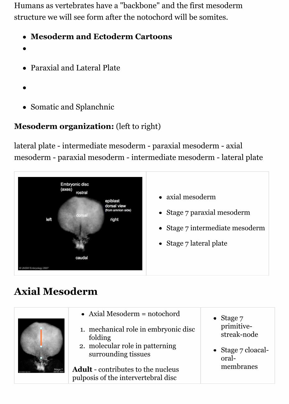
Humans as vertebrates have a "backbone" and the first mesodermstructure we will see form after the notochord will be somites.
Mesoderm and Ectoderm Cartoons
Paraxial and Lateral Plate
Somatic and Splanchnic
Mesoderm organization: (left to right)
lateral plate - intermediate mesoderm - paraxial mesoderm - axialmesoderm - paraxial mesoderm - intermediate mesoderm - lateral plate
axial mesoderm
Stage 7 paraxial mesoderm
Stage 7 intermediate mesoderm
Stage 7 lateral plate
Axial Mesoderm
Axial Mesoderm = notochord
1. mechanical role in embryonic discfolding
2. molecular role in patterningsurrounding tissues
Adult - contributes to the nucleuspulposis of the intervertebral disc
Stage 7primitive-streak-node
Stage 7 cloacal-oral-membranes

Paraxial Mesoderm
differentiates rostro-caudally (head to tail)head region - remains unsegmentedbody region - segments to form pairs of somites along thelength of the embryo.
Adult - contributes vertebral column (vertebra and IVD),dermis of the skin, skeletal muscle of body and limbs
Intermediate Mesoderm
named by position (between paraxial and lateralplate)differentiates rostro-caudally (head to tail)forms 3 sets of "kidneys" in sequence
1. pronephros2. mesonephros3. metanephros
Adult - metanephros forms the kidney
Lateral Plate Mesoderm
at edge of embryonic disc"horseshoe shaped" space forms in the middle, dividingthis region
somatic mesoderm - closest to ectodermintra-embryonic coelom - single space forms the 3major body cavities (pericardial, pleural, peritoneal)splanchnic mesoderm - closest to endoderm
Adult - body connective tissues, gastrointestinal tract(connective tissues, muscle, organs), heart
Week 4
Somite Development

Somite initially forms 2 main components
ventromedial- sclerotome forms vertebral body and intervertebraldiscdorsolateral - dermomyotome forms dermis and skeletal muscle
sclerotome and dermomyotome
dermatome and myotome
epaxial and hypaxial muscles
Sclerotome Dermatome
sclerotome later becomes subdividedrostral and caudal halves separatedlaterally by von Ebner's fissure
half somites contribute to a single vertebrallevel bodyother half intervertebral disctherefore final vertebral segmentation“shifts”
connective tissueunderlying epidermisbegins as a dorsalthickeningspreads throughoutthe body
Myotome
Body - epaxial andhypaxial musclesLimbs - flexor andextensor muscles

[Expand]
Heart
Heart Development Movies
forms initially in splanchnic mesoderm of prechordal plate region -

Mesoderm vascular development
Stage 10 Week 4, 22 - 23 days
cardiogenic regiongrowth and folding of theembryo moves heartventrallly and downwardinto anatomical position
week 3 begins as paired hearttubes that fuse to form singleheart tubebegins to beat in Humans- day22-23heart tube connects to bloodvessels forming in splanchnicand extraembryonic mesoderm
Week 2-3 pair of thin -walled tubes
Week 3 tubes fused, truncus arteriosus outflow, heart contracting
Week 4 heart tube continues to elongate, curving to form S shape
Week 5 Septation starts, atrial and ventricular
Links: Cardiac Embryology
Neural
Neural Plate
extends frombuccopharyngealmembrane toprimitive nodeforms abovenotochord andparaxial mesodermneuroectodermalcells
broad brainplatenarrower

Stage 11 neural groove to tube
spinal cord3 components form:floor plate, neuralplate, neural crest
Neural Groove
forms in the midline of theneural plate (day 18-19)either side of which are theneural folds which continues todeepen until about week 4neural folds begins to fuse,beginning at 4th somite level
Neural Tube
Neural - 3 primary vesicles
Neural Crest
population of cells at the edge of the neural plate that lie dorsallywhen the neural tube fusesdorsal to the neural tube, as a pair of streaks

Stage 14 pharyngeal arches
pluripotential, forms many different types of cellscells migrate throughout the embryo
Neural Crest Derivatives: dorsal root ganglia, autonomic ganglia,adrenal medulla, drg sheath cells, glia, pia-arachnoid sheath, skinmelanocytes, connective tissue of cardiac outflow, thyroid parafollicularcells, craniofacial skeleton, teeth odontoblasts
Head
branchial arch (Gk. branchia=gill)arch consists of all 3 trilaminarembryo layers (ectoderm-outside, mesoderm - core ofmesenchyme, endoderm -inside)
Humans have 5 arches - 1, 2, 3,4, 6 (Arch 5 does not form orregresses rapidly)from in rostro-caudal sequence,Arch 1 to 6 from week 4onwardsarch 1 and 2 appear at time ofclosure of cranial neuroporeFace - mainly arch 1 and 2Neck components - arch 3 and 4 (arch 4 and 6 fuse)

Sensory Placodes
During week 4 a series of thickened surface ectodermal patches formin pairs rostro-caudally in the head region.These sensory placodes will later contribute key components of eachof our special senses (vision, hearing and smell).Note that their initial postion on the developing head is significantlydifferent to their final position in the future sensory system
Otic placode - istage 13/14 embryo the otic placode sunk from thesurface ectoderm to form a hollow epithelial ball, the otocyst, whichnow lies beneath the surface surrounded by mesenchyme(mesoderm). The epithelia of this ball varies in thickness and hasbegun to distort, it will eventually form the inner ear membranouslabyrinth.Lens placode - lies on the surface, adjacent to the outpocketing ofthe nervous system (which will for the retina) and will form the lens.Nasal placode - has 2 components (medial and lateral) and willform the nose olefactory epithelium.
Upper and Lower Limb
Gastrointestinal Tract
Begins at buccopharyngeal membraneEnds at cloacal membrane3 distinct portions (fore-, mid- and hind-gut)liver earliest forming organ
Germ layer contributions

Stage 14 pharyngeal arches
Endoderm - epithelium and associated glandsMesoderm (splanchnic) - mesentry, connective tissues, smoothmuscle, blood vesselsEctoderm (neural crest) - enteric nervous system
Both endoderm and mesoderm will contribute to associated organs.
Gastrointestinal Tract
Week 5
Neural - 5 secondary vesicles
Heart - septation starts, atrialand ventricularVascular - 3 vascular systems(systemic, placental, vitelline)extensively remodelledRespiratory - left and right lungbuds push into thepericardioperitoneal canals(primordia of pleural cavity)Sense - Hearing cochlear partof otic vesicle elongates(humans 2.5 turns)

Septation continues, atrial septa remains open, foramen ovale

Week 6
Endocrine developmentPituitary - connecting stalkbetween pouch and oral

cavity degeneratesParathyroid - diverticulum elongate, hollow then solid, dorsal cellproliferationThymus - diverticulum elongate, hollow then solid, ventral cellproliferationAdrenal - fetal cortex forms from mesothelium adjacent to dorsalmesentery, medulla neural crest cells from adjacent sympatheticganglia
Week 7
pancreas - Week 7 to 20pancreatic hormones secretionincreases, small amountmaternal insulinlimb bones form byendochondrial ossification andthroughout embryoreplacement of cartilage withbone (week 5 onward).
Endochondral ossification inlimb
Endochondral ossification
Head Intramembranous ossification
Intramembranous ossification
Week 8
Neural - secondary vesicles

Fetal length and weight changes
Neural - early developing cortex
Gastrointestinal tract herniation
Limb - upper and lower limbs rotate in different directions (upperlimb dorsally, lower limb ventrally)
Links: Embryonic Development | Timeline human development
Fetal
First Trimester (1 - 12 weeks) -embryonic and early fetalSecond Trimester (13 - 24weeks) - organ developmentand function, growth (length)Third Trimester (25 - 40 weeks)- organ function and rapidgrowth (weight)
(see additional information fordetails)
Fetal Neural
During the fetal period there is ongoing growth in size, weight andsurface area of the brain and spinal cord. Microscopically there is

Timeline of events in Human NeuralDevelopment
ongoing: cell migration,extension of processes, celldeath and glial celldevelopment.Brain - folding of the initiallysmooth surface (insular cortex,gyral and culcal development)Neural development willcontinue after birth withsubstantial growth, death andreorganization occuring during the postnatal period
Links: neural | BGDA Lecture - Nervous System
Lung Stages
week 4 - 5 embryonicweek 5 - 17 pseudoglandularweek 16 - 25 canalicularweek 24 - 40 terminal saclate fetal - 8 years alveolar
Links: respiratory | SH Lecture - Respiratory
Fetal Genital
Gonad - ovary and testis developmentInternal genital tract - uterus and ductus deferensExternal genital tract - genital folds developmentTestis descent
Links: genital | BGDB Lecture - Genital
Fetal Renal
week 32-34 nephron development completed

term birth nephron number per kidney about 1 million (300,000 to 2million)
Links: renal
Fetal Endocrine
Many endocrine organs begin to function in the early fetal period.Pituitary hormones - HPA axis established by week 20, pituitaryfunctional throughout fetal developmentThyroid hormone - important for neural development, required formetabolic activity, also in the newborn
Remember that the Placenta also has important endocrine functionsduring development.
Links: endocrine | placenta
Critical Periods
The term "Critical Periods" refers to periods of development when specificsystems are more sensitive to teratogen exposure or developmental insults.
AdditionalInformation
See the associated BGDA Practical 6class.
Links: human timeline | firsttrimester timeline | secondtrimester timeline | third trimester timeline | fetal | Template:Movies
Abnormality Links: Introduction | Genetic | Environmental | Unknown |Teratogens | ectopic pregnancy | Cardiovascular | Coelomic Cavity | Endocrine |Gastrointestinal Tract | Genital | head abnormalities | integumentaryabnormalities | Musculoskeletal | limb abnormalities | Neural | Neural Crest |Renal | Respiratory | Placenta | Sensory | Hearing | Vision | Twinning |Developmental Origins of Health and Disease | ICD-10

[Expand]
[Expand]
Historic Embryology
Carnegie stages
BGDA: Lecture 1 | Lecture 2 | Practical 3 | Practical 6 | Practical 12 |Lecture Neural | Practical 14 | Histology Support - Female | Male |Tutorial
Glossary Links
Glossary: A | B | C | D | E | F | G | H | I | J | K | L | M | N | O | P | Q |R | S | T | U | V | W | X | Y | Z | Numbers | Symbols
Cite this page: Hill, M.A. (2018, May 13) Embryology BGDA Lecture -Development of the Embryo/Fetus 2. Retrieved fromhttps://embryology.med.unsw.edu.au/embryology/index.php/BGDA_Lecture_-_Development_of_the_Embryo/Fetus_2
What Links Here?
© Dr Mark Hill 2018, UNSW Embryology ISBN: 978 0 7334 26094 - UNSW CRICOS Provider Code No. 00098G