best1 sequence variants in italian patients with ... · pdf filebest vitelliform macular...
TRANSCRIPT
Best vitelliform macular dystrophy (OMIM # 153700; VMD) is a macular disease, which generally appears in child-hood with a yellowish yolk-like lesion in the macula [1-3]. The first pedigree was described by Best in 1905 [4].
The disease is associated with the accumulation of lipo-fuscin at the level of the retinal pigment epithelium (RPE); over time the central yellow lesions progressively disinte-grate and macular atrophy or fibrosis often develops [1,2]. The clinical picture evolves over many years producing a gradual decline of visual acuity; according to Gass [1] the macular lesions progress through various well defined stages: vitelliform, pseudohypopyon, vitelliruptive (scrambled egg), atrophic and cicatricial. Some patients develop choroidal neovascularization [1], which can be treated with photo-dynamic therapy [5,6] or intravitreal antiangiogenic drugs [7,8]. In the large majority of families the electro-oculogram (EOG) is markedly abnormal in all stages of progression and
in phenotypically normal carriers [9], even if several studies report some patients with normal EOG [10-12]. Full-field ERG is usually normal [10,11] while mfERG often presents reduced amplitudes [13]. Fundus autofluorescence (FAF) imaging shows an increased autofluorescence of the vitel-liform deposits [14] while OCT allows the visualization of the vitelliform substance and the associated alterations of the RPE and of the photoreceptors [15-17].
VMD is inherited as an autosomal dominant trait, with incomplete penetrance and highly variable clinical expres-sion. It has been associated with alterations in the gene BEST1 (OMIM # 607854; previously known as VMD2), mapped to the long arm (q13) of chromosome 11, encoding the 585-amino acid transmembrane protein bestrophin-1 [18,19]. This protein localizes to the basolateral membrane of RPE cells [20] and functions as a chloride channel [21,22] but it may act as an inhibitor of intracellular voltage-dependent Ca2+ channels, too. It has also been involved in pH and cell volume regulation and a possible role as a HCO3
- channel has been proposed too [23]. Bestrophin-1 dysfunction results in abnormal fluid and ion transport by the RPE, determining a weakened interface
Molecular Vision 2012; 18:2736-2748 <http://www.molvis.org/molvis/v18/a281>Received 2 October 2011 | Accepted 15 November 2012 | Published 17 November 2012
© 2012 Molecular Vision
2736
BEST1 sequence variants in Italian patients with vitelliform macular dystrophy
Andrea Sodi,1 Ilaria Passerini,2 Vittoria Murro,1 Roberto Caputo,3 Giacomo Maria Bacci,3 Mirela Bodoj,1 Francesca Torricelli,2 Ugo Menchini1
1Department of Specialized Surgical Sciences, Eye Clinic, University of Florence, Italy; 2Department of Genetic Diagnosis, Azienda Ospedaliero-Universitaria Careggi, Florence, Italy; 3Pediatric Ophthalmology Unit, Azienda Ospedaliero-Universitaria Meyer, Florence, Italy
Purpose: To analyze the spectrum of sequence variants in the BEST1 gene in a group of Italian patients affected by Best vitelliform macular dystrophy (VMD).Methods: Thirty Italian patients with a diagnosis of VMD and 20 clinically healthy relatives were recruited. They belonged to 19 Italian families predominantly originating from central Italy. They received a standard ophthalmologic examination, OCT scan, and electrophysiological tests (ERG and EOG). Fluorescein and ICG angiographies and fundus autofluorescence imaging were performed in selected cases. DNA samples were analyzed for sequence variants of the BEST1 gene by direct sequencing techniques.Results: Nine missense variants and one deletion were found in the affected patients; each patient carried one mutation. Five variants [c.73C>T (p.Arg25Trp), c.652C>T (p.Arg218Cys), c.652C>G (p.Arg218Gly), c.728C>T (p.Ala243Val), c.893T>C (p.Phe298Ser)] have already been described in literature while another five variants [c.217A>C (p.Ile73Leu), c.239T>G (p.Phe80Cys), c.883_885del (p.Ile295del), c.907G>A (p.Asp303Asn), c.911A>G (p.Asp304Gly)] had not previ-ously been reported. Affected patients, sometimes even from the same family, occasionally showed variable phenotypes. One heterozygous variant was also found in five clinically healthy relatives with normal fundus, visual acuity and ERG but with abnormal EOG.Conclusions: Ten variants in the BEST1 gene were detected in a group of individuals with clinically apparent VMD, and in some clinically normal individuals with an abnormal EOG. The high prevalence of novel variants and the frequent report of a specific variant (p.Arg25Trp) that has rarely been described in other ethnic groups suggests a distribution of BEST1 variants peculiar to Italian VMD patients.
Correspondence to: Vittoria Murro, Department of Specialized Surgical Sciences, University of Florence, Viale Morgagni 85, 50134 Florence, Italy; Phone: +39-055-411765; FAX: +39-055-4377749; email: [email protected]
Molecular Vision 2012; 18:2736-2749 <http://www.molvis.org/molvis/v18/a281> © 2012 Molecular Vision
2737
between RPE and photoreceptors; this may affect retinoid transport and photoreceptors outer segment phagocytosis by the RPE, finally leading to increased lipofuscin deposition [24].
In addition to VMD, BEST1 variants have been associ-ated with several other eye diseases [22] including adult-onset vitelliform macular dystrophy [25], autosomal recessive bestrophinopathy [26-28], autosomal dominant vitreoretino-choroidopathy [29,30], retinitis pigmentosa [31], microcornea, retinal dystrophy, cataract, and posterior staphyloma (MRCS syndrome) [32]. To date more than 150 BEST1 variants have been identified in VMD [22] (mutation-database; Retina international, databases accessed January 2012); the large majority of them are missense variants [22,33,34] resulting in amino acid changes in the N-terminal part of the protein.
BEST1 variants have been investigated in several different ethnic groups and in isolated Italian families [6,12,35] but at present there is no specific study on BEST1 variants in Italian VMD patients. Recently the results were published [36] of a BEST1 molecular analysis performed on a group of 23 patients, 10 of whom were from three Italian families. In the present study sequence variants of the BEST1 gene were determined in a group of Italian patients affected by VMD; these 30 patients were taken from a large sample of 19 Italian families, predominantly originating from central Italy.
METHODS
Clinical evaluation: Nineteen Italian families, of which at least one family member was affected by VMD, were recruited through the Hereditary Retinal Degenerations Referring Center of the Eye Clinic of the University of Flor-ence. Criteria for the Best phenotype included the following: 1) juvenile-to-adult onset of the disease; 2) bilateral macular dystrophy with the typical round lipofuscin lesions at the posterior pole (including patients with different stages of the disease); 3) normal ERG; 4) abnormal EOG with Arden ratio always below 1.50. We also studied some apparently healthy relatives of the patients who agreed to participate to identify possible asymptomatic carriers. The study adhered to the tenets of the Declaration of Helsinki and was approved by the Local Ethics Committee. Moreover, each patient gave written informed consent.
All the subjects included in the study were clinically evaluated by means of a standard ophthalmologic examina-tion, fundus photography, OCT scan (Topcon 3D OCT-1000, Topcon Medical Systems Inc., Oakland, NJ) and electrophysi-ological tests (EOG, ERG; Electrophysiological Diagnostic Unit Retimax, Roland Consult, Brandenburg, Germany)
performed according to the existing ISCEV Guidelines [37,38]. In most of the cases electrophysiological examina-tions were performed in our Department but in two patients we accepted examinations performed during the previous year in other hospitals and included in the medical documen-tation of the patient. Fluorescein angiography (FA; Zeiss Reti-nograph with Image Processing Software Visupac, Carl Zeiss, Dublin, CA) was performed on eight patients: to improve the diagnosis (one case), to investigate the possible presence of choroidal neovascularization (CNV) in patients complaining of reduced visual acuity and/or metamorphopsia (six cases), and on one patient presenting retinal vein occlusion. We also took into consideration three fluorescein angiographies performed in other Hospitals. ICG imaging was performed on six patients for a more refined diagnosis of possible CNV. Fundus Autofluorescence Imaging (FAF; Confocal SLO, HRA Inc., Heidelberg Engineering, Heidelberg, Germany) was performed on all the affected patients who agreed to collaborate (18 patients).
DNA extraction and PCR amplification: Following informed consent and a complete medical history of each family, 10 ml of peripheral blood were obtained from the antecu-bital vein using EDTA-containing vials. DNA was extracted from 200 μl of peripheral blood using an automated method involving the BioRobot EZ1 workstation (QIAGEN GmbH, Germany).
The PCR amplification of 11 exons and flanking intronic regions of the BEST1 gene was performed using the Core System-Robotic Station (Beckman Coulter Inc., Miami, FL). PCR products were purified by the Biomek NX station (Beckman Coulter).
Mutational analysis: Standard cycle-sequencing reaction with BigDye terminator mix v1.1 (Applied Biosystems, Foster City, CA) contained 3–10 ng purified PCR products in 20 μl and were performed with forward and reverse primers used for initial amplification. The sequencing reactions were precipitated, dried and then sequenced on a sequencer 3730 DNA Analyzer. Finally, data obtained from the Sequence Analysis Software (Applied Biosystems) were aligned with the wild-type BEST1 gene sequence (GenBank Database;). DNA samples of the probands were analyzed for mutations in all the 11 exons of the BEST1 gene by direct sequencing [28]; in the other members of the family, either clinically healthy or affected, BEST1 gene sequencing was limited to the exon in which the mutation was detected in the proband. A sequence mismatch was considered a potential disease-causing variant only if absent in 300 healthy controls, associated with amino acidic change, and confirmed by a new independent PCR (EMQN Best Practice Guidelines)
Molecular Vision 2012; 18:2736-2749 <http://www.molvis.org/molvis/v18/a281> © 2012 Molecular Vision
2738
The Alamut-1.5 software (Interactive Biosoftware, Rouen, France) was used to predict the impact of unclassified variants on the protein function; Alamut is a software suite dedicated to sequence variants interpretation and the predic-tion of pathogenicity, assembling the information provided by three scoring systems (PolyPhen, SIFT, Align GVGD) on
the basis of several parameters such as biophysical charac-teristics of amino acids and their conservation across species (EuroGenTest) [39-41].
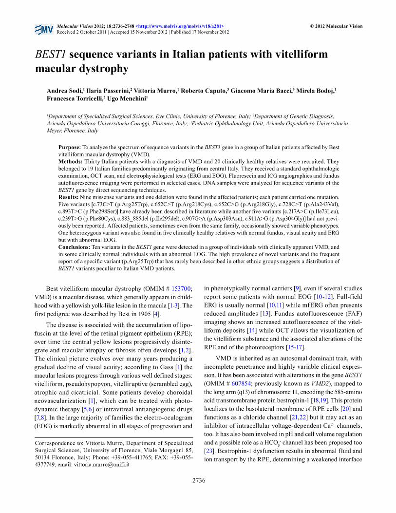
Figure 1. Different stages of VMD (Fundus photographs and OCT). A, B: Vitellifom disc (Patient R-II-1). C, D: Vitelliruptive stage (Patient C-II-1). E, F: Pseudohypopyon stage (Patient B-II-1). G, H: Macular atrophy (Patient Q-I:-1). I, J: Macular fibrosis (Patient I-II-1). OD represents the right eye, OS represents the left eye.
Molecular Vision 2012; 18:2736-2749 <http://www.molvis.org/molvis/v18/a281> © 2012 Molecular Vision
2739
RESULTS
Thirty Italian patients with a diagnosis of VMD (from 19 independent pedigrees) were clinically examined: 19 were male and 11 female. The mean age was 42.5 years (±19.1 years; range 11–82 years). The Snellen visual acuity ranged from 0.1 to 1.0, with an average value of 0.62 (±0.31). In 18/30 patients (60%) best corrected visual acuity (BCVA) was different (2 lines or greater) between the two eyes. In the eyes with the better visual acuity, BCVA was equal to or better than 0.5 in 12/13 (92.3%) of the patients younger than 40 years of age, and in 13/17 (76.4%) of the patients older than 40 years of age. In the eyes with the worse visual acuity, BCVA was equal to or better than 0.5 in 7/13 (53.8%) of the
patients younger than 40 years of age and in 6/17 (35.2%) of the patients older than 40 years of age.
All the patients showed fundus lesions according to the typical stages as described by Gass (Figure 1) [1]: 11 eyes could be classified as vitelliform lesions, 8 eyes as pseu-dohypopion stage, 16 as vitelliruptive stage, 13 as macular atrophy, and 11 as fibrotic macular scar. Stage classification was the same for both eyes in 18 patients and asymmetric in 12 patients. One patient showed a vitelliform lesion in one eye while the macula of the fellow eye presented only mild RPE dystrophy. Clinical data of our series are summarized in Table 1 and Table 2.
EOG was always abnormal with Arden ratio lower than 1.50 while ERG was always within normal limits.
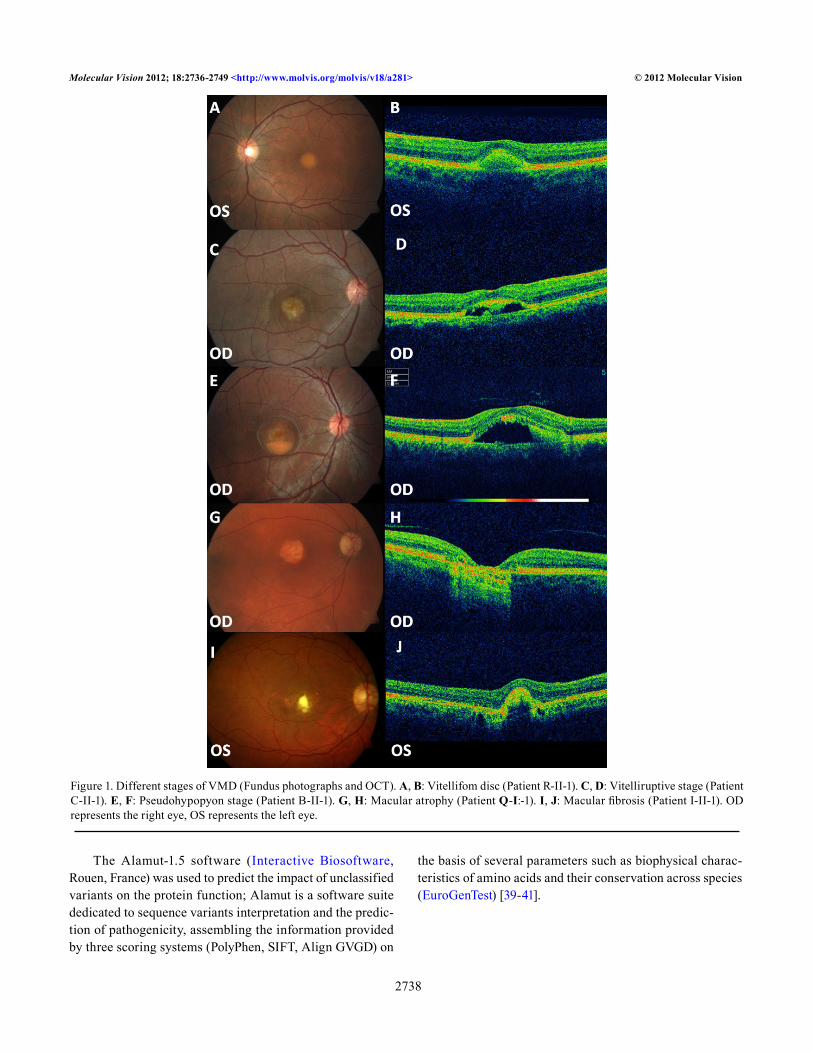
Table 1. CliniCal deTails of The VMd paTienTs inCluded in The sTudy (faMilies a-G).
Family Family member
Sex Age (years)
Onset (years)
BCVA OD
BCVA OS Fundus OD
Fundus OS
Notes
A III-3 F 11 6 0.8 1.0 VL PV UnilA II-5 M 45 // 1.0 1.0 Norm NormA II-4 F 42 // 1.0 1.0 Norm NormA III-4 M 9 // 1.0 1.0 Norm NormA II-3 F 42 // 0.6 1.0 Norm Norm Ambl ODA III-1 M 14 // 1.0 1.0 Norm NormA III-2 F 12 // 1.0 1.0 Norm NormA I-2 F 78 // 0.5 1.0 GC GC Glau OUB II-1 M 16 10 0.7 0.9 PH PHB I-1 M 44 39 0.9 1.0 VR VRB I-2 F 40 // 1.0 1.0 Norm NormC II-1 M 18 8 0.8 0.8 VR VR CNV; PC I-1 M 54 // 1.0 1.0 Norm NormC I-2 F 50 // 1.0 1.0 Norm NormD II-1 M 22 14 0.3 1.0 FI VL CNVD I-1 M 55 // 1.0 1.0 Norm NormD I-2 F 57 35 0.4 0.3 AT ATE I-1 M 62 25 0.2 0.2 AT ATE II-1 M 42 // 1.0 1.0 Norm NormF II-1 F 46 10 0.2 0.2 AT ATG I-2 M 60 29 0.3 0.1 FI FIG II-1 M 27 8 1.0 1.0 VL VLG II-2 F 25 7 1.0 1.0 VL VLG I-3 M 68 35 0.8 0.9 AT VL Mult OSG II-5 F 28 // 1.0 1.0 Norm Norm
For each patient the stage of disease is indicated. (BCVA: Best Corrected Visual Acuity; VL: vitelliform lesion; PV: pre-vitelliform lesion; GC: glaucomatous cupping; PH: pseudohypopyon stage; VR: vitelliruptive stage; AT: atrophic stage; FI: fibrotic stage; CNV: previous choroidal neovascularization; P: treated with photodynamic therapy; Unil: unilateral presentation of the disease; Mult: multifocal presen-tation of the disease; Ambl: Ambliopia; Glau: Glaucoma; OD: right eye; OS: left eye; //: unknown; OU: both eyes).
Molecular Vision 2012; 18:2736-2749 <http://www.molvis.org/molvis/v18/a281> © 2012 Molecular Vision
2740
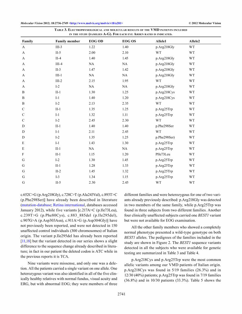
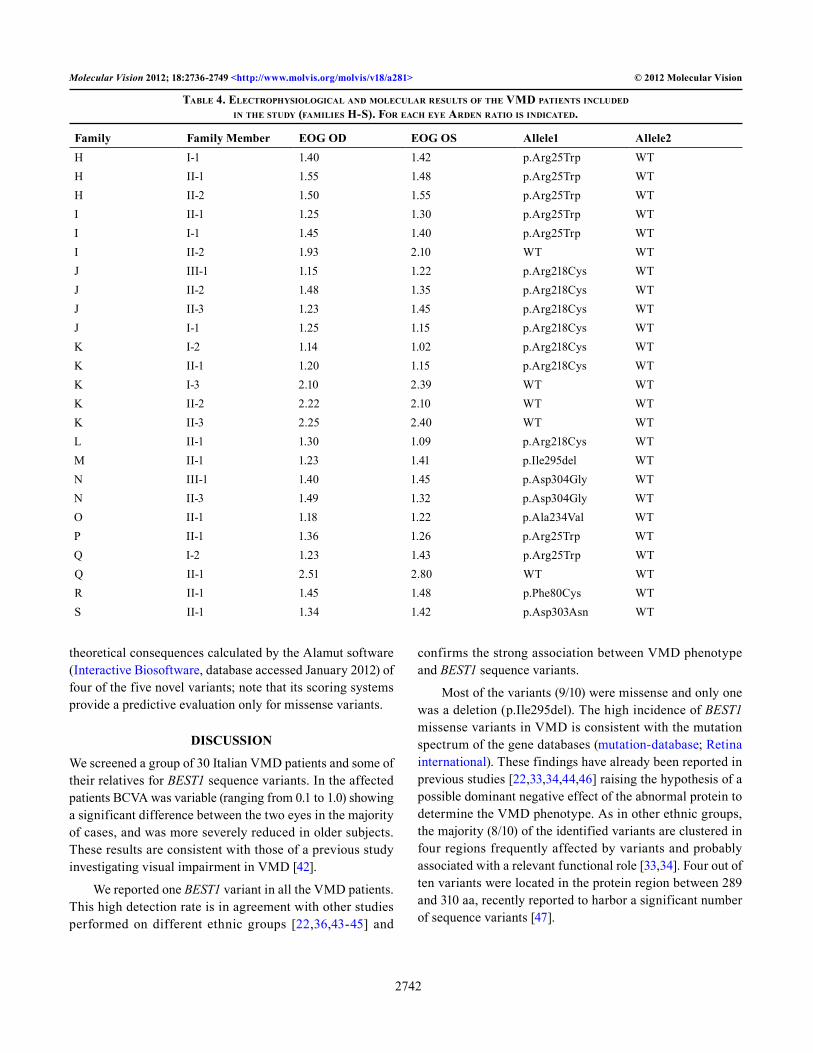
Electrophysiological results are summarized in Table 3 and Table 4.
OCT showed the accumulation of hyperreflective mate-rial in different stages of disorganization between the neuro-retina and the RPE, and allowed the visualization of fibrotic or atrophic macular alterations. None of the patients showed any sign of new vessels on fundus examination and OCT scans, or on FA and ICG when performed. In previous years five patients had been diagnosed with CNV; four patients showed sudden visual loss and macular hemorrhage while another patient presented a more intriguing clinical picture with sudden visual loss, very mild ophthalmoscopic changes and subtle FA and ICG leakage. One patient (who devel-oped CNV in 2001) was not treated, three patients received
photodynamic therapy (PDT), and one patient was treated with PDT and intravitreal bevacizumab; the three patients treated with PDT showed first a stabilization of the clinical picture and then a slow improvement of visual acuity.
All the 20 asymptomatic relatives showed a normal fundus appearance, normal visual acuity, and ERG response. Ten subjects showed a normal EOG response while five subjects showed an abnormal EOG with reduced Arden ratio. Five subjects were not available for electrophysiological testing.
Ten different BEST1 sequence variants were identified in the 30 VMD patients; each family was found to have a specific BEST1 variant that segregated with the disease. Five of these variants [c.73C>T (p.Arg25Trp), c.652C>T (p.Arg218Cys),
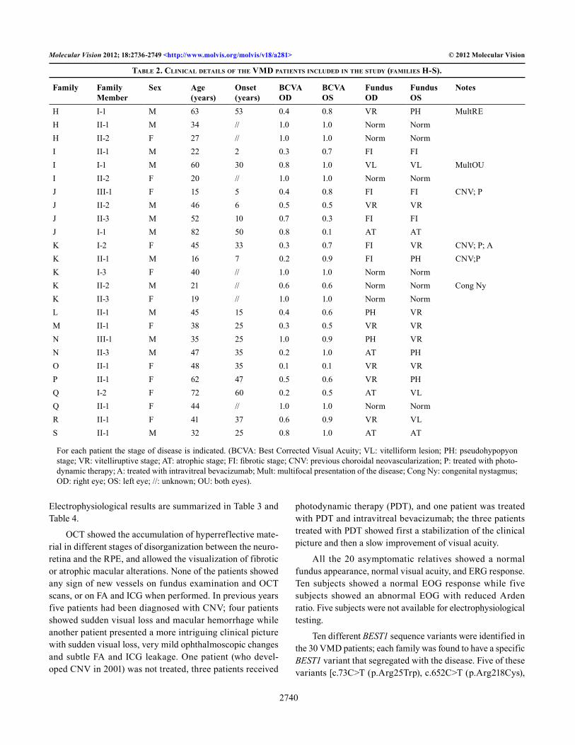
Table 2. CliniCal deTails of The VMd paTienTs inCluded in The sTudy (faMilies h-s).
Family Family Member
Sex Age (years)
Onset (years)
BCVA OD
BCVA OS
Fundus OD
Fundus OS
Notes
H I-1 M 63 53 0.4 0.8 VR PH MultREH II-1 M 34 // 1.0 1.0 Norm NormH II-2 F 27 // 1.0 1.0 Norm NormI II-1 M 22 2 0.3 0.7 FI FII I-1 M 60 30 0.8 1.0 VL VL MultOUI II-2 F 20 // 1.0 1.0 Norm NormJ III-1 F 15 5 0.4 0.8 FI FI CNV; PJ II-2 M 46 6 0.5 0.5 VR VRJ II-3 M 52 10 0.7 0.3 FI FIJ I-1 M 82 50 0.8 0.1 AT ATK I-2 F 45 33 0.3 0.7 FI VR CNV; P; AK II-1 M 16 7 0.2 0.9 FI PH CNV;PK I-3 F 40 // 1.0 1.0 Norm NormK II-2 M 21 // 0.6 0.6 Norm Norm Cong NyK II-3 F 19 // 1.0 1.0 Norm NormL II-1 M 45 15 0.4 0.6 PH VRM II-1 F 38 25 0.3 0.5 VR VRN III-1 M 35 25 1.0 0.9 PH VRN II-3 M 47 35 0.2 1.0 AT PHO II-1 F 48 35 0.1 0.1 VR VRP II-1 F 62 47 0.5 0.6 VR PHQ I-2 F 72 60 0.2 0.5 AT VLQ II-1 F 44 // 1.0 1.0 Norm NormR II-1 F 41 37 0.6 0.9 VR VLS II-1 M 32 25 0.8 1.0 AT AT
For each patient the stage of disease is indicated. (BCVA: Best Corrected Visual Acuity; VL: vitelliform lesion; PH: pseudohypopyon stage; VR: vitelliruptive stage; AT: atrophic stage; FI: fibrotic stage; CNV: previous choroidal neovascularization; P: treated with photo-dynamic therapy; A: treated with intravitreal bevacizumab; Mult: multifocal presentation of the disease; Cong Ny: congenital nystagmus; OD: right eye; OS: left eye; //: unknown; OU: both eyes).
Molecular Vision 2012; 18:2736-2749 <http://www.molvis.org/molvis/v18/a281> © 2012 Molecular Vision
2741
c.652C>G (p.Arg218Gly), c.728C>T (p.Ala243Val), c.893T>C (p.Phe298Ser)] have already been described in literature (mutation-database; Retina international, databases accessed January 2012), while five variants [c.217A>C (p.Ile73Leu), c.239T>G (p.Phe80Cys), c.883_885del (p.Ile295del), c.907G>A (p.Asp303Asn), c.911A>G (p.Asp304Gly)] have not previously been reported, and were not detected in 150 unaffected control individuals (300 chromosomes) of Italian origin. The variant p.Ile295del has already been reported [11,18] but the variant detected in our series shows a slight difference to the sequence change already described in litera-ture; in fact in our patient the deleted codon is ATC while in the previous reports it is TCA.
Nine variants were missense, and only one was a dele-tion. All the patients carried a single variant on one allele. One heterozygous variant was also identified in all of the five clin-ically healthy relatives with normal fundus, visual acuity and ERG, but with abnormal EOG; they were members of three
different families and were heterozygous for one of two vari-ants already previously described: p.Arg218Gly was detected in two members of the same family, while p.Arg25Trp was found in three subjects from two different families. Another four clinically unaffected subjects carried one BEST1 variant but were not available for EOG examination.
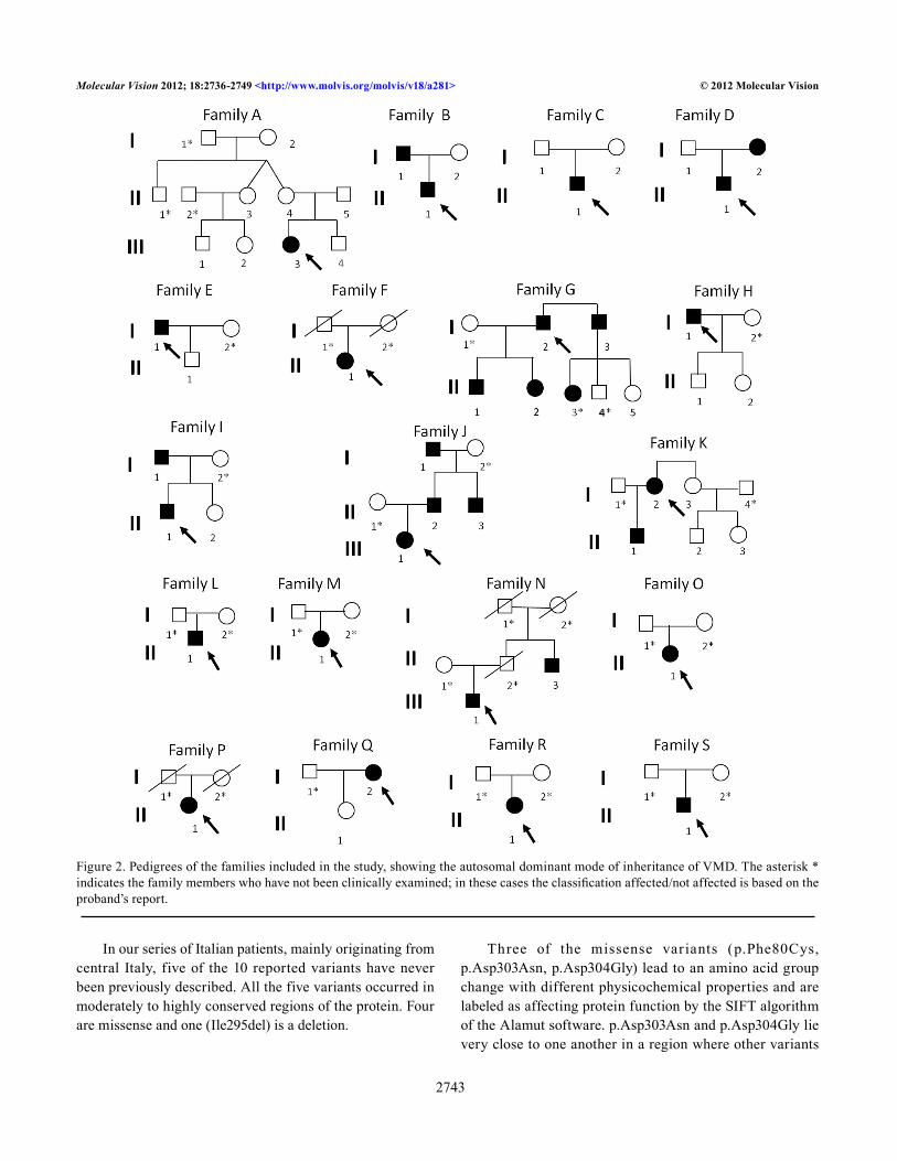
All the other family members who showed a completely normal phenotype presented a wild-type genotype on both BEST1 alleles. The pedigrees of the families included in the study are shown in Figure 2. The BEST1 sequence variants detected in all the subjects who were available for genetic testing are summarized in Table 3 and Table 4.
p.Arg218Cys and p.Arg25Trp were the most common allelic variants among our VMD patients of Italian origin. p.Arg218Cys was found in 5/19 families (26.3%) and in 12/30 (40%) patients; p.Arg25Trp was found in 7/19 families (36.8%) and in 10/30 patients (33.3%). Table 5 shows the
Table 3. eleCTrophysioloGiCal and MoleCular resulTs of The VMd paTienTs inCluded in The sTudy (faMilies a-G). for eaCh eye arden raTio is indiCaTed.
Family Family member EOG OD EOG OS Allele1 Allele2A III-3 1.22 1.40 p.Arg218Gly WTA II-5 2.00 2.10 WT WTA II-4 1.40 1.45 p.Arg218Gly WTA III-4 NA NA p.Arg218Gly WTA II-3 1.47 1.42 p.Arg218Gly WTA III-1 NA NA p.Arg218Gly WTA III-2 2.15 1.95 WT WTA I-2 NA NA p.Arg218Gly WTB II-1 1.30 1.25 p.Arg218Cys WTB I-1 1.40 1.20 p.Arg218Cys WTB I-2 2.13 2.35 WT WTC II-1 1.35 1.25 p.Arg25Trp WTC I-1 1.32 1.11 p.Arg25Trp WTC I-2 2.45 2.30 WT WTD II-1 1.40 1.40 p.Phe298Ser WTD I-1 2.11 2.45 WT WTD I-2 1.35 1.25 p.Phe298Ser) WTE I-1 1.43 1.30 p.Arg25Trp WTE II-1 NA NA p.Arg25Trp WTF II-1 1.15 1.20 PIle73Leu WTG I-2 1.30 1.45 p.Arg25Trp WTG II-1 1.28 1.33 p.Arg25Trp WTG II-2 1.45 1.32 p.Arg25Trp WTG I-3 1.34 1.15 p.Arg25Trp WTG II-5 2.30 2.45 WT WT
Molecular Vision 2012; 18:2736-2749 <http://www.molvis.org/molvis/v18/a281> © 2012 Molecular Vision
2742
theoretical consequences calculated by the Alamut software (Interactive Biosoftware, database accessed January 2012) of four of the five novel variants; note that its scoring systems provide a predictive evaluation only for missense variants.
DISCUSSION
We screened a group of 30 Italian VMD patients and some of their relatives for BEST1 sequence variants. In the affected patients BCVA was variable (ranging from 0.1 to 1.0) showing a significant difference between the two eyes in the majority of cases, and was more severely reduced in older subjects. These results are consistent with those of a previous study investigating visual impairment in VMD [42].
We reported one BEST1 variant in all the VMD patients. This high detection rate is in agreement with other studies performed on different ethnic groups [22,36,43-45] and
confirms the strong association between VMD phenotype and BEST1 sequence variants.
Most of the variants (9/10) were missense and only one was a deletion (p.Ile295del). The high incidence of BEST1 missense variants in VMD is consistent with the mutation spectrum of the gene databases (mutation-database; Retina international). These findings have already been reported in previous studies [22,33,34,44,46] raising the hypothesis of a possible dominant negative effect of the abnormal protein to determine the VMD phenotype. As in other ethnic groups, the majority (8/10) of the identified variants are clustered in four regions frequently affected by variants and probably associated with a relevant functional role [33,34]. Four out of ten variants were located in the protein region between 289 and 310 aa, recently reported to harbor a significant number of sequence variants [47].
Table 4. eleCTrophysioloGiCal and MoleCular resulTs of The VMd paTienTs inCluded in The sTudy (faMilies h-s). for eaCh eye arden raTio is indiCaTed.
Family Family Member EOG OD EOG OS Allele1 Allele2H I-1 1.40 1.42 p.Arg25Trp WTH II-1 1.55 1.48 p.Arg25Trp WTH II-2 1.50 1.55 p.Arg25Trp WTI II-1 1.25 1.30 p.Arg25Trp WTI I-1 1.45 1.40 p.Arg25Trp WTI II-2 1.93 2.10 WT WTJ III-1 1.15 1.22 p.Arg218Cys WTJ II-2 1.48 1.35 p.Arg218Cys WTJ II-3 1.23 1.45 p.Arg218Cys WTJ I-1 1.25 1.15 p.Arg218Cys WTK I-2 1.14 1.02 p.Arg218Cys WTK II-1 1.20 1.15 p.Arg218Cys WTK I-3 2.10 2.39 WT WTK II-2 2.22 2.10 WT WTK II-3 2.25 2.40 WT WTL II-1 1.30 1.09 p.Arg218Cys WTM II-1 1.23 1.41 p.Ile295del WTN III-1 1.40 1.45 p.Asp304Gly WTN II-3 1.49 1.32 p.Asp304Gly WTO II-1 1.18 1.22 p.Ala234Val WTP II-1 1.36 1.26 p.Arg25Trp WTQ I-2 1.23 1.43 p.Arg25Trp WTQ II-1 2.51 2.80 WT WTR II-1 1.45 1.48 p.Phe80Cys WTS II-1 1.34 1.42 p.Asp303Asn WT
Molecular Vision 2012; 18:2736-2749 <http://www.molvis.org/molvis/v18/a281> © 2012 Molecular Vision
2743
In our series of Italian patients, mainly originating from central Italy, five of the 10 reported variants have never been previously described. All the five variants occurred in moderately to highly conserved regions of the protein. Four are missense and one (Ile295del) is a deletion.
Three of the missense variants (p.Phe80Cys, p.Asp303Asn, p.Asp304Gly) lead to an amino acid group change with different physicochemical properties and are labeled as affecting protein function by the SIFT algorithm of the Alamut software. p.Asp303Asn and p.Asp304Gly lie very close to one another in a region where other variants
Figure 2. Pedigrees of the families included in the study, showing the autosomal dominant mode of inheritance of VMD. The asterisk * indicates the family members who have not been clinically examined; in these cases the classification affected/not affected is based on the proband’s report.
Molecular Vision 2012; 18:2736-2749 <http://www.molvis.org/molvis/v18/a281> © 2012 Molecular Vision
2744
have already been reported [18,35], suggesting a significant pathogenic impact for the alterations located in this region of the protein.
The physiopathological effect is more questionable for the other missense variant (p.Ile73Leu) because it is associ-ated with a change between amino acids that are chemically
Table 5. noVel BEST1 VarianT CharaCTerizaTion by Means of The alaMuT sofTware.
cNomen Exon pNomen Amino acid conservation
AGVGD class
SIFT (score) POLYPHEN (score)
c.217A>C 3 p.Ile73Leu Moderately C0 Tolerated (0.93) Benign (1.274)c.239T>G 3 p.Phe80Cys Highly C65 Affected protein func-
tion (0.00)Benign (2.771)
c.907G>A 8 p.Asp303Asn Highly C15 Affected protein func-tion (0.00)
Possibly damaging (1.960)
c.911A>G 8 p.Asp304Gly Highly C65 Affected protein func-tion (0.00)
Probably damaging (2.530)
Predictions of the scoring methods (Align GVGD, SIFT, Polyphen) are shown.
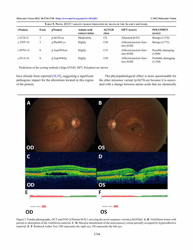
Figure 3. Fundus photographs, OCT and EOG of Patient M-II-1 carrying the novel sequence variant p.Ile295del. A, B: Vitelliform lesion with partial re-absorption of the vitelliform material. C, D: Macular detachment of the neurosensory retina partially occupied by hyperreflective material. E, F: Reduced Arden Test. OD represents the right eye, OS represents the left eye.
Molecular Vision 2012; 18:2736-2749 <http://www.molvis.org/molvis/v18/a281> © 2012 Molecular Vision
2745
similar, and it is interpreted as a benign or tolerated substitu-tion by the software; moreover, p.Ile73Leu is located outside the four regions frequently affected by mutations. However, its association with a typical VMD phenotype (Patient F-II-1) suggests a pathogenic role.
The p.Ile295del deletion found in our series is the dele-tion of codon ATC, different from codon TCA whose deletion has already been reported [11,18]. However both nucleotide variants lead to the same variation of the protein, with the
elimination of an isoleucine and an alteration of the protein sequence that is likely to impair its function. This variant does not lead to a premature stop codon in the downstream sequence of the protein, in agreement with the in vitro study showing a dominant negative effect of p.Ile295del on Cl– channel function [48]. In a previous paper [11] the variant p.Ile295del with the deletion of codon TCA was associated with reduced penetrance and normal EOG in the early stages of the disease; in our series the patient (patient M-II-1)
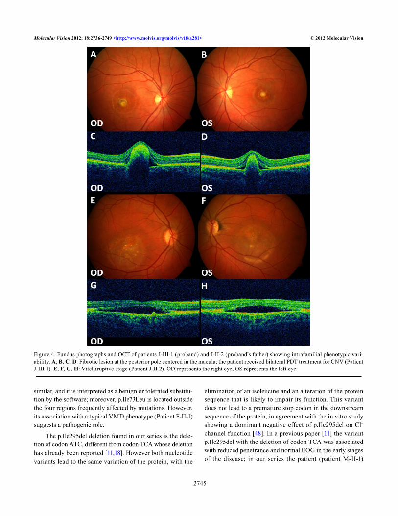
Figure 4. Fundus photographs and OCT of patients J-III-1 (proband) and J-II-2 (proband’s father) showing intrafamilial phenotypic vari-ability. A, B, C, D: Fibrotic lesion at the posterior pole centered in the macula; the patient received bilateral PDT treatment for CNV (Patient J-III-1). E, F, G, H: Vitelliruptive stage (Patient J-II-2). OD represents the right eye, OS represents the left eye.
Molecular Vision 2012; 18:2736-2749 <http://www.molvis.org/molvis/v18/a281> © 2012 Molecular Vision
2746
carrying the variant p.Ile295del with deletion of codon ATC showed a relatively severe phenotype with reduced visual acuity, abnormal EOG, and bilateral macular lesions in a vitelliruptive stage (Figure 3).
Two variants (p.Arg218Cys and p.Arg25Trp) are highly prevalent among our VMD patients and clinically normal individuals with an abnormal EOG. In our study p.Arg218Cys has been identified in 4/19 families (21%) and in 9/30 patients (30%); Arginine in 218 position is a well known mutational hotspot, suggesting that this site may also have a particular structural relevance for bestrophin function [10,33,43,44,46,49,50]. p.Arg25Trp has been found in 6/19 families (31%) and in 11/30 patients (36%); it has already been reported [6,34,36,47,51], but its high frequency in our group suggests that it may be a recurrent variant in the Italian population.
Recently 10 Italian VMD patients from 3 families were screened for BEST1 mutations [46]; among the three detected variants two were not found in our series while the third (p.Ala243Val) was identified in one of our patients, too. The variant p.Ala243Val has been reported in association with a mild VMD phenotype and a normal or near-normal EOG [25,36,52]; on the contrary, our patient carrying the same variant (patient O-II-1) showed a severe clinical picture with BCVA reduced to 0.1 in both eyes, abnormal EOG and bilateral neuroretinal detachment at the posterior pole, even though her symptoms appeared relatively late (35 years of age).
Affected members, sometimes even within the same family, occasionally showed variable phenotypes (different ages of onset, different visual loss severity, and fundus appearance). Figure 4 shows the different phenotypes of two members of the same family.
Five patients, carrying three different allelic variants: p.Arg25Trp, p.Arg218Cys, p.Phe298Ser, developed CNV during the course of the disease.
Three patients from three independent pedigrees, all carrying the same variant, p.Arg25Trp, showed multifocal vitelliform lesions in at least one eye; however, p.Arg25Trp is one of the most common variants in our series, and other affected family members do not show a multifocal clinical picture so we cannot speculate a genotype-phenotype correlation.
Variable expressivity of BEST1 mutations has already been reported [22,36], and may be due to the influence of environmental factors or unknown modifier genes [22]; this suggests that in families affected by VMD molecular genetic results must be interpreted with caution especially
when providing genetic counseling. We found one of the two variants p.Arg25Trp and p.Arg218Gly in all of the five asymptomatic carriers with EOG abnormalities without overt clinical alterations; however, our series is too small to estab-lish a different penetrance of the various genotypes.
In conclusion, we identified five novel and five previ-ously reported BEST1 sequence variants in a series of 30 VMD patients from 19 independent Italian families; to our knowledge this is the largest study in the literature inves-tigating BEST1 variants in an Italian population. The high number of novel variants and the high prevalence in our patients of a variant that is uncommon in other groups of different origin (p.Arg25Trp) suggest a difference in the spectrum of BEST1 sequence variants between Italian VMD patients and other ethnic groups. A better knowledge of the spectrum of BEST1 sequence variants in specific populations may help to improve molecular diagnostic approaches and select patients for future therapeutic options.
ACKNOWLEDGMENTS
The authors report no proprietary interests. Supported by a grant from Cassa di Risparmio di Firenze.
REFERENCES1. Gass JDM. Best’s Disease. in: Stereoscopic Atlas of Macular
Disease. Diagnosis and Treatment. Mosby. St.Louis-London-Philadelphia-Sydney-Toronto, 1997, pp.304–313.
2. Deutman AF, Hoyng CB. Macular Dystrophies. in: Retina. Ryan SJ Ed. Mosby. St.Louis-London-Philadelphia-Sydney-Toronto, 2001, pp. 1210–1257.
3. Souied E, Kaplan J, Coscas G, Soubrane G. Les dystrophies maculaires. J Fr Ophtalmol 2003; 26:743-62. [PMID: 13130265].
4. Best F. Uber eine hereditare Maculaaffektion. Beitrag zur Vererbungslehre. Z Augenheilk 1905; 13:199-212. .
5. Andrade RE, Farah ME, Costa RA. Photodynamic therapy with verteporfin for subfoveal choroidal neovasculariza-tion in Best disease. Am J Ophthalmol 2003; 136:1179-81. [PMID: 14644242].
6. Sodi A, Passerini I, Simonelli F, Testa F, Menchini U, Torricelli F. A novel mutation in the VMD2 gene in an Italian family with Best maculopathy. J Fr Ophtalmol 2007; 30:616-20. [PMID: 17646752].
7. Leu J, Schrage NF, Degenring RF. Choroidal neovascu-larisation secondary to Best’s disease in a 13-year-old boy treated by intravitreal bevacizumab. Graefes Arch Clin Exp Ophthalmol 2007; 245:1723-5. [PMID: 17605026].
8. Querques G, Bocco MC, Soubrane G, Souied EH. Intravitreal ranibizumab (Lucentis) for choroidal neovascularization
Molecular Vision 2012; 18:2736-2749 <http://www.molvis.org/molvis/v18/a281> © 2012 Molecular Vision
2747
associated with vitelliform macular dystrophy. Acta Ophthalmol (Copenh) 2008; 86:694-5. [PMID: 18752521].
9. Deutman AF. Electro-oculography in families with vitelliform dystrophy of the fovea. Detection of the carrier state. Arch Ophthalmol 1969; 81:305-16. [PMID: 5774285].
10. Renner AB, Tillack H, Kraus H, Krämer F, Mohr N, Weber BH, Foerster MH, Kellner U. Late onset is common in best macular dystrophy associated with VMD2 gene mutations. Ophthalmology 2005; 112:586-92. [PMID: 15808248].
11. Wabbels B, Preising MN, Kretschmann U, Demmler A, Lorenz B. Genotype-phenotype correlation and longitudinal course in ten families with Best vitelliform macular dystrophy. Graefes Arch Clin Exp Ophthalmol 2006; 244:1453-66. [PMID: 16612637].
12. Testa F, Rossi S, Passerini I, Sodi A, Di Iorio V, Interlandi E, Della Corte M, Menchini U, Rinaldi E, Torricelli F, Simonelli F. A normal electro-oculography in a family affected by Best disease with a novel spontaneous mutation of the BEST1 gene. Br J Ophthalmol 2008; 92:1467-70. [PMID: 18703557].
13. Palmowski AM, Allgayer R, Heinemann-Vernaleken B, Scherer V, Ruprecht KW. Detection of retinal dysfunction in vitelliform macular dystrophy using the multifocal ERG (MF-ERG). Doc Ophthalmol 2003; 106:145-52. [PMID: 12678279].
14. Boon CJF, Klevering BJ, Keunen JEE, Hoyng CB, Theelen T. Fundus autofluorescence imaging of retinal dystrophies. Vision Res 2008; 48:2569-77. [PMID: 18289629].
15. Pianta MJ, Aleman TS, Cideciyan AV, Sunness JS, Li Y, Campochiaro BA, Campochiaro PA, Zack DJ, Stone EM, Jacobson SG. In vivo micropathology of Best macular dystrophy with optical coherence tomography. Exp Eye Res 2003; 76:203-11. [PMID: 12565808].
16. Ferrara DC, Costa RA, Tsang S, Calucci D, Jorge R, Freund KB. Multimodal fundus imaging in Best vitelliform macular dystrophy. Graefes Arch Clin Exp Ophthalmol 2010; 248:1377-86. [PMID: 20414784].
17. Schatz P, Bitner H, Sander B, Holfort S, Andreasson S, Larsen M, Sharon D. Evaluation of macular structure and function by OCT and electrophysiology in patients with vitelliform macular dystrophy due to mutations in BEST1. Invest Ophthalmol Vis Sci 2010; 51:4754-65. [PMID: 20375334].
18. Marquardt A, Stohr A, Passmore LA, Kramer F, Rivera A, Weber BHF. Mutations in a novel gene, VMD2, encoding a protein of unknown properties cause juvenile-onset vitel-liform macular dystrophy (Best’s Disease). Hum Mol Genet 1998; 7:1517-25. [PMID: 9700209].
19. Petrukhin K, Koisti MJ, Bakall B, Li W, Xie G, Marknell T, Sandgren O, Forsman K, Holmgren G, Andreasson S, Vujic M, Bergen AA, McGarty-Dugan V, Figueroa D, Austin CP, Metzker ML, Caskey CT, Wadelius C. Identification of the gene responsible for Best macular dystrophy. Nat Genet 1998; 19:241-7. [PMID: 9662395].
20. Marmorstein AD, Marmorstein LY, Rayborn M, Wang X, Hollyfield J, Petrukhin K. Bestrophin, the product of the
Best vitelliform macular dystrophy gene (VMD2), localizes to the basolateral plasma membrane of the retinal pigment epithelium. Proc Natl Acad Sci USA 2000; 97:12758-63. [PMID: 11050159].
21. Sun H, Tsunenari T, Yau KW, Nathans J. The vitelliform macular dystrophy protein defines a new family of chloride channels. Proc Natl Acad Sci USA 2002; 99:4008-13. [PMID: 11904445].
22. Boon CJF, Klevering BJ, Leroy BP, Hoyng CB, Keunen JEE, den Hollander AI. The spectrum of ocular phenotypes caused by mutations in the BEST1 gene. Prog Retin Eye Res 2009; 28:187-205. [PMID: 19375515].
23. Xiao Q, Hartzell HC, Yu K. Bestrophins and Retinopathies. Pflugers Arch 2010; 460:559-69. [PMID: 20349192].
24. Hartzell HC, Qu Z, Yu K, Xiao Q, Chien LT. Molecular physi-ology of bestrophins: multifunctional membrane proteins linked to Best disease and other retinopathies. Physiol Rev 2008; 88:639-72. [PMID: 18391176].
25. Krämer F, White K, Pauleikhoff D, Gehrig A, Passmore L, Rivera A, Rudolph G, Kellner U, Andrassi M, Lorenz B, Rohrschneider K, Blankenagel A, Jurklies B, Schilling H, Schütt F, Holz FG, Weber BH. Mutations in the VMD2 gene are associated with juvenile-onset vitelliform macular dystrophy (Best disease) and adult vitelliform macular dystrophy but not age-related macular degeneration. Eur J Hum Genet 2000; 8:286-92. [PMID: 10854112].
26. Gerth C, Zawadzki RJ, Werner JS, Héon E. Detailed analysis of retinal function and morphology in a patient with auto-somal recessive bestrophinopathy (ARB). Doc Ophthalmol 2009; 118:239-46. [PMID: 18985398].
27. Burgess R, Millar ID, Leroy BP, Urquhart JE, Fearon IM, De Baere E, Brown PD, Robson AG, Wright GA, Kestelyn P, Holder GE, Webster AR, Manson FD, Black GC. Biallelic mutation of BEST1 causes a distinct retinopathy in humans. Am J Hum Genet 2008; 82:19-31. [PMID: 18179881].
28. Sodi A, Menchini F, Manitto MP, Passerini I, Murro V, Torri-celli F, Menchini U. Ocular phenotypes associated with bial-lelic mutations in BEST1 in Italian patients. Mol Vis 2011; 17:3078-87. [PMID: 22162627].
29. Yardley J, Leroy BP, Hart-Holden N, Lafaut BA, Loeys B, Messiaen LM, Perveen R, Reddy MA, Bhattacharya SS, Traboulsi E, Baralle D, De Laey JJ, Puech B, Kestelyn P, Moore AT, Manson FD, Black GC. Mutations of VMD2 splicing regulators cause nanophthalmos and autosomal dominant vitreo-retinochoroidopathy (ADVIRC). Invest Ophthalmol Vis Sci 2004; 45:3683-9. [PMID: 15452077].
30. Burgess R, MacLaren RE, Davidson AE, Urquhart JE, Holder GE, Robson AG, Moore AT, Keefe RO, Black GC, Manson FD. ADVIRC is caused by distinct mutations in BEST1 that alter pre-mRNA splicing. J Med Genet 2009; 46:620-5. [PMID: 18611979].
31. Davidson AE, Millar ID, Urquhart JE, Burgess-Mullan R, Shweikh Y, Parry N, O’Sullivan J, Maher GJ, McKibbin M, Downes SM, Lotery AJ, Jacobson SG, Brown PD, Black GC, Manson FD. Missense mutations in a retinal pigment
Molecular Vision 2012; 18:2736-2749 <http://www.molvis.org/molvis/v18/a281> © 2012 Molecular Vision
2748
epithelium protein, bestrophin-1, cause retinitis pigmentosa. Am J Hum Genet 2009; 85:581-92. [PMID: 19853238].
32. Michaelides M, Urquhart J, Holder GE, Restori M, Kayali N, Manson FD, Black GC. Evidence of genetic heterogeneity in MRCS (microcornea, rod-cone dystrophy, cataract, and posterior staphyloma) syndrome. Am J Ophthalmol 2006; 141:418-20. [PMID: 16458719].
33. Bakall B, Marknell T, Ingvast S, Koisti MJ, Sandgren O, Li W, Bergen AA, Andreasson S, Rosenberg T, Petrukhin K, Wadelius C. The mutation spectrum of the bestrophin protein functional implications. Hum Genet 1999; 104:383-9. [PMID: 10394929].
34. White K, Marquardt A, Weber BHF. VMD2 Mutations in Vitel-liform Macular Dystrophy (Best Disease) and Other Macu-lopathies. Hum Mutat 2000; 15:301-8. [PMID: 10737974].
35. Palomba G, Rozzo C, Angius A, Pierrottet CO, Orzalesi N, Pirastu M. A novel spontaneous missense mutation in VMD2 gene is a cause of Best macular dystrophy sporadic case. Am J Ophthalmol 2000; 129:260-2. [PMID: 10682987].
36. Querques G, Zerbib J, Santacroce R, Margaglione M, Delphin N, Rozet JM, Kaplan J, Martinelli D, Delle Noci N, Soubrane G, Souied EH. Functional and clinical data of Best vitelliform macular dystrophy patients with mutations in the BEST1 gene. Mol Vis 2009; 15:2960-72. [PMID: 20057903].
37. Marmor MF, Brigell MG, McCulloch DL, Westall CA, Bach M. (for the International Society for Clinical Electrophysi-ology of Vision). ISCEV standard for clinical electro-oculog-raphy (2010 update). Doc Ophthalmol 2011; 122:1-7. [PMID: 21298321].
38. Marmor MF, Fulton AB, Holder GE, Miyake Y, Brigell M, Bach M. International Society for Clinical Electrophysiology of Vision. ISCEV Standard for full-field clinical electroreti-nography (2008 update). Doc Ophthalmol 2009; 118:69-77. [PMID: 19030905].
39. Sunyaev S, Ramensky V, Koch I, Warren L III, Kondrashov AS, Boork P. Prediction of deleterious human alleles. Hum Mol Genet 2001; 10:591-7. [PMID: 11230178].
40. Kumar P, Henikoff S, Ng PC. Predicting the effects of coding non-synonymous variants on protein function using the SIFT algorithm. Nat Protoc 2009; 4:1073-81. [PMID: 19561590].
41. Mathe E, Olivier M, Kato S, Ishioka C, Hainaut P, Tavtigian SV. Computational approaches for predicting the biological effect of p53 missense mutations: a comparison of three sequence analysis based methods. Nucleic Acids Res 2006; 34:1317-25. [PMID: 16522644].
42. Fishman GA, Baca W, Alexander KR, Derlacki DJ, Glenn AM, Viana M. Visual acuity in patients with best vitelliform macular dystrophy. Ophthalmology 1993; 100:1665-70. [PMID: 8233392].
43. Marchant D, Yu K, Bigot K, Roche O, Germain A, Bonneau D, Drouin-Garraud V, Schorderet DF, Munier F, Schmidt D, Le Neindre P, Marsac C, Menasche M, Dufier JL, Fischmeister R, Hartzell C, Abitbol M. New VMD2 gene mutations identi-fied in patients affected by vitelliform macular dystrophy. J Med Genet 2007; 44:e70-[PMID: 17287362].
44. Wong RL, Hou P, Choy KW, Chiang SW, Tam PO, Li H, Chan WM, Lam DS, Pang CP, Lai TY. Novel and homozygous BEST1 mutations in Chinese patients with Best vitelli-form macular dystrophy. Retina 2010; 30:820-7. [PMID: 20057343].
45. Meunier I, Sénéchal A, Dhaenens CM, Arndt C, Puech B, Defoort-Dhelemmes S, Manes G, Chazalette D, Mazoir E, Bocquet B, Hamel CP. Systematic screening of BEST1 and PRPH2 in juvenile and adult vitelliform macular dystrophies: a rationale for molecular analysis. Ophthalmology 2011; 118:1130-6. [PMID: 21269699].
46. Krämer F, Mohr N, Kellner U, Rudolph G, Weber BH. Ten novel mutations in VMD2 associated with Best macular dystrophy (BMD). Hum Mutat 2003; 22:418-[PMID: 14517959].
47. Kinnick TR, Mullins RF, Dev S, Leys M, Mackey DA, Kay CN, Lam BL, Fishman GA, Traboulsi E, Iezzi R, Stone EM. Autosomal Recessive Vitelliform Macular dystrophy in a large cohort of vitelliform macular dystrophy patients. Retina 2011; 31:581-95. [PMID: 21273940].
48. Yu K, Qu Z, Cui Y, Hartzell HC. Chloride channel activity of bestrophin mutants associated with mild or late onset macular degeneration. Invest Ophthalmol Vis Sci 2007; 48:4694-705. [PMID: 17898294].
49. Marchant D, Gogat K, Boutboul S, Péquignot M, Sternberg C, Dureau P, Roche O, Uteza Y, Hache JC, Puech B, Puech V, Dumur V, Mouillon M, Munier FL, Schorderet DF, Marsac C, Dufier JL, Abitbol M. Identification of novel VMD2 gene mutations in patients with Best vitelliform macular dystrophy. Hum Mutat 2001; 17:235-[PMID: 11241846].
50. Atchaneeyasakul LO, Jinda W, Sakolsatayadorn N, Trina-varat A, Ruangovoravate N, Thanasombatskui N, Thong-noppakhun W, Limwongse C. Mutation analysis of the VMD2 gene in thai families with Best macular dystrophy. Ophthalmic Genet 2008; 29:139-44. [PMID: 18766995].
51. Lotery AJ, Munier FL, Fishman GA, Weleber RG, Jacobson SG, Affatigato LM, Nichols BE, Schorderet DF, Sheffield VC, Stone EM. Allelic variation in the VMD2 gene in best disease and age-related macular degeneration. Invest Ophthalmol Vis Sci 2000; 41:1291-6. [PMID: 10798642].
52. Boon CJ, Theelen T, Hoefsloot EH, van Schooneveld MJ, Keunen JE, Cremers FP, Klevering BJ, Hoyng CB. Clinical and Molecular Genetic Analysis of Best Vitelliform Macular Dystrophy. Retina 2009; 29:835-47. [PMID: 19357557].
Articles are provided courtesy of Emory University and the Zhongshan Ophthalmic Center, Sun Yat-sen University, P.R. China. The print version of this article was created on 17 November 2012. This reflects all typographical corrections and errata to the article through that date. Details of any changes may be found in the online version of the article.