behavioral paradigm development for fmri and eeg jason zevin sackler institute
TRANSCRIPT
Behavioral paradigm development for fMRI and EEG
Jason ZevinSackler Institute

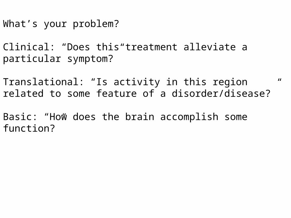
What’s your problem?
What’s your problem?
Clinical: “Does this treatment alleviate a particular symptom?”
Translational: “Is activity in this region related to some feature of a disorder/disease?”
Basic: “How does the brain accomplish some function?”
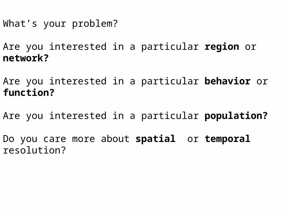
What’s your problem?
Are you interested in a particular region or network?
Are you interested in a particular behavior or function?
Are you interested in a particular population?
Do you care more about spatial or temporal resolution?
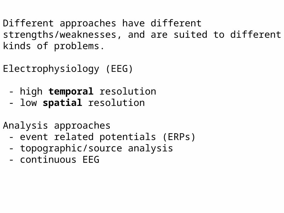
Different approaches have different strengths/weaknesses, and are suited to different kinds of problems.
Electrophysiology (EEG)
- high temporal resolution - low spatial resolution
Analysis approaches - event related potentials (ERPs) - topographic/source analysis - continuous EEG
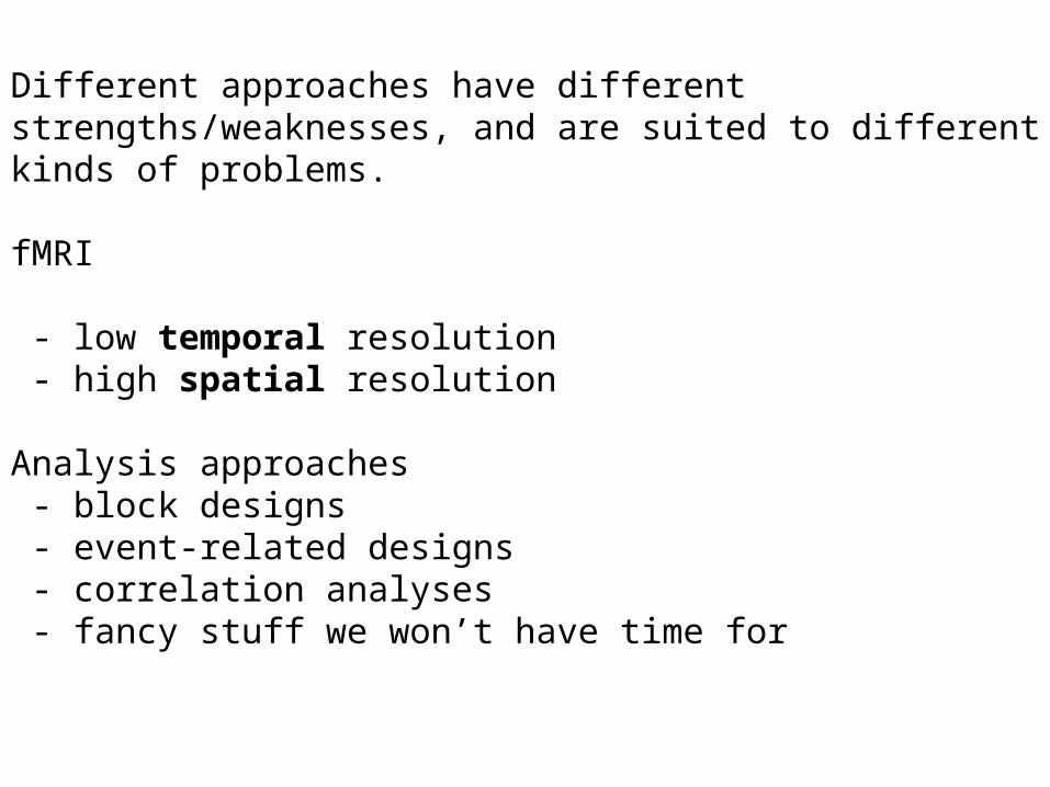
Different approaches have different strengths/weaknesses, and are suited to different kinds of problems.
fMRI
- low temporal resolution - high spatial resolution
Analysis approaches - block designs - event-related designs - correlation analyses - fancy stuff we won’t have time for

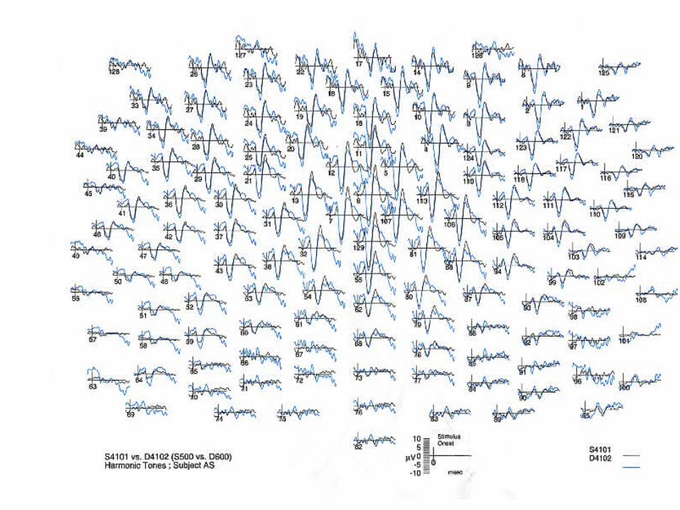
EEG
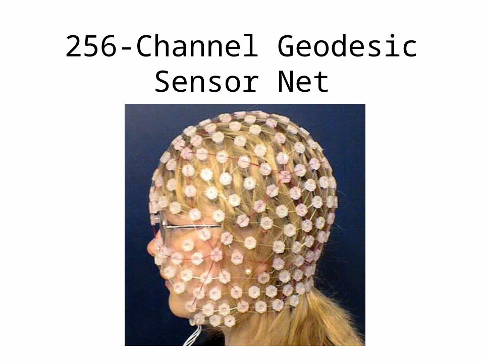
256-Channel Geodesic Sensor Net
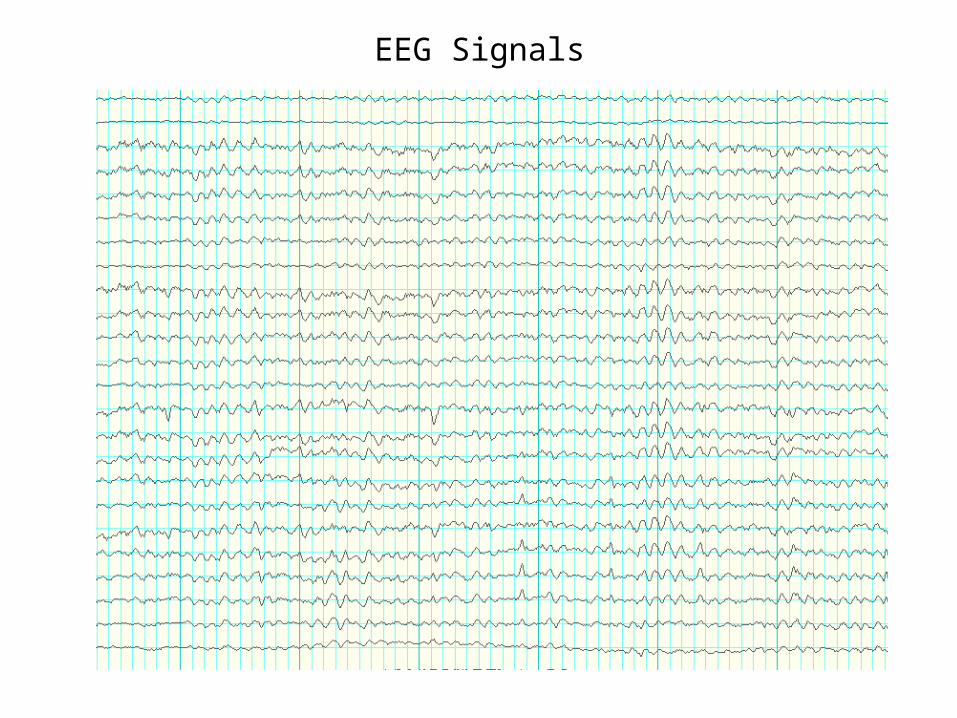
EEG Signals
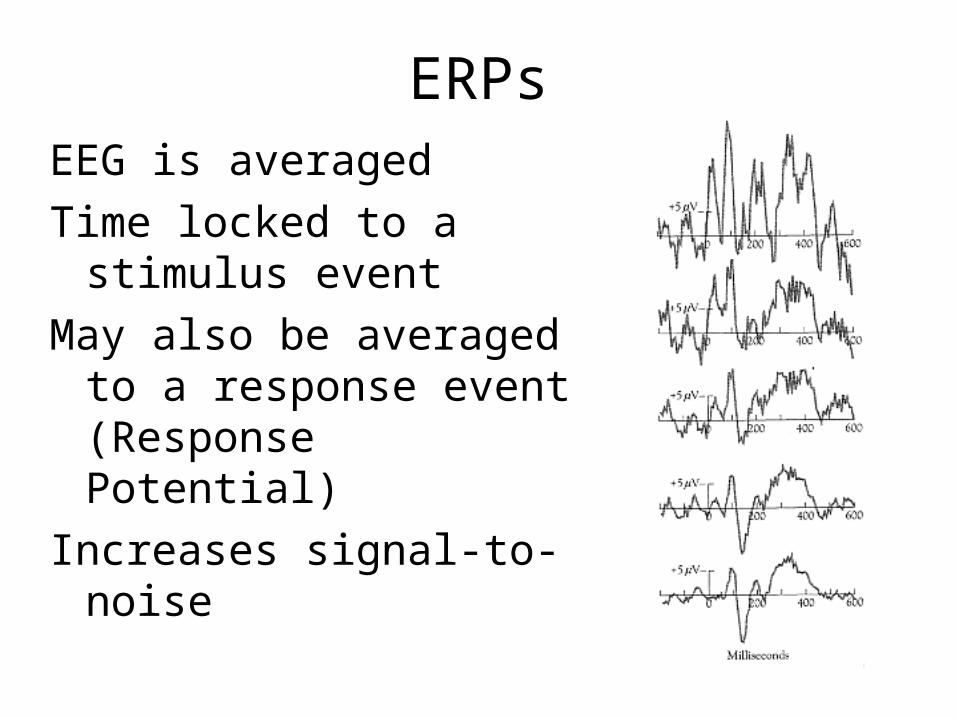
ERPsEEG is averagedTime locked to a
stimulus eventMay also be averaged
to a response event (Response Potential)
Increases signal-to-noise
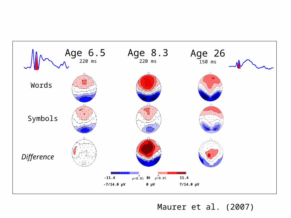
Development Print Processingin the first 200 milliseconds
Difference
Words
Symbols
Age 8.3220 ms
Age 6.5 220 ms
-7/14.0 µV 7/14.0 µV0 µV
-11.4 11.4 0t p<0.01 p<0.01
<<
Age 26150 ms
Maurer et al. (2007)
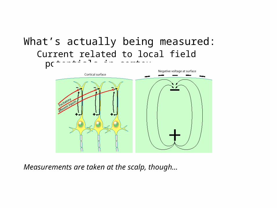
What’s actually being measured:Current related to local field potentials in
cortex
Measurements are taken at the scalp, though…
Graphics from http://www.mrc-cbu.cam.ac.uk/EEG/img/Physiological_basis_EEG.gifhttp://ww2.heartandstroke.ca/images/english/english_brain.jpg
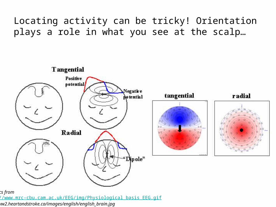
Locating activity can be tricky! Orientation plays a role in what you see at the scalp…
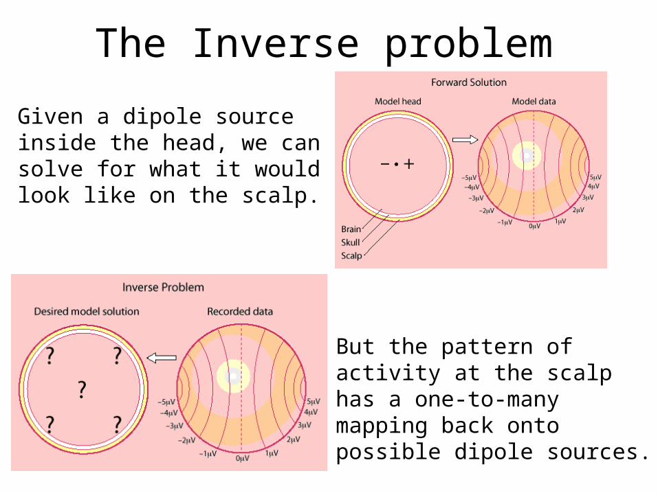
The Inverse problem
Given a dipole source inside the head, we can solve for what it would look like on the scalp.
But the pattern of activity at the scalp has a one-to-many mapping back onto possible dipole sources.

So, what’s the upside?
A more “direct” measure of neural activity. Lets you look at neat stuff like oscillation
frequency:
Alpha - strong in relaxed, awake states.
Theta - may be largely driven by hippocampus, prominent in short term memory tasks
Figures from Wikipedia entry on EEG, of all places
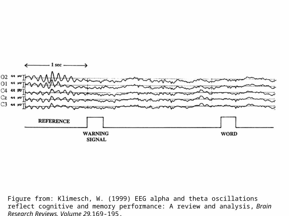
Figure from: Klimesch, W. (1999) EEG alpha and theta oscillations reflect cognitive and memory performance: A review and analysis, Brain Research Reviews, Volume 29,169-195.
Temporal resolution allows fine-grained inferences about the timing of neural events.
Molholm, S., Ritter, W., Murray, M.M., Javitt, D.C., Schroeder,C.E., and Foxe, J.J. (2002) Multisensory auditory-visual interactions during early sensory processing in humans: a high-density electrical mapping study, Cognitive Brain Research, 14, 115-128.
QuickTime™ and a decompressor
are needed to see this picture.
Some considerations for designing EEG studies:
How important is spatial resolution?
QuickTime™ and a decompressor
are needed to see this picture.
Maybe the process you care about is related to a general “brain state,” e.g. a stage of sleep.

Some considerations for designing EEG studies:
Can you get enough data to do ERPs? (typically ~100 trials)
Interestingly, kids and infants, with their thin skulls and little heads, give better EEG data and need fewer trials (but they wiggle around more).
QuickTime™ and a decompressor
are needed to see this picture.
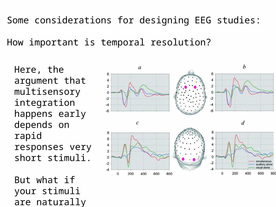
Some considerations for designing EEG studies:
How important is temporal resolution?
Here, the argument that multisensory integration happens early depends on rapid responses very short stimuli.
But what if your stimuli are naturally long, or vary in duration?
fMRI
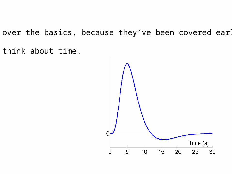
I’ll skip over the basics, because they’ve been covered earlier.
But let’s think about time.
QuickTime™ and aGIF decompressor
are needed to see this picture.
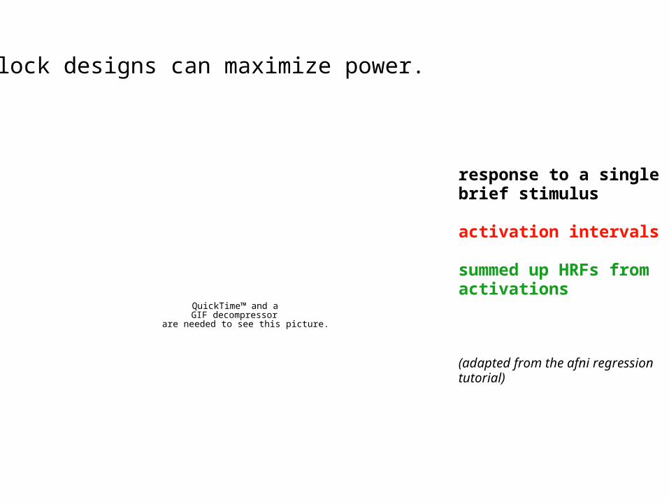
response to a single brief stimulus
activation intervals
summed up HRFs from activations
(adapted from the afni regression tutorial)
Block designs can maximize power.
Why would you ever sacrifice power?
QuickTime™ and a decompressor
are needed to see this picture.
(audience participation)
http://www.columbia.edu/cu/http://www.columbia.edu/cu/
psychology/tor/psychology/tor/
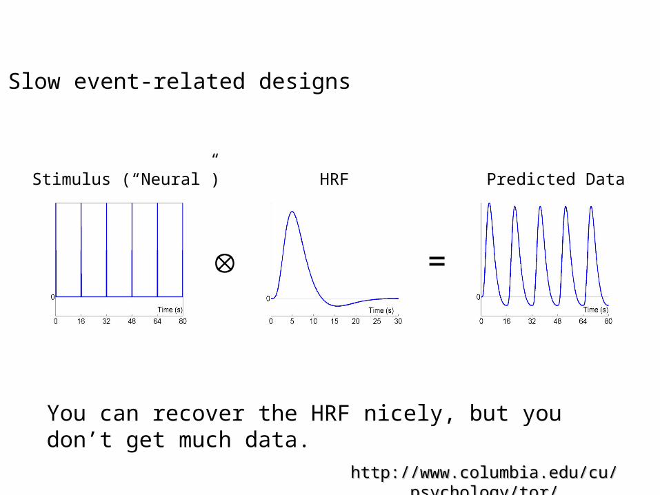
=
Stimulus (“Neural”) HRF Predicted Data
Slow event-related designs
You can recover the HRF nicely, but you don’t get much data.
http://www.columbia.edu/cu/http://www.columbia.edu/cu/
psychology/tor/psychology/tor/
=
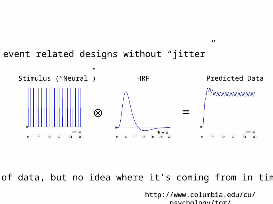
Lots of data, but no idea where it’s coming from in time.
Stimulus (“Neural”) HRF Predicted Data
Fast event related designs without “jitter”
http://www.columbia.edu/cu/http://www.columbia.edu/cu/
psychology/tor/psychology/tor/
=
Stimulus (“Neural”) HRF Predicted Data
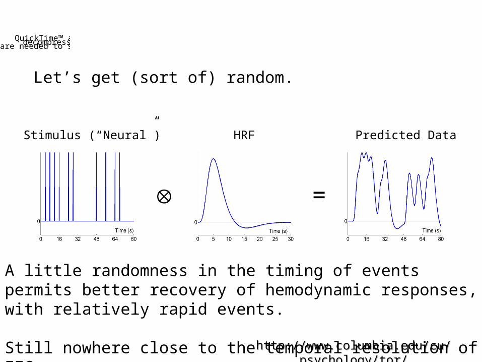
Let’s get (sort of) random.
A little randomness in the timing of events permits better recovery of hemodynamic responses, with relatively rapid events.
Still nowhere close to the temporal resolution of EEG.
QuickTime™ and a decompressorare needed to see this picture.
QuickTime™ and a decompressor
are needed to see this picture.
You don’t have to get super fancy, though, to see interesting things.
In this study, Singer et al. administered shocks to women and their partners.
Even a slow, event related design shows interesting overlap between getting shocked and watching your honey get shocked.
They also took measures of empathy using paper and pencil outside the scanner.
QuickTime™ and a decompressor
are needed to see this picture.
These correlations are probably bogus (we can talk about it if there’s time).
BUT, the principle of measuring some behavioral trait and relating it to brain responses is sound.
To do a study like this you probably want a very powerful design (or stimulus) in order to be sure you’re driving activity in the regions of interest.
QuickTime™ and a decompressor
are needed to see this picture.
But you might want to do something subtler with timing…
Here, the authors (including next week’s lecturer) wanted to measure the effect of context on responses to ambiguous faces.
They used temporal jitter to separate activity due to the context out from activity related to seeing the face.
Some considerations for designing fMRI studies:
Do you want “power” or “subtlety?”
- how much time do you have to collect data? (kids, patients sometimes kinda hate being in the scanner)
- are you trying to characterize the function of a region/network, or relate activity in some well-characterized region to a population variable?
Does dealing with the timing of stimulus presentation to get a nice HRF make your experiment awkward? Slow? What’s the impact on behavior? (This happens in EEG, too.)