bcbcr breast cancer conventional diagnosis and treatment modalities.pdf 6823
TRANSCRIPT

7/25/2019 BCBCR Breast Cancer Conventional Diagnosis and Treatment Modalities.pdf 6823
http://slidepdf.com/reader/full/bcbcr-breast-cancer-conventional-diagnosis-and-treatment-modalitiespdf-6823 1/18
17BREAST CANCER: BASIC AND CLINICAL RESEARCH 2015:9(S2)
Breast Cancer: Conventional Diagnosis and Treatment
Modalities and Recent Patents and Technologies
Supplementary Issue: Targeted Therapies in Breast Cancer Treatment
Mohamed I. Nounou1, Fatema ElAmrawy1, Nada Ahmed1, Kamilia Abdelraouf 1,Satyanarayana Goda2 and Hussaini Syed-Sha-Qhattal3
1Department of Pharmaceutics, Faculty of Pharmacy, Alexandria University, Alexandria, Egypt. 2Formurex, Inc., Stockton, CA, USA.3DPT Laboratories Ltd., San Antonio, TX, USA.
ABSTR ACT: Breast cancer is the most prevalent cancer among women worldwide. However, increased survival is due to the dramatic advances in the
screening methods, early diagnosis, and breakthroughs in treatments. Over the course of the last decade, many acquisitions have taken place in this critical
field of research in the pharmaceutical industry. Advances in molecular biology and pharmacology aided in better understanding of breast cancer, enabling
the design of smarter therapeutics able to target cancer and respond to its microenvironment efficiently. Patents and research papers investigating diagnosis
and treatment strategies for breast cancer using novel technologies have been surveyed for the past 15 years. Various nanocarriers have been introduced to
improve the therapeutic efficacy of anticancer drugs, including liposomes, polymeric micelles, quantum dots, nanoparticles, and dendrimers. Tis review
provides an overview of breast cancer, conventional therapy, novel technologies in the management of breast cancer, and rational approaches for targeting
breast cancer.
HIGHLIGHTS:1. Breast cancer is the most common cancer in women worldwide. However, surv ival rates vary widely, optimistical ly heading toward a positive trend.
Increased survival is due to the drastic shift in the screening methods, early diagnosis, and breakthroughs in treatments.
2. Different strategies of breast cancer classification and staging have evolved over the years. Intrinsic (molecular) subtyping is essential in clinical trials
and well understanding of the disease.
3. Many novel technologies are being developed to detect distant metastases and recurrent disease as well as to assess response to breast cancer
management.
4. Intensive research efforts are actively ongoing to take novel breast cancer therapeutics to potential clinical application.
5. Most of the recent research papers and patents discuss one of the following strategies: the development of new drug entities that specifical ly target
the breast tumor cells; tailor designing a novel car rier system that can multitask and multifunction as a d rug carrier, targeting vehicle and even as a
diagnostic tool, direct conjugation of a therapeutic drug moiety with a targeting moiety, diagnostic moiety or pharmacokinetics altering moiety; or
the use of innovative nontraditional approaches such as genetic engineering, stem cells, or vaccinations.
KEYWORDS: breast cancer, treatment, diagnosis, conventional modalities, novel technology, delivery systems, patents, recent studies, nanoparticles,
nanocarriers, bioconjugates, stimuli responsive particles
SUPPLEMENT: Targeted Therapies in Breast Cancer Treatment
CITATION: Nounou et al. Breast Cancer: Conventional Diagno sis and TreatmentModalities and Recent Patents and Technologies. Breast Cancer: Basic and Clinical
Research 2015:9(S2) 17–34 doi:10.4137/BCBCR.S29420.
TYPE: Review
RECEIVED: August 4, 2015. RESUBMITTED: September 8, 2015. ACCEPTED FOR
PUBLICATION: September 9, 2015.
ACADEMIC EDITOR: Goberdhan P. Dimri, Editor in Chief
PEER REVIEW: Four peer reviewers contributed to the pe er review report. Reviewers’
reports totaled 762 words, excluding any condential c omments to the academic editor.
FUNDING: Authors disclose no funding sources.
COMPETING INTERESTS: Authors disclose no potential conicts of interest.
COPYRIGHT: © the authors, publisher and licensee Libertas Academica Limited.
This is an open-access article distributed under the terms of the Creative Commons
CC-BY-NC 3.0 License.
CORRESPONDENCE: [email protected]
Paper subject to independent expert blind peer review. All editorial decisions made
by independent academic editor. Upon submission manuscript was subject to anti-
plagiarism scanning. Prior to publication all authors have given signed c onrmation of
agreement to article publication and compliance with all applicable ethical and legal
requirements, including the accuracy of author and contributor information, disclosure
of competing interests and funding sources, compliance with ethical requirements
relating to human and animal study participants, and compliance with any copyright
requirements of third parties. This journal is a member of the Committee on Publication
Ethics (COPE).
Published by Libertas Academica. Learn more about this journal.
Breast Cancer: Medical BackgroundBreast cancer history dates back to around 1,500 years
B.C. Ancient Egyptians were the first to report the disease
more than 3,500 years ago.1,2 Te condition was described
fairly accurately in both Edwin Smith3 and George Ebers4
papyri.1,2 In 460 B.C., Hippocrates, the father of Western
Medicine, described breast cancer as a humoral disease.2,5
Hippocrates was the first to define the terminology karkinos ,
a Greek word for crab/cancer. Tereafter, in 200 A.D.,
Galen, who made a detailed categorization of abnor-mal growths, wrote a treatise named “On tumors against
nature.”5 Galen believed that cancer may appear in any par t
of the body, but he had seen it more often occurring in the
breasts of women whose menstruation was either abnormal
or inconsistent.5
In this review, we will highlight the different types of
breast cancer. Te diagnosis techniques will be discussed along
with treatment strategies. Moreover, our main focus would be
exploring the recent trends and technologies in breast cancer
diagnosis and treatment as reported in recent research papers
and patents. All acronyms and abbreviations used in the man-
uscript are listed in Supplementary able 1.Prevalence of breast cancer among females. Breast
cancer is the most common cancer in women worldwide,

7/25/2019 BCBCR Breast Cancer Conventional Diagnosis and Treatment Modalities.pdf 6823
http://slidepdf.com/reader/full/bcbcr-breast-cancer-conventional-diagnosis-and-treatment-modalitiespdf-6823 2/18
Nounou et al
18 BREAST CANCER: BASIC AND CLINICAL RESEARCH 2015:9(S2)
with nearly 1.7 mil lion new cases diagnosed and 521,900
deaths in 2012 (second most common cancer overall).6 Tis
represents about 12% of all new cancer cases (14.1 million).6
Breast cancer alone accounts for 25% of all cancer cases and
15% of all cancer deaths among females.6 However, breast
cancer is not limited to females. Carcinoma of the malebreast accounts for 0.8%–1% of all breast cancers.7,8 Survival
rates vary widely, optimistically heading toward a positive
trend. Increased survival is due to the dramatic shift in the
screening methods, early diagnosis, and breakthroughs in
treatments.9
Breast cancer in men. Breast cancer is similar in men and
women; however, breast cancer in men is more frequently hor-
mone receptor positive and may be more sensitive to hormonal
therapy.8 Te risk appears to be higher with inherited BRCA2
rather than BRCA1 gene mutations.7 Men tend to be diag-
nosed at an older age than women and at a later disease stage.
umors of the male breast are more likely to express the estro-gen and progesterone receptors (PRs) and less likely to overex-
press Her-2/neu than breast cancers in women.7 Presentation is
usually a lump or nipple inversion, but is often diagnosed late,
with more than 40% of individuals diagnosed at stage III or IV
disease.7 Most tumors are ductal and 10% are ductal carcinoma
in situ. National initiatives are increasingly needed to provide
information and support for male breast cancer patients.7
Etiology and pathophysiology of breast cancer. A
meta-analysis of 52 separate epidemiological studies revealed
that 12% of women with breast cancer have one affected fam-
ily member and 1% of the patients have one or more relativesaffected.10 High-penetrance genes such as (BRCA1, BRCA2,
p53, PEN, AM, NBS1, or LKB1), low-penetrance genes
such as cytochrome P450 genes (CYP1A1, CYP2D6, CYP19),
glutathione S-transferase family (GSM1, GSP1), alcohol
and one-carbon metabolism genes (ADH1C and MHFR),
DNA repair genes (XRCC1, XRCC3, ERCC4/XPF), and
genes encoding cell signaling molecules (PR, estrogen recep-
tor (ER), NF-alpha, or heat shock protein 70 (HSP70)) are
factors contributing in the pathophysiology of breast can-
cer.10 Growth factor proteins such as HER-2/neu antigen is
overexpressed in different types of human cancers, including
breast, ovarian, lung, gastric, and oral cancers.11 In 1987, the
HER-2/neu proto-oncogene was revealed to be amplified and
overexpressed in 20%–30% of invasive breast cancers and also
shown to be associated with poorer outcome and shortened
survival.12
Classification of breast cancer. Early diagnosis and
intervention can make a transformational shift in the
statistics.13 Te intervention method varies according to the
stage, age, and the histological grade of the breast tumor.13–15
Te stage is determined by the invasion of malignancy whether
it is contained in the breast tissues or have leaked beyond the
basement membrane leading to metastasis.16
Breast cancer can be broadly categorized into in situ
carcinoma and invasive (infiltrating) carcinoma.17 Breast
carcinoma in situ is further subclassified as either ductal
(ductal carcinoma in situ [DCIS]) or lobular (lobular cancer
in situ [LCIS]).17 LCIS is believed to arise from atypical
lobular hyperplasia.17 DCIS lesions appear most often in the
mammary ducts.17 However, it is now understood that all pre-
invasive lesions originate from the DLUs.18
Still, the termslobular and ductal have persisted.17
Tere are two categories of DCIS: non-comedo and
comedo.19 Comedo-type DCIS (also referred to as Comedo-
carcinoma ) tends to be more aggressive than the non-comedo
types of DCIS.19 Te most common non-comedo types of
DCIS are19
a. Solid DCIS: cancer cells completely fill the affected
breast ducts.
b. Cribriform DCIS: cancer cells do not completely fill the
affected breast ducts, and there are gaps between the
cells.c. Papillary and micropapillary DCIS: the cancer cells
arrange themselves in a fern-like pattern within the
affected breast ducts, and micropapillary DCIS cells are
smaller than papillary DCIS cells.
Te major invasive tumor types include infiltrating/
invasive lobular (ILC) or ductal (invasive ductal carcinoma
[IDC]). ILC comprises up to 15% of all cases. 20 In the ILC
type, the cancer cells generally look quite similar to each
other.20 Te nuclei tend to be small and look alike from cell to
cell. Te growth of the tumor has several patterns20
:
a. Classic ILC: small cancer cells that invade the stroma
one-by-one in a single-file pattern.
b. Solid ILC: the cells grow in large sheets with little stroma
in between them.
c. Alveolar ILC: the cancer cells grow in groups of 20 or
more.
d. ubulolobular ILC: this subtype has some of the single-
file growth pattern of classic invasive lobular carcinoma,
but some of the cells also form small tubules.
However, some of the ILC cells can show either pleomor-
phic pattern, where the cancer cells are larger in classic ILC.
Te cells’ nuclei look different from each other (signet-ring
cell), where some tumor cells are filled with mucus that pushes
the nucleus to one side causing a signet ring appearance.20
Infiltrating IDC is, by far, the most common subtype
accounting for 70%–80% of all invasive lesions.21 Te IDC
is further subclassified into mucinous (colloid), tubular, med-
ullary, papillary, and cribriform carcinomas. However, ductal
carcinoma can be of no specific type (NOS).21 IDC is further
subclassified as either well-differentiated (grade 1), moderately
differentiated (grade 2), or poorly differentiated (grade 3)based on the levels of nuclear pleomorphism, glandular/tubule
formation, and mitotic index.21
7/25/2019 BCBCR Breast Cancer Conventional Diagnosis and Treatment Modalities.pdf 6823
http://slidepdf.com/reader/full/bcbcr-breast-cancer-conventional-diagnosis-and-treatment-modalitiespdf-6823 3/18
Novel technologies for breast cancer management
19BREAST CANCER: BASIC AND CLINICAL RESEARCH 2015:9(S2)
Molecular subtype. Breast cancer complexity has long been
known and investigated. After a first classification of the dis-
ease based on histology features and starting from the 1980s,
breast cancers have been distinguished on the basis of estrogen
receptor expression and later according to HER2. By 2000,
the microarray revolution had shown that the phenotypicdifferences between breast cancers were a reflection of their
mRNA expression profiles. Tis was confirmed using the
more recent genomic revolution.22 DNA microarrays revealed
the breast cancer molecular subtypes, which included
a Luminal A: ER positive, HER2 negative, Ki-67 protein
low, and PR high.23
b. Luminal B: ER positive, HER2 negative, and either
Ki-67 protein high or PR low.23
c. Basal-like breast cancer: typical ly lacks expression of
the molecular targets that confer responsiveness to
highly effective targeted therapies such as tamoxifenand aromatase inhibitors (AIs) or trastuzumab (HER2
amplification).24
d. riple-negative breast cancer (NBC): ER-, PR-, and
HER2-negative tumors.24 Most BRCA1 breast cancers
are basal-like NBC. riple negative also includes some
special histological types such as (typical) medullary
and adenoid cystic carcinoma with low risks of distant
recurrence.25
e. HER2+: (ERBB2+) has amplified HER2/neu. HER-2/
neu status can be analyzed by fluorescence in situ hybrid-
ization (FISH) assays. HER2-positive cancer is diag-nosed in 10%–20% of breast cancer patients. Tis cancer
is particularly aggressive and more likely to spread rap-
idly than other types of breast cancer.17
f. Claudin low: a more recently described class; often triple
negative, but distinct in that there is low expression of
cell–cell junction proteins including E-cadherin. Infil-
tration with lymphocytes is common.
Diagnosis of Breast Cancer Mammography. A mammogram is an X-ray picture
of the breast.26 Digital mammography has replaced conven-
tional (film screen) mammography in some breast screening
services.26 Potential advantages of DM include the use of
computer-aided detection, algorithm-based computer pro-
grams that alert the radiologist to possible abnormalities on
the mammogram and allowing centralized film reading.26
Mammography frequent use, however, warrants dil igent anal-
ysis of potential radiation risk. Moreover, false-positive cal ls
lead to additional imaging or histopathological assessment,
mainly percutaneous breast biopsy.26
Magnetic resonance imaging (MRI). MRI is a power-
ful imaging tool that produces high-resolution images without
requiring the application of harmful radiation. Tis techniqueis similar to nuclear magnetic resonance where a proton density
image of the tissue is studied to generate an MRI image.
MRI of the breast is not routinely used in breast diagno-
sis.26,27 National Comprehensive Cancer Network considers
breast MRI as a useful adjunct to diagnostic mammography, if
needed, in some specific situations due to poor selectivity and
its dependence contrast media.26,27 In spite of its low selec-
tivity, MRI high sensitivity enables breast cancer early diag-nosis.28 Van Goethem et al also reported the high sensitivity
of MRI in the detection of IDC and the staging of breast
cancer.27
MRI of breast depends on the enhancement of lesions
after intravenous injection of contrast agent.27 Te neovascu-
larization of the tumor tissues is characterized by high per-
meability and thus the contrast material extravasates in the
tumor tissue.27 Wide ranges of paramagnetic metal ion com-
plexes of manganese (Mn), iron (Fe), and gadolinium (Gd)
have been used as MRI contrast agents because of their para-
magnetic properties. Te use of contrast agents is associated
with well-known side effects and drawbacks. Gd has beenshown to undergo transmetallation that resulted in significant
toxicities.29 Recently, novel carrier systems and advanced tar-
geting techniques have been proposed in research papers and
patents to enhance the efficacy and minimize the toxicity of
MRI contrast agents.
A study in 2010, proposed a nucleolin-targeted multi-
modal nanoparticle (NP)-imaging probe for tracking can-
cer cells using an AS1411 aptamer (MF-AS1411).30 In a US
patent owned by the Imperial Innovations Limited, Medical
Research Council, novel liposomal NPs for tumor MRI were
described.31
Tey used gadolinium III, as a contrast agent,loaded on the novel liposomal formulation using folates as a
targeting agent to enhance the contrast agent safety, efficacy,
and selectivity.31
uretschek et al showed that ultrasmall superpara-
magnetic iron oxide particles can be used for quantitative
characterization of tumor microvessels.32 Estimates of tran-
sendothelial permeability were correlated with histologic
tumor grade, and therefore, can assess cancer micro vessel
characteristics.32
Furthermore, novel drug-delivery carriers such as lipo-
somal formulations played a critical role in MRI contrast
agent advances. Magnetoliposomes represent a type of lipo-
somal vesicular system used for imaging. Magnetoliposomes
are liposomes containing solid iron oxide particles in the
liposomal lumen.29 Te entrapped ferrofluid can serve as
MRI diagnostic agents to follow the drug carrier or can be
used for magnetic targeting or hyperthermia.29 Nonprolif-
erative cancer cells, believed to contribute to tumor recur-
rence, can be detected using micronsized superparamagnetic
iron oxide NPs, as suggested by Economopoulos et al .33 In
2008, a study by Cyran et al showed that polyethylene glycol
(PEG)-core-(Gd-DOA)-conjugated macromolecular MRI
contrast agent can used for the differentiation of humanbreast cancer from normal soft tissue with high sensitivity
and selectivity.34

7/25/2019 BCBCR Breast Cancer Conventional Diagnosis and Treatment Modalities.pdf 6823
http://slidepdf.com/reader/full/bcbcr-breast-cancer-conventional-diagnosis-and-treatment-modalitiespdf-6823 4/18
Nounou et al
20 BREAST CANCER: BASIC AND CLINICAL RESEARCH 2015:9(S2)
Iron oxide NPs have a great potential as a modern tool for
the early diagnosis of breast cancer. Google Inc. announced its
latest ambition to develop iron oxide NP diagnostics paired
with a wearable detector, to be incorporated in its next itera-
tion of Google Wear devices and operating system. 35
Molecular breast imaging (MBI). MBI uses a radioac-tive tracer that lights up cancer tissues of the breast, visualized
by a nuclear medicine scanner.36 Tis technique is also called
Miraluma test, sestamibi test, scintimammography, or specific
gamma imaging. MBI depends mainly on c-99m sestamibi,
which is approved for breast cancer imaging.36 MBI has com-
parable sensitivity to MRI and rather a higher specificity that
can detect small breast lesions.36
Breast biopsy. Te only definitive method for diagnosing
breast cancer is with a breast biopsy. Tere are several different
types of breast biopsies.37 o increase diagnostic accuracy and
eliminate as many false negative results as possible, clinical
breast examination, breast imaging, and biopsy are performedsimultaneously (triple test).37
Needle biopsy. wo types of needle biopsies are used
to diagnose breast cancer: fine needle aspiration cytology
(FNAC) and core needle biopsy (CNB).26,27
FNAC is the least invasive method of breast biopsy.38
With FNAC, a thin, hollow needle is inserted into the breast
to withdraw cells from the suspicious lesion.38 Te cells are
then submitted to a laboratory for analysis. FNAC can be con-
ducted rapidly and easily, and quick smears can be used to
assess the adequacy of the tissue sample.38
CNB uses a larger needle than FNAC, and instead ofcells, CNB removes a small cylinder of tissue (a core) about
the size of a grain of rice.39 About three to five cores are
usually removed, although more may be taken.39 Te core
tissue samples are then analyzed by a pathologist for malig-
nant cells.39
HER-2/neu detection assay. Immunohistochemistry (IHC). IHC is a technique that
uses antibodies as a tool to detect protein expression.40 Mono-
clonal or polyclonal antibodies complementary to the antigen
of interest are labeled with a marker (either visible by light
microscopy or fluorescence), allowing detection of the antibod-
ies bound to regions of protein expression in a tissue sample.40
Diagnostic IHC is widely used, for example, to detect tissue
markers associated with specific cancer.40
FISH test. FISH is a technique used to identify the
presence of specific chromosomes or chromosomal regions
through hybridization (attachment) of fluorescently labeled
DNA probes to denatured chromosomal DNA.40 Examina-
tion under fluorescent lighting detects the presence of the
hybridized fluorescent signal (and hence presence of the chro-
mosome material).40
Blood-based assay.
Serum tumor biomarkers. Breast biomarkers are CA 15-3,carcinoembryonic antigen (CEA), and CA 27-29.41 All have
low sensitivity and specificity, and thus are not helpful in the
early detection of breast cancer.41 Te American Society of
Clinical Oncology recommends the use of CEA, CA 15-3,
and CA 27–29 only in metastatic settings.42
Markers under research.
Proteins. Mammaglobin is a protein found in mammary
tissue and can be detected in serum.43
Galvis-Jimenez et al managed to detect mammaglobins in 51 breast cancer patients
using ELISA.43 Moreover, S100A11, a Ca++ binding pro-
tein, was suggested by Liu et al as an effective tool to help in
the detection of early stage breast cancer because of its high
expression in early stages.44
Cancer cells. Circulating endothelial cells (CECs) as well
as bone marrow-derived endothelial precursor cells (EPCs)
play an important role in neovascularization and tumor
growth.45 CEC and EPC are good candidates for screening
breast cancer and even better candidates for monitoring the
antiangiogenic treatment.46 Other cells that may be used are
cancer stem-like cel ls (CSCs). Chang et al reported that leptin,an obesity-associated adipokine, regulates a transcriptional
pathway to silence a genetic program of epithelial homeostasis
in breast cancer CSC that promotes malignant progression.47
DNA and RNA. Apoptosis and necrosis of the cancer tis-
sue lead to elevated free DNA/RNA in the blood of the patients
by 50-folds.41 Epigenetic analysis of abnormal DNA meth-
ylation has been promising in the detection of breast cancer.
Hypermethylation of a gene is associated with the loss of
expression and can inactivate tumor suppressor genes or other
cancer genes.41 Recently, Heyn et al in a cohort study proved
that hypermethylation of DOK7 (Docking Protein 7) occurs years before tumor diagnosis and thus acts as a powerful epi-
genetic blood-based biomarker as well as provides insights
into breast cancer pathogenesis.48
It has been demonstrated that extracellular circulat-
ing mRNA can be detected in the circulation. Circulat-
ing microRNAs (miRNAs) are present and differentially
expressed in the serum of breast cancer patients. Zhu et al
showed that R-155 miRNA is differentially expressed in the
serum of women with hormone sensitive compared to women
with hormone insensitive breast cancer.49 Screening serum for
miRNAs that predict the presence of breast cancer is feasible
and may be useful for breast cancer detection.49–51
Autoantibody. Antibodies may reflect the immune
response to the earliest cancer cells or alternatively a robust
antitumor defense associated with reduced risk of developing
cancer.41 Autoantibodies directed against tumor-associated
antigens (AAs) have been shown to be relevant tumor
markers.52 Te combination of serologic biomarkers of AAs
with autoantibodies may improve the diagnostic accuracy of
breast cancer.53 Liu et al suggest that autoantibodies against
p90/CIP2A may be a useful serum biomarker for early stage
breast cancer screening and diagnosis.52
Genomic and proteomics. Genomic studies have pro-duced a number of useful tissue-based gene signatures that
can predict prognosis.41 wo of these are already in clinical

7/25/2019 BCBCR Breast Cancer Conventional Diagnosis and Treatment Modalities.pdf 6823
http://slidepdf.com/reader/full/bcbcr-breast-cancer-conventional-diagnosis-and-treatment-modalitiespdf-6823 5/18
Novel technologies for breast cancer management
21BREAST CANCER: BASIC AND CLINICAL RESEARCH 2015:9(S2)
use for a subset of breast cancer patients: the Oncotype DX
test54 and the Mammaprint assay.55
Blood-based proteomics have identified several poten-
tial biomarkers, including HSP27, transcriptional regulator
14-3-3 σ, derivatives of the complement component C3a, and
a fragment of fibrinogen-α.41
Furthermore, numerous pro-teomic studies of breast cancer have been accomplished aim-
ing to aid the development of personalized therapies, increase
understanding of post treatment relapse, and help improve
prediction of patient prognosis.56
As many cancer proteins are heavily glycosylated, a gly-
comics approach has also been used to find glycan biomarkers
in breast cancer serum.41
Conventional Modalities of Treating Breast Cancer Te main types of treatment for breast cancer are surgery,
radiation therapy (R), chemotherapy (C), endocrine (hor-
mone) therapy (E), and targeted therapy.57
Breast conservation surgery is the trending approach in
the treatment of localized breast cancer.58 Te surgery is pre-
ceded by neoadjuvant therapy to shrink tumor bulk. Surgery
is usually followed by adjuvant therapy to ensure full recov-
ery and minimize the risk of metastases.57 Cancer cells that
may not be seen during surgery can be killed by radiation to
reduce the risk of local recurrence of cancer.57 R is a process
in which cancer cells are exposed to high levels of radiation
directly.2 R after surgery shrinks the tumor in combina-
tion with C.2 But there are some side effects of R, such as
decreased sensation in the breast tissue or under the arm, skinproblems in the treated area, for example, soreness, itching,
peeling, and/or redness, and at the end of treatment the skin
may become moist and weepy.2
Adjuvant therapy. Te decision on systemic adjuvant
treatment should be based on (i) predicted sensitivity to par-
ticular treatment methods and benefit from their use and
(ii) individual risk of relapse. Final decision should also incor-
porate the predicted treatment sequelae, the patient’s biological
age, general health status, comorbidities, and preferences.2,59–61
According to the 2011 and 2013 St Gallen guidelines,62 the
decision on systemic adjuvant therapies should be based on the
surrogate intrinsic phenotype determined by ER/PR, HER-2,
and Ki-67 assessment with the selective help of first-generation
genomic tests when available.62
For special histological types,St Gallen 2013 recommends E for endocrine-responsive
histology (cribriform, tubular, and mucinous) and C for
endocrine-nonresponsive (apocrine, medullary, adenoid cystic,
and metaplastic).62 Figure 1 illustrates the adjuvant therapy
options according to the intrinsic subtypes.25,62
Endocrine therapy. Te purpose of E is either balancing
or blocking hormones.57 E is indicated in all patients with
detectable ER expression, defined as 1% of invasive cancer
cells, irrespective of C and/or targeted therapy.57 Te choice
of medication is primari ly determined by patient’s menopausal
status. Other factors include differences in efficacy and side
effect profile.57
Premenopausal patients. amoxifen (ER antagonist)
20 mg/day for 5–10 years is a standard. Te use of tamoxifen
is associated with increased risk of thromboembolic compli-
cations, endometrial hyperplasia, and endometrial cancer.63
Combination of ovarian ablation/GnRH-agonist (eg, gosere-
lin) and tamoxifen in ER-positive patients is at least as effective
as cyclophosphamide/methotrexate/fluorouracil (CMF)-type
C and may be used as an alternative.64
Te optimal duration of ovarian suppression is not
known, although it is usually administered for two to five
years.65
Combining ovarian suppression and AI demonstratedno benefit compared with combination with tamoxifen in the
ABCSG-12 trial and cannot be recommended outside clini-
cal trials.66 For patients with contraindications to the use of
tamoxifen, a GnRH agonist alone or in combination with an
AI can be used.67
Postmenopausal patients. AIs (both nonsteroidal and ste-
roidal) and tamoxifen are valid options.61 A recently published
ALAS study demonstrated an advantage of 10 years rather
Figure 1. The adjuvant therapy options according to the intrinsic subtypes.

7/25/2019 BCBCR Breast Cancer Conventional Diagnosis and Treatment Modalities.pdf 6823
http://slidepdf.com/reader/full/bcbcr-breast-cancer-conventional-diagnosis-and-treatment-modalitiespdf-6823 6/18
Nounou et al
22 BREAST CANCER: BASIC AND CLINICAL RESEARCH 2015:9(S2)
than 5 years of tamoxifen, although the optimal duration and
regimen of adjuvant E remain unknown.61
Chemotherapy. Te benefit from C is more pronounced
in ER-negative tumors. C is recommended in the vast major-
ity of NBC, HER2-positive breast cancers, and in high-risk
luminal tumors. In ER-positive tumors, C at least partiallyexerts its effect by induction of ovarian failure.68 Four cycles
of AC (doxorubicin, cyclophosphamide) are considered equal
to six cycles of CMF, whereas six cycles of three-drug anthra-
cycline-based regimens are superior.68 Te addition of tax-
anes improves the efficacy of C, independently of age, nodal
status, tumor size or grade, steroid receptor expression, or
tamoxifen use, but at the cost of increased noncardiotoxicity.69
Overall, C regimens based on anthracyclines and taxanes
reduce breast cancer mortality by about one-third.69
Nonanthracycline and taxane-based regimens (such
as four cycles of taxotere and cyclophosphamide [C]) may
be used in selected patients such as those at risk of cardiaccomplications as an alternative to four cycles of anthracycline-
based C.70 C is usually administered for 12–24 weeks (four
to eight cycles), depending on the individual recurrence risk
and the selected regimen.25 Te use of dose-dense schedules,
with granulocyte colony-stimulating factor support, should be
considered, in particular, in highly proliferative tumors.25
HER2-directed therapy. rastuzumab combined with
C in patients with HER2 overexpression/amplification
reduces the reoccurrence risk by approximately one half when
compared with C alone.71 rastuzumab is approved in
patients with node-positive disease and in N0 patients withtumors 2 cm.72 In most studies, trastuzumab is adminis-
tered for one year.71 Due to its cardiotoxicity, trastuzumab
should not be routinely administered concomitantly with
anthracyclines.73 Combination with taxanes is safe and
has been demonstrated to be more effective than sequential
treatment.74 rastuzumab may also be safely combined with
R and E.74 rastuzumab (Herceptin®) still leads the mar-
ket, but its dominance will end soon, with biosimilar and
new-generation agents now on the horizon.75
In the neoadjuvant setting, dual anti-HER2 blockade
associated with C (trastuzumab/lapatinib, or trastuzumab/
pertuzumab) has led to improvements in the outcomes when
compared with C associated with one anti-HER2 agent.76
However, long-term outcomes are not known and such a
treatment cannot be recommended outside of clinical trials.
Pertuzumab plus trastuzumab plus docetaxel regimen now is
a first-line therapy for patients with HER2-positive metastatic
breast cancer.77 Te treatment cost of Perjeta (pertuzumab),
first-line treatment of metastatic HER2+ breast cancer, is
$5,838/month.75
On February 22, 2013, Food and Drug Administra-
tion (FDA) approved the first antibody–drug conjugate for
the treatment of HER2 metastatic breast cancer, Kadcyla(rastuzumab–Emtansine).78 It is used as a treatment for the
recurrence of HER2+ and costs $10,439/month.75
Phosphatidylinositol 3-kinase/mammalian target of rapamy-
cin (PI3K/mOR) pathway. Phosphatidylinositol 3-kinase/
mammalian target of rapamycin (PI3K/mOR) pathway
is commonly dysregulated in breast cancer.79 Inhibitors to
mOR have demonstrated antitumor activity in a variety of
cancer types, including hormone receptor positive.80
Affinitor(everolimus) is the only FDA-approved inhibitor of mOR to
be used in combination with Aromasin (exemestane) to treat
postmenopausal women with advanced HR +, HER2− breast
cancer.81
PI3K pathway activation occurs frequently in NBC
and confers susceptibility to mOR inhibitors.82 Gonzalez-
Angulo et al investigated the addition of everolimus to pacli-
taxel in the neoadjuvant setting for the treatment of NBC
and showed that downregulation of mOR was achieved after
48 hours.83
Cycline-dependent kinases (CDKs) targeting. CDK target-
ing for treatment of cancer have been emerging in the last few years for treating hormone-positive breast cancer.84 CDK is
evidence-based proven for reestablishing cell cycle control.84–86
On February 3, 2015, Pfizer Inc. announced the accelerated
approval of Palbociclib (Ibrance®) in combination with letro-
zole for the treatment of postmenopausal women with ER +/
HER2− advanced breast cancer as initial endocrine-based
therapy for their metastatic disease.84,87 On the same aspect,
abemaciclib, a potent inhibitor of CDK4/6, is under investiga-
tion by Lilly Inc.88
Bisphosphonates. Some data suggest a beneficial anti-
cancer effect of bisphosphonates, especially when used in alow-estrogen environment (women undergoing ovarian sup-
pression or postmenopausal), although study results are equiv-
ocal and such a treatment cannot be routinely recommended
in women with normal bone mineral density. In patients with
treatment-related bone loss, bisphosphonates decrease the risk
of skeletal complications.89–92
Neoadjuvant therapy. In locally advanced and large
operable cancers, in particular, when mastectomy is required
due to tumor size, neoadjuvant therapy may al low for achiev-
ing operability or decreasing the extent of surgery.93 All
modalities (C, E, and targeted therapy) used in adjuvant
treatment may also be used preoperatively.93 In HER2-
positive breast cancer, trastuzumab therapy should be started
in the neoadjuvant setting in association with the taxane part
of the C regimen, thus increasing the probability of achiev-
ing a pathologic complete response.25,93
Te C regimens to be used in the neoadjuvant setting
are the same ones used in the adjuvant setting.93 Unfortu-
nately, there are no validated predictive markers to allow the
tailoring of the regimen to the individual patient.68 It is there-
fore recommended that a sequential regimen of anthracyclines
and taxanes be used.68
Supplementary Figure 1 illustrates the evolution ofbreast cancer therapy through the years from 1970 and till
2014.

7/25/2019 BCBCR Breast Cancer Conventional Diagnosis and Treatment Modalities.pdf 6823
http://slidepdf.com/reader/full/bcbcr-breast-cancer-conventional-diagnosis-and-treatment-modalitiespdf-6823 7/18
Novel technologies for breast cancer management
23BREAST CANCER: BASIC AND CLINICAL RESEARCH 2015:9(S2)
Novel Strategies and Nanotechnology in BreastCancer Management Te significant C adverse effects such as hair loss, gastrointes-
tinal disturbances, neutropenia, and depressed immunity 94 have
significant negative impact on health related quality of life. Tis
necessitates the development of selective drug-delivery systemsand novel treatment carriers. Tese selective drug-delivery
systems are important approach with great potential for over-
coming problems associated with the systemic toxicity and poor
bioavailability of antineoplastic drugs.57 Nanotechnology plays
a pivotal role by delivering drugs in a targeted manner to the
malignant tumor cells, thereby reducing the systemic toxicity
of the anticancer drugs and reducing health-related quality of
life.57,95 Nanotechnology refers to the interactions of cellular
and molecular components and engineered materials; typically,
clusters of atoms, molecules, and molecular fragments into
incredibly small particles, between 1 and 100 nm.96
In the past 10 years, the major advances in nanotechnol-ogy and novel drug carriers paved the road toward safer and
more effective breast cancer treatment strategies compared
with conventional modalities. Furthermore, advances in
molecular biology and pharmacology aided in better under-
standing of breast cancer, enabling the design of smarter
therapeutics able to target cancer and respond to its microen-
vironment efficiently. Patents and research papers investigat-
ing diagnosis and treatment strategies for breast cancer using
novel technologies have been surveyed for the past 10 years
and listed in Supplementary ables 2 and 3. In the following
sections, we will provide some examples within the surveyedpatents and research papers. More examples are l isted in Sup-
plementary ables 2 and 3.
Passive targeting. argeted drug-delivery systems for
antitumor drugs have demonstrated great potential to lower
cytotoxicity and increase therapeutic effects.57 Te enhanced
cell targeting may be passive or active. Te passive target-
ing depends on taking advantage of physical and chemical
properties of cancer tissue.57 Cancer tissues have large fenes-
trations in the cancer vasculature resulted from imbalanced
angiogenesis,97 which are wide enough to let large NPs pass
and accumulate in cancer tissue.97 Tese large fenestrations
lead to enhanced passive cancer targeting and drug retention
in the cancer site.97 Te vascular permeability is referred to
as enhanced permeability and retention (EPR) effect.98 How-
ever, the EPR effect is the main concept of passive targeting
for tumor selective delivery of macromolecular drugs.98 Te
EPR effect of tumor tissue is frequently inhomogeneous. Te
heterogeneity of the EPR effect may reduce the tumor delivery
of macromolecular drugs.98
Nanomedicines primarily aim to improve the circula-
tion time of the conjugated or entrapped (chemo-) therapeutic
drugs. Nanomedicines use the pathophysiological cancer t is-
sues exploit, where solid tumors tend to present with a tortuousand poorly differentiated vasculature in contrast to the vascu-
lature in healthy tissues. Such exploit enables nanomedicines
to extravasate into the cancer tissues selectively in a passive
manner with sizes of up to several hundreds of nanometers.99
Using nanocarriers that respond only to cancer tissue
conditions and release the drug at the cancer site only is an
example on passive targeting.100 Te pH around cancer cells is
slightly acidic (pH 6.7) due to the high metabolism of cancercells and accumulation of acidic by-products.100 Nogueira et al
designed a pH sensitive nanoparticulate formulations using
anionic lysine-based surfactant 77 KL and chitosan loaded
with methotrexate for the treatment of breast cancer, which
promised efficient selective intracellular drug delivery.101
Active targeting. Active targeting was proposed for
improved targeting efficacy.57 A targeting moiety such as a
protein or an antibody is conjugated to the nanoparticulate
system or the drug moiety directly targeting specific recep-
tors on the cancer cells.102,103 Tis approach is based on spe-
cific interactions of ligand–receptor and antibody–antigen.57
An overexpression of receptors or antigens in cancer acts as apotential target to achieve efficient drug uptake via receptor-
mediated endocytosis.57 In some cases, a supplemental ligand
is conjugated to the NPs, such as folic acid, to target, which
is required for cancer growth and have its cellular transporter
hyper expressed by the cancer cells (Folate receptor in case of
folic acid).104
In a recent patent owned by Oregon Providence
Health & Services, alumina NP–autophagosome conjugates
adopted the active targeting strategy to manage breast cancer.
Te autophagosome, derived from a tumor cell, includes
defective ribosomal products of the target antigen.105
New active pharmaceutical entities and targeting moi-
eties. Te advancement in molecular pharmacology of cancer,
phytochemistry, medicinal chemistry, computer-aided drug
design, and docking-based drug synthesis contributed to the
generation of new active pharmaceutical entities and the tai-
lored designing of active targeting moieties for breast cancer
management.
In a patent owned by GW Pharma Limited and Otsuka
Pharmaceutical Co. Limited, tetrahydrocannabinol and phy-
tocannabinoid cannabidiol were proposed for the treatment of
breast cancer.106 Cannabinoids have been shown to have an
antiproliferative effect on different cancer cell lines and were
also shown to inhibit id-1 gene expression in some aggressive
forms of breast cancer.106
A novel fusion protein (human prolactin antagonist-
interleukin 2 [hPRLA-IL-2]) was developed by Greenville
Hospital System in a registered US patent.107 hPRLA-IL-2
acts as positive immunomodulator, combining apoptosis
induction and immunotherapy to combat breast and prostate
cancer.107
In a US patent owned by Martin Slade and Raoul Charles
described a gamma secretase inhibitor 1 (GSI1) for effective
kill ing of breast cancer cell l ines by inhibiting the productionof the substrate binding component (nicastrin, Nct, a single
span membrane protein with a large, heavily glycosylated

7/25/2019 BCBCR Breast Cancer Conventional Diagnosis and Treatment Modalities.pdf 6823
http://slidepdf.com/reader/full/bcbcr-breast-cancer-conventional-diagnosis-and-treatment-modalitiespdf-6823 8/18
Nounou et al
24 BREAST CANCER: BASIC AND CLINICAL RESEARCH 2015:9(S2)
extracellular domain), which was particularly effective in
selectively targeting breast cancer cell lines.108 RNA interfer-
ence (RNAi) of GS components showed that only Nct RNAi
caused inhibition of cell proliferation and consequent cell
death in breast cancer cell lines, with minimal effect on nor-
mal breast cancer cells.108
In a US patent owned by Eos Biotechnology, Inc., an
inhibitor of breast cancer protein activity for inhibiting breast
cancer cells was designed.109 Later in 2003, they owned
another patent on novel methods for diagnosing breast can-
cer, compositions, and methods of screening for breast can-
cer modulators via breast cancer modulating protein (BCX3)
molecular targeting.110 Inhibiting the activity of BCX3 can
provide anticancer activity.110
A patent by University of Maryland, Baltimore, proposed
another novel combination of anticancer agents, retinamides
(retinoic acid metabolism blocking agents), which inhibited
the growth of established breast and prostate tumor xenografts via apoptosis and cel l cycle arrest.111 Te novel retinamide can
be used in the treatment of breast and prostate cancer.111
Oncotherapy Science, Inc. owned a patent discussing the
use of nucleic acid molecules as promising prospective targets
for effective cancer therapy.112 Te patent describes human
genes A7322 and F3374 (SEQ ID no: 79) whose expression
is markedly elevated in breast cancer.112 Tese encoded genes
and polypeptides can be used, for example, in the diagnosis
of breast cancer, and as target molecules for developing drugs
against breast cancer.112
In a US patent owned by Steven P. Linke, roy M. Bremer,and Cornelius A. Diamond, diagnostic markers (CDKN1B
and others) for breast cancer treatment and diagnosis were
described.113 Te patent contemplates a multiple molecular
marker diagnostic; the values of each assayed marker collectively
interpolated by a nonlinear algorithm, to predict the outcomes
of endocrine, particularly tamoxifen therapy for breast cancer
in consideration of multiple molecular biomarkers.113 Moreover,
the patent provides an expanded panel model that incorporates
the marker CDKN1B and additional markers, which are deter-
mined by an algorithm weighing individual marker interactions
relating to outcome.113 Tis can be beneficial in predicting likely
survival for various time periods when C is given alone or in
conjugation with E.113
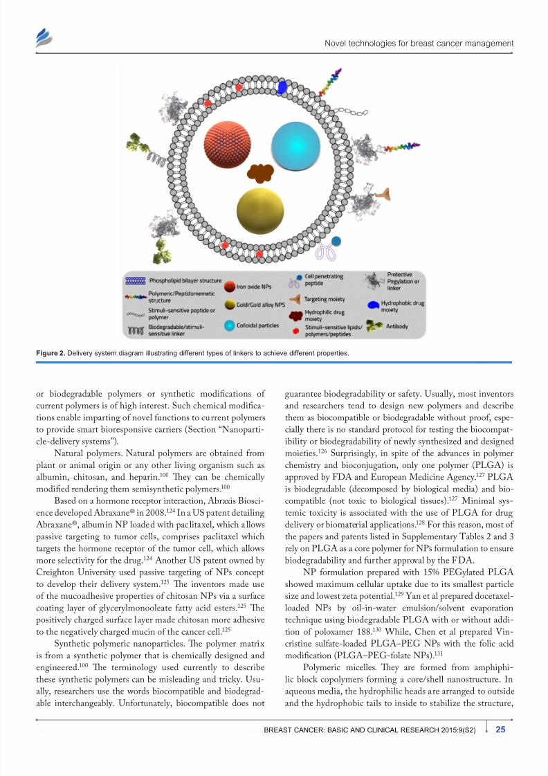
Nanoparticle-delivery systems. NPs provide many
favorable properties to the drug including longer elimina-
tion time; increase drug-site contact time, and reducing drug
resistance.114–118 Te NP drug carriers consist of at least two
materials, one of them is the active drug.119,120 Te other
material(s) form the NP system and may be used to enhance
system targeting.121 NPs with size larger than 100 nm are
more sufficient as drug carr iers because they have higher drug
loading capacity.121
NPs represent versatile tools to encapsulate various typesof drugs, either hydrophilic or hydrophobic altering their
physicochemical parameters and pharmacokinetics profile.
Furthermore, NPs represent a platform for custom-tailored
novel therapy design, through ease of conjugation of various
helping moieties via a linker such as stimuli-responsive pep-
tide or polymers, protective PEGylation layer, cell-penetrating
peptide, targeting moiety, or antibodies. Tey also enable the
creation of a multifunctional platform incorporating multiple-therapeutic agents for efficient treatment. Dual function-
ing diagnostic/imaging/therapeutic system via simultaneous
incorporation of MRI contrast agents along with anticancer
active pharmaceutical ingredient (API) was also described
(Fig. 2). Longer circulation time with the delivery system
can be achieved by conjugating the NPs with PEG.114,115 Te
PEG-coated NPs can escape the mononuclear phagocytic
system and circulate in the body for a longer time increas-
ing the chance of reaching the target and thereby the effect
of the loaded drug.114,115 Carrier modulations for modifying
the pharmacokinetic characteristics of the active therapeutic
agents will be discussed in detail in Section “SEALH®technology and pharmacokinetics manipulation in breast can-
cer management.” Beside increasing drug-site contact time,
some polymers used in NPs formulations like polylactic-
co-glycolic acid (PLGA) have high cell adhesion property,
increasing the drug concentration gradient at the adhesion site
by longer drug carrier contact time with the targeted cells.116
As for reducing drug resistance chances, NPs are taken up
by cells through receptor-mediated endocytosis and remain
inside the cell in endosomes. Tus, they bypass the recogni-
tion of P-glycoprotein that is responsible for drug resistance
and therapy failure.117,118
However, NPs can show some serious adverse effects.122
Adverse effects of NPs depend on individual factors such as
genetics, existing disease conditions, exposure, NP chem-
istry, size, shape, agglomeration state, and electromagnetic
properties.122 Te key to understanding the toxicity of NPs is
their size and NPs are smaller than cells and cellular organ-
elles, which allow them to penetrate these biological struc-
tures, disrupting their normal function.122 Examples of toxic
effects include tissue inflammation and altered cellular redox
balance toward oxidation, causing abnormal function or cell
death.122 Furthermore, a major drawback is the difficulty in
scaling up the formulation and its transformation from bench
to bedside, due to high cost and instability during storage.
NPs can be synthesized by different materials including
polymers, lipids, organometallic compounds, and viruses.123
Te NP materials must be biocompatible and safe when
administered.123 Tey can be classified according to material
nature into the following.
Polymeric nanoparticles. Polymers used in the formulation
of NPs can be either synthetic or natural. Te nature of the
chosen polymer and the NP formulation technique can cre-
ate diverse types of polymeric NPs. Te drug loaded to the
polymeric NPs is either physically entrapped or covalentlybounded to the polymer matrix depending on the method of
formulation.100 Currently, the design of novel biocompatible
7/25/2019 BCBCR Breast Cancer Conventional Diagnosis and Treatment Modalities.pdf 6823
http://slidepdf.com/reader/full/bcbcr-breast-cancer-conventional-diagnosis-and-treatment-modalitiespdf-6823 9/18
Novel technologies for breast cancer management
25BREAST CANCER: BASIC AND CLINICAL RESEARCH 2015:9(S2)
or biodegradable polymers or synthetic modifications of
current polymers is of high interest. Such chemical modifica-tions enable imparting of novel functions to current polymers
to provide smart bioresponsive carriers (Section “Nanoparti-
cle-delivery systems”).
Natural polymers. Natural polymers are obtained from
plant or animal origin or any other living organism such as
albumin, chitosan, and heparin.100 Tey can be chemically
modified rendering them semisynthetic polymers.100
Based on a hormone receptor interaction, Abraxis Biosci-
ence developed Abraxane® in 2008.124 In a US patent detailing
Abraxane®, albumin NP loaded with paclitaxel, which a llows
passive targeting to tumor cells, comprises paclitaxel which
targets the hormone receptor of the tumor cell, which allows
more selectivity for the drug.124 Another US patent owned by
Creighton University used passive targeting of NPs concept
to develop their delivery system.125 Te inventors made use
of the mucoadhesive properties of chitosan NPs via a surface
coating layer of glycerylmonooleate fatty acid esters.125 Te
positively charged surface layer made chitosan more adhesive
to the negatively charged mucin of the cancer cell.125
Synthetic polymeric nanoparticles. Te polymer matrix
is from a synthetic polymer that is chemically designed and
engineered.100 Te terminology used currently to describe
these synthetic polymers can be misleading and tricky. Usu-ally, researchers use the words biocompatible and biodegrad-
able interchangeably. Unfortunately, biocompatible does not
guarantee biodegradability or safety. Usually, most inventors
and researchers tend to design new polymers and describethem as biocompatible or biodegradable without proof, espe-
cially there is no standard protocol for testing the biocompat-
ibility or biodegradability of newly synthesized and designed
moieties.126 Surprisingly, in spite of the advances in polymer
chemistry and bioconjugation, only one polymer (PLGA) is
approved by FDA and European Medicine Agency.127 PLGA
is biodegradable (decomposed by biological media) and bio-
compatible (not toxic to biological tissues).127 Minimal sys-
temic toxicity is associated with the use of PLGA for drug
delivery or biomaterial applications.128 For this reason, most of
the papers and patents listed in Supplementary ables 2 and 3
rely on PLGA as a core polymer for NPs formulation to ensure
biodegradability and further approval by the FDA.
NP formulation prepared with 15% PEGylated PLGA
showed maximum cellular uptake due to its smallest particle
size and lowest zeta potential.129 Yan et al prepared docetaxel-
loaded NPs by oil-in-water emulsion/solvent evaporation
technique using biodegradable PLGA with or without addi-
tion of poloxamer 188.130 While, Chen et al prepared Vin-
cristine sulfate-loaded PLGA–PEG NPs with the folic acid
modification (PLGA–PEG-folate NPs).131
Polymeric micelles. Tey are formed from amphiphi-
lic block copolymers forming a core/shell nanostructure. Inaqueous media, the hydrophilic heads are arranged to outside
and the hydrophobic tails to inside to stabilize the structure,
Figure 2. Delivery system diagram illustrating different types of linkers to achieve different properties.

7/25/2019 BCBCR Breast Cancer Conventional Diagnosis and Treatment Modalities.pdf 6823
http://slidepdf.com/reader/full/bcbcr-breast-cancer-conventional-diagnosis-and-treatment-modalitiespdf-6823 10/18
Nounou et al
26 BREAST CANCER: BASIC AND CLINICAL RESEARCH 2015:9(S2)
which is suitable for IV injections.132 Onyuksel et al devel-
oped a grafted sterically stabilized phospholipid nanomicelles
of 17-allylamino-17-demethoxy geldanamycin as a novel-
targeted nanomedicine for breast cancer.133
Dendrimers. Dendrimers (ree in Latin) are multiple
highly branched synthetic polymer macromolecules. Den-drimers are flexible modifiable systems with monodisperse
size distribution. Tey are easy to manipulate and to conjugate
with different therapeutic agents. A patent in 2013 suggested
a novel antibreast-cancer Her2 vaccine with dendrimers of
lysine and cysteine backbone structure as a carrier system.134
Lipid-based drug carriers (liposomes). Liposomal (lipo-
some: lipid vesicle in latin) nanocarrier systems are vesicu-
lar lipid bilayer colloidal spheres formed by self-assembly.135
Liposomal formulations are the first novel controllable carrier
system to be sold in market for cancer (Doxil©, PEGylated
liposomal formulation encapsulating doxorubicin).136–139
Doxil© was the first novel carrier system for breast cancerapproved by the FDA on 1995.136 Currently, many others
are undergoing evaluation in clinical tria ls. Tis could be due
to the ensured safety of the phospholipid bilayer structure
of the liposomal lipids. Anthracyclines doxorubicin (Doxil,
Myocet) and daunorubicin (DaunoXome) were approved for
treating metastatic breast cancer and Kaposi’s sarcoma.140,141
PEGylated liposomal doxorubicin (PLD) HCl (CAELYX™/
Doxil®) lowered cardiotoxicity in phase III trials, which was
compared to conventional doxorubicin as a first-line treatment
in metastatic breast cancer.142 Also, Delek Keskin owned a
World Intellectual Property Organization patent of targetedPEGylated nanosized liposomal system conjugated with tar-
geting antibody (immunoliposome) for in vivo delivery of
COX II inhibitors, celecoxib.143
In a recent patent, muramyl tripeptide phosphatidyletha-
nolamine wrapped in liposomes (L-MP-PE) and loaded
with the antitumor agents was developed by the Institute of
Mataria Medica, the Chinese Academy of Medical Sciences.144
Te invention is a dual functional conjugate combining C
and immunotherapy. L-MP-PE synergizes the release of
cytokines, which can enhance the antitumor therapy.144
Viral nanoparticles. After emptying the virus from its
genetic material, drugs can be loaded into the empty virus
capsid.145 Tis carrier system takes invaluable advantages from
its nanostructure and biologically active capsid surface.145
Cowpea mosaic virus, cowpea chlorotic mottle virus, canine
parvovirus, and bacteriophages are generally used as viral
NPs.145 For example, a viral carrier system in conjugation with
radiation or a cytotoxic agent was the invention in a registered
patent by the University of Sydney.146 Te mechanism of the
system is genetic modification to increase the expression of
insulin-like growth factor binding protein-5 by the cell to an
apoptosis inducing amount.146
Carbon nanotubes. Tey are composed of benzene ringsforming carbon cylinders. Tey have very low solubility that is
overcome by linking water soluble ligands such as proteins and
peptides as well as therapeutic agents.147 Chemotherapeutic
agents linked to them showed more tendency to accumulate in
targeted cells compared to the free drug alone.148 In a patent
owned by the board of regents of the University of Oklahoma,
the inventors linked a protein or a peptide such as annexin V
or other annexins to carbon nanotubes such as single-walledcarbon nanotubes (SWNs) to form a protein–CN com-
plex for the treatment of breast cancer.149 Furthermore,
Oraki Kohshour et al proposed the ablation of breast cancer
cells using trastuzumab-functionalized multiwalled carbon
nanotubes and trastuzumab–diphtheria toxin conjugate.150
Moreover, Mohammadi et al used SWNs functionalized
with aptamer and piperazine–polyethylenimine derivative
for targeted short interfering RNA (siRNA) delivery into
breast cancer cells.151 Adopting another interesting approach,
Al Faraj et al used carbon nanotubes as a preferential magnetic
targeting tool for noninvasive tracking of breast cancer.152
Nanoshells. Nanoshells are optically tunable core/shellNPs that can be fabricated from gold or gold alloy to strongly
absorb in the near-infrared (NIR) region where light trans-
mits deeply into tissue.153 When injected systemically, these
particles have been shown to accumulate in the tumor due to
the EPR effect and induce photothermal ablation of the tumor
when irradiated with NIR laser.153 In a recent patent owned
by Te regents of the University of California, the inventors
described degradable silica nanoshells for ultrasonic imaging
and therapy.154
Inorganic nanoparticles. Drug delivery, magnetic reso-
nance and fluorescence imaging, magnetic influence, and celltargeting are concurrently feasible using multifunctional inor-
ganic NPs such as mesoporous silica NP, superparamagnetic
iron oxide NPs, calcium phosphosilicate NPs, gold NPs, and
others.155 Water-insoluble anticancer drugs can be delivered
into human breast cancer cells via surface conjugation with
cancer-specific targeting agents increasing the uptake into
cancer cells relative to that in noncancerous fibroblasts. Inor-
ganic particles often exhibit novel physical properties as their
size approaches nanometer scale. For example, the unique
electronic and optical properties of nanocrystalline quantum
dots may lead to future applications in electro-optic devices
and biomedical imaging.156
reating and imaging the primary and metastatic tumors
using inorganic NPs were the main target of several patents
in the last few years. In a US patent by Tomas Morgan et al,
nonaggregating resorbable calcium phosphosilicate NPs
(CPNPs) were used to formulate a bioconjugate for selective
targeting of cells.157 Te drug comprises a targeting molecule,
polypeptide, antibody, ligand or receptor, bound to a PEG-
maleimide molecule.157 Inside the targeted cell, the intercellu-
lar pH dissolves the CPNPs, releasing the chemotherapeutic/
imaging agent allowing selective treatment/imaging.157
In another patent, owned by Johns Hopkins University andNanomaterials echnology PE Ltd, surfactant-coated iron
oxide NPs for breast cancer diagnosis and treatment prepared

7/25/2019 BCBCR Breast Cancer Conventional Diagnosis and Treatment Modalities.pdf 6823
http://slidepdf.com/reader/full/bcbcr-breast-cancer-conventional-diagnosis-and-treatment-modalitiespdf-6823 11/18
Novel technologies for breast cancer management
27BREAST CANCER: BASIC AND CLINICAL RESEARCH 2015:9(S2)
via high gravity controlled precipitation were described.158 In
another patent owned by Hanwha Chemical Corporation, a
method of preparing iron oxide NPs coated with hydrophilic
material (carboxymethyl dextran) for MRI was described.159
Te University Of Louisville Research Foundation, Inc.
proposed novel anti-nucleolin (AS1411)-targeted gold NPsfor imaging, diagnosis, and treatment of breast cancer.160
argeted NPs in this patent comprise at least one member
selected from the group consisting of gold, platinum, iridium,
and palladium.160
Bioconjugates-delivery systems. Te main aim of bio-
conjugation is to form a stable biologically cleavable covalent
link between two molecules, at least one of which is a biomol-
ecule.161 Bioconjugation aims to increase stability, protect drug
from proteolysis, or to enhance the targeting properties of the
delivery system.161 Inspite of the historic fact that bioconju-
gates are older than NPs, research is currently being diverted
back to it.162 Tis could be contributed to its ease of synthesis,high scale up yield, ease of bench-to-bedside transformation,
ease of formulation, and final formulation stability.162 Bio-
conjugation reactions are generally categorized by the general
reactivity or functional group that is involved in the associ-
ated conjugation process, such as amine reactions, thiol reac-
tions, carboxylate reactions, hydroxyl reactions, aldehyde and
ketone reactions, active hydrogen reactions, photochemical
reactions, and cycloaddition reactions.162 Te design of a useful
bioconjugate will depend mainly on its use, purpose, and the
desired properties needed. Tus, one could choose a suitable mol-
ecule and suitable cross-linker to form the bioconjugate.163
Bio-conjugates can be synthesized by using cross-linkers (couplers)
that have been designed for this purpose or by using a reactive
group on one of the two molecules to facilitate the reaction161
(Fig. 3). Most of these reactions couplers are used to enhance the
conjugation yield such as 1-ethyl-3-(3-dimethylaminopropyl)
carbodiimide (EDC) and N , N ′-dicyclohexylcarbodiimide.162
EDC is often used in combination with N -hydroxysuccinimide
for the immobilization of large biomolecules.162 Bioconjugation
can also be carried out by using a secondary activating agent
that forms an intermediate reactive group on one of the two
molecules and thus facilitates the coupling reaction.161 Tus, the
key to forming a successful bioconjugate is choosing the suit-
able cross-linker between the molecules.161 Cross-linkers may
be classified as follows161:
– Zero-length cross-linkers: this kind of bioconjugates
involves the formation of a covalent bond between the
two molecules without the addition of any additional
atoms or spacer.
– Homobifunctional cross-linkers: this type involves the
formation of symmetrical bioconjugates of same func-
tionality at both ends.
– Heterobifunctional cross-linkers: have two differentgroups on both ends and thus attaching two reactive
groups different in nature.
– rifunctional cross-linkers: it involves a spacer of threereactive sites to attach three different molecules.
Dendrimers: it acts as a multivalent bioconjugation cross-
linker due to its structure, which resembles a tree. As described
earlier in NP design, bioconjugates are usually tailored
designed to provide the function of interest. Te active drug
entity can be linked to a diagnostic agent, targeting moiety,
pharmacokinetics-modifying agent such as PEG, biorespon-
sive or stimuli-sensitive agent, an aptamer or antibody (Fig. 3).
Furthermore, the choice of the proper linker can impart new
functions and smart characteristics to the bioconjugate system.
In the recent literature and patents, numerous examples
of bioconjugates have been described for the treatment and
diagnosis of cancer, including breast cancer. In a patent owned
by Phigenix, Inc., a bioconjugate linking tamoxifen and
trastuzumab to an antibody, a receptor, or a ligand to target
breast cancer tumor tissue was described.164
In a Johns Hopkins University owned patent, novel bio-
conjugate systems synthesized via in situ complexation of two
or more delivery components by the bioorthogonal click reac-
tions were described.165 wo novel bioconjugate systems were
descried. Te first bioconjugate system constituted of multiple
azido-functionalized or tetrazine-functionalized monoclonalantibody) and multiple cyclooctyne-functionalized nanocar-
rier bioconjugate. Te second bioconjugate system components
were trans-cyclooctene functionalization and bovine serum
albumin (BSA) substituted with chemotherapeutics, such as
paclitaxel.165 Tis system can be used for both treatment and
diagnosis of cancer depending on the substitution of the BSA.165
Research Development Foundation owned a patent pro-
posing a bioconjugate of monoclonal antibody and a cytotoxic
biological response modifying moiety selected from tumor
necrosis factor tumor tumor necrosis factor-alpha (NF-α),
NF-β, and IL-1 for the effective treatment of breast cancer.166
A patent, owned by Dartmouth College, described a tar-
geted bioconjugate that consists of a metal NP such as a gold,
Figure 3. Bioconjugate general structural design.
7/25/2019 BCBCR Breast Cancer Conventional Diagnosis and Treatment Modalities.pdf 6823
http://slidepdf.com/reader/full/bcbcr-breast-cancer-conventional-diagnosis-and-treatment-modalitiespdf-6823 12/18
Nounou et al
28 BREAST CANCER: BASIC AND CLINICAL RESEARCH 2015:9(S2)
silver, copper, nickel, aluminum, zinc, calcium, platinum,
palladium, or iron NPs bound to at least one peptide that is
modified at its N-terminal with a myristoyl group.167 In cer-
tain embodiments, the bioconjugate further comprises a sec-
ond therapeutic agent attached to the NP.167 Such proposed
design could be used for breast cancer diagnosis and treatment.Stimuli-responsive drug-delivery systems. Te approach
of using stimuli-responsive drug-delivery system had been
applied in 1950168 by studying stimuli-responsive hydrogels in
drug release and in 1970 by thermosensitive liposomes in drug
release.169 Te stimuli-responsive carrier could be attached with
a ligand to ensure the active targeting to the cancer cell before
triggering the drug release.170 Tey are classified according to
the stimuli as follows:
1. Systems triggered by external stimuli: such as light,
ultrasound, temperature change, and magnetic field.
2. Systems triggered by internal stimuli: such as pH, redoxpotential, and concentrations of enzymes.
3. Multiresponsive-delivery systems: developed so as to
respond to more than one stimulus. For example, there is
a great interest for combining a pH-stimuli system with a
receptor-mediated active targeting system to form multi-
functional polymeric micelles.171
Several recent patents described stimulus-responsive sys-
tems for breast cancer treatment and diagnosis. Chilkoti et al
designed a drug-delivery system with stimulus-responsive
biopolymers in a patent owned by Duke University.172
Tebiopolymer is a temperature-sensitive targeted bioconjugate
of API (radionuclides, chemotherapeutic agents, cytotoxic
agents, and diagnostic agents) and temperature sensitive block
copolymer (elastin-like polypeptide).172
In another patent owned by Magnamedics Gmbh,173 a
thermosensitive biocompatible polymer for breast cancer man-
agement was described. Te polymer has variable features for
therapy, diagnostics, and analytics.173 Termosensitive bio-
polymer particles begin to swell in tumor tissues, to finally
reach their equilibrium swelling state after a few minutes.173
In this state, the swollen polymer carriers exert emboliza-
tion function, that is, they are able to block the blood vessels,
thereby counteract tumor formation.173 Te thermosensitive
polymer described was either hydroxyalkylcellulose, isopro-
pylcellulose, polyoxyethylene thylene, or poly (ethylene glycol-
lactide-glycolide) copolymers.173
A US patent owned by the Industry Academic Coop-
eration Foundation (Younsei University) proposed a stimulus-
sensitive magnetic nanocomposite using pyrene polymer and
different contrast agents.174 Te stimuli-sensitive magnetic
nanocomposite has target specificity and serves as a contrast
composition or therapeutic composition.174
“Formulation of stealth thermosensitive liposomal drug-delivery system(s) for cancer therapy” was the title of a Chinese
patent owned by Zhengzhou University.175 In this patent,
thermosensitive liposomes contained thermal and chemother-
apeutic agents in a ratio of 1:3 was described.175 Termal agent
may include SWNs and their derivatives, multiwalled car-
bon nanotubes, graphite kaesa and its derivatives, derivatives
of nano gold, nano silver and its derivatives, lysine carboxyl
carbon nanotubes, and/or organic polymers.175
Nucleic acid technologies.Short interfering RNA. RNAi is an endogenous path-
way for posttranscriptional silencing of gene expression that
is triggered by double-stranded RNA, including endogenous
miRNA and synthetic siRNA.176 By activating this pathway,
siRNAs can silence the expression of virtually any gene with
high efficiency and specificity, including targets tradition-
ally considered to be undruggable.176 Peer suggest that RNAi
world is promising to manipulate the function of virtually any
gene in the human genome, opening new avenues to the per-
sonalized treatment of many types of diseases.177
Te siRNA was the target of recent papers and patentslately. Te University of Utah Research Foundation owned
patent that proposed an RNAi agent covalently coupled to the
alpha or omega end of a pH-dependent membrane-destabilizing
polymer.178 Te polymer bioconjugate further comprises one or
more PEG moiety applying passive targeting to tumor cells.178
Liu et al used doxorubicin and siRNA-loaded
heptapeptide-conjugated NPs to enhance chemosensitization
against epidermal growth factor receptor, which is overex-
pressed on breast cancer cells.179 In this study, PEG/PLGA
NPs were used to deliver doxorubicin and siRNA to the tumor
tissue.179
Also, eIF3c-siRNA was studied as a potential thera-peutic target for cancer by Emmanuel et al.180
Aptamer. Aptamers are nonbiological oligonucleotides that
can bind to protein targets.181 Aptamers can be used for thera-
peutic purposes in the same way as monoclonal antibodies.181
However, unlike traditional methods for producing monoclo-
nal antibodies, no organisms are required for the in vitro selec-
tion of oligonucleotides.181 For this reason, aptamers avoid the
immunogenicity of antibodies retaining all their properties.181
However, there still remain largely unknown pharmacokinetic
properties, which make them harder to develop than any given
therapeutic antibody.181
A series of aptamers currently in development may change
how nucleic acid therapeutics are perceived. In a patent owned by
Ecosynthetix Ltd., aptamer targeted cross-linked biocompatible
NPs were premeditated for the purpose of targeting doxorubi-
cin to breast cancer tumor cells.182 Tis structure is proposed to
function as a controlled release-delivery system via the swell ing
of the NP core and the enzymatic breakdown of the NP.182
Furthermore, Reyes-Reyes et al designed a PEGylated
anti-MUC1 aptamer–doxorubicin complex for targeted drug
delivery to MCF7 breast cancer cells.183 Aravind et al pro-
posed another example of the use of aptamers in breast cancer
management.184 Aravind et al described an AS1411 aptamertagged PLGA–lecithin–PEG NPs for tumor cell targeting
and drug delivery of paclitaxel.184 AS1411 aptamer, which is
7/25/2019 BCBCR Breast Cancer Conventional Diagnosis and Treatment Modalities.pdf 6823
http://slidepdf.com/reader/full/bcbcr-breast-cancer-conventional-diagnosis-and-treatment-modalitiespdf-6823 13/18
Novel technologies for breast cancer management
29BREAST CANCER: BASIC AND CLINICAL RESEARCH 2015:9(S2)
a 26-nucleotide guanosine-rich DNA sequence commonly
known as anti-nucleolin aptamers, was selected as the target-
ing moiety.30,183–187 It can bind to the nucleolin receptors nor-
mally seen over expressed on the tumor cells.
In a recent publication, Wang et al described specific
stimuli sensitive (pH sensitive and NIR-triggered release (pho-tothermal)) aptamer-conjugated mesoporous silica–carbon NPs
for HER2-targeted chemo-photothermal combined therapy.188
Te aptamer used in this study was HB5, an 86-nucleotide
DNA molecule, which is bound to an epitope peptide of
HER2.189
Biological technologies. umor targeting appears to
be controlled by tumor-specific and circulating cell-specific
factors. Consequently, targeted cancer therapy using cancer
cells could be a promising field of investigation. University of
Florida Research Foundation, Inc. owned a patent describ-
ing radiated cancer cells as a vehicle for cancer nanotherapy.190
Biological cellular carr iers were loaded with stimuli-responsiveparticles.190 Te agent-loaded radiated cancer vehicles migrate
to a primary or a metastatic tumor and deliver the therapeutic
agent to the primary or metastatic tumor following electro-
magnetic radiation.190
STEALTH® Technology and PharmacokineticsManipulation in Breast Cancer Management Since their emergence in 1971, liposomes have been investi-
gated as important targeted drug carriers for cancer C.191 Te
pharmacokinetics and biodistribution of liposomal prepara-
tions were extensively studied in vivo. However, a major limi-tation for the use of systemic liposomal preparations for drug
delivery was soon perceived; the clearance of the liposomes
following intravenous administration was very rapid (usually
within minutes). Tis was accompanied by the rapid release
of a large fraction of encapsulated content into circulation
resulting in reduced bioavailability and impairment of target-
ing. Te rapid clearance was likely attributed to the uptake of
the liposomes by the Kupffer cells of the reticuloendothelial
system (RES) following opsonization of liposomes by plasma
proteins.192,193
Several interventions were previously employed to pro-
long the circulation time of liposomes and thus improve
targeting. For example, the use of high phase-transition
temperature lipids and cholesterol.194 Te reduction in RES
accumulation observed was, however, modest. In early
1990s, PEGylated polymeric vesicles were introduced by
Yokoyama et al,195 which represented an important mile-
stone in the synthesis of long-circulating liposomal formula-
tions (SEALH® liposomes). Conjugation of PEG moiety
through a lipid anchor to the liposomes successfully reduced
their RES uptake, which could be attributed, at least in part,
to the reduced opsonization by plasma proteins.192 An impor-
tant application of the SEALH® liposomes technology was the development of PLD.196,197 PLD (Caelyx™, Doxil®)
is superior to the conventional doxorubicin preparation
showing reduced cardiotoxicity and prolonged activity.
PEGylation has been shown to alter the pharmacokinetics of
doxorubicin considerably; the total clearance (CL) was sig-
nificantly reduced. Mean CL values following administration
of PLD were as low as 0.03–0.041 L/hours/m2 compared
with 24–35 L/hours/m2
for the conventional doxorubi-cin.196 As a consequence to the reduced clearance, the total
systemic exposure estimated by the area under the plasma
concentration–time curve for doxorubicin was increased
by more than 100-folds.198 Te volume of distribution (V d)
of PLD was ∼2 L/m2 compared with 700–1,100 L/m2
for the conventional formulation.196 Te reduction in V d
implies that PLD is retained in plasma compartment with
minimal diffusion into noncancerous cells, thus reducing the
adverse effects. Tis could be attributed to the large size of
PLD particles (∼100 nm in diameter). On the other hand, the
leaky nature of cancerous vasculature would allow the selec-
tive diffusion of PLD into cancerous tissue.198
Modifying the pharmacokinetics profile of existing anti-
cancer drugs has been the main focus of some pharmaceutical
companies, such as Nektar Terapeutics. Nektar develops new
drug candidates by applying its proprietary three-dimensional
(3D) four-armed branched PEGylation and advanced polymer
conjugate technologies to modify chemical structure of vari-
ous APIs. It is a PEGylation technology supplier to a number
of pharmaceutical companies, including Affymax, Amegen,
Merck, Pfizer, and UCB Pharma.199
Nektar therapeutics has several patents on PEGylation
bioconjugation aiming to modifying the pharmacokineticprofile.200–206 Four of their patents described a novel etiri-
notecan (topoisomerase I inhibitor, or its active metabolite
SN-38) prodrug 3D four-armed PEG conjugate in a polymer
form, (Etirinotecan Pegol [NKR-102]).199–201,203–206 NKR-
102 has two main advantages over irinotecan.200,203–206 Te
first is the enlarged irinotecan molecular size to prevent its
transport across through the normal endothelia but enables
its penetration through the leaky tumor endothelia (passive
targeting strategy).199–201,203–206 Te second advantage of this
novel NKR-102 prodrug is its ability to modify the distri-
bution of irinotecan, decreasing the C-max while prolonging
the plasma circulation time compared with the unconjugated
irinotecan.199–201,203–206 Such technology extended the SN-38
1/2 to 168 hours.207 Nektar therapeutics Inc. also invented
a treatment for breast cancer and metastases using 3D four-
armed PEG20k -glycine-docetaxel bioconjugate.202 Te 3D
four-armed PEG20k -glycine-docetaxel bioconjugate offered
the same two advantages as NKR-102.202
Conclusions and Future Directions argeting cancer cells while avoiding noncancerous cells is the
Holy Grail of cancer therapy. Many different systems and strat-
egies have been designed for drug targeting to tumors over the years. Improved insights into the genetic and (patho-) physi-
ological processes contributing to malignant transformation

7/25/2019 BCBCR Breast Cancer Conventional Diagnosis and Treatment Modalities.pdf 6823
http://slidepdf.com/reader/full/bcbcr-breast-cancer-conventional-diagnosis-and-treatment-modalitiespdf-6823 14/18
Nounou et al
30 BREAST CANCER: BASIC AND CLINICAL RESEARCH 2015:9(S2)
and tumorigenesis have resulted in the development of several
novel chemotherapeutic drugs and strategies.99
Over the course of the last decade, many acquisitions
have taken place in this critical field of research in the phar-
maceutical industry. For these reasons, emerging pharma-
ceutical and nanotechnology companies try to increase theirpatent portfolio to increase their commercial value for possible
buyouts by big pharmaceutical firms. Such acquisitions show
the importance of this area of research and, most importantly,
highlight the global need for effective and safe pharmaceu-
tical chemotherapeutic agents that have the potential to tar-
get tumors like breast cancer with minimal toxicity and side
effects. Unfortunately, these factors affected the quality and
value of patents registered currently for the management of
breast cancer. Most of the reviewed patents were extremely
broad within their scope, unclear, and unorganized to avoid
any specifications of the invention. Tis prevents researchers
and scientists from reproducing the patents results and con-clusions. Tis could also be attributed to the companies urge
to gain maximum intellectual properties rights. Furthermore,
multiple patents were found to be describing the same prod-
uct exactly. Such flaws in the patents system need more strict
regulations during patents filing to protect consumers as well
as inventors.
Surveying the published research papers and patents
also revealed other major drawbacks in the methodology
design, which is the lack of standardization. Most stud-
ies only depended on M assay depends on M dyes
(3-(4,5-dimethylthiazol-2-yl)-2,5-diphenyltetrazolium bro-mide) X assay depend on X dye (2,3-bis-(2-methoxy-
4-nitro-5-sulfophenyl)-2H-tetrazolium-5-carboxanilide)
cell viability studies as an efficacy evaluation tool for the
antineoplastic property. Te use of a single test could never
describe the true efficacy pattern, especially that M
only depends on mitochondrial activity as a marker for cell
death. Multiple evaluation techniques should be adopted
for safety and efficacy evaluation of antineoplastic agents.
Furthermore, most in vitro cell culture studies were only
performed on two cell lines, MCF-7s and MDA-MB-231,
neglecting the newer more aggressive breast cancer cell vari-
ants such as JIM-1 cell line. Tis could provide mislead-
ing conclusions for the efficacy of the proposed treatment
strategies. Another major flaw is the lack of proper in vivo
models in the majority studies. Only few studies deployed a
truly breast cancer model in animals. Te establishment of
an in vivo animal model with recent breast cancer cell lines
from patients is an invaluable tool in the true assessment of
the efficacy and safety of novel strategies in breast cancer
management.
Another major debate stil l ongoing, as revealed by the
recent patents and papers portfolio, is the preferential advan-
tage of active targeting over passive targeting. Several impor-tant pitfalls in active tumor-targeted drug delivery were
identified by Lammers et al.99 Tey suggested that future
efforts should also address some of the conceptual drawbacks
of drug targeting to tumors, and that strategies should be
developed to overcome these shortcomings.99 It was shown
that passive targeting could yield very comparable results to
active targeting.99,208 Furthermore, the slightly increased
cellular drug uptake in tumor active targeting strategies doesnot justify the extremely higher cost, difficulty in bench-to-
bedside transformation, and the high complexity level of the
carrier system.99,208 It was shown that slight shape and par-
ticles characteristics modifications, such as drifting from
the traditional spherical carrier structures, can yield a much
effective passive targeting. Tis could be attributed to the fact
that irregular particles are easily trapped in cancerous leaky
angiogenic blood vessels compared to spherical traditional
particles.209
Finally, a major limitation impeding the entry of novel
nanomedicines for breast cancer into the market is that new
concepts and innovative research ideas within academiaare not being developed and exploited in collaboration
with the pharmaceutical industry. An integrated bench-to-
clinic approach, realized through a structural collaboration
between industry and academia, would strongly stimulate the
progression of tumor-targeted nanomedicines toward clinical
application.208 A major focus should be the transformation of
such novel technologies from bench to bedside. Developing
novel complexes and sophisticated systems that could never
reach the market due to high cost, inability of scaling up the
system, or instability of the final formulation is a major hurdle.
Major process and formulation development concerns exist with respect to scale up process of complex nanoparticluate
carriers. Most of the reagents and inactive moieties in the for-
mulation of such novel therapeutic systems are not included
in the FDA approved inactive ingredient database. Use of
click chemistry, NP formulations, ligand postinsertion, and
labeling techniques need to be extensively researched for ease
of scale up and proper bench to bedside transformation.
Consequently, many patents currently focus heavily on
simple bioconjugate structures, which are easily synthesized
with high yield, reduced cost, and high stability profile of the
final formulation. Tis could provide a practical direction for
the development of novel management tools and therapeutics
for breast cancer for researchers worldwide, paving the road
to affordable, scalable, stable, efficient, and safe management
strategies.
Author Contributions Wrote the first draft of the manuscript: MIN and FE.
Contributed to the writing of the manuscript: NA, KA,
SG, and HSQ. Agree with manuscript results and conclu-
sions: MIN, FE, NA, KA, SG, and HSQ. Jointly developed
the structure and arguments for the paper: MIN, FE, NA,
and KA. Made critical revisions and approved final version:MIN and FE. Al l authors reviewed and approved of the final
manuscript.

7/25/2019 BCBCR Breast Cancer Conventional Diagnosis and Treatment Modalities.pdf 6823
http://slidepdf.com/reader/full/bcbcr-breast-cancer-conventional-diagnosis-and-treatment-modalitiespdf-6823 15/18
Novel technologies for breast cancer management
31BREAST CANCER: BASIC AND CLINICAL RESEARCH 2015:9(S2)
Supplementary MaterialsSupplementary table 1. Acronyms and abbreviations
used in the manuscript.
Supplementary figure 1. Clinical tria ls for the evolution
of breast cancer treatment since 1970 till 2014.
Supplementary table 2. Recent patents on trends andtechnologies in breast cancer diagnosis and treatment.
Supplementary table 3. Recent papers reporting trends
and technologies in breast cancer diagnosis and treatment.
REFERENCES
1. Ebeid NI. Egyptian Medicine in the D ays of the Pharaohs . General Egyptian BookOrganization, Cairo, Egypt; 1999.
2. Akram M, Siddiqui SA. Breast cancer management: past, present and evolving. Indian J Cancer . 2012;49(3):277–282.
3. Sanchez GM, Meltzer ES. Te Edwin Smith Papyrus: Updated ranslation of therauma reatise and Modern Medical Commentaries . Lockwood Press, Atlanta,
GA; 2012. 4. Brayn CP, Smith GE. Te Papyrus Ebers, ranslated from the German Version .Letchworth, Herts: Te Guardian City Press LD; 1930.
5. Papavramidou N, Papavramidis , Demetriou . Ancient Greek and Greco-
Roman methods in modern surgica l treatment of cancer. Ann Surg Oncol . 2010;17(3):665–667.
6. orre LA, Bray F, Siegel RL, Ferlay J, Lorte t-ieulent J, Jemal A. Global cancerstatistics, 2012. CA Cancer J Clin. 2015;65(2):87–108.
7. Gomez-Raposo C, Zambrana evar F, Sereno Moyano M, Lopez Gomez M,Casado E. Male breast cancer. Cancer reat Rev. 2010;36(6):451–457.
8. Giordano SH, Buzdar AU, Hortobagyi GN. Breast cancer in men. Ann Inter n Med . 2002;137(8):678–687.
9. Ferlay J SI, Ervik M, Dikshit R, Eser S, Mathers C, Rebelo M, Parkin DM,Forman D, Bray F. Globocan 2012, Cancer Incidence and Mortality World-
wide: I ARC. Cancer Fact Sheets 2013; v1.0. Available at: http://globocan.iarc.fr.
Accessed May 2, 2015. 10. Dumitrescu RG, Cotarla I. Understanding breast cancer risk—where do we
stand in 2005? J Cell Mol Med . 2005;9(1):208–221. 11. Gancberg D, Lespagnard L, Rouas G, et al. Sensitivity of HER-2/neu antibodies
in archival tissue samples of invasive breast carcinomas. Correlation with onco-
gene amplification in 160 cases. Am J Clin Pathol . 2000;113(5):675–682. 12. Hung MC, Lau YK. Basic science of HER-2/neu: a review. Semin Oncol . 1999;
26(4 suppl 12):51–59. 13. Malhotra GK, Zhao X, Band H, Band V. Histological, molecular and functional
subtypes of breast cancers. Cancer Biol Ter . 2010;10(10):955–960. 14. Breastcancer.org. ypes of breast cancer. Breastcancer . Ardmore, PA: Breastcancer.
org; 2013. 15. Weigelt B, Horlings HM, Kreike B, et al. Refinement of breast cancer classifica-
tion by molecular characterization of histological special types. J Pathol . 2008;216(2):141–150.
16. Phipps AI, Li CI. Breast cancer biology and clinical characteristics. In: Li CI, ed.
Breast Cancer Epidemiology . New York, NY: Springer; 2010:21–46. 17. Logan GJ, Dabbs DJ, Lucas PC, et al. Molecular drivers of lobular carcinoma
in situ. Breast Cancer Res . 2015;17(1):76. 18. Wellings SR. A hypothesis of the origin of human breast cancer from the termi-nal ductal lobular unit. Pathol Res Pract . 1980;166(4):515–535.
19. Norton KA, Wininger M, Bhanot G, Ganesan S, Barnard N, Shinbrot . A 2Dmechanistic model of breast ductal carcinoma in situ (DCIS) morphology andprogression. J Teor Bio l . 2010;263(4):393–406.
20. McCart Reed AE, Kutasovic JR, Lakhani SR, Simpson P. Invasive lobularcarcinoma of the breast: morphology, biomarkers and ‘omics. Breast Cancer Res .2015;17(1):12.
21. Lester SC, Bose S, Chen YY, et al. Protocol for the examination of specimensfrom patients with invasive carcinoma of the breast. Arch Pathol Lab Med . 2009;133(10):1515–1538.
22. Sonnenblick A, Fumagalli D, Sotiriou C, Piccart M. Is the differentiation intomolecular subtypes of breast cancer important for staging, local and systemic
therapy, and follow up? Cancer reat Rev. 2014;40(9):1089–1095. 23. Inic Z, Zegarac M, Inic M, et al. Difference between luminal A and lumi-
nal B subtypes according to Ki-67, tumor size, and progesterone receptor
negativity providing prognostic information. Clin Med Insights Oncol . 2014;8:107–111.
24. oft DJ, Cryns VL. Minireview: basal-like breast cancer: from molecular pro-
files to targeted therapies. Mol Endocr inol . 2011;25(2):199–211.
25. Senkus E, Kyriakides S, Penault-Llorca F, et al. Primary breast cancer: ESMOclinical practice guidelines for diagnosis, treatment and follow-up. Ann Oncol .2013;24(suppl 6):vi7–vi23.
26. Kerlikowske K, Hubbard RA, Miglioretti DL, et al; Breast Cancer Surveillance
Consortium. Comparative effectiveness of digital versus fi lm-screen mammogra-phy in community practice in the United States: a cohort study. Ann Intern Med .2011;155(8):493–502.
27. Van Goethem M, jalma W, Schelfout K, Verslegers I, Biltjes I, Parizel P.
Magnetic resonance imaging in breast cancer. Eur J Surg Oncol . 2006;32(9):901–910.
28. Sinha S, Sinha U. Recent advances in breast MRI and MRS. NMR Biomed . 2009;22(1):3–16.
29. Mody VV, Nounou MI, Bikram M. Novel nanomedicine-based MRI con-trast agents for gynecological malignancies. Adv Drug Deliv Rev. 2009;61(10):
795–807. 30. Hwang do W, Ko HY, Lee JH, et al. A nucleoli n-targeted multimodal nanopar-
ticle imaging probe for tracking cancer cells using an aptamer. J Nucl Med . 2010;51(1):98–105.
31. Kamaly N, Kalber L, Kenny GD, Tanou M, Miller AD, Bell J, Inventors;Imperial Innovations Limited, Medical Research Council, assignee. Novel
liposome nanoparticles for tumor magnetic resonance imaging. US PatentUS20130129636-A1, 2013.
32. uretschek K, Roberts P, Floyd E, et al. umor microvascular character-ization using ultrasmall superparamagnetic iron oxide particles (USPIO)
in an experimental breast cancer model. J Magn Reson Imaging . 2001;13(6):882–888. 33. Economopoulos V, Chen Y, McFadden C, Foster PJ. MRI detect ion of nonpro-
liferative tumor cells in lymph node metastases using iron oxide particles in amouse model of breast cancer. ransl Oncol . 2013;6(3):347–354.
34. Cyran CC, Fu Y, Raatschen HJ, et al. New macromolecular polymeric MRI
contrast agents for application in the differentiation of cancer from benig n softtissues. J Magn Reson Imaging . 2008;27(3):581–589.
35. Drahl C. Google’s nanoparticle diagnostic vision (Google X). C&EN . 2014;92(47):26–28.
36. O’Connor M, Rhodes D, Hruska C. Molecular breast imaging. Expert Rev Anti-
cancer Ter . 2009;9(8):1073–1080.
37. Palmer ML, sangari s N. Breast biopsy in women 30 years old or less. Am JSurg . 1993;165(6):708–712.
38. Chaiwun B, Torner P. Fine needle aspiration for evaluation of breast masses.Curr Opin Obstet Gynecol . 20 07;19(1):48–55.
39. Neal L, Sandhu NP, Hieken J, et al. Diagnosis and management of benign,
atypical, a nd indeterminate breast lesions detected on core needle biopsy. MayoClin Proc . 2014;89(4):536–547.
40. Kroese M, Zimmern RL, Pinder SE. HER2 status in breast cancer—an exampleof pharmacogenetic testing. J R Soc Med . 2007;100(7):326–329.
41. Brooks M. Breast cancer screening and biomarkers. Methods Mol Biol . 2009;
472:307–321. 42. Bast RC Jr, Ravdin P, Hayes DF, et al; American Society of Clinical Oncology
umor Markers Exper t Pane l. 20 00 update of recommendations for the use oftumor markers in breast and colorectal cancer: clinical pr actice guidelines of the
American Socie ty of Clin ical Oncolog y. J Clin Oncol . 20 01;19(6):1865–1878. 43. Galvis-Jimenez JM, Curtidor H, Patarroyo MA, Monterrey P, Ramirez-
Clavijo SR. Mammaglobin peptide as a novel biomarker for breast cancerdetection. Cancer Biol Ter . 2013;14(4):327–332.
44. Liu XG, Wang XP, Li WF, et al. Ca2+-binding protein S100A11: a novel diag-nostic marker for breast carcinoma. Oncol Rep . 2010;23(5):1301–1308.
45. Fürstenberger G, von Moos R, Lucas R, et al. Circulating endothelial cells and
angiogenic serum factors during neoadjuvant chemotherapy of primary breastcancer. Br J Cancer . 2006;94(4):524–531. 46. wardowski PW, Smith-Powell L, Carroll M, et al. Biologic markers of angio-
genesis: circulating endothelial cells in patients with advanced malignanciestreated on phase I protocol with metronomic chemotherapy and celecoxib. Cancer
Invest . 2008;26(1):53–59.
47. Chang CC, Wu MJ, Yang JY, Camaril lo IG, Chang CJ. Leptin-SA3-G9asignaling promotes obesity-mediated breast cancer progression. Cancer Res .2015;75(11):2375–2386.
48. Heyn H, Carmona FJ, Gomez A, et al. DNA methylation profiling in breastcancer discordant identical tw ins identifies DOK7 as novel epigenetic biomarker.
Carcinogenesis . 2013;34(1):102–108. 49. Zhu W, Qin W, Atasoy U, Sauter ER. Circulati ng microRNAs in breast cancer
and healthy subjects. BMC Res Notes . 2009;2:89. 50. Park KH, Yong YR, Kang HJ, Kim G, Park DH, Lee MY, Inventors; Samsung
Electronics Co., Ltd, assignee. Composition and kit for diagnosing breast cancer
including miRNAs w ithin vesicle, and method of diagnosing breast cancer using
the same. US Patent EP2746406–A1, 2014. 51. Richer J, Cochrane D, Anderson SM, Inventors; Te Regents Of Te University
Of Colorado, assignee. MiRNAs dysregulated in triple-negative breast cancer.US Patent US8507195–B2, 2013.

7/25/2019 BCBCR Breast Cancer Conventional Diagnosis and Treatment Modalities.pdf 6823
http://slidepdf.com/reader/full/bcbcr-breast-cancer-conventional-diagnosis-and-treatment-modalitiespdf-6823 16/18
Nounou et al
32 BREAST CANCER: BASIC AND CLINICAL RESEARCH 2015:9(S2)
52. Liu X, Chai Y, Li J, et al. Autoantibody response to a novel tumor-assoc iatedantigen p90/CIP2A in breast cancer immunodiagnosis. umour Biol . 2014;35(3):2661–2667.
53. Dong X, Yang M, Sun H, et al. Combined measurement of CA 15-3 with novel
autoantibodies improves diagnostic accuracy for breast cancer. Onco argets Ter .2013;6:273–279.
54. Paik S, Shak S, ang G, et al. A multigene assay to predict recurrence oftamoxifen-treated, node-negative breast cancer. N Engl J Med . 2004;351(27):
2817–2826. 55. Buyse M, Loi S, van’t Veer L, et al. Validation and clinical utility of a 70-gene
prognostic signature for women with node-negative breast cancer. J Natl Cancer Inst . 2006;98(17):1183–1192.
56. Zeidan BA, ownsend PA, Garbis SD, Copson E, Cutress RI. Clinical pro-teomics and breast cancer. Surgeon. 2015;13(5):271–278.
57. Dhankhar R, Vyas SP, Jain AK, Arora S, Rath G, Goyal AK. Advances in noveldrug delivery strategies for breast cancer therapy. Artif Cells Blood Subst it ImmobilBiotechnol . 2010;38(5):230–249.
58. Matsen CB, Neumayer LA. Breast cancer: a review for the general surgeon. JAM A Surg . 2013;148(10):971–979.
59. Joensuu H, Bono P, Kataja V, et al. Fluorourac il, epirubic in, and cyclophospha-
mide with either docetaxel or vinorelbine, with or without trastuzu mab, as adju- vant treatments of breast cancer : fina l resu lts of t he FinHer ria l. J Clin Oncol .2009;27(34):5685–5692.
60. Houssami N, Cuzick J, Dixon JM. Te prevention, detection, and management
of breast cancer. Med J Aust . 2006;184(5):230–234. 61. Davies C, Pan H, Godwin J, et al. Long-term effects of continuing adjuvanttamoxifen to 10 years versus stopping at 5 years after diagnosis of oestrogenreceptor-positive breast cancer: ALAS, a randomised trial. Lancet . 2013;381(9869):805–816.
62. Goldhirsch A, Winer EP, Coates AS, et al; Panel members. Personalizing the
treatment of women with early breast cancer: highlights of the St Ga llen interna-tional expert consensus on the prima ry therapy of early breast cancer 2013. AnnOncol . 2013;24(9):2206–2223.
63. Hirsimaki P, Aaltonen A, Mantyla E. oxicity of antiestrogens. Breast J . 2002;8(2):92–96.
64. Puhalla S, Brufsky A, Davidson N. Adjuvant endocrine therapy for premeno-
pausal women with breast cancer. Breast . 2009;18(suppl 3):S122–S130. 65. Christinat A, Di Lascio S, Pagani O. Hormonal therapies in young breast cancer
patients: when, what and for how long? J Torac Dis . 2013;5(suppl 1):S36–S46. 66. Gnant M, Mlineritsch B, Stoeger H, et al; Austrian Breast and Colorectal Cancer
Study Group, Vienna, Austria. Adjuvant endocrine t herapy plus zoledronic acid
in premenopausal women with early-stage breast cancer: 62-month follow-upfrom the ABCSG-12 randomised trial. Lancet Oncol . 2011;12(7):631–641.
67. Dowset t M, Stein RC, Coombes RC. Aromatization inhibit ion alone or in com-bination with GnRH agonists for the treatment of premenopausal breast cancerpatients. J Steroid Biochem Mol Biol . 1992;43(1–3):155–159.
68. Shao N, Wang S, Yao C, et al. Sequential versus concurrent anthracyc linesand taxanes as adjuvant chemotherapy of early breast cancer: a meta-analysis ofphase III randomized control trials. Breast . 2012;21(3):389–393.
69. Early Breast Cancer rialists’ Collaborative Group (EBCCG), Peto R,Davies C, et al. Comparisons between different polychemotherapy regimens forearly breast ca ncer: meta-analyses of long-term outcome among 100,000 women
in 123 randomised trials. Lancet . 2012;379(9814):432–444. 70. Valero V. Docetaxel and c yclophosphamide in patients with advanced solid
tumors. Oncology (Williston Park). 1997;11(6 suppl 6):21–23. 71. Callahan R, Hurvitz S. HER2-positive breast cancer: current management of
early, advanced, and recurrent disease. Curr Opin Obstet Gynecol . 2011;23(1):
37–43. 72. O’Sullivan CC, Bradbury I, Campbell C, et al. Efficacy of adjuvant trastuzumabfor patients with human epidermal growth factor receptor 2-positive early breastcancer and tumors2 cm: a meta-analysis of the randomized trastuzumab trials. J Clin Oncol . 2015;33(24):2600–2608.
73. Ewer SM, Ewer MS. Cardiotoxicity profile of trastuzumab. Drug Saf . 2008;
31(6):459–467. 74. urner N, Biganzoli L, Di Leo A. Continued value of adjuvant anthracyclines as
treatment for early breast cancer. Lancet Oncol . 2015;16(7):e362–e369. 75. Kjel J, Lee B, Radha M, Biase SD. Innovation in Cancer Care and Implications
for Health Systems: Global Oncology rend Re port 2014 . Parsippany, USA: IMS
Institute for Healthcare Informatics; 2014. 76. sang RY, Finn RS. Beyond trastuzumab: novel therapeutic strategies in HER2-
positive metastatic breast cancer. Br J Cancer . 2012;106(1):6–13. 77. Swain SM, Im YH, Im SA, et al. Safety profile of pertuzumab with trastuzumab
and docetaxel in patients from Asia with human epidermal growth factor recep-
tor 2-positive metastatic breast cancer: results from t he phase III t rial CLEOPA-
RA. Oncologist . 2014;19(7):693–701. 78. XXXX. FDA Approves Roche’s Kadcyla (rastuzumab Emtansine), the First
Antibody-Drug Conjugate for reating HER2-Positive Metastatic Breast Cancer .Basel: Roche; 2013.
79. Vinayak S, Carlson RW. mOR inhibitors in the treatment of breast cancer.Oncology (Williston Park). 2013;27(1):38–44,46,48assim.
80. Yardley DA. Combining mOR inhibitors with chemotherapy and other tar-geted therapies in advanced breast cancer: rationale, clinical experience, and
future directions. Breast Cancer (Auckl). 2013;7:7–22. 81. U.S. Food and Drug Administration. FDA Approves Afinitor for Advanced Breast
Cancer . Maryland, USA: FDA News Release; 2012. 82. Mancini P, Angeloni A, Risi E, Orsi E, Mezi S. Standard of care and prom-
ising new agents for triple negative metastatic breast cancer. Cancers . 2014;6(4):2187–2223.
83. Gonzalez-Angulo AM, Akcaka nat A, Liu S, et al. Open-label randomizedclinical trial of standard neoadjuvant chemotherapy with paclitaxel followedby FEC versus the combination of paclitaxel and everolimus followed by FECin women with triple receptor-negative breast cancerdagger. Ann Oncol . 2014;
25(6):1122–1127. 84. Mayer EL. argeting breast cancer with CDK inhibitors. Curr Oncol Rep .
2015;17(5):443. 85. Finn RS, Crown JP, Lang I, et al. Te cyclin-dependent kinase 4/6 inhibitor
palbociclib in combination with letrozole versus letrozole alone as first-linetreatment of oestrogen receptor-positive, HER2-negative, advanced breast
cancer (PALOMA-1/RIO-18): a randomised phase 2 study. Lancet Oncol .2015;16(1):25–35.
86. Cadoo KA, Gucalp A, raina A. Palbociclib: an evidence-based review ofits potential in the treatment of breast cancer. Breast Cancer (Dove Med Press).
2014;6:123–133. 87. Pfizer Receives U.S. FDA Accelerated Approval of IBRA NCE® (palboc iclib). 2015; Availab le at: http://www.pfizer.com/news/press-release/press-release-detail/pfizer_receives_u_s_fda_accelerated_approval_of_ibrance_palbociclib
88. Gelbert LM, Cai S, Lin X, et al. Preclinica l characteriz ation of the CDK4/6inhibitor LY2835219: in-vivo cell cycle-dependent/independent anti-tumor
activities alone/in combination with gemcitabine. Invest New Drugs . 2014;32(5):825–837.
89. Pavlakis N, Stockler M. Bisphosphonates for breast cancer. Cochrane DatabaseSyst Rev. 2002;1:CD003474.
90. Fick EM, Anzeneder , Katalinic A, Waldmann A. Bisphosphonates and theirrole in therapy for breast cancer—results from the PAH biobank. GeburtshilfeFrauenheilkd . 2013;73(5):412–421.
91. Mathew A, Brufsky A. Bisphosphonates in breast cancer. Int J Cancer . 2 015;137(4):753–764.
92. Mathew A, Brufsky A. Bisphosphonates in breast cancer: a triple winner? Oncol-
ogy (Williston Park). 2015;29(1):37–39.
93. Papadimitriou K, Ardavanis A, Kountourakis P. Neoadjuvant therapy for locallyadvanced breast cancer: focus on chemotherapy and biological targeted treat-ments’ armamentarium. J Torac Dis . 2010;2 (3):160–170.
94. Lemieux J, Maunsell E, Provencher L. Chemotherapy‐induced alopecia andeffects on quality of life among women with breast cancer: a literature review.
Psychooncology . 2 008;17(4):317–328. 95. Acharya S, Dilnawaz F, Sahoo SK. argeted epidermal growth factor receptor
nanoparticle bioconjugates for breast cancer therapy. Biomaterials . 2009;30(29):5737–5750.
96. Buzea C, Pacheco I, Robbie K. Nanomaterials and nanoparticles: sources andtoxicity. Biointerphases . 2007;2:MR17–MR71.
97. Danhier F, Feron O, Preat V. o exploit the tumor microenvi ronment: passiveand active tumor targeting of nanocarriers for anti-cancer drug delivery. J ControlRelease . 2010;148(2):135–146.
98. Maeda H. Vascular permeability in cancer and infection as related to macromo-lecular drug delivery, with emphasis on the EPR effect for tu mor-selective drug
targeting. Proc Jpn Acad Ser B Phys Biol Sci . 2012;88(3):53–71. 99. Lammers , Kiessling F, Hennink WE, Storm G. Drug targeting to tumors:principles, pitfalls and (pre−) clinical progress. J Control Release . 2012;161(2):175–187.
100. Cho K, Wang X, Nie S, Chen ZG, Shin DM. Terapeutic nanoparticles for drugdelivery in ca ncer. Clin Cancer Res . 2008;14(5):1310–1316.
101. Nogueira DR, avano L, Mitjans M, Perez L, In fante MR, Vinardell MP.In vitro antitumor activity of methotrexate via pH-sensitive chitosan nanopar-ticles. Biomaterials . 2013;34(11):2758–2772.
102. Nobs L, Buchegger F, Gurny R, Allemann E. Poly(lactic acid) nanoparticleslabeled with biologically active neutravidin for active targeting. Eur J Pharm Bio- pharm. 2004;58(3):483–490.
103. Prinzen L, Miserus RJ, Dirksen A, et al. Optical and magnetic resonance imag-ing of cell death and platelet activation using annexin a5-functionalized quan-tum dots. Nano Lett . 2007;7(1):93–100.
104. Sudimack J, Lee RJ. argeted drug delivery via the folate receptor. Adv DrugDeliv Rev. 20 00;41(2):147–162.
105. Hu HM, Li H, Jiao J, Inventors; Hong-Ming Hu, Haiyan Li and Jun Jiao,assignee. Alumina nanoparticle bioconjugates and methods of stimulating animmune response using said bioconjugates. US Patent US20130209502-A1,2013.

7/25/2019 BCBCR Breast Cancer Conventional Diagnosis and Treatment Modalities.pdf 6823
http://slidepdf.com/reader/full/bcbcr-breast-cancer-conventional-diagnosis-and-treatment-modalitiespdf-6823 17/18
Novel technologies for breast cancer management
33BREAST CANCER: BASIC AND CLINICAL RESEARCH 2015:9(S2)
106. Sanchez C, Guzman M, Wright S, et al., Inventors; GW Pharma Limited,Otsuka Pharmaceutical Co., Limited, assignee. Phytocannabinoids for use inthe treatment of breast cancer. US Patent EP2768493-A1, 2014.
107. Chen W Y, Wagner E, Inventors; Greenville Hospital System, assignee.
Bi-functional cancer treatment agents. US Patent US7201905-B2, 2007. 108. Slade M, Coombes RC, Inventors; Martin Slade and Raoul Charles Coombes,
assignee. Breast cancer methods, medicaments and agents. 2010. 109. Mack D, Gish KC, Inventors ; Eos Biotechnology, Inc., assignee. Novel methods
of diagnosing and treating breast cancer, compositions, and methods of screeningfor breast cancer modulators. US Patent CA2368152-A1, 2000.
110. Gish K, Mack D, Inventors; Eos Biotechnology, Inc., assignee. Novel methodsof diagnosing breast cancer, compositions, and methods of screening for breastcancer modulators. 2003.
111. Njar VCO, Gediya LK, Khandelwal A, Inventors; University of Maryland, Bal-
timore, assignee. Novel retinamide retinoic acid metabolism blocking agents. USPatent WO2010036404-A2, 2010.
112. Nakamura Y, Katagiri , Nakatsuru S, Inventors; Oncotherapy Science,Inc., assignee. Genes and polypeptides relating to breast cancers. US PatentUS20140228238-A1, 2014.
113. Linke SP, Bremer M, Diamond CA, Inventors; Steven P. Linke, roy M.
Bremer and Cornelius A. Diamond, assignee. Diagnostic markers of breastcancer treatment and progression and methods of use thereof. US PatentUS20090061422-A1, 2009.
114. Bazile D, Prud’homme C, Bassoullet M, Marlard M, Spenlehauer G, Veillard M.
Stealth Me.PEG-PLA nanoparticles avoid uptake by the mononuclear phago-cytes system. J Pharm Sci . 1995;84(4):493–498. 115. Niidome , Yamagata M, Okamoto Y, et al. PEG-modified gold nanorods with a
stealth character for in v ivo applications. J Control Rele ase . 200 6;114(3):343–347. 116. Weissenbock A, Wirth M, Gabor F. WGA-grafted PLGA-nanospheres: prepa-
ration and association with Caco-2 single cells. J Control Release . 2004;99(3):
383–392. 117. Brigger I, Dubernet C, Couvreur P. Nanoparticles in cancer therapy and diagno-
sis. Adv Drug Deliv Rev. 2002;54(5):631–651. 118. Zhang P, Ling G, Sun J, et al. Multifunctional nanoassemblies for vincristine
sulfate delivery to overcome multidrug resistance by escaping P-glycoproteinmediated efflux. Biomaterials . 2011;32(23):5524–5533.
119. Duncan R . Te dawning era of polymer therapeutics. Nat Rev Drug Discov.2003;2(5):347–360.
120. Ferrari M. Cancer nanotechnology: opportunities and challenges. Nat Rev Cancer .2005;5(3):161–171.
121. Dowling A, Clift R, Grobert N, et al. Nanoscienc e and Nanotechnolog ies: O ppor-
tunities and Uncertainties . London: Te Royal Society & Te Royal Academy ofEngineering Report; 2004:61–64.
122. Wesselinova D. Current major cancer targets for nanoparticle systems. CurrCancer Drug argets . 2011;11(2):164–183.
123. De Jong WH, Borm PJ. Drug delivery and nanoparticles: applications and haz-
ards. Int J Nanomedic ine . 2008;3(2):133–149. 124. Desai NP, Soon-Shiong P, Inventors; Abraxis Bioscience, assignee. Breast can-
cer therapy based on hormone receptor status with nanoparticles comprising tax-ane. US Patent WO2008076373-A1, 2008.
125. Dash AK, rickler WJ, Inventors; Creighton University, assignee. Mucoadhesivenanoparticles for cancer treatment. US Patent US8242165-B2, 2012.
126. Zhang Y, Yang J. Design stra tegies for fluorescent biodegradable polymeric bio-materials. J Mater Chem B Mater Biol Med . 2013;1(2):132–148.
127. Danhier F, Ansorena E, Silva JM, Coco R, Le Breton A, Preat V. PLGA-based nanoparticles: an overview of biomedical applications. J Control Release .2012;161(2):505–522.
128. Kumari A, Yadav SK, Yadav SC. Biodegradable polymeric nanoparticles baseddrug delivery systems. Colloids Surf B Biointerfaces . 2010;75(1):1–18. 129. Pamujula S, Hazari S, Bolden G, et al. Cellular delivery of PEGylated PLGA
nanoparticles. J Pharm Pharmacol . 2012 ;64(1):61–67. 130. Yan F, Zhang C, Zheng Y, et al. Te effect of poloxamer 188 on nanopa rtic le
morphology, size, cancer cell uptake, and cytotoxicity. Nanomedic ine . 2010;6(1):
170–178. 131. Chen J, Li S, Shen Q, He H, Zhang Y. Enhanced cellular uptake of folic acid-
conjugated PLGA-PEG nanoparticles loaded with vincr istine sulfate in humanbreast cancer. Drug Dev Ind Pharm. 2011;37(11):1339–1346.
132. Adams ML, Lavasanifar A, Kwon GS. Amphiphilic block copolymers for drug
delivery. J Pharm Sci . 2003;92(7):1343–1355. 133. Onyuk sel H, Mohanty PS, Rubinstein I. VIP-gra fted sterically stabili zed
phospholipid nanomicellar 17-allylamino-17-demethoxy geldanamycin: anovel targeted nanomedicine for breast cancer. Int J Pharm. 2009;365(1–2):157–161.
134. Nankai University, assignee. Novel anti-breast-cancer Her2 vaccine with den-
drimer as carrier. US Patent CN103191423 A, 2013. 135. Perche F, orchilin VP. Recent trends in multifunctional liposomal nanocarriers
for enhanced tumor targeting. J Drug Deliv. 2013;2013:705265. 136. XX XX. DOXIL approved by FDA. AIDS Patient Care . 1995;9(6):306.
137. James ND, Coker RJ, omlinson D, et al. Liposomal doxorubic in (Doxi l): aneffective new treatment for Kaposi’s sarcoma in AIDS. Clin Oncol (R Coll Radiol).1994;6(5):294–296.
138. Muggia FM. Doxil in breast cancer. J Clin Oncol . 1998;16(2):811–812.
139. Porche DJ. Liposomal doxorubicin (Dox il). J Assoc Nurses AIDS Care . 1996;7(2):55–59.
140. Markman M. Pegylated liposomal doxorubicin in the treatment of cancers of thebreast and ovary. Expert Opin Pharmacother . 2006;7(11):1469–1474.
141. Rosenthal E, Poizot-Martin I, Saint-Marc , Spano JP, Cacoub P, Group DNXS.Phase IV study of liposomal daunorubicin (DaunoXome) in AIDS-relatedKaposi sarcoma. Am J Clin Oncol . 20 02;25(1):57–59.
142. O’Brien ME, Wigler N, Inbar M, et al; CAELYX Breast Cancer StudyGroup. Reduced cardiotoxicity and comparable efficacy in a phase III trial ofpegylated liposomal doxorubicin HCl (CAELYX/Doxil) versus conventional
doxorubicin for first-line treatment of metastatic breast cancer. Ann Oncol .2004;15(3):440–449.
143. Banerjee S, ezcancer A, Erdog A, Inventors; Dilek Keskin, assig nee. umortargeted liposomal drug delivery system. US Patent WO2014046630-A1, 2014.
144. Liu G, Zhao N, Ma Y, Inventors; Institute Of Mataria Medica, Chinese Acad-emy Of Medical Sciences, assignee. Chemical s ynthesis and anti-tumor and anti-
metastatic effects of dual functional conjugate. 2011. 145. Wu Z, Chen K, Yildiz I, et al. Development of viral nanoparticles for efficient
intracellular delivery. Nanoscale . 2012;4(11):3567–3576. 146. Charles BR, Joy BA, Inventors; Baxter Robert Charles and Butt Alison Joy, Uni-
versit y of Sydney, assignee. Method of i nducing apoptosis in cancer cells. USPatent WO2003006029-A1, 2003. 147. Bianco A, Kostarelos K, Prato M. Applications of carbon nanotubes in dr ug
delivery. Curr Opin Chem Biol . 2005;9(6):674–679. 148. Wu W, Wieckowsk i S, Pastorin G, et al. argeted deliver y of amphoteri-
cin B to cells by using functionalized carbon nanotubes. Angew Chem Int Ed .
2005;44(39):6358–6362. 149. Harri son RG, Resasco DE, Neves LFF, Inventors; Te Board Of Regents Of
Te Univ ersi ty Of Ok lahom a, ass ignee. Composit ions and methods for ca ncertreatment using targeted carbon nanotubes. US Patent US20100184669,2013.
150. Oraki Kohshour M, Mirzaie S, Zeinali M, et al. Ablation of breast cancer cells
using trastuzumab-functionalized multi-walled carbon nanotubes and trastu-zumab-diphtheria toxin conjugate. Chem Biol Drug Des . 2014;83(3):259–265.
151. Mohammadi M, Salmasi Z, Hashemi M, Mosaffa F, Abnous K, Ramezani M.Single-walled carbon nanotubes functionalized with aptamer and piperazine-polyethylenimine derivative for targeted siRNA delivery i nto breast cancer cells.
Int J Pharm . 2015;485(1–2):50–60. 152. Al Faraj A, Shaik AS, Al Sayed B. Preferential magnetic targeting of carbon
nanotubes to cancer sites: noninvasive tracking using MRI in a murine breastcancer model. Nanomedic ine . 2015;10(6):931–948.
153. Morton JG, Day ES, Halas NJ, West JL. Nanoshel ls for phototherma l cancer
therapy. Methods Mol Biol . 2010;624:101–117. 154. rogler WC, Kummel AC, Wu Z, et al., Inventors; Te Regents Of Te Univer-
sity Of California, assignee. Degradable silica nanoshells for ultrasonic imaging/therapy. US Patent WO2014052911 A1, 2014.
155. Yezhelyev M, Yacoub R, O’Regan R. Inorganic nanoparticles for predictiveoncology of breast cancer. Nanomedi cine . 2009;4(1):83–103.
156. Wang LW, Peng CW, Chen C, Li Y. Quantum dots-based tissue and in vivoimaging in breast cancer researches: current status and future perspectives. BreastCancer Res reat . 2015;151(1):7–17.
157. Morgan , Barth BM, Adair JH, et al., Inventors ; Tomas . Morgan et al.,assignee. Bioconjugation of calcium phosphosilicate nanoparticles for selective
targeting of cells in vivo. 2011. 158. Ivkov R, Goh YW, Ng M, Shen Z, Yun SLJ, Inventors; Te Johns HopkinsUniversity, Nanomaterials echnology Pte Ltd, assignee. A process for mak-ing iron oxide nanoparticle preparations for cancer hyperthermia. US Patent
WO2014085651 A1, 2014. 159. Hyeon , Park YI, Piao Y, Inventors ; Hanwha Chemical Corporation, assig nee.
Method of preparing iron oxide nanoparticles coated with hydrophilic mate-rial, and magnetic resonance imaging contrast agent using the same. US Patent
WO2012108648 A3, 2012. 160. Bates PJ, Malik M, Kang KA, Inventors; Te University Of Louisville
Research Foundation, Inc., assignee. Anti-nucleolin agent-conjugated nanopar-
ticles. US Patent WO2012167173-A1, 2012. 161. Hermanson G. Chapter 1—Introduction to Bioconjugation. In: Hermanson G,
ed. Bioconjugate echniques (Tird edition). Boston: Academic Press; 2013:1–125. 162. Werengowska-Cie, #x107, wierz K, et al. Te Chemistry of Bioconjugation in
Nanoparticles-Based Drug Delivery System. Advances in Conde nsed Matter Phys-ics . 2015;2015:27.
163. Duncan R. Polymer conjugates as anticancer nanomedicines. Nat Rev Cancer .2006;6(9):688–701.
164. Donald CD, Inventor; Phigenix, Inc., assignee. argeting PAX2 for the treat-ment of breast cancer. US Patent US8461101-B2, 2013.
7/25/2019 BCBCR Breast Cancer Conventional Diagnosis and Treatment Modalities.pdf 6823
http://slidepdf.com/reader/full/bcbcr-breast-cancer-conventional-diagnosis-and-treatment-modalitiespdf-6823 18/18
Nounou et al
34 BREAST CANCER: BASIC AND CLINICAL RESEARCH 2015:9(S2)
165. Hapuarachchige S, Artemov D, Inventors; Te Johns Hopkins University,assignee. Bioorthogonal two-component delivery systems for enhanced internal-ization of nanotherapeutics. US Patent WO2014138186-A1, 2014.
166. Rosenblum MG, Wellen CW, Inventors ; Research Development Foundation,
Houston, X, assignee. A novel antibody deliver y system for biological responsemodifiers. US Patent EP0396387-B1, 1993.
167. Mukhopadhyay D, Muk herjee P, Spaller M, Inventors; Dar tmouth College,assignee. Peptides and nanopart icles for therapeutic and diagnostic applications.
2014. 168. Kuhn W, Hargitay B, Katchalsky A, Eisenberg H. Reversible dilation and
contraction by changing t he state of ionization of high-polymer acid networks. Nature . 1950;165(4196):514–516.
169. Yatvin MB, Weinstein JN, Dennis WH, Blumenthal R. Design of liposomesfor enhanced local release of drugs by hyperthermia. Science . 1978;202(4374):
1290–1293. 170. You JO, Almeda D, Ye GJ, Auguste D. Bioresponsive matrices in drug delive ry.
J Biol Eng . 2010;4:15. 171. Liu Y, Wang W, Yang J, Zhou C, Sun J. pH-sensitive polymer ic micelle s trig-
gered drug release for extracellular and intracellular drug targeting delivery.
Asian J Pharm Sci . 2013;8(3):159–167.
172. Chilkoti A, Dreher MR, Meyer DE, Inventors; Duke University, assignee. Drugdelivery with stimulus responsive biopolymers. US Patent US 20100189643 A1,2010.
173. Müller-Schulte DD, Inventor; Magnamedics Gmbh, assignee. Termosensitive,
biokompatible Polymerträger mit veränderbarer physikalischer Struktur für die Terapie, Diagnosti k und Analyt ik Termosens itive biocompat ible polymer sup-port with variable physical structure for therapy, diagnostics and analytics. USPatent DE 10350248 A1, 2005.
174. Haam SJ, Suh JS, Huh YM, Yang JM, Lim EK, Inventors; Industr y AcademicCooperation Foundation Younsei University, assignee. Stimulus Sensitive
Magnetic Nanocomposite Using Pyrene Polymer, and Contrast MediumComposition Contain ing the Nanocomposite. US Patent US20130183249 A1,2013.
175. Zhengzhou University, assignee. Preparation and drug delivery systems astealth thermosensitive liposomes in cancer therapy application. US Patent CN103520726 B, 2015.
176. Kanasty R, Dorkin JR , Vegas A, Anderson D. Delivery materials for siRNAtherapeutics. Nat Mater . 2013;12(11):967–977.
177. Peer D. Harnessing RNAi nanomedicine for precision therapy. Mol Cell Ter .2014;2:5.
178. Johnson P, Stayton PS, Hoffman AS, et al, Inventors; University Of
Washington and Phaserx , assignee. argeted polymer bioconjugates. US Patent WO2009140423-A2, 2009.
179. Liu CW, Lin WJ. Using doxorubic in and siRNA-loaded heptapeptide-conjugatednanoparticles to enhance chemosensitization in epidermal growth factor receptorhigh-expressed breast cancer cells. J Drug arget . 2013;21(8):776–786.
180. Emmanuel R, Weinstein S, Landesman-Milo D, Peer D. eIF3c: a potentialtherapeutic target for cancer. Cancer Lett . 2013;336(1):158–166.
181. Keefe AD, Pai S, Ellington A. Aptamers as therapeutics. Nat Rev Drug Di scov.2010;9(8):660–660.
182. Bloembergen S, McLennan IJ, Jones N, et al, Inventors; Ecosynthetix Ltd., assignee. Aptamer bioconjugate drug delivery device. US Patent US20120141551-A1,
2012. 183. Reyes-Reyes EM, eng Y, Bates PJ. A new parad igm for aptamer therapeutic
AS1411 action: uptake by mac ropinocytosis and i ts st imulat ion by a nuc leolin-dependent mechanism. Cancer Res . 2010;70(21):8617–8629.
184. Arav ind A, Jeyamohan P, Nair R, et al. AS1411 aptamer tagged PLGA-lecith in-
PEG nanoparticles for tumor cell targeting a nd drug delivery. Biotechnol Bioeng .2012;109(11):2920–2931. 185. Lale SV, Aswathy RG, Aravind A, Kuma r DS, Koul V. AS1411 aptamer and
folic acid functionalized pH-responsive ARP fabricated pPEGMA-PCL-pPEGMA polymeric nanoparticles for targeted drug delivery in cancer therapy.
Biomacromolecules . 2014;15(5):1737–1752.
186. Wu J, Song C, Jiang C, Shen X, Qiao Q, Hu Y. Nucleolin target ing AS1411modified protein nanoparticle for antitumor drugs delivery. Mol Pharm.2013;10(10):3555–3563.
187. Zhou W, Zhou Y, Wu J, et al. Aptamer-nanopartic le bioconjugates enhance intra-cellular delivery of v inorelbine to breast cancer cells. J Drug arget . 2014;22(1):
57–66.
188. Wang K, Yao H, Meng Y, Wang Y, Yan X, Huang R. Spec ific aptamer-conjugatedmesoporous silica-carbon nanoparticles for HER2-targeted chemo-photothermalcombined therapy. Acta Biomater . 2015;16:196–205.
189. Liu Z, Duan JH, Song YM, et al. Novel HER2 aptamer selectively deliv-
ers cytotoxic drug to HER2-positive breast cancer cells in vitro. J ransl Med .2012;10:148.
190. Brown SC, Grobmyer SR, Moudgil BM, ZHOU G, Hahn M, Sharma P, Inven-tors; University Of Florida Research Foundation, Inc., assignee. Radiated cancer
cells as a vehicle for cancer nanotherapy. US Patent WO2013020040 A2, 2013. 191. Gregoriadis G, Ryman BE. Liposomes as carriers of enzymes or drug s: a new
approach to the treatment of storage diseases. Biochem J . 1971;124(5):58. 192. Moghimi SM, Szebeni J. Stealth liposomes and long circulating nanoparticles:
critical issues in pharmacokinetics, opsonization and protein-binding properties.
Prog Lipid Res . 2003;42(6):463–478.
193. Hoekstra D, Scherphof G. Effect of fetal calf serum and serum protein fractionson the uptake of liposomal phosphatidylcholine by rat hepatocytes in primarymonolayer culture. Biochim Biophys Acta . 1979;551(1):109–121.
194. Kirby C, Gregoriadis G. Te effect of the cholesterol content of small unila-mellar liposomes on the fate of their lipid components in vitro. Life Sci . 1980;27(23):2223–2230.
195. Yokoyama M, Miyauchi M, Yamada N, et al. Characterization and anticanceractivity of the micelle-forming polymeric anticancer drug adriamycin-conjugated poly(ethylene glycol)-poly(aspartic acid) block copolymer. Cancer Res .1990;50(6):1693–1700.
196. Duggan S, Keating GM. Pegylated liposomal doxorubicin: a review of its usein metastatic breast cancer, ovaria n cancer, multiple myeloma and AIDS-relatedKaposi’s sarcoma. Drugs . 2011;71(18):2531–2558.
197. Minotti G, Menna P, Salvatorelli E, Cairo G, Gianni L. Anthracycli nes: molec-ular advances and pharmacologic developments in antitumor activity and cardio-toxicity. Pharmacol Rev. 2004;56(2):185–229.
198. Vail DM, Amantea MA, Colbern G, Martin FJ, Hilger R A, Working PK.Pegylated liposomal doxorubicin: proof of principle using preclinical animalmodels and pharmacokinetic studies. Semin Oncol . 2004;31(6 suppl 13):16–35.
199. Terapeut ics N. Etir inotecan Pegol (NKR-102): A Next-Generation opoi-somerase I Inhibitor Being Developed in Breast, Ovarian and Colorectal Can-cers. Etirinotecan Pegol (NKR-102) 2013. Available at: http://www.nektar.com/
pdf/pipeline/NKR-102/NKR-102_Product_Fact_Sheet.pdf , 2013. 200. Zhao X, Bentley MD, Ren Z, Viegas X, Inventors; Nektar Terapeutics,
assignee. Multi-arm polymer prodrugs. US Patent US20130158062 A1,US8394365, US20090074704, 2013.
201. Zhang W, Inventor; Nektar therapeutics, assignee. Method for preparing a poly-
mer conjugate. US Patent US8541608-B2, 2013. 202. Minamitani EL, Zappe H, Bossard MJ, Roczniak SO, Liu X, Inventors; Nek-
tar therapeutics, assignee. Polymer conjugates of kiss1 peptides. US PatentUS2011/0171160-A1, 2011.
203. Hoch U, Eldon MA, Leung ACF, Inventors; Nektar Terapeutics, assignee.
reatment of pa tients suffer ing from cancer. US Patent WO 2013063286 A1,2013.
204. Fishburn CS, Lechuga-Ballesteros D, Viegas , et al, Inventors; Nektar Tera-peutics, assignee. Chemically modified small molecules. US Patent US8067431B2, US20060182692, US20100210676, US20120088745, 2011.
205. Eldon MA, Harite SS, Barker L, Inventors; Nektar Terapeutics, assignee.
Compositions and Methods for Achieving Sustained Terapeutic Drug Concen-trations in a Subject. US Patent US20110269789 A1, 2011.
206. Chia YL, Hoch U, Hannah A, Eldon MA, Inventors; Nektar Terapeutics,assignee. Method for assessing and predicting efficacy of breast cancer treatment
with a long-acting topoisomerase i inhibi tor. US Patent WO2014085571-A1,
2014. 207. Hoch U, Staschen CM, Johnson RK , Eldon MA. Nonclinical pharmacoki-netics and activity of etirinotecan pegol (NKR-102), a long-acting topoi-somerase 1 inhibitor, in multiple cancer models. Cancer Chemother Pharmacol .2014;74(6):1125–1137.
208. Lammers , Hennink WE, Storm G. umour-targeted nanomedicines: prin-
ciples and practice. Br J Cancer . 2008;99(3):392–397. 209. Devarajan PV, Jindal AB, Patil RR, Mulla F, Gaikwad RV, Samad A. Particle
shape: a new design parameter for passive targeting in splenotropic drug delivery. J Pharm Sci . 2010;99(6):2576–2581.