bayesian and frequentist methods for compartmental models in perfusion mri
TRANSCRIPT
Bayesian and Frequentist Methods forCompartmental Models in Perfusion MRI
Brandon Whitcher PhD CStatMango Solutions, London, UKwww.mango-solutions.com
PaSiPhIC – 14 July 2011
[email protected] Compartmental Models in Perfusion MRI
The Menu Today
Imaging biomarkers for oncology
Compartmental modelling in medical imaging
Magnetic resonance imaging (MRI)
Dynamic contrast-enhanced MRI
Discussion
[email protected] Compartmental Models in Perfusion MRI
Imaging Biomarkers for Oncology
The characterization of perfusion in tissue is a useful endpointin clinical trials for drug development.
AngiogenesisBlood-brain-barrier integrity
Imaging techniques may be used to assess perfusionnon-invasively.
Exogenous and endogenous contrast
The characterization of diffusion in tissue has become apopular tool.
Endogenous contrast
[email protected] Compartmental Models in Perfusion MRI


Compartmental Models in Medical Imaging
For example:
Dynamic contrast-enhanced MRI for perfusion
Kinetic modelling in dynamic PET
Arterial spin labelling for perfusion
What is the experimental design?
Duration
Contrast agent
An approximation to the underlying biological system/process(es).
[email protected] Compartmental Models in Perfusion MRI

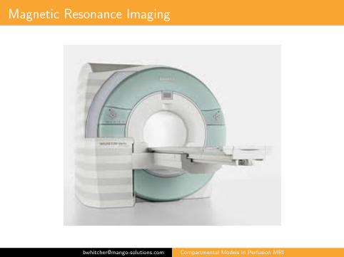
Magnetic Resonance Imaging
A constant, homogeneous magnetic field (the B0 field) is usedto polarize spins.
The exposure of nuclei to a radio frequency (RF) pulse (theB1 field) at the Larmor frequency causes the nuclei in thelower energy state to jump to the higher energy state.
Macroscopic level: this causes net magnetization to spiral awayfrom the B0 field.After time, the magnetization vector becomes perpendicular tothe main B0 field.
MR imaging is based on the relaxation that takes place afterthe RF pulse has stopped.
It is repeated for many different levels of phase encoding tobuild up a matrix in k-space.
A 2D Fourier transform is performed, resulting in a single slicefrom an MRI acquisition.
[email protected] Compartmental Models in Perfusion MRI

Dynamic Contrast-Enhanced MRI
Primary breast cancer example from the PSSC
K trans ve
[email protected] Compartmental Models in Perfusion MRI

Dynamic Contrast-Enhanced MRI
RIDER Neuro MRI data from Daniel Barboriak’s lab at Duke
[email protected] Compartmental Models in Perfusion MRI
Dynamic Contrast-Enhanced MRI
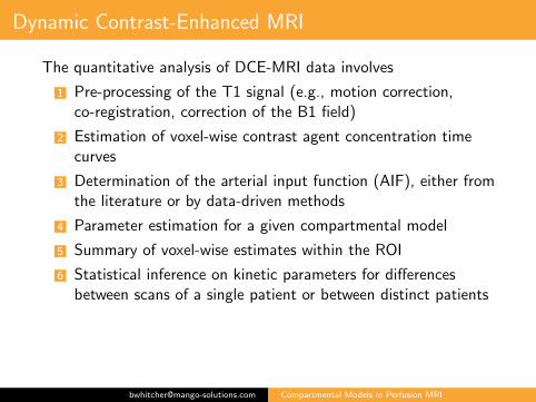
The quantitative analysis of DCE-MRI data involves
1 Pre-processing of the T1 signal (e.g., motion correction,co-registration, correction of the B1 field)
2 Estimation of voxel-wise contrast agent concentration timecurves
3 Determination of the arterial input function (AIF), either fromthe literature or by data-driven methods
4 Parameter estimation for a given compartmental model
5 Summary of voxel-wise estimates within the ROI
6 Statistical inference on kinetic parameters for differencesbetween scans of a single patient or between distinct patients
[email protected] Compartmental Models in Perfusion MRI
Dynamic Contrast-Enhanced MRI
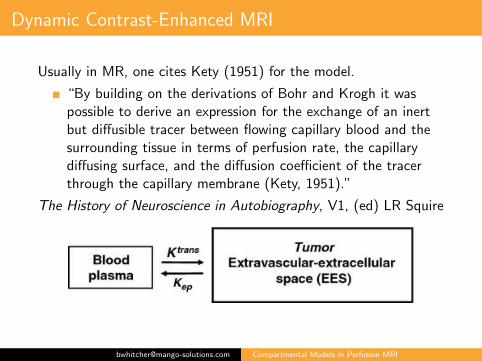
Usually in MR, one cites Kety (1951) for the model.
“By building on the derivations of Bohr and Krogh it waspossible to derive an expression for the exchange of an inertbut diffusible tracer between flowing capillary blood and thesurrounding tissue in terms of perfusion rate, the capillarydiffusing surface, and the diffusion coefficient of the tracerthrough the capillary membrane (Kety, 1951).”
The History of Neuroscience in Autobiography, V1, (ed) LR Squire
[email protected] Compartmental Models in Perfusion MRI
Dynamic Contrast-Enhanced MRI
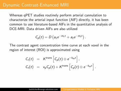
Whereas qPET studies routinely perform arterial cannulation tocharacterize the arterial input function (AIF) directly, it has beencommon to use literature-based AIFs in the quantitative analysis ofDCE-MRI. Data driven AIFs are also utilized
Cp(t) = D(a1e
−m1t + a2e−m2t
).
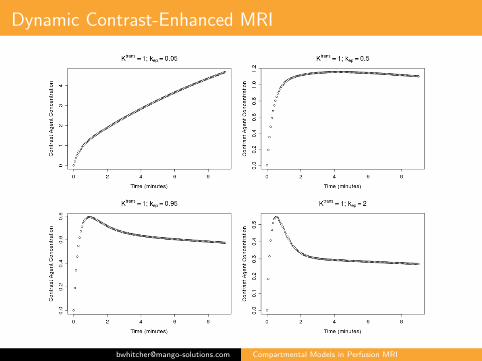
The contrast agent concentration time curve at each voxel in theregion of interest (ROI) is approximated using
Ct(t) = K trans[Cp(t) ⊗ e−kept
],
Ct(t) = vpCp(t) + K trans[Cp(t) ⊗ e−kept
].
[email protected] Compartmental Models in Perfusion MRI
Dynamic Contrast-Enhanced MRI
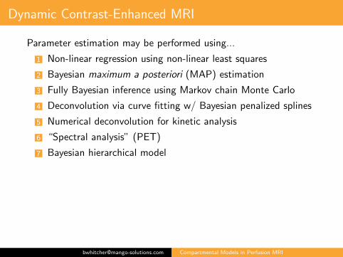
Parameter estimation may be performed using...
1 Non-linear regression using non-linear least squares
2 Bayesian maximum a posteriori (MAP) estimation
3 Fully Bayesian inference using Markov chain Monte Carlo
4 Deconvolution via curve fitting w/ Bayesian penalized splines
5 Numerical deconvolution for kinetic analysis
6 “Spectral analysis” (PET)
7 Bayesian hierarchical model
[email protected] Compartmental Models in Perfusion MRI
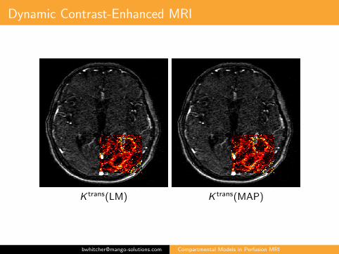
Dynamic Contrast-Enhanced MRI
K trans(LM) K trans(MAP)
[email protected] Compartmental Models in Perfusion MRI
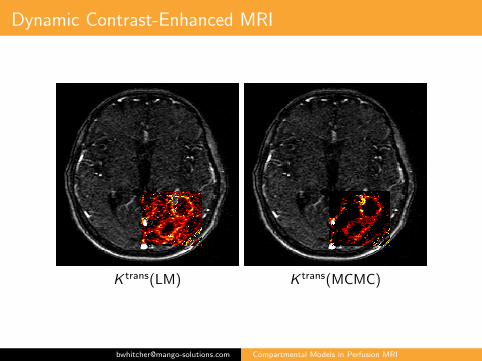
Dynamic Contrast-Enhanced MRI
K trans(LM) K trans(MCMC)
[email protected] Compartmental Models in Perfusion MRI
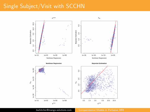
Single Subject/Visit with SCCHN
1e−01 1e+02 1e+05 1e+08
0.5
1.0
2.0
5.0
10.0
20.0
Ktrans
Nonlinear Regression
Bay
esia
n E
stim
atio
n
1e−09 1e−06 1e−03 1e+00
0.5
1.0
2.0
5.0
kep
Nonlinear Regression
Bay
esia
n E
stim
atio
n
1e−01 1e+02 1e+05 1e+08
1e−
091e
−06
1e−
031e
+00
Nonlinear Regression
Ktrans
k ep
0.5 1.0 2.0 5.0 10.0 20.0
0.5
1.0
2.0
5.0
Bayesian Estimation
Ktrans
k ep
[email protected] Compartmental Models in Perfusion MRI

Discussion
Compartmental models are utilized in many medical imageexperiments (MRI and PET)
Research is, unfortunately, done in isolation
Which is the best method?
Prior informationMore complex models (demanding acquisition protocol)Differentiating flow from permeability?Smoothing
What can we (image modellers) learn from pharmacometrics?
How can we merge PK or PD information from imaging withnon-imaging sources?
PET biodistribution studies?
Small molecules versus large molecules
[email protected] Compartmental Models in Perfusion MRI