balance of th1/th2 cytokines associated with the preventive effect of incomplete freund's...
TRANSCRIPT
Journal of Autoimmunity (2001) 17, 289–295doi:10.1006/jaut.2001.0552, available online at http://www.idealibrary.com on
Balance of Th1/Th2 Cytokines Associated with thePreventive Effect of Incomplete Freund’s Adjuvant on theDevelopment of Adjuvant Arthritis in LEW Rats
Aslam Hossain, Cong Long Zheng, Akiko Kukita and Osamu Kohashi
Department of Microbiology,Saga Medical School, 5-1-1 Nabeshima,Saga 849-8501, Japan
Received 11 May 2001Accepted 13 September 2001
Key words: AA, cytokine, IFA,rats, Th1/Th2
Complete Freund’s adjuvant (CFA) could induce adjuvant arthritis (AA) inLEW rats and incomplete Freund’s adjuvant (IFA) could induce oil inducedarthritis (OIA) in DA but not in LEW rats. Lymph node cells (LNCs) from theseAA and OIA rats showed increased mRNA expression of IFN-�, IL-2 andTNF-� but not IL-4. LNCs from IFA immunized LEW rats showed increasedexpression of IL-4, reduced expression of IFN-� and TNF-� and no IL-2, incontrast to IFA immunized DA rats. The pretreatment of IFA before CFAchallenge could completely prevent AA in LEW rats and their LNCs showedincreased expression of IL-4 and IFN-� but not IL-2 and TNF-�. In F1(LEW×DA) rats, IFA could not induce OIA but the pretreatment of IFA beforeCFA challenge could induce very mild AA with 80% incidence, LNCs showingan elevated expression of all the above cytokines. These findings suggest thatincreased Th1 cytokine expression is associated with disease development andthat increased IL-4 expression or the balance of Th2 over Th1 cytokineexpression plays an important regulatory role in disease development.
© 2001 Academic Press
Correspondence to: Professor Osamu Kohashi, MD, Ph.D., Depart-ment of Microbiology, Saga Medical School, 5-1-1 Nabeshima,Saga 849-8501, Japan. Fax: (+) 81 952 342015; E-mail:[email protected]
Introduction
Adjuvant arthritis (AA) in LEW rats is commonlyused as an animal model of rheumatoid arthritis (RA).AA can be induced in LEW rats by a single intra-dermal injection of complete Freund’s adjuvant (CFA)consisting of heat-killed Mycobacterium tuberculosis(Mt) and incomplete Freund’s adjuvant (IFA) [1]. Onthe other hand, an intradermal injection of IFA alonecan induce oil induced arthritis (OIA) in DA rats butnot in LEW rats [2]. Since AA and OIA can bepassively transferred to either naive or irradiatedrecipients by the lymph node cells (LNCs) derivedfrom the arthritis rats, it is believed that both AA andOIA are T cell-mediated diseases [3, 4].
CD4+ T cells have been classified into two majortypes, T helper type 1 (Th1) and Th2 type on the basisof their distinct pattern of cytokine profiles. Th1 cellsproduce predominantly IFN-� and IL-2, whereas Th2cells produce predominantly IL-4, IL-5, IL-10 andIL-13 [5]. In experimental animal models of arthritis,Th1 phenotype and/or other pro-inflammatorycytokines are considered to be responsible for diseaseprogression, while Th2 type cytokines are associated
2890896–8411/01/080289+07 $35.00/0
with disease resolution [6–13]. Generally, the balanceof the cytokines produced by these two subsets of Tcells plays a very important role in the induction andpropagation of diseases. The mRNA expression ofseveral cytokines during the various phases of AAindicated that the increased expression of Th1 andinflammatory cytokines were associated with diseaseprogression, while the subsiding of the diseases wasparalleled by increased Th2-like cytokines [9]. Alter-natively, some drugs could completely prevent orsuppress the development of AA by inhibiting theproduction of Th1 type cytokines and inflammatorycytokines, suggesting the importance of Th1 type andinflammatory cytokines in AA [14, 15]. Furthermore,anti-inflammatory cytokine therapy (such as anti-TNF-� and anti-IL-1) can also suppress the develop-ment of AA in rats [16]. The systemic administrationof murine IL-4 or intra-articular inoculation of IL-4gene can prevent the development of AA in rats,suggesting that Th2 type cytokines are important indisease resolution [17, 18]. As for AA, Th1 type andinflammatory cytokines also play an important role inthe pathogenesis of OIA [12, 13].
We reported earlier that pretreatment with IFA 28days before CFA challenge could completely preventthe development of AA in LEW rats and suggested thepossible regulatory role of Th2 cells in Th1 mediatedAA [19]. In order to understand the role of Th1 andTh2 cytokines in the induction or prevention of AA
© 2001 Academic Press
290 M. A. Hossain et al.
and OIA, we have studied the mRNA expression ofIFN-� and IL-2 (Th1 type cytokines), IL-4 and IL-5(Th2 type cytokines) and TNF-� (pro-inflammatorycytokine) in the inguinal lymph node cells (ILNCs)of LEW, DA and F1 (LEW×DA) rats by reversetranscriptase-polymerase chain reaction (RT-PCR).
Materials and Methods
Animals
Inbred LEW rats were obtained from Charles RiverInc., Tokyo, Japan and DA rats from Japan SLC Inc.,Shizuoka, Japan. Rats were housed at the center forLaboratory Animals, Saga Medical School, fed a sterilecommercial diet and water ad libitum. They weremaintained by continuous brother and sister mating,and for F1 (LEW×DA) rats by cross-mating betweenLEW and DA rats under specific pathogen-free con-ditions. At the age of 7–10 weeks only female LEWand DA rats and both sexes of F1 rats were used inthe experiments. All the animal experiments werecarried out under clean room facilities in accordancewith Saga Medical School Guidelines for AnimalExperiments.
Adjuvant
Mycobacterium tuberculosis H37 Ra (Mt) and IFA werepurchased from Difco Laboratories (Detroit, MI,USA).
Preparation of CFA
CFA was prepared as follows: 10 mg of H 37 Ra wasground with a mortar and pestle and IFA was addeddrop by drop to a final volume of 1 ml. This prep-aration was kept in an incubator at 42°C for 72 h toimprove the solubility of Mt in IFA. An equal volumeof phosphate-buffered saline (PBS) was added drop bydrop with continuous mixing in this preparation tomake a good water-in-oil emulsion.
Immunization
LEW and F1 rats were intradermally injected at thebase of the tail with 0.1 ml of CFA (CFA group). LEWand F1 rats were intradermally injected at the left hindfoot pad with 50 �l of IFA (IFA group). LEW and F1rats were intradermally injected at the left hind footpad with 50 �l of IFA and 0.1 ml of CFA was chal-lenged intradermally at the base of the tail after 28days of IFA pretreatment (IFA/CFA group). For com-parison of cytokine profiles between LEW and DArats, they were intradermally injected at the base ofthe tail with 0.2 ml of IFA. For the induction of OIA,F1 rats were intradermally injected at the base of thetail with 0.2 ml of IFA.
Clinical evaluation of arthritis
The severity of arthritis was evaluated according to aprevious paper [20]. In brief, each paw was gradedfrom 0 to 4 at intervals of 0.5, where 0 was no obviousarthritis and 4 was equal to full swelling of almost allpaw-joints. The highest achievable clinical scoreswould be 16 but in case of preinjection into the lefthind foot pad, the highest achievable clinical scorewould be 12 (excluding the preinjected left hind paw).The rats were observed and scored every other day ina blinded fashion.
Collection of ILNCs for RT-PCR analysis
ILNCs were isolated separately from the individualrats for RT-PCR analysis in all experiments. Eachgroup of LEW rats consisted of 5 rats and each groupof F1 rats consisted of 4 rats. ILNCs were harvestedfrom differently treated LEW and F1 rats as follows:CFA group, ILNCs were isolated on day 11 from LEWrats and on day 10 from F1 rats after CFA injection;IFA group, ILNCs were isolated on day 28 from LEWand F1 rats after IFA injection. In the IFA/CFA group,ILNCs were isolated on day 11 from LEW rats and onday 10 from F1 rats after CFA challenge. In the naivegroup, ILNCs were isolated from the age-matchednaive LEW and F1 rats. For the comparison ofcytokine profiles between IFA immunized DA andLEW rats, ILNCs were isolated from DA and LEW ratson day 15 after IFA injection. ILNCs were also isolatedfrom the age-matched naive DA and LEW rats, ascontrol groups. Each group consisted of 4 rats in thiscomparative study.
RT-PCR analysis of cytokine mRNA expression inILNCs
ILNCs of the individual rats were homogenized tomake a single-cell suspension in PBS and washedthree times with PBS. Total RNA was extracted usinga commercial kit (Isogen, Nippon gene, Toyama,Japan). Single stranded cDNA was synthesized from1 �g of total RNA and was subjected to PCR using aRT-PCR kit (Takara, Tokyo, Japan) with specific ratprimers as shown in Table 1. The amplification ofcDNA with specific primers was 30 cycles for IL-2,IFN-�, TNF-� and GADPH and 36 cycles for IL-4 andIL-5. Following amplification, the PCR products wereseparated by electrophoresis on 1.5% agarose geland visualized by ethidium bromide staining. Wemeasured the mRNA expression of cytokines repeat-edly (4 to 5 times), from individual rats among thegroups, to get repeatable results throughout all theexperiments.
Statistical analysis
Statistical analysis was carried out using the commer-cially available microcomputer program Stat View.Statistical significance of differences between the two

Th1/Th2 cytokines in the prevention of AA 291
groups was assessed by the Mann-Whitney U-test andthe P value <0.05 was taken as significant.
Results
Preventive effect of IFA on the development ofAA in LEW rats
As shown in Figure 1, we confirmed our previousreport [19] that a single intradermal injection ofCFA could induce severe arthritis in all the rats andthe pretreatment of IFA could completely prevent thedevelopment of AA in all the rats.
Table 1. PCR primers used for RT-PCR analysis
Type ofmRNA Primer sequence Sequence based on
accession no.Basepairs Reference
GADPH 5′-CATGGAGAAGGCTGGGGCTC-3′ X 02231 414 [21]5′-AACGGATACATTGGGGTAG-3′
IFN-� 5′-CCCTCTCTGGCTGTTACTGC-3′ AF 010466 415 [9]5′-CTCCTTTTCCGCTTCCTTAG-3′
IL-2 5′-TACAGCATGCAGCTCGCATCCTGTG-3′ M 22899 414 [22]5′-CAGAAATTCCACCACAGTTGCTGGC-3′
IL-4 5′-ACCTTGCTGTCACCCTGTTCTGC-3′ X 16058 352 [23]5′-GTTGTGAGCGTGGACTACTTCACG-3′
IL-5 5′-TGACGAGCAATGAGACGATG-3′ AJ 011299 248 [9]5′-TCATCACGCCAAGGAACTCT-3′
TNF-� 5′-GGCAGGTCTACTTTGGAGTCATTGC-3′ D 00475 319 [24]5′-ACATTCGGGGATCCAGTGAGTTCCG-3′
28
11
Days after intradermal injection of CFA
Mea
n c
lin
ical
sco
res
10
9
7
5
3
10
2624222018161412–28
IFA
CFA
CFAIFA/CFA
Figure 1. Preventive effect of IFA on the development of AAin LEW rats. CFA was intradermally injected at the base ofthe tail of all the LEW rats either without any pretreatmentor the injection of 50 �l IFA intradermally into the left hindfoot pad 28 days before CFA challenge. Each groupconsisted of 10 rats.
Figure 2. mRNA expression of cytokines by the LNCs in thepreventive effect of IFA on AA in LEW rats. LNCs wereisolated from individual rats on day 11 after CFA injectionwithout IFA pretreatment (lane 1), with IFA pretreatment 28days before CFA injection (lane 2), on day 28 after IFAinjection (lane 3) and naive control (lane 4), respectively.Total RNA was extracted from the LNCs of individual rats,reverse transcribed, cDNA was amplified using specific ratprimers and the PCR products were subjected to gel electro-phoresis stained with ethidium bromide. Each groupconsisted of 5 rats.
mRNA expression of cytokines by LNCs in thepreventive effect of IFA on AA in LEW rats
As shown in Figure 2, LNCs derived from CFAimmunized LEW rats (around the onset of the disease)(lane 1), showed increased mRNA expression of
IFN-�, IL-2 and TNF-� and similar mRNA expressionof IL-4 to naive rats (lane 4), indicating the relation-ship between increased expression of Th1 typecytokines and disease development. LNCs derivedfrom IFA immunized rats (no sign of arthritis at all)(lane 3), showed increased mRNA expression of IL-4
292 M. A. Hossain et al.
and TNF-�, reduced mRNA expression of IFN-� andsimilar mRNA expression of IL-2 to naive rats (lane 4),suggesting the important regulatory role of IL-4 in thedevelopment of the disease. LNCs derived from IFApretreated and subsequently CFA challenged rats (AAprevented rats) (lane 2), showed increased mRNAexpression of IL-4 and IFN-� and similar mRNAexpression of IL-2 and TNF-� to naive rats (lane 4),suggesting higher mRNA expression of Th2 than Th1cytokines. There was apparently no difference inmRNA expression of IL-5 among the immunized andnon-immunized naive LEW rats (lanes 1–4).
mRNA expression of cytokines by LNCs amongDA and LEW rats after IFA immunization
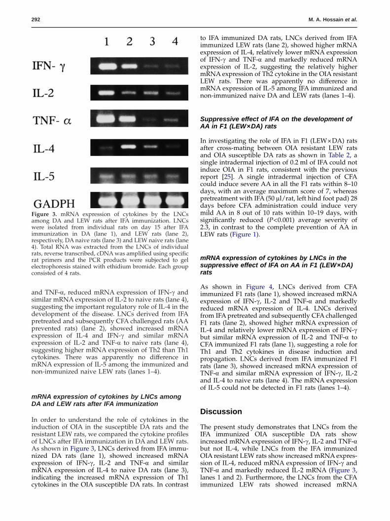
In order to understand the role of cytokines in theinduction of OIA in the susceptible DA rats and theresistant LEW rats, we compared the cytokine profilesof LNCs after IFA immunization in DA and LEW rats.As shown in Figure 3, LNCs derived from IFA immu-nized DA rats (lane 1), showed increased mRNAexpression of IFN-�, IL-2 and TNF-� and similarmRNA expression of IL-4 to naive DA rats (lane 3),indicating the increased mRNA expression of Th1cytokines in the OIA susceptible DA rats. In contrast
to IFA immunized DA rats, LNCs derived from IFAimmunized LEW rats (lane 2), showed higher mRNAexpression of IL-4, relatively lower mRNA expressionof IFN-� and TNF-� and markedly reduced mRNAexpression of IL-2, suggesting the relatively highermRNA expression of Th2 cytokine in the OIA resistantLEW rats. There was apparently no difference inmRNA expression of IL-5 among IFA immunized andnon-immunized naive DA and LEW rats (lanes 1–4).
mRNA expression of cytokines by LNCs in thesuppressive effect of IFA on AA in F1 (LEW×DA)rats
As shown in Figure 4, LNCs derived from CFAimmunized F1 rats (lane 1), showed increased mRNAexpression of IFN-�, IL-2 and TNF-� and markedlyreduced mRNA expression of IL-4. LNCs derivedfrom IFA pretreated and subsequently CFA challengedF1 rats (lane 2), showed higher mRNA expression ofIL-4 and relatively lower mRNA expression of IFN-�but similar mRNA expression of IL-2 and TNF-� toCFA immunized F1 rats (lane 1), suggesting a role forTh1 and Th2 cytokines in disease induction andpropagation. LNCs derived from IFA immunized F1rats (lane 3), showed increased mRNA expression ofTNF-� and similar mRNA expression of IFN-�, IL-2and IL-4 to naive rats (lane 4). The mRNA expressionof IL-5 could not be detected in F1 rats (lanes 1–4).
Figure 3. mRNA expression of cytokines by the LNCsamong DA and LEW rats after IFA immunization. LNCswere isolated from individual rats on day 15 after IFAimmunization in DA (lane 1), and LEW rats (lane 2),respectively, DA naive rats (lane 3) and LEW naive rats (lane4). Total RNA was extracted from the LNCs of individualrats, reverse transcribed, cDNA was amplified using specificrat primers and the PCR products were subjected to gelelectrophoresis stained with ethidium bromide. Each groupconsisted of 4 rats.
Discussion
The present study demonstrates that LNCs from theIFA immunized OIA susceptible DA rats showincreased mRNA expression of IFN-�, IL-2 and TNF-�but not IL-4, while LNCs from the IFA immunizedOIA resistant LEW rats show increased mRNA expres-sion of IL-4, reduced mRNA expression of IFN-� andTNF-� and markedly reduced IL-2 mRNA (Figure 3,lanes 1 and 2). Furthermore, the LNCs from the CFAimmunized LEW rats showed increased mRNA
Suppressive effect of IFA on the development ofAA in F1 (LEW×DA) rats
In investigating the role of IFA in F1 (LEW×DA) ratsafter cross-mating between OIA resistant LEW ratsand OIA susceptible DA rats as shown in Table 2, asingle intradermal injection of 0.2 ml of IFA could notinduce OIA in F1 rats, consistent with the previousreport [25]. A single intradermal injection of CFAcould induce severe AA in all the F1 rats within 8–10days, with an average maximum score of 7, whereaspretreatment with IFA (50 �l/rat, left hind foot pad) 28days before CFA administration could induce verymild AA in 8 out of 10 rats within 10–19 days, withsignificantly reduced (P<0.001) average severity of2.3, in contrast to the complete prevention of AA inLEW rats (Figure 1).

Th1/Th2 cytokines in the prevention of AA 293
Table 2. Suppressive effect of IFA on the development of AA in F1 (LEW×DA) rats
Pretreatment Injection Disease incidence(diseased/total rats)
Day ofonset
Maximum score(mean±SE)a
None IFA 0/10 — —None CFA 10/10 8–10 7.0±0.27IFA CFA 8/10 10–19 2.3±0.59*
F1 rats were intradermally injected at the base of the tail with 0.2 ml of IFA for the induction of OIA. CFA wasinjected intradermally at the base of the tail of F1 rats either without any pretreatment or 50 �l IFA wasintradermally injected at the left hind foot pad 28 days before CFA challenge. Arthritis score was measuredevery other day as described in Materials and Methods. SE, standard error; amean of maximum severity of thediseased rats. *P<0.001 versus CFA immunized F1 rats.
Figure 4. mRNA expression of cytokines by the LNCs in thesuppressive effect of IFA on AA in F1 (DA×LEW) rats. LNCswere isolated from individual rats on day 10 after CFAinjection without IFA pretreatment (lane 1), with IFA pre-treatment 28 days before CFA injection (lane 2), on day 28after IFA injection (lane 3) and naive control (lane 4),respectively. Total RNA was extracted from the LNCs ofindividual rats, reverse transcribed, cDNA was amplifiedusing specific rat primers and the PCR products weresubjected to gel electrophoresis stained with ethidiumbromide. Each group consisted of 4 rats.
expression of IFN-�, IL-2 and TNF-� but not IL-4,similar to the cytokine profiles of the OIA susceptibleDA rats. Interestingly, the LNCs from the IFA pre-treated and subsequently CFA challenged LEW ratsshowed increased mRNA expression of IL-4 andmarkedly reduced mRNA expression of IL-2 andTNF-� compared with CFA immunized LEW rats(Figure 2, lanes 1 and 2). These findings with regard tothe mRNA expression of Th1 and Th2 cytokinessuggest that OIA and AA are Th1 mediated diseases.The present study further demonstrates that IFA can-not induce OIA in F1 (LEW×DA) rats but pretreat-ment with IFA before CFA challenge can induce very
mild AA in F1 rats, with 80% incidence. The LNCsfrom CFA immunized F1 rats showed increasedmRNA expression of IFN-�, IL-2 and TNF-� andmarkedly reduced mRNA expression of IL-4, whilethe LNCs from the IFA pretreated and subsequentlyCFA challenged rats showed increased mRNA expres-sion of IL-4 and reduced mRNA expression of IFN-�but similar mRNA expression of IL-2 and TNF-� toCFA immunized rats (Figure 4, lanes 1 and 2). Thus,the present study suggests that the increased IL-4mRNA expression, or the balance of Th2 over Th1cytokine mRNA expression, plays an important regu-latory role in the development of OIA and AA. Thefact that IFA could induce higher Th1 cytokine expres-sion in DA rats and rather higher Th2 cytokine ex-pression in LEW rats can be explained by the diseaseregulatory genes between DA and LEW rats [25]and/or by an important role of dendritic cells (DCs) inTh1 and Th2 differentiation [26]. If the latter is thecase, CD8�+ DCs are associated with Th1 differen-tiation in DA rats, while CD8�− DCs are associatedwith Th2 differentiation in LEW rats following immu-nization with IFA in DA and LEW rats.
As with the regulatory role of IL-4 in the inductionof Th1 mediated experimental arthritis, continuousadministration of exogenous murine IL-4 could sup-press the development of collagen-induced arthritis(CIA) in mice [8]. Similarly, systemic administration ofrecombinant IL-4 could also suppress the develop-ment of streptococcal cell wall induced arthritis in rats[27]. Bober et al. demonstrated that systemic admin-istration of murine IL-4 could suppress the pawedema and bone destruction of AA in rats [17]. Inaddition, the increased expression of IL-4 via IL-4gene therapy in rats could also reduce the clinical andhistological parameters of AA [18]. Interestingly, CFAcould not induce AA in mice but treatment with amonoclonal antibody against IL-4 could successfullydo so [28]. Furthermore, the regulatory role of IL-4has also been reported in OIA, where IFA co-immunization with ovalbumin cannot induce OIA inDA rats because of the increased mRNA expression ofIL-4 [7, 12]. It is thus considered that IL-4 plays animportant regulatory role in the disease induction andpropagation. TNF-� has also been proven to have apivotal role in the pathogenesis of RA and experimen-tal arthritis. Anti-TNF-� therapy has been reported to

294 M. A. Hossain et al.
inhibit the development of AA in rats and of CIA inmice [16, 29]. The strong mRNA expression of TNF-�induced by IFA was important for the induction ofOIA in DA rats [12]. IL-2 also played an important rolein the induction of AA and OIA. Haynes et al. dem-onstrated that cyclosporin A and lobenzarit couldprevent the development of AA in rats by specificallyinhibiting the production of IL-2, TNF-� and IFN-�[14]. In addition, the recipients treated with anti-IL-2receptor antibody before transfer of CFA sensitizedspleen cells or in vitro culture of these sensitizedspleen cells with this antibody failed to develop thedisease, suggesting the important pathological role ofIL-2 in AA [30]. Moreover, Svelander et al. reportedthat IFA triggering polyclonal IL-2 dependent Th1cells played an important role for the induction ofOIA in DA rats [13]. The present findings, therefore,suggest that increased expression of IL-4 mRNA andreduced expression of IL-2 mRNA (Figure 2, lane 3;Figure 3, lane 2) may play an important regulatoryrole for the induction of OIA in LEW rats after IFAimmunization. Our previous findings [19] suggestedthat IFA immunization by itself was not enough tostimulate the Th1 and Th2 cells to induce or preventAA in LEW rats, while subsequent CFA challenge inthe IFA pretreated rats stimulated Th2 cells ratherthan Th1 cells, resulting in the prevention of AA.Taken altogether, the present study suggests that theincreased mRNA expression of IL-4 and the markedlyreduced mRNA expression of TNF-� and IL-2 areresponsible for the prevention of AA in the IFApretreated and subsequently CFA challenged LEWrats, through the balance of Th2 over Th1 cytokines.Alternatively, the preventive effect of IFA on AA inLEW rats can be explained by the regulatory T cells. Itis possible that IFA induces inflammation [19] result-ing in the upregulation of self heat shock protein(hsp60) [31] and that the regulatory T cells which canrespond to both self hsp60 and mycobacterial hsp65in CFA are activated to prevent AA, throughmechanisms which are still unknown [32, 33].
With regard to F1 rats, the present study confirmsthat IFA cannot induce OIA [25] and adds a newfinding that the pretreatment of IFA before CFA chal-lenge can induce very mild AA (P<0.001) with 80%incidence and delayed onset (Table 2). Holmdahl et al.postulated that OIA was genetically restricted, influ-enced by recessive genes in DA and suppressive genesin LEW rats [25]. The present findings, therefore,suggest that LEW suppressive genes may play adominant role in the suppressive effect of IFA on thedevelopment of AA in F1 rats. In terms of cytokineprofiles, the LNCs of CFA immunized in F1 ratsshowed increased mRNA expression of Th1 typecytokines, similar to that of AA LEW rats (Figure 2,lane 1; Figure 4, lane 1). IFA could induce TNF-�expression in F1 rats (Figure 4, lane 3) but the elevatedmRNA expression of TNF-� may not be enough forthe induction of OIA, as described above in LEW rats.The increased mRNA expression of IL-4 and therelatively reduced mRNA expression of IFN-� in theIFA pretreated and subsequently CFA challenged F1rats (Figure 4, lane 2) may provide an explanation for
the suppressive effect of IFA on AA. Furthermore, theregulatory T cells may also be involved in the sup-pression of AA in F1 rats, like LEW rats, as describedabove.
Finally, the present study showed no difference inmRNA expression of IL-5 in immunized and non-immunized LEW and DA rats (Figures 2 and 3). ThemRNA expression of IL-5 by the inguinal LNCs ofCFA immunized and non-immunized LEW rats in thispresent study is consistent with the previous report[9]. The elevated mRNA expression of IL-5 in thespleen at the later stage of AA is associated withdisease resolution [9]. Although there is no report ofIL-5 in OIA, further analysis of IL-5 mRNA expressionof LNCs from the early to the later stages of AA orOIA is necessary.
In conclusion, the present findings of the mRNAexpression of Th1 and Th2 cytokines support thetheory that AA and OIA are Th1 mediated diseases.The increased mRNA expression of IL-4 and thereduced mRNA expression of IL-2 and TNF-� areresponsible for the prevention of AA in the IFApretreated and subsequently CFA challenged LEWrats, through the balance of Th2 over Th1 cytokineresponses. Further studies are continuing to isolate theT cell clone responsible for either induction or preven-tion of AA for future therapeutic models of humanrheumatoid arthritis.
Acknowledgements
We thank Dr K. Ohki of the Department of Microbiol-ogy, Saga Medical School, for his help and suggestionsduring the work and Ms I. Nanbu for her excellentsecretarial assistance. We also thank Dr M. Morimotoof Saga Medical School Animal Care Center forarranging the animal experimental facilities.
References1. Pearson C.M. 1996. Development of arthritis,
periarthritis and periostitis in rats given adjuvant.Proc. Soc. Exp. Biol. Med. 96: 95–101
2. Kleinau S., Erlandsson H., Holmdahl R., Klareskog L.1991. Adjuvant oils induce arthritis in the DA rat. I.Characterization of the disease and evidence for animmunological involvement. J. Autoimmun. 4: 871–880
3. Taurog J.D., Sandberg G.P., Mahowald M.L. 1983. Thecellular basis of adjuvant arthritis II. Characterizationof the cells mediating passive transfer. Cell Immunol.80: 198–204
4. Kleinau S., Klareskog L. 1993. Oil-induced arthritis inDA rats: passive transfer by T cells but not withserum. J. Autoimmun. 6: 449–458
5. Abbas A.K., Murphy K.M., Sher A. 1996. Functionaldiversity of helper T lymphocytes. Nature 383: 787–793
6. Mauri C., Williams R.O., Walmsley M., Feldmann M.1996. Relationship between Th1/Th2 cytokine patternsand arthritogenic response in collagen-inducedarthritis. Eur. J. Immunol. 26: 1511–1518
7. Mussener A., Lorentzen J.C., Kleainau S., Klareskog L.1997. Altered Th1/Th2 balance associated with
Th1/Th2 cytokines in the prevention of AA 295
non-major histocompatibility complex genes incollagen-induced arthritis in resistant and non-resistantrat strains. Eur. J. Immunol. 27: 695–699
8. Horsfall A.C., Butler D.M., Marinova L., Warden P.J.,Williams R.O., Maini R.N., Feldmann M. 1997.Suppression of collagen-induced arthritis bycontinuous administration of IL-4. J. Immunol. 159:5687–5696
9. Schmidt-Weber C.B., Pohlers D., Siegling A., SchadlichH., Buchner E., Volk H., Palombo-Kinne E., EmmrichF., Kinne R.W. 1999. Cytokine gene activation insynovial membrane, regional lymph nodes and spleenduring the course of rat adjuvant arthritis. CellImmunol. 195: 53–65
10. Ayer L.M., Issekutz A.C., Waterhouse C.C.M., StadnykA.W. 2000. Cytokine mRNA in the joints and draininglymph nodes of rats with adjuvant arthritis and effectsof cyclosporin A. Inflammation 24: 447–461
11. Boyle D.L., Nguyen K.H.Y., Zhuang S., Shi Y.,McCormack J.E., Chada S., Firestein G.S. 1999.Intra-articular IL-4 gene therapy in arthritis:anti-inflammatory effect and enhanced Th2 activity.Gene Therapy 6: 1911–1918
12. Mussener A., Klareskog L., Lorentzen J.C., Kleainau S.1995. TNF-� dominates cytokine mRNA expression inlymphoid tissues of rats developing collagen- andoil-induced arthritis. Scand. J. Immunol. 42: 128–134
13. Svelander L., Mussener A., Erlandsson-Harris H.,Kleinau S. 1997. Polyclonal Th1 cells transferoil-induced arthritis. Immunology 91: 260–265
14. Haynes D.R., Gadd S.J., Whitehouse M.W., MayrhoferG., Vernon-Roberts B. 1996. Complete prevention ofthe clinical expression of adjuvant-induced arthritis inrats by cyclosporin-A and lobenzarit: the regulation oflymph node cell populations and cytokine production.Inflamm. Res. 45: 159–165
15. Ohta Y., Yamane M., Sohda T., Makino H. 1997.TAK-603 selectively suppresses Th1-type cytokineproduction and inhibits the progression of adjuvantarthritis. Immunology 92: 75–83
16. Issekutz A.C., Meager A., Otterness I., Issekutz T.B.1994. The role of tumor necrosis factor-alpha and IL-1in polymorphonuclear leukocyte and T lymphocyterecruitment to joint inflammation in adjuvant arthritis.Clin. Exp. Immunol. 97: 26–32
17. Bober L.A., Rojas-triana A., Jackson J.V., Leach M.W.,Manfra D., Narula S.K., Grace M.J. 2000. Regulatoryeffects of interleukin-4 and interleukin-10 on humanneutrophil function ex vivo and on neutrophil influxin a rat model of arthritis. Arthritis Rheum. 43:2660–2667
18. Woods J.M., Katschke K.J. Jr, Volin M.V., Ruth J.H.,Woodruff D.C., Amin M.A., Connors M.A., Kurata H.,Arai K., Haines III G.K., Kumar P., Koch A.E. 2001.IL-4 adenoviral gene therapy reduces inflammation,proinflammatory cytokines, vascularization and bonydestruction in rat adjuvant-induced arthritis. J.Immunol. 166: 1214–1222
19. Zhang L., Mia M.Y., Zheng C.L., Hossain M.A.,Yamasaki F., Tokunaga O., Kohashi O. 1999. Thepreventive effects of incomplete Freund’s adjuvant andother vehicles on the development of adjuvant-induced arthritis in Lewis rats. Immunology 98: 267–272
20. Wood F.D., Pearson C.M., Tanaka A. 1967. Capacity ofmycobacterial wax D and its subfractions to induceadjuvant arthritis in rats. Int. Arch. Allergy. Appl.Immunol. 35: 456–467
21. Yun Tso J., Sun X., Kao T., Reece K.S., Wu R. 1985.Isolation and characterization of rat and humanglyceraldehyde-3-phosphate dehydrogenase cDNAs:genomic complexity and molecular evolution of thegene. Nucleic Acids Research 13: 2485–2502
22. Zipris D., Greiner D.L., Malkani S., Whalen B., MordesJ.P., Rossini A.A. 1996. Cytokine gene expression inislets and thyroids of BB rats. IFN-� and IL-12p40mRNA increase with age in both diabetic andinsulin treated nondiabetic BB rats. J. Immunol. 156:1315–1321
23. Ide K., Hayakawa H., Yagi T., Sato A., Koide Y.,Yoshida A., Uchijima M., Suda T., Chida K., NakamuraH. 1999. Decreased expression of Th2 cyotkine mRNAcontributes to the lack of allergic bronchialinflammation in aged rats. J. Immunol. 163: 396–402
24. Mitsui Y., Okamoto K., Martin D.P., Schmelzer J.D.,Low P.A. 1999. The expression of proinflammatorycytokine mRNA in the sciatic-tibial nerve ofischemia-reperfusion injury. Brain Research 844: 192–195
25. Holmdahl R., Goldschmidt T.J., Kleinau S., Kvick C.,Jonsson R. 1992. Arthritis induced in rats withadjuvant oil is a genetically restricted, �� T-celldependent autoimmune disease. Immunology 76:197–202
26. Rengarajan J., Szabo S.J., Glimcher L.H. 2000.Transcriptional regulation of Th1/Th2 polarization.Immunol. Today 21: 479–483
27. Allen J.B., Wong H.L., Costa G.L., Bienkowski M.J.,Wahl S.M. 1993. Suppression of monocyte functionand differentiation regulation of IL-1 and IL-1 ra byIL-4 contribute to resolution of experimental arthritis.J. Immunol. 151: 4344–4351
28. Yoshino S., Murata Y., Ohsawa 1998. Successfulinduction of adjuvant arthritis in mice by treatmentwith a monoclonal antibody against IL-4. J. Immunol.161: 6904–6908
29. Williams R.O., Feldmann M., Maini R.N. 1992.Anti-tumor necrosis factor ameliorates joint disease inmurine collagen-induced arthritis. Proc. Natl. Acad. Sci.USA 89: 9784–9788
30. Ferguson K.M., Osawa H., Diamantstein T., OronskyA.L., Kerwar S.S. 1988. Treatment with anti-interleukin2 receptor antibody protects rats from passive but notactive adjuvant arthritis. Int. J. Immunotherapy IV:29–33
31. Anderton S.M., van Der Zee R., Goodacre J.A. 1993.Inflammation activates self hsp-specific T cells. Eur. J.Immunol. 23: 33–38
32. Anderton S.M., van Der Zee R., Prakken B., NoordzijA., van Eden W. 1995. Activation of T cellsrecognizing self 60-kDa heat shock protein can protectagainstexperimental arthritis. J. Exp. Med. 181: 943–952
33. Anderton S.M., van Der Zee R., Noordzij A., van EdenW. 1994. Differential mycobacterial 65-kDa heat shockprotein T cell epitope recognition after adjuvantarthritis-inducing or protective immunizationprotocols. J. Immunol. 152: 3656–3664