azelnidipine prevents cardiac dysfunction in - biomed central
TRANSCRIPT

ORIGINAL INVESTIGATION Open Access
Azelnidipine prevents cardiac dysfunction instreptozotocin-diabetic rats by reducingintracellular calcium accumulation, oxidativestress and apoptosisVasundhara Kain†, Sandeep Kumar† and Sandhya L Sitasawad*
Abstract
Background: Numerous evidences suggest that diabetic heart is characterized by compromised ventricularcontraction and prolonged relaxation attributable to multiple causative factors including calcium accumulation,oxidative stress and apoptosis. Therapeutic interventions to prevent calcium accumulation and oxidative stresscould be therefore helpful in improving the cardiac function under diabetic condition.
Methods: This study was designed to examine the effect of long-acting calcium channel blocker (CCB),Azelnidipine (AZL) on contractile dysfunction, intracellular calcium (Ca2+) cycling proteins, stress-activated signalingmolecules and apoptosis on cardiomyocytes in diabetes. Adult male Wistar rats were made diabetic by a singleintraperitoneal (IP) injection of streptozotocin (STZ). Contractile functions were traced from live diabetic rats toisolated individual cardiomyocytes including peak shortening (PS), time-to-PS (TPS), time-to-relengthening (TR90),maximal velocity of shortening/relengthening (± dL/dt) and intracellular Ca2+ fluorescence.
Results: Diabetic heart showed significantly depressed PS, ± dL/dt, prolonged TPS, TR90 and intracellular Ca2+
clearing and showed an elevated resting intracellular Ca2+. AZL itself exhibited little effect on myocyte mechanicsbut it significantly alleviated STZ-induced myocyte contractile dysfunction. Diabetes increased the levels ofsuperoxide, enhanced expression of the cardiac damage markers like troponin I, p67phox NADPH oxidase subunit,restored the levels of the mitochondrial superoxide dismutase (Mn-SOD), calcium regulatory proteins RyR2 andSERCA2a, and suppressed the levels of the anti-apoptotic Bcl-2 protein. All of these STZ-induced alterations werereconciled by AZL treatment.
Conclusion: Collectively, the data suggest beneficial effect of AZL in diabetic cardiomyopathy via alteringintracellular Ca2+ handling proteins and preventing apoptosis by its antioxidant property.
Keywords: Azelnidipine, Apoptosis, Ca2+ homeostasis; diabetic cardiomyopathy, mitochondria, oxidative stress
BackgroundIndividuals with diabetes develop cardiomyopathy inde-pendent of coronary artery disease, hypertension oratherosclerosis [1-3]. This ‘diabetic cardiomyopathy’ ischaracterized in the early stages by reduced relaxationrates (diastolic dysfunction) while in later stages the sys-tolic dysfunction becomes more prominent [4-6]. Also,
hyperglycemia-induced defective intracellular Ca2+
([Ca2+]i) homeostasis and increased reactive oxygenspecies (ROS) production have been implicated in thisimpaired electromechanical performance [7,8]. A combi-nation of these events ultimately leads to diabetic cardio-myopathy [9,10]. Accumulating evidences implicate thatROS plays pivotal role in the pathogenesis of cardiac dys-function during diabetes, and is likely to be a causativeagent for the disturbance in intracellular Ca2+ signaling.Several ion-transport pathways are highly sensitive to
* Correspondence: [email protected]† Contributed equallyNational Centre for Cell Science, NCCS Complex, Pune University Campus,Ganeshkhind Road, Pune-411007, Maharashtra, India
Kain et al. Cardiovascular Diabetology 2011, 10:97http://www.cardiab.com/content/10/1/97
CARDIOVASCULAR DIABETOLOGY
© 2011 Kain et al; licensee BioMed Central Ltd. This is an Open Access article distributed under the terms of the Creative CommonsAttribution License (http://creativecommons.org/licenses/by/2.0), which permits unrestricted use, distribution, and reproduction inany medium, provided the original work is properly cited.

redox regulation and oxidative stress directly impedesintracellular Ca2+ homeostasis [11].In the diabetic heart, abnormal Ca2+ handling during
the contractile cycle results in a decreased upstrokephase of the Ca2+ transient due to reduction in therelease of Ca2+ from the sarcoplasmic reticulum (SR) byryanodine receptor (RyR2) [12]. In addition, the diastolicdecline of the Ca2+ transient is diminished due toreduced activity of the sarco(endo)plasmic reticulumCa2+-ATPase (SERCA)2a pump [13]. Recent evidencesindicate that ventricular dysfunction secondary to myo-cardial infarction in diabetic rat model was attenuatedby restoring the balance of calcium regulatory proteins[14]. As far as the endogenous sources of ROS are con-sidered, NADPH oxidase and mitochondria are theimportant centers of ROS production and essentiallydetermine the redox state of the myocardium [15-20].Also, higher myocardial NADPH oxidase activity andincreased mitochondrial ROS generation have beendetected in diabetes way before diastolic dysfunction isdetected indicating a subtle role of hyperglycemia ingeneration of ROS [21-24]. More importantly, NADPHoxidase activity is markedly increased by high glucoselevels [25]. Therefore, improving the abnormal Ca2+ fluxin the heart with calcium channel blockers (CCBs) thatpossesses additional antioxidant property is an attractivestrategy to effectively normalize the disturbed Ca2+ tran-sients and improve contractile function.Long-acting CCBs have been reported to be effective in
treating ischemic heart disease; however, their effects ondiabetic cardiomyopathy are still unclear. Our previousstudy showed beneficial effects of AZL in the animalmodel of STZ-induced diabetes on the circulating mar-kers of cardiac damage, oxidative stress, homocysteine,pro- and anti-inflammatory cytokines [26]. The presentstudy was designed to examine the effect of AZL on car-diomyocyte contractility and intracellular Ca2+ homeo-static defects in the streptozotocin (STZ)-diabetic ratmodel with special relevance to oxidative stress andapoptosis.
MethodsDevelopment and characterization of diabetic ratsSix to eight-week-old male Wistar rats (NCCS, Pune,India), weighing 250 to 280 g, were made diabetic by sin-gle intra-peritoneal (IP) injection of streptozotocin (STZ)(55 mg/kg, Sigma, St. Louis, MO). Control animals weretreated with vehicle (0.1 mol/L sodium citrate buffer, pH4.5). Hyperglycemia (blood glucose > 200 mg/dL) wasconfirmed 3 days post STZ injection using a glucometer(AccuCheck; Roche, Germany). Diabetic animals weretreated with single dose of 5 mg/kg AZL suspended in1% carboxy methyl cellulose, administered orally bygavage, starting the 4th day of STZ treatment (n = 12)
daily for a period of 12 weeks. Blood glucose and bodyweight were measured weekly and at the end of thestudy. All procedures were approved by Institutional Ani-mal Care and Use Committee and were performed inaccordance to the standards for the care and use of ani-mal subjects, as stated in the Guide for the Care and Useof Laboratory Animals (Institute of LaboratoryResources, National Academy of Sciences, Bethesda,MD).
Measurement of cardiac contractility in vivoUrethane (1 g/kg bw IP) was selected as an anestheticagent as its single dose induces long-term anesthesia andanalgesia with minimal cardiovascular and respiratorysystem depression [27]. The right carotid artery was can-nulated with a microtip pressure transducer (SPR-671,Millar Instruments) connected to 8-channel PowerLabinstrument via bridge amplifier (AD Instrument). Thepressure-tip transducer catheter was then advanced intothe left ventricle for the evaluation of ventricular pres-sures. LV systolic and end-diastolic pressures, the maxi-mum rate of LV systolic pressure rise (+ΔP/Δtmax) andminimum rate of LV systolic pressure decay (-ΔP/Δtmin)were monitored and recorded using Chart 5.5. Rectaltemperature was maintained at 36-38°C throughout theprocedure [28].
Measurement of cell shortening/relengthening andintracellular Ca2+ fluorescence in isolated cardiacmyocytesAdult rat ventricular myocytes (ARVMs) were isolatedas described previously [29]. The mechanical propertiesof ventricular myocytes from control and treated ratswere assessed using a SoftEdge MyoCam system(IonOptix Corp., Milton, MA, USA) [29]. Cell shorten-ing and relengthening were assessed using the followingindices: peak shortening (PS)–indicative of peak ventri-cular contractility, time to PS (TPS)–indicative of con-traction duration, time to 90% relengthening (TR90)–representing cardiomyocyte relaxation duration, andmaximal velocities of shortening (+dL/dt) and relength-ening (-dL/dt)–indicators of maximal velocities of ven-tricular pressure rise/fall. Briefly, myocytes were loadedwith Fura-2AM (0.5 μM) for 10 min and fluorescencemeasurements were recorded. Resting calcium, qualita-tive changes in the intracellular calcium, and fluores-cence decay time (Tau) were also measured. Bothsingle- and bi-exponential curve-fit programs wereapplied to calculate the intracellular Ca2+ decay constant[29]. At least 25 individual myocytes were used for datacollection. Changes in [Ca]i were calculated by deter-mining the rise in [Ca]i relative to basal levels measuredimmediately before that particular experimentalmaneuver.
Kain et al. Cardiovascular Diabetology 2011, 10:97http://www.cardiab.com/content/10/1/97
Page 2 of 12

Determination of intracellular superoxide (O2·-) levels in
diabetic ARVMsROS generation was measured using fluorescent probeDHE, an O2·
--sensitive probe [30,31]. DHE at a final con-centration of 2 μM was added to the ARVMs from con-trol and diabetic rat, and the staining was carried out at37°C. The cells were washed using phosphate-bufferedsaline (PBS) and fixed with 4% buffered paraformalde-hyde. The coverslip was mounted with antifade on a glassslide and observed using a confocal laser-scanning micro-scope (Zeiss 510; Zeiss GmbH, Oberkochen, Germany).Quantitative determination of DHE fluorescence wasdone using fluorimetry. Briefly, post treatment, the cellswere washed with PBS, and re-suspended in HEPES buf-fer (5 mM HEPES, pH 7.4; 5 mM KCl, 140 mM NaCl,2 mM CaCl2, 1 mM MgCl2 and 10 mM glucose), stainedwith DHE for 20 min and their fluorescence intensitieswere acquired by fluorimetery (SpectraMaxPro, USA).
Western blot analysisVentricular tissue was homogenized into radioimmuno-precipitation assay lysis buffer (120 mM NaCl, 1.0% Tri-ton X-100, 20 mM Tris-HCl, pH 7.5, 10% glycerol, 2 mMEDTA, protease inhibitor cocktail (Roche GmbH,Germany) and the protein concentration for each samplewas determined using a Bradford-based protein assay kit(Bio-Rad, Hercules, USA). For immunoblotting, 50-60 μgof protein lysate per sample was denatured in 2× SDS-PAGE sample buffer and resolved on SDS-PAGE (4% to10%), transferred to a PVDF membrane (Millipore,Germany), blocked with non-fat milk, and probed forTroponin I (Cell Signaling, USA), RYR2, Mn-SOD(Sigma), Bcl-2 and p-67phox (Santa Cruz Biotechnology,Inc, USA), SERCA2 ATPase (Affinity Bioreagents, USA)and b-actin (ICN Biomedicals Inc. USA) and HRP-conju-gated appropriate secondary antibody (Bio-Rad, Hercules,USA). The enhanced chemiluminescence was detectedusing chemiluminescence detection system (Pierce Che-mical, Rockford, IL, USA). Membranes were stripped andreprobed with b-actin (ICN Biomedicals, USA) primaryantibody (1:10, 000) as a protein loading control.
Terminal Transferase dUTP Nick End Labeling (TUNEL)AssayApoptotic cell death in cardiomyocytes in heart wasdetected by in situ terminal deoxynucleotidyl transferase-mediated dUTP nick-end labeling. TUNEL staining wasperformed on the cardiac tissue sections using the fluor-escent In situ Cell Death Detection Kit (Roche DiagnosticGmbH, Mannheim, Germany) according to the manufac-turer’s instructions. TUNEL-positive nuclei were countedin a minimum of 150 cells per group by fluorescencemicroscopy and an apoptotic index (AI) was determinedas the percentage of TUNEL-positive nuclei which was
scored blindly by two evaluators. The statistical analysisrevealed a good correlation (Pearson’s correlation coeffi-cient 0.91, p < 0.0001).
Statistical analysesAt least six to seven rats were used per group for eachtreatment (control and diabetic with or without AZL) formechanical and intracellular Ca2+ recordings. For eachexperimental series, data are presented as means ± SE.Statistical significance (P <0.05) for each variable wasestimated by ANOVA by using Tukey-Kramer post testsusing Prism 4.0 GraphPad software (GraphPad, SanDiego, CA, USA). A p value less than 0.05 was consideredstatistically significant.
ResultsAZL improves cardiac contractile dysfunction in diabeticrat heartI) In vivoSTZ-induced diabetic animals showed stable signs of dia-betes, including hyperglycemia, reduced levels of insulin.Also there was a noted increase heart/body weight ratio(H/BW). Diabetic rats treated with AZL showedimprovement in these physiological parameters. STZgroup showed reduced rate of contraction (+ Δp/Δt) andrate of relaxation (- Δp/Δt) as compared with controlgroup. A significant reduction in the heart rate andimpairment in left ventricular pressure (LVP) as well asleft ventricular end diastolic pressure (LVEDP) wasobserved in STZ-diabetic animals. Table 1 shows AZL-treated diabetic rats significantly improved left ventricu-lar parameters.II) In isolated single myocytes from diabetic ratThe contractile dysfunction observed in vivo could bepartly due to extrinsic factors, such as changes in circu-lating metabolites or hormones. In isolated myocytes, theinfluence of extrinsic factors is eliminated, which allowsfor the evaluation of intrinsic contractile dysfunction.Therefore, contractile function was examined in isolatedmyocytes from control, diabetic and AZL-treated groups.Peak shortening (PS) amplitude normalized to cell
length was significantly decreased in ventricular myocytesunder STZ-induced diabetes (27.82% ± 8.17, p < 0.05).Myocytes from the diabetic group also demonstrated sig-nificantly prolonged time-to-peak shortening (TPS,(29.30% ± 11.2, p < 0.05) and time-to-90% relengthening(TR90, (25.72% ± 20.67, p < 0.05) compared with control.AZL treatment completely abolished the diabetes-induced abnormalities of PS, TPS and TR90 (Figure 1A-D). The maximal velocities of shortening (+dl/dt) andrelengthening (-dl/dt) were significantly reduced by dia-betes and AZL treatment restored the diabetes-induceddysfunction (Figure 1A and 1B). Myocytes isolated from12-week AZL-treated diabetic rats had significantly
Kain et al. Cardiovascular Diabetology 2011, 10:97http://www.cardiab.com/content/10/1/97
Page 3 of 12

smaller deviation from corresponding values when com-pared to the myocytes isolated from the control groupsuggesting role of AZL in maintaining ventricular func-tion of the heart along with preserving the contractileproperties of individual myocytes.
AZL maintains global Ca2+ homeostasis in diabetic ratheartOur data indicated enhanced level of resting intracellu-lar Ca2+ in STZ induced diabetic rat myocytes. The riseof intracellular Ca2+ in response to electrical stimuli wassignificantly reduced. “Diabetic” myocytes showedreduced intracellular Ca2+ clearing rate (single and bi-exponential decay). Furthermore, 12-weeks of AZLtreatment significantly ablated intracellular Ca2+
abnormalities in STZ treated diabetic rats. Consistentwith its response in cardiomyocyte shortening, AZLtreatment improved diabetes induced changes in Ca2+
homeostasis including elevated resting intracellular Ca2+
levels, depressed intracellular Ca2+ rise in response toelectrical stimuli and prolonged intracellular Ca2+ decay(Figure 2 A-C).
AZL reduces superoxide (O2·-) from diabetic ARVMs
Superoxide overproduction in the cellular systems is animportant feature of diabetic cardiomyopathy. A signifi-cant increase in the DHE fluorescence was observed inisolated myocytes from the STZ diabetic rats indicatinggeneration of superoxide radicals in comparison to themyocytes isolated from control rats. Myocytes from 12-week AZL-treated diabetic rats showed significantdecrease in the fluorescent levels indicating that AZL canreduce the superoxide production (Figure 3A).Qualitative analysis of DHE fluorescent intensity showed
that there was a 5.6 ± 0.5 fold (p < 0.001) increase in theDHE fluorescence in the “diabetic” myocytes when com-pared to the “control” myocytes. The fluorescence inten-sity of DHE showed a 2.7 ± 0.8 fold (p < 0.001) decreasein the AZL treated group when compared to the STZ dia-betic rats (Figure 3B). These results suggest that AZLtreatment prevents diabetes-induced accumulation ofsuperoxide in the myocytes.
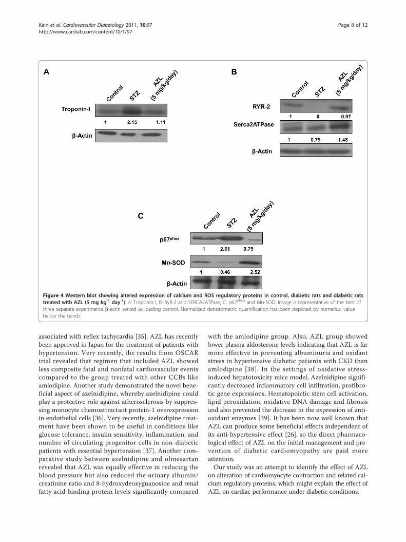
AZL neutralizes the increased expression levels ofcontractile proteins (Troponin I) in diabetic heartSince mechanical dysfunction that characterizes diabeticcardiomyopathy plays an essential role in the Ca2+ regu-lation of muscle contraction, we studied the effect ofAZL on the expression level of cardiac Troponin I in ourexperimental model. Our western blot results showed a2.15-fold increase (p < 0.05) in the expression of Tropo-nin I in the heart of STZ treated animal when comparedwith the control. The increased cardiac Troponin Iexpression counter-balanced in the cardiac tissue fromAZL treated diabetic rats (Figure 4A). These findingssuggest that AZL treatment under diabetic condition pre-vents cardiac damage by reducing the expression of car-diac Troponin I.
AZL regulates the expression of calcium regulatoryreceptors and channels in diabetic heartDownregulation of key Ca2+-handling proteins like sarco(endo)plasmic reticulum Ca2+-ATPase (SERCA)2a andryanodine receptor (RyR2) is one of the major cause ofabnormal Ca2+ homeostasis in diabetic cardiomyopathy[13,32-34]. The alteration in SERCA2a and RyR2 expres-sion results in altered cytosolic Ca2+ transients, leading toabnormal contraction. Our western blot results indicated acomplete loss of expression of RYR2 and a significantreduction in SERCA-2a in STZ-treated diabetic rats com-pared to the controls (Figure 4B). Reduction in the RyR2expression induced by uncontrolled diabetes was attenu-ated with AZL treatment. Similarly, the expression ofSERCA- 2a was also restored after AZL treatment (Figure4B). These findings indicate that AZL treatment underdiabetic condition prevents dysregulation of calcium regu-latory proteins in the heart.
AZL induces downregulation of NADPH oxidases andprevents oxidative stressWe determined effect of AZL treatment on the endo-genous pro-oxidants and antioxidants like p67phox andMn-SOD. Our western blot results showed increasedexpression of p67phox in diabetic heart. This indicatedthat endogenous pro-oxidant system was triggered
Table 1 Hemodynamic parameters from control, diabetic rats and diabetic rats treated with AZL (5 mg kg-1 day-1)
Parameters Control(n = 10)
STZ*(n = 10)
AZL** (5 mg/kg/day) (n = 12)
Systolic Pressure (mm Hg) 138.1 ± 4.7 93.8 ± 4.1 124.6 ± 8.9
Diastolic Pressure (mm Hg) 85.8 ± 6.2 64.0 ± 3.5 82.5 ± 7.1
Systolic duration (s) 0.085 ± 0.01 0.140 ± 0.01 0.084 ± 0.01
Diastolic duration (s) 0.117 ± 0.02 0.297 ± 0.02 0.127 ± 0.01
Heart rate (BPM) 323.3 ± 20.9 244.9 ± 10.8 312.2 ± 14.1
LV+dp/dt (mm Hg/s) 5861.4 ± 729.5 3219.3 ± 297.8 6098.6 ± 395.4
LV-dp/dt (mm Hg/s) -6846.8 ± 752.1 -3553.0 ± 437.3 -7015.9 ± 391.5
Kain et al. Cardiovascular Diabetology 2011, 10:97http://www.cardiab.com/content/10/1/97
Page 4 of 12

under diabetic condition which may further aggravateoxidative stress. Treatment with AZL significantly atte-nuated the p67phox expression (Figure 4C). On the otherhand, a significant decrease (0.46 fold, p < 0.05) in theexpression of Mn-SOD in STZ-diabetic heart wasobserved as compared to control hearts. Treatment withAZL resulted in a 2.5-fold (p < 0.05) increase in theexpression of Mn-SOD in the diabetic condition. Theseresults indicate that AZL exerts its protective effects bytargeting the NADPH oxidase and mitochondrial redoxenzymes (Figure 4C).
AZL prevents STZ-induced cardiac apoptosisApoptosis was also evaluated in the cardiac tissue byTUNEL assay. Diabetic rats showed significant myocardialapoptotic cell death manifested by a 6 fold increase in the
percent TUNEL-positive cell labeling compared with con-trol rats (Figure 5A and 5B). The counts of TUNEL-posi-tive nuclei significantly decreased in AZL-treated group.Under oxidative stress, mitochondria play an importantrole in apoptosis and a decrease in the level of bcl-2 isobserved. This decreasing bcl-2 expression is one of thehallmarks of apoptosis through mitochondrial pathway.STZ diabetic animal showed elevated cardiac apoptosis, asindicated by decreased bcl-2 protein expression, comparedto the control animals. AZL-treated diabetic rats expressedenhanced level of bcl-2 in the heart lysates indicating thatAZL plays protective role in cardiac apoptosis.
DiscussionThe key findings of our present study demonstrated thatAZL treatment for 12 weeks in diabetic animal inhibits
Figure 1 Contractile properties of cardiomyocytes isolated from control, diabetic rats and diabetic rats treated with AZL (5 mg kg-1 day-1).A: Maximal velocity of shortening (+dL/dt, A) and relengthening (-dL/dt, B) of ventricular myocytes isolated from control, diabetic rats and diabetic ratstreated with AZL (5 mg kg-1 day-1). B. Graph illustrates Peak shortening (PS, normalized to cell length) of the myocytes isolated from control, diabeticrats and diabetic rats treated with AZL. C. Graph illustrates time-to-peak shortening (TPS) of the myocytes isolated from control, diabetic rats anddiabetic rats treated with AZL. D. Graph illustrates time-to-90% relengthening (TR90) of the myocytes isolated from control, diabetic rats and diabeticrats treated with AZL. Values are means ± SE; n = 151-163 cells from 5-7 rats per group, *P < 0.05 vs. control group; **P < 0.05 vs. diabetic group.
Kain et al. Cardiovascular Diabetology 2011, 10:97http://www.cardiab.com/content/10/1/97
Page 5 of 12

the development of early characteristics of diabetic cardi-omyopathy, such as, prolonged relaxation and abnormalE-C coupling in vivo in the intact myocardium as well asin the isolated ventricular myocytes. Delayed diastolicrelaxation in diabetic cardiomyopathy is related to dimin-ished removal of [Ca2+]i from cardiomyocytes after thesystolic contraction event. Treatment with AZL showedimprovement in the systolic and diastolic duration. Alsothe markers for diastolic dysfunction, viz., maximal riseand decay in the blood pressure showed improvement.These mechanical abnormalities may be underscored byaltered intracellular Ca2+ homeostasis that was associatedwith enhanced oxidative stress. We found that AZLprevents maladaptive ventricular remodeling. Untreateddiabetes further accelerated oxidative stress at molecularlevel by upregulating the endogenous NADPH oxidaseslike p67phox and downregulation of Mn-SOD. Treatmentwith AZL normalized the p67phox and Mn-SOD
expression. Also, AZL treatment normalized the levels ofcardiac troponin I, RyR2, and SERCA2a. The ability ofAZL to restore all the parameters to the control levelprovides a plausible explanation for its ability to preventdiabetes-induced defects in calcium signaling. The intrin-sic antioxidant activity of AZL might thus contribute toits beneficial effects on LV dysfunction and cardiacfailure.Abnormalities in the myocardial calcium handling can
contribute to deranged cardiac mechanics in the diabeticheart. Diabetes impairs sarcoplasmic reticular calciumpump activities, which reduces the rate of Ca2+ removalfrom the cytoplasm in diastole [7]. Such alterations maycontribute to the increased diastolic stiffness characteristicof diabetic cardiomyopathy. Intracellular retention of cal-cium in diabetes is associated with the depletion of high-energy phosphate stores, derangement of cellular ultra-structure and can lead to cardiac dysfunction. Calcium
Figure 2 Intracellular Ca2+ transient properties of ventricular myocytes isolated from control, diabetic rats and diabetic rats treatedwith AZL (5 mg kg-1 day-1). A: Baseline intracellular Ca2+ concentrations; B: Increase in intracellular Ca2+ transient in response to electricalstimulus; C: rate of cytosolic free Ca2+ decrease (Tau). Values are means ± SEM; n = 104 to 126/group. *P < 0.05 vs. control group; **P < 0.05 vs.diabetic group.
Kain et al. Cardiovascular Diabetology 2011, 10:97http://www.cardiab.com/content/10/1/97
Page 6 of 12

channel blockers in conjunction with antioxidants canreverse the intracellular calcium defects and prevent dia-betes induced myocardial changes. The present studyinvestigates Ca2+-dependent regulation of cellular functionin diabetic cardiomyocytes and highlights role of AZL in
prevention of early onset of diastolic dysfunction at cellularand organ level.AZL is a novel dihydropyridine calcium channel
blocker which has a long lasting anti-hypertensive action[35]. It is generally well tolerated and its use is not
Figure 3 Generation of O2·- in ventricular myocytes isolated from control, diabetic rats and diabetic rats treated with AZL (5 mg kg-1
day-1). A. Representative confocal laser scanning microscopy images of cells fluorescently stained with DHE. B. Graph shows quantification ofDHE fluorescence emission (Arbitrary Units) of the myocytes isolated from control, diabetic rats and diabetic rats treated with AZL after stainingwith DHE. Data represents Means ± SEM; n = 104 to 126/group. *P < 0.05 vs. control group; **P < 0.05 vs. diabetic group.
Kain et al. Cardiovascular Diabetology 2011, 10:97http://www.cardiab.com/content/10/1/97
Page 7 of 12
associated with reflex tachycardia [35]. AZL has recentlybeen approved in Japan for the treatment of patients withhypertension. Very recently, the results from OSCARtrial revealed that regimen that included AZL showedless composite fatal and nonfatal cardiovascular eventscompared to the group treated with other CCBs likeamlodipine. Another study demonstrated the novel bene-ficial aspect of azelnidipine, whereby azelnidipine couldplay a protective role against atherosclerosis by suppres-sing monocyte chemoattractant protein-1 overexpressionin endothelial cells [36]. Very recently, azelnidipine treat-ment have been shown to be useful in conditions likeglucose tolerance, insulin sensitivity, inflammation, andnumber of circulating progenitor cells in non-diabeticpatients with essential hypertension [37]. Another com-parative study between azelnidipine and olmesartanrevealed that AZL was equally effective in reducing theblood pressure but also reduced the urinary albumin/creatinine ratio and 8-hydroxydeoxyguanosine and renalfatty acid binding protein levels significantly compared
with the amlodipine group. Also, AZL group showedlower plasma aldosterone levels indicating that AZL is farmore effective in preventing albuminuria and oxidantstress in hypertensive diabetic patients with CKD thanamlodipine [38]. In the settings of oxidative stress-induced hepatotoxicity mice model, Azelnidipine signifi-cantly decreased inflammatory cell infiltration, profibro-tic gene expressions, Hematopoietic stem cell activation,lipid peroxidation, oxidative DNA damage and fibrosisand also prevented the decrease in the expression of anti-oxidant enzymes [39]. It has been now well known thatAZL can produce some beneficial effects independent ofits anti-hypertensive effect [26], so the direct pharmaco-logical effect of AZL on the initial management and pre-vention of diabetic cardiomyopathy are paid moreattention.Our study was an attempt to identify the effect of AZL
on alteration of cardiomyocyte contraction and related cal-cium regulatory proteins, which might explain the effect ofAZL on cardiac performance under diabetic conditions.
Figure 4 Western blot showing altered expression of calcium and ROS regulatory proteins in control, diabetic rats and diabetic ratstreated with AZL (5 mg kg-1 day-1). A: Troponin I, B: RyR-2 and SERCA2ATPase, C: p67phox and Mn-SOD. Image is representative of the best ofthree separate experiments. b-actin served as loading control. Normalized densitometric quantification has been depicted by numerical valuebelow the bands.
Kain et al. Cardiovascular Diabetology 2011, 10:97http://www.cardiab.com/content/10/1/97
Page 8 of 12

Diabetes is characterized by consistently elevated bloodglucose levels, decreased insulin levels and increasedheart/body weight ratio indicative of hypertrophied heartwas observed in the STZ diabetic rat. Cardiac hypertro-phy involves remodeling of entire heart especially in theleft ventricular region which eventually leads to impaireddiastolic function, further causing deterioration of cardiacmorphology and function. The important finding of thepresent study is that STZ-induced hyperglycemia leads todilapidated cardiac function further leading to diabeticcardiomyopathy. Our STZ diabetic rats showed left ven-tricular dysfunction. This study along with our previousreport [26] provides evidence that hyperglycemia-inducedleft ventricular dysfunction due to oxidative stressinduced by reactive oxygen species (ROS) and reactivenitrogen species (RNS) and defective antioxidant system
contributing to the development of cardiomyopathy.Apoptosis induced by hyperglycemia is an early event inthe pathophysiology of diabetic cardiomyopathy [40].Hyperglycemia and insulin resistance independently con-tribute to functional alteration in the heart [41-44]. AZLtreatment in streptozotocin diabetic rats has been shownto improve these functional cardiac abnormalities per-haps through tyrosine kinase-dependent increases inintracellular [Ca2+]i removal after systole. In the presentstudy, treatment with AZL showed improvement in thesystolic and diastolic duration. Also the markers for dia-stolic dysfunction, viz., maximal rise and decay in theblood pressure showed improvement. In the presentstudy, we found that AZL prevents ventricular remodel-ing accompanied by cardiac dysfunction. We alsodemonstrated that AZL did not alter blood pressure and
Figure 5 Apoptosis in diabetic rat hearts and treated with AZL. A: representative photographs of in situ detection of apoptosis in hearttissue from controls, diabetic rats treated with vehicle, and diabetic rats treated with AZL (5 mg kg-1day-1). Total nuclei were labeled with DAPI(blue), and apoptotic nuclei were detected by TUNEL staining (green). B: average number of percent TUNEL-positive nuclei in tissue sectionsfrom each group (n = 4 to 5 sections each per group). *P < 0.05 vs. control group; **P < 0.05 vs. STZ treated group. C: Western blot showingbcl-2 expression in hearts from the same groups. Image is representative of the best of three separate experiments. b-actin served as loadingcontrol. Normalized densitometric quantification has been depicted by numerical value below the bands.
Kain et al. Cardiovascular Diabetology 2011, 10:97http://www.cardiab.com/content/10/1/97
Page 9 of 12

this suggests that AZL has preventive effects on cardiacdysfunction beyond its antihypertensive effects. Oxidativestress might play an important role in the progression ofLV dysfunction and failure, the data somewhat is consis-tent with previous finding using STZ diabetic models[45-47]. These mechanical abnormalities may be under-scored by altered intracellular Ca2+ homeostasis that wasassociated with enhanced oxidative stress. The intrinsicantioxidant activity of AZL might thus be a contributorto its beneficial effects on LV dysfunction in diabetic car-diomyopathy. Although these findings are of interest, noclinical trials to date have investigated the effect of AZLon the development and progression of congestive heartfailure in diabetic patients.In our study, certain diabetes-induced mechanical
defects were not improved or protected by AZL treatment.For example, AZL improved the diabetes-induced reduc-tion in PS but not Ca2+-induced Ca2+ release. Althoughthe underlying mechanism is largely unknown, the abilityof AZL to enhance myofilament Ca2+ sensitivity may playa role. This is somewhat supported by the results shownin Figure 1, where myocytes from the AZL-treated groupexhibit an improved PS compared with the myocytes fromthe diabetic group. The results from this study revealedthat AZL treatment lowered the resting intracellular Ca2+
levels in the diabetic group. This AZL-induced reductionin resting intracellular Ca2+ level may be associated withan enhanced SERCA Ca2+ clearing ability in the AZL-trea-ted group (Figure 2) and is consistent with the vasodilatoryand cardioprotective effect against Ca2+ overload underpathological conditions such as heart failure. The fre-quency-PS relationship was improved by AZL-treated dia-betic group (Figure 1), indicating a preserved sarcoplasmicreticulum (SR)-replenishing function in diabetic hearts.One possible explanation is that AZL may significantlyaugment the basal SR Ca2+ load in the diabetic group. Theimpaired intracellular Ca2+ homeostasis may be associatedwith a reduction in the main Ca2+-regulating proteinSERCA2 and ryanodine receptor (RyR) proteins indirectlywith reduced levels of Troponin I under the diabetic state[48]. Interestingly, the STZ diabetes-induced oxidativestress, apoptosis and alterations in oxidative stress-relatedsignaling molecule p67phox NADPH oxidase were effec-tively alleviated by AZL treatment. It also improved thelevels of Troponin I, RyR2, and SERCA2a. Because SERCAand RYR2 contributes to ~92% of the cytosolic Ca2+
removal workload in rat hearts [49], our finding of anovert reduction in SERCA2a protein level in STZ-induceddiabetic hearts should have provided one of the most com-pelling explanations for the slowed intracellular Ca2+
clearing and prolonged duration of relaxation (TR90). Theability of AZL to restore all the parameters to the controllevel provides a plausible explanation for its ability to pre-vent diabetes-induced defects in calcium signaling.
Further, restoration of TPS after AZL treatment indicatesthat AZL may have a significant effect on the key rate-lim-iting components determining the length of contractionduration such as SR Ca2+ release, troponin, tropomyosin,and actin-myosin cross-bridge linking. These observationsare consistent with the functional data of improved intra-cellular Ca2+ clearing and duration of relengthening(TR90) after AZL treatment. These results suggest thatAZL treatment may ameliorate contractile disturbances incardiomyocytes from diabetic animals and could providetherapeutic potential in the treatment of diabeticcardiomyopathy.Since AZL did not affect the hyperglycemic condition
in diabetes, our data suggest that STZ-induced diabetesmay elicit cardiac contractile dysfunction and intracellu-lar Ca2+ mishandling likely through enhanced oxidativestress and cell injury.Increased oxidative stress is believed to be an initial and
important step in the development of cardiac dysfunctionand cardiomyopathy. NADPH oxidase and mitochondriaare considered to be important sources of ROS [15-20]and are critical determinants of the redox state of the dia-betic myocardium. Previous studies reported that mem-brane translocation of p67phox and the increasedexpression of p22phox was prevented by N-acetyl Lcysteine [50]. Therefore, we further tested whether AZLexerts its antioxidative properties by modulating theexpression and function of NADPH oxidase subunitp67phox and mitochondrial ROS-eliminating enzyme Mn-SOD. The results from the isolated cardiomyocytes studyshowed that hyperglycemia leads to increased oxidativestress by enhancing the O2·
- generation, by decreasing theexpression of antioxidant enzyme Mn-SOD and byincreasing expression of p67phox. The main new findingsof this study are that AZL treatment prevents theincreased expression of p67phox, and enhances Mn-SODexpression, thus reducing myocardial superoxide forma-tion in the diabetic rat hearts. This reduction of O2·
-
generation and normalization of p67phox and Mn-SODafter AZL treatment indicate that AZL reduces diabeticcardiac damage by targeting the redox signaling pathways.Moreover, an increase in the expression of p67phox anddecrease in the expression of Mn-SOD as well as bcl-2and normalization of these expressions by AZL indicates amutual functional relationship between NADPH oxidaseand mitochondria. AZL in our study not only improvescardiac contractile function but also offers protectionagainst oxidative stress, apoptosis and ultimately leadingdiabetic cardiomyopathy.
Limitations of the studyWe did not test the effect of known antioxidants incomparison to AZL in the present study. We observedthe changes after the treatment duration of 12 weeks.
Kain et al. Cardiovascular Diabetology 2011, 10:97http://www.cardiab.com/content/10/1/97
Page 10 of 12

Some of the changes could be result of the overall func-tional improvement due to AZL treatment and may notbe directly attributable to AZL treatment. The hemody-namic parameters were evaluated at the end of thestudy, and comparisons were made with comparing withdiabetic and non-diabetic control in the experimentaldesign. As a result of which the signals were recordedonly at the end of the experimental period, leading tothe lack of baseline values in the same animals at thestart of the study.
ConclusionIn conclusion, the present study reveals the beneficialeffects of AZL treatment on diabetes induced early leftventricular dysfunction. AZL exhibited additional anti-oxidant properties in addition to its calcium channelblocking activity. This intrinsic antioxidant property ofAZL may provide a promising advantage over other cal-cium channel blockers in the management of compro-mised heart function especially under diabetes.
AcknowledgementsWe thank Dr. G.C. Mishra, Director, NCCS, Pune, for encouragement andsupport; the Department of Biotechnology, Government of India, forproviding financial support; the Council for Scientific and Industrial Researchand the University Grants Commission, New Delhi, for providing fellowships.We acknowledge the technical help of Ms. Ashwini Atre in confocalmicroscopy and the staff of the experimental animal facility at NationalCentre for Cell Science.
Authors’ contributionsVK and SK contributed equally to the experimental designing and benchwork. SLS conceived and designed the study. All authors have read,discussed and approved the final manuscript.
Competing interestsThe authors declare that they have no competing interests.
Received: 15 July 2011 Accepted: 4 November 2011Published: 4 November 2011
References1. Rubler S, Dlugash J, Yuceoglu YZ, Kumral T, Branwood AW, Grishman A:
New type of cardiomyopathy associated with diabeticglomerulosclerosis. Am J Cardiol 1972, 30(6):595-602.
2. Asmal AC, Leary WP, Thandroyen F: Diabetic heart disease. S Afr Med J1980, 57(19):788-790.
3. Regan TJ: Congestive heart failure in the diabetic. Annu Rev Med 1983,34:161-168.
4. Hamby RI, Zoneraich S, Sherman L: Diabetic cardiomyopathy. Jama 1974,229(13):1749-1754.
5. Regan TJ, Lyons MM, Ahmed SS, Levinson GE, Oldewurtel HA, Ahmad MR,Haider B: Evidence for cardiomyopathy in familial diabetes mellitus. J ClinInvest 1977, 60(4):884-899.
6. Mahgoub MA, Abd-Elfattah AS: Diabetes mellitus and cardiac function.Mol Cell Biochem 1998, 180(1-2):59-64.
7. Pierce GN, Russell JC: Regulation of intracellular Ca2+ in the heart duringdiabetes. Cardiovasc Res 1997, 34(1):41-47.
8. Choi KM, Zhong Y, Hoit BD, Grupp IL, Hahn H, Dilly KW, Guatimosim S,Lederer WJ, Matlib MA: Defective intracellular Ca(2+) signalingcontributes to cardiomyopathy in Type 1 diabetic rats. Am J Physiol HeartCirc Physiol 2002, 283(4):H1398-1408.
9. Cai L, Wang Y, Zhou G, Chen T, Song Y, Li X, Kang YJ: Attenuation bymetallothionein of early cardiac cell death via suppression ofmitochondrial oxidative stress results in a prevention of diabeticcardiomyopathy. J Am Coll Cardiol 2006, 48(8):1688-1697.
10. Cai L: Suppression of nitrative damage by metallothionein in diabeticheart contributes to the prevention of cardiomyopathy. Free Radic BiolMed 2006, 41(6):851-861.
11. Boudina S, Abel ED: Diabetic cardiomyopathy revisited. Circulation 2007,115(25):3213-3223.
12. Teshima Y, Takahashi N, Saikawa T, Hara M, Yasunaga S, Hidaka S, Sakata T:Diminished expression of sarcoplasmic reticulum Ca(2+)-ATPase andryanodine sensitive Ca(2+)Channel mRNA in streptozotocin-induceddiabetic rat heart. J Mol Cell Cardiol 2000, 32(4):655-664.
13. Ganguly PK, Pierce GN, Dhalla KS, Dhalla NS: Defective sarcoplasmicreticular calcium transport in diabetic cardiomyopathy. Am J Physiol 1983,244(6):E528-535.
14. Rodrigues B, Rosa K, Medeiros A, Schaan B, Brum P, De Angelis K,Irigoyen M: Hyperglycemia can delay left ventricular dysfunction but notautonomic damage after myocardial infarction in rodents. CardiovascDiabetol 2011, 10(1):26.
15. Isabelle M, Vergeade A, Moritz F, Dautreaux B, Henry JP, Lallemand F,Richard V, Mulder P, Thuillez C, Monteil C: NADPH oxidase inhibitionprevents cocaine-induced up-regulation of xanthine oxidoreductase andcardiac dysfunction. J Mol Cell Cardiol 2007, 42(2):326-332.
16. Heymes C, Bendall JK, Ratajczak P, Cave AC, Samuel JL, Hasenfuss G,Shah AM: Increased myocardial NADPH oxidase activity in human heartfailure. J Am Coll Cardiol 2003, 41(12):2164-2171.
17. Li JM, Gall NP, Grieve DJ, Chen M, Shah AM: Activation of NADPH oxidaseduring progression of cardiac hypertrophy to failure. Hypertension 2002,40(4):477-484.
18. Xiao L, Pimentel DR, Wang J, Singh K, Colucci WS, Sawyer DB: Role ofreactive oxygen species and NAD(P)H oxidase in alpha(1)-adrenoceptorsignaling in adult rat cardiac myocytes. Am J Physiol Cell Physiol 2002,282(4):C926-934.
19. Zhao K, Zhao GM, Wu D, Soong Y, Birk AV, Schiller PW, Szeto HH: Cell-permeable peptide antioxidants targeted to inner mitochondrialmembrane inhibit mitochondrial swelling, oxidative cell death, andreperfusion injury. J Biol Chem 2004, 279(33):34682-34690.
20. Nagy N, Malik G, Tosaki A, Ho YS, Maulik N, Das DK: Overexpression ofglutaredoxin-2 reduces myocardial cell death by preventing bothapoptosis and necrosis. J Mol Cell Cardiol 2008, 44(2):252-260.
21. Wold LE, Ceylan-Isik AF, Fang CX, Yang X, Li SY, Sreejayan N, Privratsky JR,Ren J: Metallothionein alleviates cardiac dysfunction in streptozotocin-induced diabetes: role of Ca2+ cycling proteins, NADPH oxidase, poly(ADP-Ribose) polymerase and myosin heavy chain isozyme. Free RadicBiol Med 2006, 40(8):1419-1429.
22. Wendt MC, Daiber A, Kleschyov AL, Mulsch A, Sydow K, Schulz E, Chen K,Keaney JF Jr, Lassegue B, Walter U, et al: Differential effects of diabetes onthe expression of the gp91phox homologues nox1 and nox4. Free RadicBiol Med 2005, 39(3):381-391.
23. Shen X, Zheng S, Metreveli NS, Epstein PN: Protection of cardiacmitochondria by overexpression of MnSOD reduces diabeticcardiomyopathy. Diabetes 2006, 55(3):798-805.
24. Boudina S, Sena S, Theobald H, Sheng X, Wright JJ, Hu XX, Aziz S, Johnson JI,Bugger H, Zaha VG, et al: Mitochondrial energetics in the heart in obesity-related diabetes: direct evidence for increased uncoupled respiration andactivation of uncoupling proteins. Diabetes 2007, 56(10):2457-2466.
25. Privratsky JR, Wold LE, Sowers JR, Quinn MT, Ren J: AT1 blockade preventsglucose-induced cardiac dysfunction in ventricular myocytes: role of theAT1 receptor and NADPH oxidase. Hypertension 2003, 42(2):206-212.
26. Kain V, Kumar S, Puranik AS, Sitasawad SL: Azelnidipine protectsmyocardium in hyperglycemia-induced cardiac damage. CardiovascDiabetol 2010, 9:82.
27. Maggi CA, Meli A: Suitability of urethane anesthesia forphysiopharmacological investigations in various systems. Part 2:Cardiovascular system. Experientia 1986, 42(3):292-297.
28. Kajstura J, Fiordaliso F, Andreoli AM, Li B, Chimenti S, Medow MS, Limana F,Nadal-Ginard B, Leri A, Anversa P: IGF-1 overexpression inhibits thedevelopment of diabetic cardiomyopathy and angiotensin II-mediatedoxidative stress. Diabetes 2001, 50(6):1414-1424.
Kain et al. Cardiovascular Diabetology 2011, 10:97http://www.cardiab.com/content/10/1/97
Page 11 of 12

29. Ren J, Wold LE: Measurement of Cardiac Mechanical Function in IsolatedVentricular Myocytes from Rats and Mice by Computerized Video-BasedImaging. Biol Proced Online 2001, 3:43-53.
30. Kumar S, Kain V, Sitasawad SL: Cardiotoxicity of calmidazolium chloride isattributed to calcium aggravation, oxidative and nitrosative stress, andapoptosis. Free Radic Biol Med 2009, 47(6):699-709.
31. Murugavel P, Pari L, Sitasawad SL, Kumar S: Cadmium inducedmitochondrial injury and apoptosis in vero cells: protective effect ofdiallyl tetrasufide from garlic. Int J Biochem Cell Biol 2007, 39(1):161-170.
32. Trost SU, Belke DD, Bluhm WF, Meyer M, Swanson E, Dillmann WH:Overexpression of the sarcoplasmic reticulum Ca(2+)-ATPase improvesmyocardial contractility in diabetic cardiomyopathy. Diabetes 2002,51(4):1166-1171.
33. Bidasee KR, Dincer UD, Besch HR Jr: Ryanodine receptor dysfunction inhearts of streptozotocin-induced diabetic rats. Mol Pharmacol 2001,60(6):1356-1364.
34. Shao CH, Rozanski GJ, Patel KP, Bidasee KR: Dyssynchronous (non-uniform)Ca2+ release in myocytes from streptozotocin-induced diabetic rats. JMol Cell Cardiol 2007, 42(1):234-246.
35. Oizumi K, Nishino H, Koike H, Sada T, Miyamoto M, Kimura T:Antihypertensive effects of CS-905, a novel dihydropyridine Ca++channel blocker. Jpn J Pharmacol 1989, 51(1):57-64.
36. Matsui T, Yamagishi S, Nakamura K, Inoue H: Azelnidipine, a New Long-acting Calcium-channel Blocker, Inhibits Tumour Necrosis Factor–induced Monocyte Chemoattractant Protein-1 Expression in EndothelialCells. The Journal of International Medical Research 2006, 34(6):671-675.
37. Fukao K, Shimada K, Hiki M, Kiyanagi T, Hirose K, Kume A, Ohsaka H,Matsumori R, Kurata T, Miyazaki T, et al: Effects of calcium channelblockers on glucose tolerance, inflammatory state, and circulatingprogenitor cells in non-diabetic patients with essential hypertension: acomparative study between Azelnidipine and amlodipine on Glucosetolerance and ENdothelial function - a crossover Trial (AGENT).Cardiovasc Diabetol 2011, 10(1):79.
38. Abe M, Maruyama N, Okada K, Matsumoto S, Matsumoto K, Soma M:Additive antioxidative effects of azelnidipine on angiotensin receptorblocker olmesartan treatment for type 2 diabetic patients withalbuminuria. Hypertens Res 2011, 34(8):935-941.
39. Ohyama T, Sato K, Kishimoto K, Yamazaki Y, Horiguchi N, Ichikawa T,Kakizaki S, Takagi H, Izumi T, Mori M: Azelnidipine is a calcium blockerthat attenuates liver fibrosis and may increase antioxidant defence. Brit JPharmacol 2011, no-no.
40. Sarkar K, Mehta JL: Apoptosis in Atherosclerosis: Virtue or Vice. CurrentCardiology Reviews 2005, 1:141-152.
41. Oliveira PJ: Cardiac mitochondrial alterations observed in hyperglycaemicrats–what can we learn from cell biology? Curr Diabetes Rev 2005,1(1):11-21.
42. Schaffer SW, Croft CB, Solodushko V: Cardioprotective effect of chronichyperglycemia: effect on hypoxia-induced apoptosis and necrosis. Am JPhysiol Heart Circ Physiol 2000, 278(6):H1948-1954.
43. Border WA, Brees D, Noble NA: Transforming growth factor-beta andextracellular matrix deposition in the kidney. Contrib Nephrol 1994,107:140-145.
44. Verma S, Maitland A, Weisel RD, Li SH, Fedak PW, Pomroy NC, Mickle DA,Li RK, Ko L, Rao V: Hyperglycemia exaggerates ischemia-reperfusion-induced cardiomyocyte injury: reversal with endothelin antagonism. JThorac Cardiovasc Surg 2002, 123(6):1120-1124.
45. Ren J, Davidoff AJ: Diabetes rapidly induces contractile dysfunctions inisolated ventricular myocytes. Am J Physiol 1997, 272(1 Pt 2):H148-158.
46. Norby FL, Wold LE, Duan J, Hintz KK, Ren J: IGF-I attenuates diabetes-induced cardiac contractile dysfunction in ventricular myocytes. Am JPhysiol Endocrinol Metab 2002, 283(4):E658-666.
47. Ye G, Metreveli NS, Ren J, Epstein PN: Metallothionein prevents diabetes-induced deficits in cardiomyocytes by inhibiting reactive oxygen speciesproduction. Diabetes 2003, 52(3):777-783.
48. Liu X, Takeda N, Dhalla NS: Troponin I phosphorylation in hearthomogenate from diabetic rat. Biochim Biophys Acta 1996, 1316(2):78-84.
49. Bers DM: Cardiac excitation-contraction coupling. Nature 2002,415(6868):198-205.
50. Guo Z, Xia Z, Jiang J, McNeill JH: Downregulation of NADPH oxidase,antioxidant enzymes, and inflammatory markers in the heart of
streptozotocin-induced diabetic rats by N-acetyl-L-cysteine. Am J PhysiolHeart Circ Physiol 2007, 292(4):H1728-1736.
doi:10.1186/1475-2840-10-97Cite this article as: Kain et al.: Azelnidipine prevents cardiac dysfunctionin streptozotocin-diabetic rats by reducing intracellular calciumaccumulation, oxidative stress and apoptosis. Cardiovascular Diabetology2011 10:97.
Submit your next manuscript to BioMed Centraland take full advantage of:
• Convenient online submission
• Thorough peer review
• No space constraints or color figure charges
• Immediate publication on acceptance
• Inclusion in PubMed, CAS, Scopus and Google Scholar
• Research which is freely available for redistribution
Submit your manuscript at www.biomedcentral.com/submit
Kain et al. Cardiovascular Diabetology 2011, 10:97http://www.cardiab.com/content/10/1/97
Page 12 of 12