award number: w81xwh-11-1-0468 title regulation and ... · presenting with anemia, ... the overall...
TRANSCRIPT

AD_________________
Award Number: W81XWH-11-1-0468 TITLE: Regulation and Function of TIFAB in Myelodysplastic Syndrome PRINCIPAL INVESTIGATOR: Daniel Starczynowski CONTRACTING ORGANIZATION: Cincinnati Children’s Hospital Medical Center Cincinnati, OH 45229 REPORT DATE: June 2012 TYPE OF REPORT: Annual report PREPARED FOR: U.S. Army Medical Research and Materiel Command Fort Detrick, Maryland 21702-5012 DISTRIBUTION STATEMENT: Approved for Public Release; Distribution Unlimited The views, opinions and/or findings contained in this report are those of the author(s) and should not be construed as an official Department of the Army position, policy or decision unless so designated by other documentation.

REPORT DOCUMENTATION PAGE Form Approved OMB No. 0704-0188
Public reporting burden for this collection of information is estimated to average 1 hour per response, including the time for reviewing instructions, searching existing data sources, gathering and maintaining the data needed, and completing and reviewing this collection of information. Send comments regarding this burden estimate or any other aspect of this collection of information, including suggestions for reducing this burden to Department of Defense, Washington Headquarters Services, Directorate for Information Operations and Reports (0704-0188), 1215 Jefferson Davis Highway, Suite 1204, Arlington, VA 22202-4302. Respondents should be aware that notwithstanding any other provision of law, no person shall be subject to any penalty for failing to comply with a collection of information if it does not display a currently valid OMB control number. PLEASE DO NOT RETURN YOUR FORM TO THE ABOVE ADDRESS. 1. REPORT DATE
2. REPORT TYPE
3. DATES COVERED
4. TITLE AND SUBTITLE
Regulation and Function of TIFAB in Myelodysplastic Syndrome
5a. CONTRACT NUMBER
5b. GRANT NUMBER
5c. PROGRAM ELEMENT NUMBER
6. AUTHOR(S) Betty Diamond
5d. PROJECT NUMBER
5e. TASK NUMBER
E-Mail:
5f. WORK UNIT NUMBER 7. PERFORMING ORGANIZATION NAME(S) AND ADDRESS(ES)
AND ADDRESS(ES)
8. PERFORMING ORGANIZATION REPORT NUMBER
9. SPONSORING / MONITORING AGENCY NAME(S) AND ADDRESS(ES) 10. SPONSOR/MONITOR’S ACRONYM(S) U.S. Army Medical Research and Materiel Command
Fort Detrick, Maryland 21702-5012 11. SPONSOR/MONITOR’S REPORT NUMBER(S) 12. DISTRIBUTION / AVAILABILITY STATEMENT Approved for Public Release; Distribution Unlimited 13. SUPPLEMENTARY NOTES
14. ABSTRACT Myelodysplastic syndromes (MDS) are clonal bone marrow failure (BMF) disorders defined by blood cytopenias due to ineffective hematopoiesis, genomic instability, and a predisposition to acute myeloid leukemia (AML). The most commonly recurring genomic alteration in MDS is deletion of chromosome 5q (del(5q)). MDS patients with an isolated del(5q) presenting with anemia, neutropenia, and elevated platelets associated with dysplastic megakaryocytes are considered to have 5q- syndrome. The majority of MDS patients with del(5q) do not exhibit these particular symptoms and, instead, are referred to as “del(5q) MDS”. We have recently identified miR-146a, which target the TRAF6 arm of the innate immune pathway, a gene within the deleted region in del(5q) MDS. We posit that multiple genes on chr 5q coordinate TRAF6 activation in del(5q) MDS. A search of annotated genes within or near the CDRs revealed a known inhibitor of TRAF6, TIFAB, on band q31.1. We hypothesize that deletion of TIFAB promotes activation of the TRAF6 complex in human CD34+ cells resulting in hematopoietic defects resembling MDS and AML with del(5q). The overall objectives of this proposal are to (1) determine whether loss of TIFAB in human CD34+ cells contributes to MDS in mice; (2) to investigate whether deletions of TIFAB and miR-146a cooperate to activate TRAF6 in MDS; and (3) to determine the consequences of TIFAB deletion on signal transduction in human CD34+ cells, and whether these could explain features of MDS. In preliminary data from the first year of the proposal, we have evidence that TIFAB exhibits tumor suppressor-like functions in human hematopoietic cells. Our key observations show that knockdown of TIFAB in human CD34+ hematopoietic stem/progenitor cells results in increased survival and altered hematopoietic progenitor function, that TIFAB inhibits TRAF6 protein expression and activation, resulting in lower NF-κB activation, and that TIFAB expression impacts leukemic cell survival, growth and progenitor function. Given that TIFAB is deleted in many MDS and AML patients, these findings could have major implications in MDS and AML subtypes with deletions of chr 5q. The observation that del(5q) results in inappropriate activation of TRAF6 provides a strong rationale to study the contribution of TIFAB to deregulation of the TRAF6 pathway in MDS.
15. SUBJECT TERMS
16. SECURITY CLASSIFICATION OF:
17. LIMITATION OF ABSTRACT
18. NUMBER OF PAGES
19a. NAME OF RESPONSIBLE PERSON USAMRMC
a. REPORT U
b. ABSTRACT U
c. THIS PAGE U
UU
19b. TELEPHONE NUMBER (include area code)
1 Jun 2011 - 30 May 2012Annual01-06-2012
W81XWH-11-1-0468
No subject terms provided.
24
Daniel Starczynowski
Cincinnati Children’s Hospital Medical Center Cincinnati, OH 45229

Table of Contents
Page
Introduction…………………………………………………………….………..….. 1
Body………………………………………………………………………………….. 1
Key Research Accomplishments………………………………………….…….. 5
Reportable Outcomes……………………………………………………………… 6
Conclusion…………………………………………………………………………… 6
References……………………………………………………………………………. 8
Appendices…………………………………………………………………………… 9

! 1!
INTRODUCTION
Myelodysplastic syndromes (MDS) are clonal bone marrow failure (BMF) disorders defined by blood cytopenias due to ineffective hematopoiesis, genomic instability, and a predisposition to acute myeloid leukemia (AML). The most commonly recurring genomic alteration in MDS is deletion of chromosome 5q (del(5q)). MDS patients with an isolated del(5q) presenting with anemia, neutropenia, and elevated platelets associated with dysplastic megakaryocytes are considered to have 5q- syndrome. The majority of MDS patients with del(5q) do not exhibit these particular symptoms and, instead, are referred to as “del(5q) MDS”. We have recently identified miR-146a, which target the TRAF6 arm of the innate immune pathway, a gene within the deleted region in del(5q) MDS. We posit that multiple genes on chr 5q coordinate TRAF6 activation in del(5q) MDS. A search of annotated genes within or near the CDRs revealed a known inhibitor of TRAF6, TIFAB, on band q31.1. We hypothesize that deletion of TIFAB promotes activation of the TRAF6 complex in human CD34+ cells resulting in hematopoietic defects resembling MDS and AML with del(5q). The overall objectives of this proposal are to (1) determine whether loss of TIFAB in human CD34+ cells contributes to MDS in mice; (2) to investigate whether deletions of TIFAB and miR-146a cooperate to activate TRAF6 in MDS; and (3) to determine the consequences of TIFAB deletion on signal transduction in human CD34+ cells, and whether these could explain features of MDS. In preliminary data from the first year of the proposal, we have evidence that TIFAB exhibits tumor suppressor-like functions in human hematopoietic cells. Our key observations show that knockdown of TIFAB in human CD34+ hematopoietic stem/progenitor cells results in increased survival and altered hematopoietic progenitor function, TIFAB inhibits TRAF6 protein expression and activation, resulting in lower NF-κB activation, and TIFAB expression impacts leukemic cell survival, growth and progenitor function. Given that TIFAB is deleted in many MDS and AML patients, these findings could have major implications in MDS and AML subtypes with deletions of chr 5q. The observation that del(5q) results in inappropriate activation of TRAF6 provides a strong rationale to study the contribution of TIFAB to deregulation of the TRAF6 pathway in MDS.
BODY Task 1. Plasmid constructs and validation (months 1-4): 1a. For knockdown of TIFAB in human CD34+ or MDS/AML cell lines, we used RNAi-mediated gene silencing. Lentiviral vectors encoding two independent shRNAs targeting human TIFAB were purchased from OpenBiosystems. The bicistronic lentiviral vector contains a microRNA-adapted shRNA and a CMV-driven turboGFP (Figure 1A)1. 1c. For qRT-PCR and immunoblot analysis to determine TIFAB knockdown, two independent shRNAs targeting TIFAB (#88 and #89) and a scrambled control vector (shCTL) were transduced into human HL60 and THP1 leukemia cell lines. Two days post transduction, cells were sorted for GFP expression and expanded for an additional 2 days in culture (Figure 1B). RNA was collected and examined for TIFAB knockdown by qRT-PCR (Figure 1C). Our preliminary data indicates that TIFAB, a gene within the deleted segment on chr 5q2, is expressed approximately half in marrow cells from del(5q) MDS patients as compared to age-matched normal control marrow cells (in original grant proposal). To mimic haploinsufficiency
! 2!
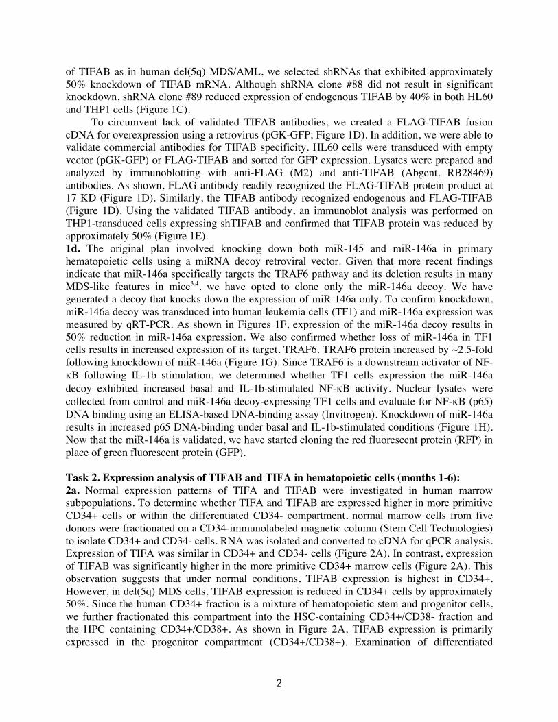
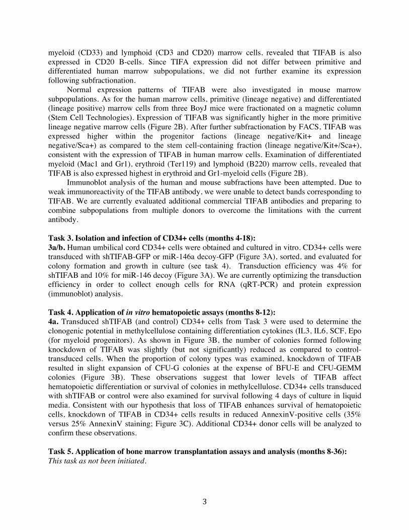
of TIFAB as in human del(5q) MDS/AML, we selected shRNAs that exhibited approximately 50% knockdown of TIFAB mRNA. Although shRNA clone #88 did not result in significant knockdown, shRNA clone #89 reduced expression of endogenous TIFAB by 40% in both HL60 and THP1 cells (Figure 1C). To circumvent lack of validated TIFAB antibodies, we created a FLAG-TIFAB fusion cDNA for overexpression using a retrovirus (pGK-GFP; Figure 1D). In addition, we were able to validate commercial antibodies for TIFAB specificity. HL60 cells were transduced with empty vector (pGK-GFP) or FLAG-TIFAB and sorted for GFP expression. Lysates were prepared and analyzed by immunoblotting with anti-FLAG (M2) and anti-TIFAB (Abgent, RB28469) antibodies. As shown, FLAG antibody readily recognized the FLAG-TIFAB protein product at 17 KD (Figure 1D). Similarly, the TIFAB antibody recognized endogenous and FLAG-TIFAB (Figure 1D). Using the validated TIFAB antibody, an immunoblot analysis was performed on THP1-transduced cells expressing shTIFAB and confirmed that TIFAB protein was reduced by approximately 50% (Figure 1E). 1d. The original plan involved knocking down both miR-145 and miR-146a in primary hematopoietic cells using a miRNA decoy retroviral vector. Given that more recent findings indicate that miR-146a specifically targets the TRAF6 pathway and its deletion results in many MDS-like features in mice3,4, we have opted to clone only the miR-146a decoy. We have generated a decoy that knocks down the expression of miR-146a only. To confirm knockdown, miR-146a decoy was transduced into human leukemia cells (TF1) and miR-146a expression was measured by qRT-PCR. As shown in Figures 1F, expression of the miR-146a decoy results in 50% reduction in miR-146a expression. We also confirmed whether loss of miR-146a in TF1 cells results in increased expression of its target, TRAF6. TRAF6 protein increased by ~2.5-fold following knockdown of miR-146a (Figure 1G). Since TRAF6 is a downstream activator of NF-κB following IL-1b stimulation, we determined whether TF1 cells expression the miR-146a decoy exhibited increased basal and IL-1b-stimulated NF-κB activity. Nuclear lysates were collected from control and miR-146a decoy-expressing TF1 cells and evaluate for NF-κB (p65) DNA binding using an ELISA-based DNA-binding assay (Invitrogen). Knockdown of miR-146a results in increased p65 DNA-binding under basal and IL-1b-stimulated conditions (Figure 1H). Now that the miR-146a is validated, we have started cloning the red fluorescent protein (RFP) in place of green fluorescent protein (GFP). Task 2. Expression analysis of TIFAB and TIFA in hematopoietic cells (months 1-6): 2a. Normal expression patterns of TIFA and TIFAB were investigated in human marrow subpopulations. To determine whether TIFA and TIFAB are expressed higher in more primitive CD34+ cells or within the differentiated CD34- compartment, normal marrow cells from five donors were fractionated on a CD34-immunolabeled magnetic column (Stem Cell Technologies) to isolate CD34+ and CD34- cells. RNA was isolated and converted to cDNA for qPCR analysis. Expression of TIFA was similar in CD34+ and CD34- cells (Figure 2A). In contrast, expression of TIFAB was significantly higher in the more primitive CD34+ marrow cells (Figure 2A). This observation suggests that under normal conditions, TIFAB expression is highest in CD34+. However, in del(5q) MDS cells, TIFAB expression is reduced in CD34+ cells by approximately 50%. Since the human CD34+ fraction is a mixture of hematopoietic stem and progenitor cells, we further fractionated this compartment into the HSC-containing CD34+/CD38- fraction and the HPC containing CD34+/CD38+. As shown in Figure 2A, TIFAB expression is primarily expressed in the progenitor compartment (CD34+/CD38+). Examination of differentiated
! 3!
myeloid (CD33) and lymphoid (CD3 and CD20) marrow cells, revealed that TIFAB is also expressed in CD20 B-cells. Since TIFA expression did not differ between primitive and differentiated human marrow subpopulations, we did not further examine its expression following subfractionation. Normal expression patterns of TIFAB were also investigated in mouse marrow subpopulations. As for the human marrow cells, primitive (lineage negative) and differentiated (lineage positive) marrow cells from three BoyJ mice were fractionated on a magnetic column (Stem Cell Technologies). Expression of TIFAB was significantly higher in the more primitive lineage negative marrow cells (Figure 2B). After further subfractionation by FACS, TIFAB was expressed higher within the progenitor factions (lineage negative/Kit+ and lineage negative/Sca+) as compared to the stem cell-containing fraction (lineage negative/Kit+/Sca+), consistent with the expression of TIFAB in human marrow cells. Examination of differentiated myeloid (Mac1 and Gr1), erythroid (Ter119) and lymphoid (B220) marrow cells, revealed that TIFAB is also expressed highest in erythroid and Gr1-myeloid cells (Figure 2B). Immunoblot analysis of the human and mouse subfractions have been attempted. Due to weak immunoreactivity of the TIFAB antibody, we were unable to detect bands corresponding to TIFAB. We are currently evaluated additional commercial TIFAB antibodies and preparing to combine subpopulations from multiple donors to overcome the limitations with the current antibody. Task 3. Isolation and infection of CD34+ cells (months 4-18): 3a/b. Human umbilical cord CD34+ cells were obtained and cultured in vitro. CD34+ cells were transduced with shTIFAB-GFP or miR-146a decoy-GFP (Figure 3A), sorted, and evaluated for colony formation and growth in culture (see task 4). Transduction efficiency was 4% for shTIFAB and 10% for miR-146 decoy (Figure 3A). We are currently optimizing the transduction efficiency in order to collect enough cells for RNA (qRT-PCR) and protein expression (immunoblot) analysis. Task 4. Application of in vitro hematopoietic assays (months 8-12): 4a. Transduced shTIFAB (and control) CD34+ cells from Task 3 were used to determine the clonogenic potential in methylcellulose containing differentiation cytokines (IL3, IL6, SCF, Epo (for myeloid progenitors). As shown in Figure 3B, the number of colonies formed following knockdown of TIFAB was slightly (but not significantly) reduced as compared to control-transduced cells. When the proportion of colony types was examined, knockdown of TIFAB resulted in slight expansion of CFU-G colonies at the expense of BFU-E and CFU-GEMM colonies (Figure 3B). These observations suggest that lower levels of TIFAB affect hematopoietic differentiation or survival of colonies in methylcellulose. CD34+ cells transduced with shTIFAB or control were also examined for survival following 4 days of culture in liquid media. Consistent with our hypothesis that loss of TIFAB enhances survival of hematopoietic cells, knockdown of TIFAB in CD34+ cells results in reduced AnnexinV-positive cells (35% versus 25% AnnexinV staining; Figure 3C). Additional CD34+ donor cells will be analyzed to confirm these observations. Task 5. Application of bone marrow transplantation assays and analysis (months 8-36): This task as not been initiated.

! 4!
Task 6. Identification of changes in TRAF6 activation and NF-κB signaling by TIFAB (months 10-14): 6a. It was previously reported that TIFAB may function by suppressing TRAF6-mediated NF-κB activation5,6. However, the mechanism by which TIFAB may inhibit TRAF6 and/or NF-κB is not known. To investigate the consequences of TIFAB expression on TRAF6 activation, HL60 cells were transduced with FLAG-TIFAB retrovirus (pGK-GFP). A convenient measure of TRAF6 activation is lysine (K)-63 linked autoubiquitination7. To measure TRAF6 autoubiquitination, TRAF6 was immunoprecipitated (IP) with a TRAF6 antibody and immunoblotted for ubiquitin (Ub). In cells overexpressing TIFAB, TRAF6 ubiquitination was significantly reduced (Figure 4A), an indication of less active TRAF6. In HL60 and THP1 cells expressing TIFAB, we also detected less phosphorylated IKKβ (the catalytic subunit of the NF-κB kinase complex) (Figure 4B). Interestingly, we observed that TRAF6 protein levels were also reduced in TIFAB-expressing cells (Figure 4A). Since it has not been reported that TIFAB affects TRAF6 protein expression, we examined this observation in more detail. To determine whether knockdown of TIFAB results in increased TRAF6 levels, we examined HL60 cells expressing shTIFAB and measured TRAF6 protein by immunoblotting. As shown in Figure 4C, HL60 cells with knockdown of TIFAB exhibited approximately 3-fold increase in TRAF6 protein. To exclude the possibility that the effect of TIFAB on TRAF6 is an artifact of leukemic cells or regulation of the TRAF6 promoter, we cloned TIFAB and TRAF6 into pcDNA3.1 expression vectors and transiently cotransfected A293 cells. As expected, transfection of TRAF6 results in increased active (e.g., ubiquitinated) forms of TRAF6 (Figure 4D). However, co-transfection of TRAF6 and TIFAB resulted in a dose-dependent inhibition of TRAF6 autoubiquitination by TIFAB (Figure 4D). On the same cells, we also examined the levels of total TRAF6 protein. Consistent with less active TRAF6, transfection of TIFAB coincided with less total TRAF6 protein (Figure 4E) despite expression from a constitutively active promoter. Future experiments will determine the effects of TIFAB on TRAF6 in normal CD34+ and on TRAF6 target genes (IL-6 and TNFa) in CD34+ and leukemic cell lines. 6b. One of the main pathways activated by TRAF6 is the NF-κB complex. Although multiple signals converge on NF-κB (e.g., TLR4 and TNFR), downstream modulators instruct the appropriate signal to NF-κB. For example, TRAF6 mediates NF-κB activation following LPS stimulation of TLR4, but not via TNFR stimulation. In contrast, TRAF2 mediates NF-κB activation following TNFa simulation of TNFR (Figure 4F). Conditions are being optimized to allow for measuring NF-κB activation in primary CD34+ cells. In the mean time, we have performed experiments in human cell lines to better understand the role of TIFAB on NF-κB. First we wanted to determine whether TIFAB is a general inhibitor of NF-κB or is a specific inhibitor of TRAF6-mediated activation of NF-κB. For these experiments, we transfected A293 cells with a κB-site containing NF-κB luciferase reporter and TIFAB. Following a 24 hr transfection, cells were treated with LPS (1 ug/ml) or TNFa (1 ng/ml) for an additional 6 hours and then assessed for luciferase activity. As shown in Figure 4G, stimulation with LPS resulted in 3-fold increase in κB-luciferase activity. Cotransfection of TIFAB did not affect basal κB-luciferase activity. However, cotransfection of TIFAB significantly repressed LPS-mediated activation of κB-luciferase activity. We next examined whether TIFAB affects TNFa-mediated activation of NF-κB, which does not depend on TRAF6. Stimulation with TNFa resulted in 20-fold increase in κB-luciferase activity (Figure 4G). Interestingly, cotransfection of TIFAB did not suppress TNFa-mediated activation of κB-luciferase activity. This finding clearly reveals that

! 5!
TIFAB inhibits NF-κB downstream of the TLR4 receptor, but not downstream of the TNFR, implicating TRAF6 as a key target of TIFAB. As discussed above, stable expression of TIFAB in THP1 and HL60 cells significantly reduced NF-κB activation as measured by phosphorylated IKKβ (Figure 4B). Current efforts are focused on utilizing alternative measures of NF-κB activation in CD34+ and cell lines, including nuclear localization and DNA-binding of the NF-κB subunit p65, immunoblotting for phosphorylated versions of p65, and measuring select target genes by qRT-PCR. Although we are capable of transducing primary CD34+ cells (Figure 3A), our initial attempts resulted in transduction efficiencies that were too low to yield enough cells for biochemical assays. Once the transduction efficiencies are improved (which we anticipate to happen within the next 2-4 weeks), we will perform the NF-κB biochemical assays in the primary CD34+ cells. Task 7. Gene expression analysis (months 16-24). This task as not been initiated. Task 8. Validation of targets (months 24-36) This task as not been initiated. Extra Tasks: Measuring the cellular consequences of TIFAB expression on malignant cell growth and survival.
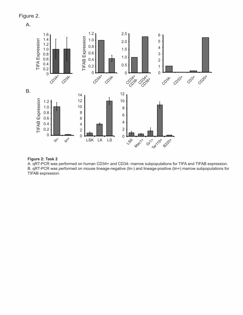
Our first year of experiments evaluating the role of TIFAB in normal and malignant hematopoietic cell function revealed striking effects on TRAF6 and NF-κB. Although not directly proposed in the Tasks within the first year, we opted to determine the cellular consequences of TIFAB on malignant cell growth, cell cycle, and survival. For these experiments, HL60 cells were transduced with FLAG-TIFAB and cultured in normal growth serum conditions (10% FBS) or under starved conditions (1% FBS) (Figure 5A). Expression of TIFAB resulted in increased AnnexinV-positive cells under serum rich (9.2% versus 18.6% AnnexinV) or starved culture (14.4% versus 24.6% AnnexinV) conditions (Figure 5A). Consistent with reduced survival, TIFAB-expressing HL60 cells grew significantly slower in liquid culture when evaluated for 6 days (Figure 5B). In addition, cell cycle status was evaluated by propidium iodide incorporation. TIFAB expression resulted in fewer cells in the S- and G1/M-phase of the cell cycle (Figure 5C). To determine whether TIFAB affects leukemic progenitor function, HL60 and THP1 cells expressing TIFAB were plated into methylcellulose for colony formation. Although TIFAB only slightly reduced colony formation in HL60, TIFAB significantly reduced colony formation in THP1 cells (Figure 5D). Interestingly, knockdown of TIFAB enhanced colony formation in HL60 cells, but not in THP1 cells (Figure 5E). These results indicate that these cells are sensitive to variations in TIFAB expression, and support our hypothesis that TIFAB functions as a tumor suppressor-like protein in hematopoietic cells.
KEY RESEARCH ACCOMPLISHMENTS
- Identified an shRNA lentiviral vector containing a GFP reporter that knocks down the expression of human TIFAB by approximately 50%.

! 6!
- Successfully transduced normal CD34+ cells and human leukemic cell lines to evaluate TIFAB knockdown with the shRNA.
- Measured expression of TIFA and TIFAB in normal human and mouse hematopoietic subpopulations. Identified that TIFAB is enriched in hematopoietic progenitor cells.
- In preliminary experiments, knockdown of TIFAB in human CD34+ cells resulted in increased CD34+ survival (less AnnexinV+ cells) and altered progenitor differentiation in methylcellulose.
- Discovered that TIFAB suppresses active TRAF6 and reduces TRAF6 protein expression in human cell lines.
- Determined that TIFAB suppress NF-κB activation following LPS/TRAF6-mediated stimulation, but not TNFa/TNFR.
- Show that TIFAB expression in human leukemic cells results in impaired growth, survival, and leukemic progenitor function, underscoring a tumor suppressor role for TIFAB.
REPORTABLE OUTCOMES
- Accepted Review Article: Fang J, M Varney, and DT Starczynowski. Implication of miRNAs in the pathogenesis of MDS. Current Pharmaceutical Design. May 7, 2012 - Accepted Research Article: Fang J, G Rhyasen, L Bolanos, C Rasch, M Varney, G Jansen, J Cloos, C Rigolino, A Cortelezzi, EN Oliva, M Cuzzola, DT Starczynowski. Cytotoxic effects of Bortezomib in Myelodysplastic syndrome/Acute Myeloid Leukemia depend on autophagy-mediated lysosomal degradation of TRAF6 and repression of PSMA1. Blood. June 8, 2012 - Abstracts at Conferences: Varney M, L Bolanos, J Fang, G Rhyasen, J Inoue, DT Starczynowski. Deciphering the role of TIFAB in del(5q) Myelodysplastic Syndromes. Midwest Blood Club, Indianapolis, IN. March 15, 2012 Varney M, L Bolanos, J Fang, G Rhyasen, J Inoue, DT Starczynowski. Deciphering the role of TIFAB in del(5q) Myelodysplastic Syndromes. Myeloid Meeting, Cincinnati, OH. May 7, 2012
CONCLUSIONS
The first year of the project has yielded many interesting results, most of which support
our original hypothesis, and allowed us to continue with the majority of goals in the Statement of Work. Overall, the experiments suggest that TIFAB, a novel and uncharacterized protein, exhibits tumor suppressor-like functions in human hematopoietic cells. Our key observations show that (1) TIFAB is primarily expressed in hematopoietic progenitor cells (rather than in primitive hematopoietic stem cells or mature blood cells; (2) knockdown of TIFAB in human CD34+ hematopoietic stem/progenitor cells results in increased survival and altered hematopoietic progenitor function; (3) TIFAB inhibits TRAF6 protein expression and activation, resulting in lower NF-κB activation; and (4) TIFAB expression impacts leukemic cell survival, growth and progenitor function. Given that TIFAB is deleted in many MDS and AML patients,

! 7!
these findings could have major implications in MDS and AML subtypes with deletions of chr 5q.
As indicated above, majority of the goals in the Statement of Work have been accomplished for the first year of the proposal. In addition, our ongoing experiments support our hypothesis, permitting us to continue with our original outline of experiments. However, there have been a few minor alterations to the Statement of Work:
- We have limited our analysis of TIFA as it appears that TIFAB has a major effect on TRAF6 independent of TIFA levels. In addition, TIFA expression did not differ in hematopoietic subpopulations. As such, we propose to delay examining TIFA’s potential role in linking TIFAB and TRAF6.
- We have opted to focus on miR-146a and not on miR-145 and miR-146a. At the time of original submission, we expected that knocking down both miRNAs is necessary for mediating an MDS-like disease. However, more recent publications indicate that knockdown of miR-146a is sufficient to induce an MDS-like disease in mice. As such, we propose to focus on the cooperation of miR-146a and TIFAB loss in human hematopoietic cells.
- In the Statement of Work we did not propose to use human cell lines to validate gain/loss of TIFAB. As shown in our preliminary data (“Extra Tasks”), we find convincing and reproducible effects of TIFAB on human MDS/AML cell line function. We propose to include human cell lines (in addition to human CD34+ as originally proposed) to investigate the role of TIFAB on survival, proliferation, and TRAF6/ NF-κB signaling.

! 8!
REFERENCES 1. Gobel F, Taschner S, Jurkin J, et al. Reciprocal role of GATA-1 and vitamin D receptor in human myeloid dendritic cell differentiation. Blood. 2009;114:3813-3821. 2. Starczynowski DT, Vercauteren S, Telenius A, et al. High-resolution whole genome tiling path array CGH analysis of CD34+ cells from patients with low-risk myelodysplastic syndromes reveals cryptic copy number alterations and predicts overall and leukemia-free survival. Blood. 2008;112:3412-3424. 3. Zhao JL, Rao DS, Boldin MP, Taganov KD, O'Connell RM, Baltimore D. NF-kappaB dysregulation in microRNA-146a-deficient mice drives the development of myeloid malignancies. Proc Natl Acad Sci U S A. 2011;108:9184-9189. 4. Boldin MP, Taganov KD, Rao DS, et al. miR-146a is a significant brake on autoimmunity, myeloproliferation, and cancer in mice. J Exp Med. 2011;208:1189-1201. 5. Matsumura T, Kawamura-Tsuzuku J, Yamamoto T, Semba K, Inoue J. TRAF-interacting protein with a forkhead-associated domain B (TIFAB) is a negative regulator of the TRAF6-induced cellular functions. J Biochem. 2009;146:375-381. 6. Matsumura T, Semba K, Azuma S, et al. TIFAB inhibits TIFA, TRAF-interacting protein with a forkhead-associated domain. Biochem Biophys Res Commun. 2004;317:230-234. 7. Lamothe B, Besse A, Campos AD, Webster WK, Wu H, Darnay BG. Site-specific Lys-63-linked tumor necrosis factor receptor-associated factor 6 auto-ubiquitination is a critical determinant of I kappa B kinase activation. J Biol Chem. 2007;282:4102-4112.

! 9!
APPENDICES - Figures 1-5 - Curriculum Vitae

0.0
0.5
1.0
1.5
Rel
ativ
e TI
FAB
Exp
ress
ion
TIFAB
TIFAB
GAPDH
Vector
FLAG
shC
tl
shTI
FAB
TIFAB
GAPDH
0.81.01.2
00.20.40.6
Vect
or
miR
-146
deco
y
Rel
ativ
e m
iR-1
46a
Vect
or
miR
-146
deco
y
0
5
10
25
15
20
p65
DN
A B
indi
ng(x
103 p
g/m
l)
- + - +vector miR-146
decoy
**
**
:IL-1`
TRAF6
GAPDH
shRNAPuroIRESGFP
UTR
UTRpGIPZ
CMV
FHA
FLAG TIFAB
1 170
_F _T
A.B.
Figure 1.
C.
D. E.
F. G. H.
sh#8
8
shC
TL
THP1HL60
sh#8
9
sh#8
8
shC
TL
sh#8
9
Figure 1: Task 1A. Schematic representation of the pGIPZ lentiviral vector for knockdown of TIFAB is shown. On The bicistronic miRNA-based shRNA is driven by a CMV promoter and also expresses GFP for tracking expression in transduced cells. For simplicity, cPPT, WRE, Amp, pUC, and Ori are not shown. B. 1x10⁶ HL60 and THP1 cells were transduced with control shRNA or shTIFAB (clone #88 and #89) and analyzed by FACS for transduction efficiency. Numbers represent percent GFP.C. qRT-PCR was performed on HL60 and THP1 cells transduced with the indicated shRNA-containing lentiviral vectors. D. A schematic representation of FLAG-TIFAB protein. The FLAG sequences (yellow) and fork-associated domain �)+$��DUH�LQGLFDWHG��$QWLERGLHV�GLUHFWHG�DJDLQVW�)/$*��Į)��DQG�7,)$%��DPLQR�DFLGV��������Į7��DUH�KLJKOLJKWHG��Numbers indicate the amino acid position. FLAG-TIFAB was subcloned into pGK-GFP retroviral vector and trans-duced into HL60 cells. Protein lysates were evaluated for immunobloting with the indicated antibodies. E. THP1 cells were transduced with control shRNA or shTIFAB and analyzed for TIFAB expression by immunoblot-ting with the indicated antibodies.F. pGK-miR-146a-GFP decoy was transduced into TF1 cells. RNA was collected and analyzed by qRT-PCR for miR-146a expression. G. Protein lysates from miR-146a-decoy expressing cells were analyzed for TRAF6 expression by immunoblotting. H. TF1 cells transduced with miR-146a decoy were stimulated with IL-1b for 6 hours. Nuclear lysates were collected and NF-kB (p65) DNA binding was measured by an ELISA-based assay. *, P < 0.05
GFP
FSC
shCTL shTIFAB#88 shTIFAB#89
THP1
HL6063% 25% 43%
4% 3% 3%
neg control0%
0%
1086420
1412
TIFA
B E
xpre
ssio
n
LSK LK LS
12
10
8
6
4
0
2
LSK
Mac1+
Gr1+
Ter11
9+B22
0+
1.21.00.80.60.40.2
0
1.4
lin-
lin+
A.
B.
Figure 2.
CD34+
CD34-
1.21.00.80.60.40.2
0
TIFA
Exp
ress
ion
1.61.4
1.21.00.80.60.40.2
0
2.5
2.0
1.5
1.0
0.5
0
65432
01
CD34+
CD34-
CD34+
CD38-
CD34+
CD38+
CD34-
CD33+
CD3+CD20
+
TIFA
B E
xpre
ssio
n
Figure 2: Task 2A. qRT-PCR was performed on human CD34+ and CD34- marrow subpopulations for TIFA and TIFAB expression. B. qRT-PCR was performed on mouse lineage-negative (lin-) and lineage-positive (lin+) marrow subpopulations for TIFAB expression.

100
80
60
40
20
0
Per
cent
Col
onie
s
shTI
FAB
shC
TL
BFU-ECFU-MCFU-GCFU-GMCFU-GEMM
10
8
6
4
2
0
12
14
16
# C
olon
ies
shTI
FAB
shC
TL
shControl
shTIFAB
% M
ax
AnnexinV-APC
shTI
FAB
shC
TL
40
30
20
10
0
% A
nnex
inV
*
A.
B.
Figure 3.
C.
GFP
FSC pGK pGK-miR-146 decoy
Figure 3: Task 3&4A. 1x10⁶ human CD34+ cells were transduced with control shRNA or shTIFAB (clone #89), or pGK-GFP or pGK-miR-146 decoy-GFP and analyzed by FACS for transduction efficiency. Numbers represent percent GFP.B. 5x10⁴ transduced CD34+ were plated in methylcellulose and analyzed for colony formation. Colonies were scored after 14 days.C. Transduced CD34+ cells were cultured for 4 days and analyzed for AnnexinV staining by FACS. The histogram is a summary of 3 replicate experiments.
shCTL shTIFAB#89
4%16%
10%20%

TRAF6
GAPDH
shC
tl
shT
IFA
B
pIKK`
IKK`
Flag-TIFAB
GAPDH
TIFABVector TIFABVector
THP1HL60
75
150
IB:GAPDH
IP: TRAF6IB: Ub
IB:TRAF6
TIF
AB
Vect
or
TR
AF
6-U
bn
vector:
TRAF6:
TIFAB:
++ + +
+ ++
- - --- -
100
250
TR
AF
6-U
bn
IB:GAPDH
IP: TRAF6IB: Ub
vector:
TRAF6:
TIFAB:
++ + +
+ ++
- - --- -
++++
--+
-
TRAF6
TIFAB
GAPDH
0
5
10
15
20
25
0
1
2
3
4
vector:
TIFAB:
LPS:
+ ++ + ++
+ + +
- - -- -- -
vector:
TIFAB:
TNF_:
+ ++ + ++
+ + +
- - -- -- -
Re
lativ
e k
B-lu
cife
rase
Re
lativ
e k
B-lu
cife
rase
***
*
TLR4
IKK
p65
TNFR
LPS TNF_
TRAF6 TRAF2
A. B.
Figure 4.
C.
D. E.
F. G.
Figure 4: Task 6A. HL60 cells were transduced with empty vector or FLAG-TIFAB. TRAF6 was immunoprecipitated (IP) and lysates blotted with anti-ubiquitin. Pre-IP lysates (IB) were evaluated for TRAF6 and GAPDH expression.B. HL60 and THP1 cells were transduced with empty vector or FLAG-TIFAB and analyzed by immunoblotting with the indicated antibodies. C. HL60 cells were transduced with control shRNA or shTIFAB and analyzed by immunoblotting for TRAF6 expres-sion.D. A293 cells were transfected with pcDNA3.1 (vector, 250 ng), pcDNA3.1-TRAF6 (250 ng), pcDNA3.1-FLAG-TIFAB (250 and 500 ng). TRAF6 was IP and lysates blotted with anti-ubiquitin. Pre-IP lysates (IB) were evaluated for GAPDH expression.E. A293 cells were transfected with pcDNA3.1 (vector, 250 ng), pcDNA3.1-TRAF6 (250 ng), pcDNA3.1-FLAG-TIFAB (250, 500, 1000 ng). Lysates were analyzed by immunoblotting with the indicated antibodies. )��0RGHO�GLVWLQJXLVKLQJ�/36�7/5��DQG�71)D�71)5�DFWLYDWLRQ�RI�1)�ț%��:H�SURSRVH�WKDW�7,)$%�VHOHFWLYHO\�LQKLELWV�TRAF6. *��$����FHOOV�ZHUH�WUDQVIHFWHG�ZLWK�SF'1$�����YHFWRU������QJ��RU�SF'1$����)/$*�7,)$%�����������QJ���DQG�țB-luciferase. Following transfection, cells were simulated with either LPS (1 ug/ml) or TNFa (1 ng/ml) for 6 hours. Values represent relative luciferase.
0
10
20
30
40
50
60
70
80
90
G1 S G1/M
vector
TIFAB
Per
cent
Cel
ls
AnnexinV-APC
PI
9.2
14.4
18.6
24.6
vector TIFAB
+serum
-serum Day 0 Day 3 Day 6
Rel
ativ
e ce
ll #
0
6
8
10
4
2
vectorTIFAB
0
20
40
60
80
0
500
1000
0
100
200
300
400
500
1000
500
0
THP1HL60
# co
lone
s/40
00 c
ells
# co
lone
s/40
00 c
ells
TIFABVector TIFABVector
****
shTIFABshCTL shTIFABshCTL
*THP1HL60
# co
lone
s/40
00 c
ells
# co
lone
s/40
00 c
ells
A. B.
Figure 5.
C.
D.
E.
Figure 5: Extra TasksA. HL60 cells were transduced with empty vector or FLAG-TIFAB, sorted for GFP expression, and allowed to recover for 1 week. 1x10⁵ cells were cultured with (10% FBS) or without (1% FBS) serum for 48 hours and then analyzed for AnnexinV by FACS. Shown is a representative experiment. B. HL60 cells expressing TIFAB were plated (1x10⁵ cells/ml) and counted every 3 days. Viable cells were measured by Trypan blue exclusion. Shown is the summary of 2 independent experiments.C. HL60 cells expression TIFAB were evaluated for cell cycle with propidium iodide. Shown is the summary of 3 independent replicates.D. 4x10³ HL60 and THP1 cells expressing TIFAB were put into methylcellulose. After 10 days, colonies were counted. Shown is the summary of 3 independent replicates.E. 4x10³ HL60 and THP1 cells expression shTIFAB were put into methylcellulose. After 10 days, colonies were counted. Shown is the summary of 3 independent replicates.

1
Daniel T. Starczynowski
Division of Experimental Hematology, Cincinnati Children’s Hospital Medical Center Department of Cancer and Cell Biology, University of Cincinnati
3333 Burnet Avenue, Cincinnati, OH 45229 (513) 803-5317
Education and Training 2005-2010 Postdoctorate
BC Cancer Research Centre/University of British Columbia, Vancouver, Canada
2000-2005 Ph.D., Molecular Biology, Cell Biology and Biochemistry Boston University, Boston, MA, USA.
1996-2000 B.Sc., Honors Biology
Concentrations in Chemistry and Biotechnology Fairleigh Dickinson University, Teaneck, NJ, USA.
Research and Professional Experience
2010- present Assistant Professor, Division of Experimental Hematology, Cincinnati Children’s Hospital Medical
Center, Cincinnati, OH 2010-present Affiliate Assistant Professor, Department of Cancer and Cell Biology, University of Cincinnati,
Cincinnati, OH 2010- present Lecturer, Ulm University, Germany 2005-2010 Postdoctoral Fellow
Research Focus: Identification and functional analysis of genetic and molecular determinants of hematological malignancies: deregulation of miR-146/TRAF6 signaling in Myelodysplastic syndromes Advisor: Dr. Aly Karsan, BC Cancer Research Centre/University of British Columbia
2001-2005 Graduate Research Assistant
Dissertation: A mutational and functional analysis of C-terminal sequences of transcription factor REL: their role in cellular transformation and transcriptional activation
Advisor: Dr. Thomas D. Gilmore, Boston University 2000 Summer Research Assistant Research Focus: Functional analysis of androgen receptor in prostate cancer
Advisor: Dr. Marianne Sadar, BC Cancer Research Centre 1999-2000 Honors Research
Dissertation: Inhibitory effects of somatostatin on the viability of a cell line Advisor: Dr. Anjali Saxena, Fairleigh Dickinson University

2
1997-1999 Research Assistant Research Focus: Development of enzyme-friendly biosensors for lactate detection Advisor: Dr. Mihaela Leonida, Fairleigh Dickinson University
Pedagogical Experience 2012 Lectures “Critical data presentation III”, Department of Cancer and Cell Biology, University of Cincinnati,
OH 2010 Lectures “Innate Immunity and Cancer”, Master Online Program in Advanced Oncology. Ulm University. Developed a lecture on innate immunity and its role in human cancer for postgraduates of
medicine. 2010 Individual Lecture “MicroRNAs in cancer”, Medical Oncology Residency Training Program. BC Cancer Agency.
Organized and conducted a lecture for 1st and 2nd year residents of medical training on microRNAs and their role in clinical oncology.
2009 Individual Lecture
“MicroRNAs in hematological malignancies”, Hematology Fellows Series. Department of Medicine, University of British Columbia. Organized and conducted a lecture for hematology and pathology fellows on the emerging role of microRNAs in hematological malignancies.
2009 Workshop
“MicroRNAs: small RNAs with big impact”. Terry Fox Laboratory, BC Cancer Research Centre. Organized and co-lead a workshop on microRNAs, their biogenesis, diversity, and mechanism as related to gene regulation and to human disease.
2005 Teaching Assistant
“Molecular Biology Laboratory”, Boston University. Prepared and delivered weekly labs for advanced (500-level) molecular biology students.
2000-2001 Teaching Fellow
“Life Sciences Chemistry I and II”, Boston University Organized and delivered weekly labs for introductory (100-level) undergraduate science students to supplement course material. Prepared and evaluated exams and final grades. Held weekly office hours.
1998 Teaching Assistant “Chemistry for Health Science”, Fairleigh Dickinson University. Organized and delivered weekly labs for introductory (100-level) undergraduate science students.
Awards and Honors 2011-14 American Society of Hematology Scholar Award, Basic Research Junior Faculty 2010 National Institute of Allergy and Infectious Disease, Keystone Symposium Scholarship
3
2008 Travel Award, American Society of Hematology 2008 Eugene Cronkite Award: New Investigator Award (1st place). International Society of Experimental Hematology 2008 Travel Grant, International Society of Experimental Hematology Scientific Meeting 2007-2010 Postdoctoral Fellowship, Canadian Institute of Health Research 2006 Frank A. Belamarich Award; Outstanding Scholarship and Performance in Graduate Studies,
Biology Department, Boston University 2006-2009 Postdoctoral Fellowship, Michael Smith Foundation for Health Research 2001-2005 Graduate Research Fellowship, Boston University 2002-2004 Postgraduate Research Scholarship, Natural Sciences and Engineering Research Council of Canada 2002 Travel Scholarship, NF-κB: Bench to Bedside - Keystone Symposium 2000-01, 2005 Teaching Fellowships, Boston University 2000 J.M. Warren Summer Research Scholarship, British Columbia Cancer Agency 2000 ECAC Award: Graduating athlete with highest cumulative grade point average, Fairleigh
Dickinson University, NJ 2000 Summa Cum Laude, Fairleigh Dickinson University 2000 Phi Omega Epsilon, Honor Society 1999 Phi Zeta Kappa, Honor Society 1999 Charter Day Scholarship, Fairleigh Dickinson University 1998 University-College Dean’s Award, Fairleigh Dickinson University 1996-2000 University Honors List, Fairleigh Dickinson University 1996-2000 Tennis Scholarship, Fairleigh Dickinson University
Memberships and Committees 2012-present Early Career Reviewer (ECR) at the Center for Scientific Review, National Institute of Health 2011 (Sept) NIH State of the Science Symposium: Myelodysplastic Syndrome Working Group, Bethesda MD
(Invited member) 2011-present Graduate student recruitment committee, Cancer and Cell Biology Graduate Program, University
of Cincinnati 2011-present Immunobiology Graduate Program, Cincinnati Children’s Hospital (Training Faculty)

4
2010-present Cancer and Cell Biology Graduate Program, University of Cincinnati (Training Faculty) 2008 Campus Provost Search Committee, Fairleigh Dickinson University-Vancouver 2008-present International Society of Experimental Hematology (Member) 2005-present American Society of Hematology (Associate Member) 2006-2011 Board of Directors, Fairleigh Dickinson University-Vancouver (Member) 1999-2000 TriBeta Biological Society, Fairleigh Dickinson University Chapter (Vice President) 1997-2000 University Honors Program, Fairleigh Dickinson University
University Service
2012 Qualifying Exam Committee, Graduate Program in Cancer and Cell Biology, University of
Cincinnati (Standing Member) 2011 (Sept) CCB Graduate Program Student Symposium, University of Cincinnati (Faculty co-organizer) 2011 (Feb) Research Ethics, University of Cincinnati (Discussion leader) 2011-12 Cancer and Blood Diseases Institute Seminar Series (Co-coordinator) 2011 Scholarship Oversight Committee, Hematology/Oncology Clinical Fellowship (Committee
Member) 2011 Immunohematology Club Seminar Series (Speaker)
Patents
Aly Karsan and Daniel Starczynowski, “TRAF6 as a therapeutic target and predictive biomarker for lung and colorectal cancer,” U.S. provisional patent, September 2010 Aly Karsan and Daniel Starczynowski, “Novel methods to predict therapeutic response to Lenalidomide and related drugs,” U.S. provisional patent, January 2008
Peer-Reviewed Publications
1. Gilmore TD, M-E Gapuzan, D Kalaitzidis, and D Starczynowski. (2002) Rel/NF-kB/IkB signal transduction in
the generation and treatment of human cancer. Cancer Letters, 181: 1-9. PMID:12430173 (Review) *Top 25 Hottest Article” on ScienceDirect within Cancer Letters (April-June 2005) 2. Leonida MD, DT Starczynowski, R Waldman, and B Aurian-Blajeni. (2003) Polymeric FAD used as enzyme-
friendly mediator in lactate detection. Analytical and Bioanalytical Chemistry, 376: 832-837. PMID: 12811450 3. Starczynowski DT, JG Reynolds, and TD Gilmore. (2003). Deletion of either C-terminal transactivation
subdomain enhances the in vitro transforming activity of human transcription factor REL. Oncogene, 22: 6929-6936. PMID: 14534540

5
4. Gilmore TD, D Kalaitzidis, M-C Liang, and DT Starczynowski. (2004). The c-Rel transcription factor and B-
cell proliferation: a deal with the devil. Oncogene, 23: 2275-2286. PMID: 14755244 (Review) 5. Gilmore TD, D Kalaitzidis, and DT Starczynowski. (2004). RELevant gene amplification in B-cell lymphomas.
Blood. 103: 3243. PMID: 15070712 (Letter) 6. Kalaitzidis D, J Ok, L Sulak, DT Starczynowski, and TD Gilmore. (2004). Characterization of a human REL-
estrogen receptor fusion protein with a reverse conditional transforming activity in chicken spleen cells. Oncogene, 23: 7580-7587. PMID:15326488
7. Starczynowski DT, JG Reynolds, and TD Gilmore. (2005). Mutations of tumor necrosis factor a-responsive
serine residues 460 and 471 within the C-terminal transactivation domain of human transcription factor REL can enhance its in vitro transforming ability. Oncogene, 24: 7355-7368. PMID: 16027730
8. Starczynowski DT, H Trautmann, C Pott, L Harder, N Arnold, R Siebert, and TD Gilmore. (2007). Mutation of
an IKK phosphorylation site within the transactivation domain of REL in two patients with B-cell lymphoma enhances REL’s in vitro transforming activity. Oncogene, 26: 2685-2694. PMID: 17072339
*Oncogene’s Featured Article (April 2007) 9. Starczynowski DT, S Vercauteren, S Sung, A Brooks-Wilson, J Spinelli, C Eaves, A Eaves, D Horsman, W
Lam, and A Karsan. (2008). High-resolution array comparative genomic hybridization of CD34+ cells from patients with low-risk myelodysplastic syndromes predicts overall and leukemia-free survival. Blood, 112(8): 3412-3424. PMID: 18663149
10. Starczynowski DT, F Kuchenbauer, B Argiropoulos, S Sung, R Morin, A Muranyi, D Hogue, R Wells, M
Marra, WL Lam, K Humphries, and A Karsan. (2010). Identification of miR-145 and miR-146a as microRNAs involved in the pathogenesis of 5q- syndrome. Nature Medicine, 16(1): 49-58. PMID: 19898489
*Comment in Nature Medicine. 2010. Myelodysplasia: Battle in the Bone Marrow. 16(1): 30-32. *Featured in The Hematologist. 2010. Clarifying the genetic underpinnings of the 5q- syndrome. July/Aug:7(4). 11. Starczynowski DT and A Karsan. (2010). Deregulation of innate immune signaling in myelodysplastic
syndromes is associated with deletion of chromosome arm 5q. Cell Cycle, 11; 9(5). PMID: 20160505 (Review) 12. Starczynowski DT and A Karsan. (2010). Innate immune signaling in Myelodysplastic Syndromes.
Hematology/Oncology Clinics of North America, 24(2):343-359. PMID: 20359630 (Review) 13. Vercauteren SM, S Sung, DT Starczynowski, WL Lam, H Bruyere, DE Horsman, P Tsang, H Leitch, and A
Karsan. (2010). Cryptic alterations detected by array comparative genomic hybridization in bone marrow CD34+ cells are not present in peripheral blood granulocytes of patients with myelodysplastic syndrome. American Journal of Clinical Pathology, 134(1):119-126. PMID: 20551276
14. Starczynowski DT, S Vercauteren, S Sung, A Brooks-Wilson, J Spinelli, C Eaves, A Eaves, D Horsman, W
Lam, and A Karsan. (2010). Evidence of somatic alterations of genomic copy number variants in clonally derived cells from patients with low-risk myelodysplastic syndromes. Leukemia Research, Aug 27. Epub. PMID: 20801506
15. Starczynowski DT, RD Morin, A McPherson, J Lam, R Chari, J Wegrzyn, A Delaney, AL Prabhu, Y Zhao, M
Hirst, W Lam, MA Marra, and A Karsan. (2011). Genome-wide identification of human microRNAs located in leukemia-associated genomic alterations. Blood, Oct 20. Epub. PMID: 20962326
6
16. Starczynowski DT, F Kuchenbauer, J Wegrzyn, K Humphries, and A Karsan. (2011). Increased expression of microRNA-146 disrupts differentiation and survival of hematopoietic progenitor cells. Experimental Hematology, Oct 5. Epub. PMID: 20933052
17. Kuchenbauer F, SM Mah, M Heuser, A McPherson, J Ruschmann, A Rouhi, T Berg, L Bullinger, B
Argiropoulos, RD Morin, D. Lai, DT Starczynowski, A Karsan, CJ Eaves, A Watahiki, Y Wang, SA Aparicio, A Ganser, J Krauter, H Doehner, K Doehner, MA Marra, FD Carmargo L Palmquist, C Buske, and RK Humphries. (2011). Comprehensive analysis of mammalian miRNA* species and their role in myeloid cells. Blood, ePub. PMID: 21628414
18. Rhyasen G and DT Starczynowski*. (2011). Deregulation of microRNAs in Myelodysplastic Syndromes.
Leukemia, Epub. PMID: 21852786 19. Starczynowski DT, WL Lockwood, S Delehouzee, S Lam, M-S Tsao, AF Gazdar, W Lam, and A Karsan.
TRAF6 is an amplified oncogene bridging the Ras and nuclear factor-kB cascade in lung cancer. (2011). Journal of Clinical Investigation, Epub. PMID: 21911935.
20. Vercauteren S, DT Starczynowski, S Sung, K McNeil, C Salski, C-L Jensen, W Lam, A Karsan. (2011). T cells of patients with Myelodysplastic syndrome are frequently derived from the malignant clone. British Journal of Haematology, Epub. 21. Fang J, M Varney, and DT Starczynowski*. Implication of miRNAs in the pathogenesis of MDS. (2012) Current Pharmaceutical Design. In Press 22. Fang J, G Rhyasen, L Bolanos, C Rasch, M Varney, G Jansen, J Cloos, C Rigolino, A Cortelezzi, EN Oliva, M Cuzzola, DT Starczynowski*. Cytotoxic effects of Bortezomib in Myelodysplastic syndrome/Acute Myeloid Leukemia depend on autophagy-mediated lysosomal degradation of TRAF6 and repression of PSMA1. (2012) Blood. In Press. *Corresponding author
Invited Talks
Midwest Blood Club Symposium, Indianapolis, IN (March, 2012). Oral Abstract. American Society of Hematology, San Diego, CA (December, 2011): New Therapies in Myelodysplastic Syndromes. Oral Abstract Session. MD Anderson, Leukemia Group (November, 2011) Japanese Society of Hematology, Nagoya, Japan (October, 2011). Plenary Session. Baltic Stem Cell Meeting, Szczecin, Poland (May, 2011). Plenary Session. Midwest Blood Club Symposium, Cincinnati, OH (April, 2011). Plenary Session. Cepheid Inc., Sunnyvale, CA (March, 2011). Taussig Cancer Institute, Cleveland Clinic, Cleveland, OH (March, 2011). Translational Hematology and Oncology Research Lecture Series.

7
Ulm University, Ulm, Germany (February, 2011). Workshop on Translational Research: Cellular and Molecular Biology of Cancer. James Graham Brown Cancer Center, University of Louisville, Louisville, KY (February, 2011). Keystone Symposia, NF-κB in Inflammation and Disease, Santa Fe, NM (January 2010). Workshop: NF-κB in Disease Pathogenesis. Department of Cancer and Cell Biology, University of Cincinnati, OH (November, 2009). Department Seminar Series. Department of Cytogenetics, Vancouver Hospital and Health Sciences Centre, Vancouver General Hospital, Vancouver, BC (March 2009). Cytogenetics Department Seminar Series. Center for Advanced Biotechnology and Medicine, Rutgers University, New Brunswick, NJ (February 2009). Seminar Series. Centre for Blood Research, University of British Columbia, Vancouver, BC (February 2009). Centre for Blood Research Seminar Series. American Society of Hematology, San Francisco, CA (December 2008). Myelodysplastic Syndromes: Basic Biology: Ribosomal Proteins, MicroRNAs and Animal Models. Oral Abstract Session. Stem Cell Network, Annual General Meeting. Vancouver, BC (November 2008). Plenary Session. BC Cancer Agency Annual Conference, Vancouver, BC (November 2008). Knowledge Translation - Basic vs. Clinical Research: Hematological Malignancies. International Society of Experimental Hematology. Boston, MA. (July 2008). New Investigator Session.
Editorial Board 2012- Leukemia 2012- PLoS One
Ad Hoc Reviewer PLoS One, Genes and Cancer, Current Pharmaceutical Review, Experimental Hematology, Leukemia, Blood, International Journal of Cancer, Haematologica