author's personal copy - neuroregenerationneuroregeneration.org/s/hallett et al nbd...
TRANSCRIPT


Author's personal copy
Alpha-synuclein overexpressing transgenic mice show internal organ pathology andautonomic deficits
Penelope J. Hallett a,⁎, Jesse R. McLean a, Andrew Kartunen a, J. William Langston b, Ole Isacson a
a Center for Neuroregeneration Research, McLean Hospital/Harvard Medical School, 115 Mill Street, Belmont, MA 02478, USAb The Parkinson's Institute and Clinical Center, 675 Almanor Avenue, Sunnyvale, CA 94085, USA
a b s t r a c ta r t i c l e i n f o
Article history:Received 6 February 2012Revised 6 April 2012Accepted 11 April 2012Available online 19 April 2012
Keywords:Alpha-synucleinSynucleinopathyParkinson's diseaseConstipationGastrointestinalAutonomicAxonopathy
While studying transgenic mice that overexpress human wildtype alpha-synuclein (Thy1-ASO, ASO) for typ-ical brain alpha-synucleinopathy and central nervous system neuropathology, we observed progressive func-tional changes in the gastrointestinal and other peripheral organs. A more systematic study revealed that thegastrointestinal tract in ASO mice showed severe distension and blockage of the large intestine by 9–12 months of age. Functional assessments demonstrated a reduction in fecal water content and fecal pelletoutput, and increased whole gut transit time, in ASO mice compared to wildtype littermates, indicative ofconstipation, a symptom commonly reported by Parkinson's disease (PD) patients. Food intake was increasedand body weight was decreased in 12 month old ASO mice, suggestive of metabolic abnormalities. Post-mortem histological analyses showed that human alpha-synuclein protein was robustly expressed in axonalfibers and in occasional cell bodies of the enteric nervous system, and in the heart of ASO mice. Accumulationof proteinase-K insoluble alpha-synuclein, reminiscent of neurodegenerative processes in PD was also ob-served. The functional and pathological changes we document here in ASO mice could relate to the autonom-ic deficits also seen in idiopathic and alpha-synuclein-mediated genetic forms of PD. These experimental dataprovide a foundation for therapeutic modeling of autonomic changes in PD and related alpha-synucleinopathies.
© 2012 Elsevier Inc. All rights reserved.
Introduction
The non-motor symptomatology of Parkinson's disease (PD) in-cludes autonomic dysfunction, hyposmia, and sleep abnormalities(Chaudhuri et al., 2006). Such symptoms commonly experienced byPD patients are often detected before clinical motor symptoms, pro-gressively become very disabling and significantly impact the qualityof life for patients (Savica et al., 2010). Dysfunction of gastrointestinaland cardiovascular systems are significant autonomic features of PD,and post-mortem studies have identified PD neuropathologythroughout the autonomic nervous system including the gastrointes-tinal tract, heart and sympathetic ganglia (Beach et al., 2010;Goldstein, 2010).
PD has multiple etiologies, including mitochondrial dysfunctionand oxidative stress, and genes involved in such pathways havebeen identified (e.g. DJ-1, PINK1) (Cookson and Bandmann, 2010).Toxin-based and genetic approaches to induce mitochondrial
dysfunction and oxidative stress in animals have been widely usedto model central nervous system (CNS) dysfunction and the selectiveloss of midbrain dopamine neurons (Cannon and Greenamyre, 2010;Magen and Chesselet, 2010). These models have also indicated in-volvement of the peripheral nervous system (PNS). MPTP, a complexI inhibitor selective for the dopaminergic system, alters gastrointesti-nal dopaminergic transmission and related function, and reduces do-pamine neurons in the enteric nervous system (Anderson et al., 2007;Chaumette et al., 2009; Natale et al., 2010; Tian et al., 2008). Systemicadministration of rotenone, a general mitochondrial complex I inhib-itor, in rats, induces clear gastrointestinal dysfunction and neuropa-thology in the enteric nervous system, similar to PD (Drolet et al.,2009; Greene et al., 2009). These toxin-based models demonstratethat mechanisms involved in CNS dysfunction in PD can also elicitfunctional deficits and pathology in the PNS. While such models de-pend on external agents that induce, for example, oxidative stress, amajor genetic and pathologic component of PD is associated withthe synaptic protein, alpha-synuclein. Mutations in, or multiplicationof SNCA leads to early onset PD and diffuse Lewy body disease, andpolymorphisms in the regulatory elements of SNCA can predisposeto PD (Cookson and Bandmann, 2010). In both sporadic and familialPD, alpha-synuclein is a component of proteinacious Lewy body in-clusions and neurites found throughout the CNS and also in thePNS. Alpha-synuclein is a highly expressed presynaptic protein,
Neurobiology of Disease 47 (2012) 258–267
⁎ Corresponding author. Fax: +1 617 855 3284.E-mail addresses: [email protected] (P.J. Hallett),
[email protected] (J.R. McLean), [email protected](A. Kartunen), [email protected] (J.W. Langston), [email protected](O. Isacson).
Available online on ScienceDirect (www.sciencedirect.com).
0969-9961/$ – see front matter © 2012 Elsevier Inc. All rights reserved.doi:10.1016/j.nbd.2012.04.009
Contents lists available at SciVerse ScienceDirect
Neurobiology of Disease
j ourna l homepage: www.e lsev ie r .com/ locate /ynbd i
Author's personal copy
involved in SNARE protein assembly (Burre et al., 2010), and with animportant role in neurotransmitter release (Nemani et al., 2010).Overexpression of either wildtype or mutant alpha-synuclein, orknockdown of alpha-synuclein together with beta and gamma synu-cleins, causes neuronal dysfunction and degeneration (Burre et al.,2010; Chung et al., 2009; Greten-Harrison et al., 2010; Nemani etal., 2010; Ulusoy et al., 2010). A variety of transgenic mouse modelsof alpha-synucleinopathy exist and although CNS deficits have beenthe focus of studies in such models (Chesselet, 2008; Chesselet etal., 2008; Dawson et al., 2010; Fleming et al., 2004, 2008; McLean etal., 2012; Rockenstein et al., 2002; Song et al., 2004; Watson et al.,2009), more recently, functional deficits in cardiac and gastrointesti-nal systems have also been described (Fleming et al., 2007, 2009;Kuo et al., 2010; Wang et al., 2008).
We first noticed evidence for autonomic dysfunction in transgenicmice that overexpress human wildtype alpha-synuclein on the Thy1promoter (ASO, Thy1-ASO) by their extreme sensitivity to thealpha2-adrenergic agonist, xylazine, used as a routine anestheticagent (Hallett et al., unpublished data). We also observed at post-mortem examination, clear gut and bladder distension in older(>12 months) ASO mice. Based on these observations we investigat-ed gastrointestinal functional deficits, and neuropathology of the gas-trointestinal system in ASO mice.
Materials and methods
Animals
All animal procedures were performed in accordance with theguidelines of the National Institute of Health and were approved bythe Institutional Animal Care and Use Committee (IACUC) at McLeanHospital, Harvard Medical School. Animals were housed according tostandard conditions, in a dark/light cycle of 12 h, with ad libetum ac-cess to food and water. Transgenic mice overexpressing human wild-type alpha-synuclein under the Thy1 promoter have been previouslydescribed (Rockenstein et al., 2002). Mice hemizygous for alpha-synuclein overexpression were maintained on a mixed C57BL/6-DBA/2 background by breeding female hemizygous mice with maleBDF1 hybrids (Charles River, Wilmington, USA). Genotypes were ver-ified using polymerase chain reaction amplification of tail DNA. For allexperimentation only male ASO and wildtype littermate mice wereused.
Beam traversal test
Motor coordination and balance in 12–15 month old ASO and WTlittermate mice was tested using a challenging beam traversal test, aspreviously described (Fleming et al., 2004). In brief, a plexiglass beam(Plastics Zone, Woodland Hills, CA), 1 m length total, and comprisingof four (25 cm length) sections that gradually decreased in diameterfrom 3.5 cm to 0.5 cm in 1 cm increments was used. Animals weretrained to traverse the beam (from widest to narrowest) directlyinto the animal's home cage. Each mouse received two days of train-ing (5 trials each) followed by testing on the third day. During thetesting phase, a wire mesh grid (1 cm2) of corresponding beamwidth was placed over the beam. Animals were videotaped while tra-versing the beam, over 5 trials. Videotaped were analyzed at slow-motion, by an investigator blinded to the genotype of the animalsand the time taken to traverse the beam, and number of footslipsoff the beam were determined over the 5 trials and averaged.
Assessment of fecal water content
Fecal pellet output and water content was assessed in 2–4 and9–12 month old ASO and WT littermate mice as previously described(Anderson et al., 2007; Kuo et al., 2010; Taylor et al., 2009) (n=10–
15 per group). Fecal water content provides an indication of constipa-tion (reduced fecal water content resulting from reduced gastrointes-tinal motility and subsequent increased reabsorption of water),diarrhea (increased fecal water content) and malabsorption. Micewere placed individually in a novel environment (clean cage) and ob-served for 60 min. Testing was performed at the same time each day(2–3 PM). Fecal pellets from individual mice were collected immedi-ately after expulsion in a pre-weighed sealed 2 mL microcentrifugetube. The number of pellets expulsed by each mouse was recorded.Tubes were weighed to obtain the wet weight of the stool. Stoolwas then dried overnight at 60 °C and reweighed to obtain the dryweight. The difference in wet and dry weight was expressed overthe wet stool weight to calculate fecal water content.
Whole gut transit time
Whole gut transit time (WGTT) was assessed in 2–4 and 12 monthold ASO and WT littermate mice as previously described (Kuo et al.,2010) (n=7–14 per group). Animals were placed in individualcages and food deprived for 1 h prior to receiving 0.3 mL 6% (w/v)carmine solution in 0.5% methylcellulose by oral gavage. The timetaken for each mouse to produce a red fecal pellet after the adminis-tration of carmine dye was recorded. Animals which did not producea pellet containing red dye after 12 h were recorded as >12 h. WGTTwas calculated as time (min) per 20 g animal bodyweight.
Food and water intake
Food and water intake were measured over 3 consecutive days in2 and 12 month old ASO andWT littermate mice. Individually housedmice were provided with a preweighed amount of rodent chow,placed in a heavy glass jar in the homecage. Remaining chow wasweighed at the same time each day, over the next 3 days. Similarly,mice were provided with a premeasured volume of water in thedrinking bottle, and the volume of water consumed was determinedby calculating the remaining volume at the same time each dayover 3 consecutive days. Food and water intake were determined astotal consumption over 3 days, and as average consumption per20 g animal bodyweight.
Immunohistochemistry
12–15 month old mice (n=5 WT, n=6 ASO) were terminallyanesthetized by interaperitoneal injection of sodium pentobarbital(130 mg/kg) and intracardially perfused with heparinized saline fol-lowed by 4% paraformaldehyde in PBS. The brain, heart and gastroin-testinal tissues were quickly dissected (gastrointestinal tissue wereflushed with saline) and post-fixed overnight in 4% paraformalde-hyde at 4 °C for 8 h, before cryoprotection in 30% sucrose/PBS untilequilibration. Brains were sectioned at 40 μm on a freezing micro-tome, and serially collected in PBS. Gastrointestinal tract and heartwere cryosectioned at 30 μm and collected directly onto slides in 4 se-ries. Whole mount sections of the myenteric plexus were also pre-pared from PFA-fixed gastrointestinal tissues by manual separationof the myenteric plexus attached to longitudinal muscle layers, fromthe submucosal and mucosal layers.
Free-floating brain sections, whole-mount and slide-mounted tis-sue sections of heart and gastrointestinal tract were permeabilized in0.1 or 0.5% Triton X-100/PBS. Non-specific labeling was blocked by in-cubating sections in 10% donkey or goat serum in 0.1 or 0.5% Triton X-100/PBS for 1 h at room temperature. Sections were then incubatedwith primary antibodies for 12 h at 4 °C, washed in PBS and incubatedfor 1 h at room temperature with secondary antibody. For immuno-fluroescent staining, anti-rabbit, anti-rat, anti-sheep and anti-goatAlexaFluor 488, 568 and 680-conjugated fluorescent secondary anti-bodies were used (1:500, Molecular Probes), followed by DAPI
259P.J. Hallett et al. / Neurobiology of Disease 47 (2012) 258–267

Author's personal copy
staining to visualize nuclei. For peroxidase-based detection sectionswere incubated in 3% in hydrogen peroxide prior to the serum block-ing step. Biotinylated anti-rabbit, anti-rat, anti-sheep and anti-goatsecondary antibodies (1:300, Vector Labs) were used followed byABC complexes and DAB substrate (Vector Labs). The following com-mercially available primary antibodies were used for both fluores-cence and peroxidase immunostanings: sheep anti-alpha-synuclein(Millipore, AB5336P) at 1:1500; rat anti-alpha-synuclein 15G7(Axxora, ALX-804-258) at 1:10; mouse anti-alpha-synuclein Syn211(Millipore, 36-008); sheep or rabbit anti-tyrosine hydroxylase (Pel-Freez, P60101 or P40101 respectively), 1:300; rabbit anti-vasoactiveintestinal peptide (AbCam, AB22736), 1:300; rabbit anti-peripherin(Millipore, AB1530), 1:500; goat anti-vesicular acetylcholine trans-porter (VAChT) (Millipore, AB1578), 1:750; rabbit neuronal nitricoxide synthase (nNOS) (Abcam, ab76067), 1:100). NeuroTrace™ redfluorescent Nissl stain (Invitrogen) (1:500) was used for labelingneurons in brain tissue.
Proteinase-K digestion
For assessing accumulation of insoluble alpha-synuclein, aproteinase-K digestion step was included prior to immunostainingwith alpha-synuclein and DAB visualization. Proteinase-K digestionwas performed as previously described with slight modifications(Fernagut et al., 2007). Briefly, free-floating sections of tissue, or tis-sue sections mounted onto slides, were incubated with Proteinase-K(Promega) at 10 μg/mL in PBS for 15 min at room temperature. Sec-tions were then washed in PBS and processed as described above.
Imaging
Confocal analysis of immunofluorescent immunostainings wasperformed using a Zeiss LSM510/Meta station. Light microscopy wasvisualized using a Zeiss Axiovert microscope coupled to an OptronicsMicrofire digital camera. For quantification of neuronal number in thedorsalmotor nucleus of vagus, the total number of Nissl-stained neuronsin the dorsalmotor nucleus of vagus in every 6th section containing this
nucleuswere counted. For quantification of peripherin-immunoreactiveneurons in the myenteric plexus, the average number of neurons in15–20 myenteric ganglia per animal was quantified.
Statistical analyses
A Student's t-test was used to compare two groups on a single de-pendent measure. 1-way ANOVA was used to compare more than 2groups on the same dependent measure followed by a Bonferronipost-hoc test. All analyses were conducted using GraphPad Prism(Version 4.0) (GraphPad Software, Inc). All tests were considered sig-nificant at pb0.05.
Results
Assessment of CNS functional deficits and pathology in ASO mice
To verify involvement of the CNS in ASO mice, behavioral analysesof sensorimotor function, using the challenging beam traversal test,and post-mortem analysis to examine proteinase-K resistant alpha-synuclein immunostaining in the brain, was performed in a cohortof 12–15 month old ASO and WT littermate mice, as previously de-scribed (Fernagut et al., 2007; Fleming et al., 2004). In the beam tra-versal test (Figs. 1A, B), ASO mice had a significantly increasednumber of footslips off the beam, compared to WT mice (pb0.01, un-paired T-test). Post-mortem analyses were performed in the same co-hort of animals. A proteinase-K pretreatment of brain tissue sectionsfollowed by immunostaining with a pan-alpha-synuclein antibody(detecting both mouse and human forms of the protein), was usedto reveal proteinase-K resistant accumulations of alpha-synuclein, aspreviously described (Fernagut et al., 2007) (Figs. 1C–E). Followinga proteinase-K digestion, no alpha-synuclein immunostaining wasobserved in the substantia nigra pars reticulata of WT mice(Fig. 1C), whereas robust accumulations of alpha-synuclein weredetected in ASO mice in the SNr (Fig. 1D) and throughout the brain.At higher magnification, structures that morphologically resembledswollen neurites were observed in ASO mice (Fig. 1E).
Fig. 1. Functional motor deficits and CNS alpha-synuclein pathology in ASO mice. (A and B) Functional testing of motor performance in 12–15 month old ASO and wildtype (WT)mice on the challenging beam traversal test, shows that the average number of foot slips was significantly increased in ASO mice (B). The average time taken to traverse the beamwas not significantly increased in this group of animals (A). ** pb0.01, unpaired T-test. (C–E) Proteinase-K-resistant alpha-synuclein immunostaining in the substantia nigra parsreticulata of 12–15 month old WT and ASO mice, using an antibody that detects both human and endogenous mouse alpha-synuclein, reveals robust staining of proteinase-K in-soluble alpha-synuclein in ASO mice (D and E), but not in WT mice (C). Panel E is a high magnification of the boxed area in panel D, and shows a structure that morphologicallyresembles a swollen neurite. SNr = substantia nigra pars reticulata. Scale bar=100 μm (C and D) and 20 μm (E).
260 P.J. Hallett et al. / Neurobiology of Disease 47 (2012) 258–267

Author's personal copy
Assessment of gastrointestinal gross pathology and function in ASO mice
Initial observations of gross pathology of freshly dissected WT andASOmice revealed clear abnormalities (Fig. 2). On inspection, the intes-tinal tract was of different pallor and size, and showed distinct areas ofenlargement in ASO mice (Fig. 2A). These changes were already appar-ent in 2 month old mice, and in older (15 month old) mice, severe dis-tension and blockage was observed. The urinary bladder was alsonoticeably enlarged in some, but not all, 15 month old ASO mice, butwas never enlarged in WT littermates (Fig. 2B). We also observed uri-nary bladder enlargement in 9 month old ASO mice (data not shown).Of note, it was apparent during dissections that the urinary bladderwas frequently expelled in WT mice, or if full, was no larger than~0.75 cm wide (see Fig. 2B). In comparison, the urinary bladder wasrarely empty in ASOmice, and on occasion, as described above, was no-ticeably enlarged compared to WT mice (see Fig. 2B).
Based on our observations of gross pathology, we next examinedmore quantitative measures of gastrointestinal function. The totalnumber of fecal pellets expelled in individual mice over a one hourperiod was determined (Figs. 2C, D) and was reduced in older (9–12 months) ASO mice compared to WT littermates, whereas therewas no significant different in younger (2–4) month old mice. Fecalwater content was determined by calculating the water content infecal pellets collected over a one hour period. Between 2 and4 months of age, the fecal water content (Fig. 2E) was not significant-ly different in ASO and WT mice. However, in older mice at 9–12 months of age, fecal water content was significantly reduced inASO mice compared to WT mice (Fig. 2F). We also measured thetotal wet weight of a 3.5 cm sample of small intestine isolated from2–4 and 9–12 month old mice and found that this was significantlyincreased in 2–4 month ASO mice (66.6±7.2 mg WT vs. 113.7±5.0 mg ASO, pb0.001, unpaired T-test), suggesting early signs of in-testinal dysfunction. The intestinal weight was not significantly dif-ferent between ASO and WT mice at 9–12 months of age (132.1±6.5 mg WT vs. 150.3±10.5 mg ASO, p>0.05, unpaired T-test), sug-gesting an overall slowing of gastrointestinal function or motility inWT mice as they age. Interestingly, when examining comparable dis-sected samples of small intestine in ASO and WT littermate mice atboth age-groups, it was noticeable that distinct localized areas offecal matter were observed in ASO mice, which differed to a more ho-mogenous distribution of fecal matter throughout the small intestinein WT mice. In addition, during observations of older mice (9+months of age) during collections of fecal pellets, and during dissec-tions of the gastrointestinal tract, it was apparent that the fecal pelletsin ASO mice were abnormally hard (data not shown).
We also measured whole-gut transit time (WGTT) in WT and ASOmice as a marker of gastrointestinal motility (Figs. 2G, H). A red car-mine dye was administered directly into the stomach using oral ga-vage, and the time until excretion was monitored. Although therewas no overall significant different in WGTT between ASO and WTmice at 2–4 months of age, 10 out of 14 ASO mice showed a longerWGTT than the average WGTT measured in WT mice. At 12 monthsof age, WGTT in ASO mice was significantly increased compared toWTmice, and 6 out of 7 mice showed a longerWGTT than the averageWGTT in age-matched WT mice.
In order to determine whether gastrointestinal functional deficitsin ASO mice such as reduced fecal water content and increasedWGTT, were a result of reduced food consumption, we measureddaily food and water consumption over a total of 3 days in 2 and12 month old ASO and WT mice, as well as determining animalweight (Figs. 2I–N). As has been previously described (Fleming etal., 2004), ASO mice have a significantly reduced body weight com-pared to age-matched WT littermates at both 2 and 12 months ofage (Figs. 2 I, L). In ASO mice at 12 months of age, both the averagefood intake over 3 days (expressed per 20 g bodyweight), and totalfood intake over 3 days, was significantly increased compared to WT
mice (Figs. 2M–N). Food intake was not altered in younger(2 month old) animals (Figs. 2J–K). Water intake was not significantlychanged in ASO mice although average water intake showed a ten-dency to increase in 12 month old ASO mice (Figs. 2J–K, M–N).
Histological assessment of the gastrointestinal system in ASO mice
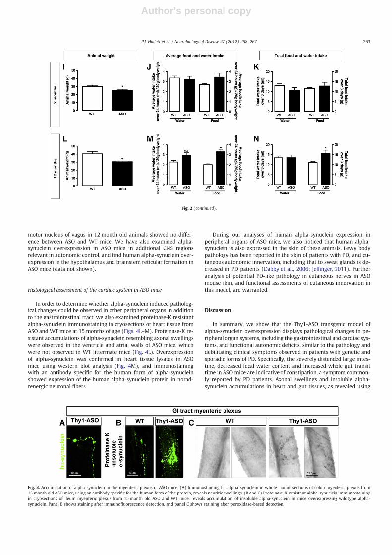
Our observations on abnormal gastrointestinal pathology andfunction, together with previously published work in ASO mice sup-porting our functional findings (Wang et al., 2008), led us to furtherinvestigate whether neuronal pathology at the histological levelcould be observed in ASO mice. We examined whole mounts andalso cryosectioned PFA-fixed tissue from the gastrointestinal tract(ileum and distal colon) of 15 month old WT and ASO mice. We firstconfirmed expression of the human alpha-synuclein protein inwhole mount sections of colon, using a monoclonal rat antibody spe-cific for the human form of the protein (directed against amino acids116–131 of human alpha-synuclein) (Neumann et al., 2000) (Fig. 3A).We observed structures resembling axonal swellings that were im-munoreactive for human alpha-synuclein in ASO mice, and thereforenext determined whether there was evidence for accumulation of in-soluble alpha-synuclein. A proteinase-K digestion was performed fol-lowed by immunolabeling with a polyclonal sheep antibody thatdetects both mouse and human forms of alpha-synuclein (directedagainst amino acids 108–120 of human alpha-synuclein). In mice at15 months of age, small accumulations of insoluble alpha-synucleinwere observed in the myenteric plexus of wildtype mice, however,these appeared larger and more frequent in ASO mice (Figs. 3B, C).Overexpression of alpha-synuclein in peripheral tissues (heart andileum) in ASO mice, was also confirmed using western blot analysisof total alpha-synuclein protein (Supplementary Fig. 1).
Next, we examined further the expression of human alpha-synuclein protein in whole mount sections of colon, (Fig. 4). Alpha-synuclein-immunoreactive fibers and occasional cell bodies, thatwere colocalized with the pan-neuronal (intermediate filament)marker peripherin (Fig. 4B), were localized in the myenteric plexus.Using cryosections of ileum, we also observed human alpha-synuclein immunoreactive fibers in the submucosal plexus and inthe villi (data not shown). There was no positive immunolabelingfor human alpha-synuclein in WT mice (Fig. 4A). Coimmunolabelingfor human alpha-synuclein with neuronal markers of extrinsicand intrinsic enteric neuron innervation (Figs. 4D–K), shows littlecolocalization of human alpha-synuclein with noradrenergic (TH-immunoreactive), cholinergic (VAChT-immunoreactive) and VIP-immunoreactive neurons in ASO mice. Although human alpha-synuclein was not colocalized also with nNOS immunoreactiveneurons, the human alpha-synuclein puncta were localized aroundnNOS cell bodies, suggesting innervation of this population cells.Quantification of peripherin-immunoreactive neurons in myentericganglia showed no significant change in neuronal number in15 month old ASO mice compared to age-matched wildtype litter-mate control mice (1951±226 neurons/mm2, WT mice; 2399±87neurons/mm2, ASO mice; p>0.05, unpaired T-test).
The dorsal motor nucleus of vagus located in the brainstem pro-vides parasympathetic preganglionic extrinsic innervation to entericneurons, and alpha-synuclein pathology and neuronal degenerationhas been reported in this nucleus in PD patients (Braak et al., 2001;Gai et al., 1992). Immunofluorescence labeling with a human-specific alpha-synuclein antibody combined with a fluorescent Nisslstain to label all neurons, was used to examine the dorsal motor nu-cleus of vagus in 2 and 12–15 month old ASO and wildtype littermatemice. There was an absence of human alpha-synuclein labeling in thisregion in ASO mice at both ages studied, despite intense labeling forhuman alpha-synuclein in the adjacent hypoglossal nucleus (Supple-mentary Figure 2). Quantification of neuronal number in the dorsal
261P.J. Hallett et al. / Neurobiology of Disease 47 (2012) 258–267

Author's personal copy
Fig. 2. Gastrointestinal and urinary bladder pathology in ASO mice. Macroscopic assessment of the large intestine (A) and urinary bladder (B) in wildtype (WT) and ASOmice. Com-parable regions of the large intestine were dissected from 15 month old ASO and WT littermate mice and photographed. Urinary bladders were photographed immediately prior todissection of the gastrointestinal tract. Note the pronounced distension of the large intestine and urinary bladder in ASO mice. Scale bar=0.5 cm (A) and =1 cm (B). (C and D) Thetotal number of fecal pellets expelled in 2–4 month (C) and 9–12 month (D) WT and ASO mice over a 1 h period after mice was individually placed in a clean cage. (E and F) Fecalwater content was then determined in the collected fecal pellets expelled over 1 h. Water content was significantly reduced in 9–12 month old ASO mice compared to WT litter-mates. (G and H) Whole gut transit time (WGTT), defined as the time taken for animals to produce a red pellet after receiving a red carmine dye by oral gavage, was determined in2–4 and 9–12 month old WT and ASO mice. WGTT was significantly increased in 9–12 month ASO mice compared to WT littermates * pb0.05, unpaired T-test. (I–N) Animal weightand assessment of food and water intake in 2 and 12 month old WT and ASO mice. ASO mice have a significantly lower body weight than age-matched WT littermate mice at both 2and 12 months old age (I and L). Food and water intake was measured over 3 days in 2 and 12 month old animals. The average food intake over 3 days, expressed over body weight(J and M), and total food intake over 3 days (K and N), is significantly increased in 12 month old ASO mice compared to WT mice. * pb0.05, ** pb0.01, unpaired T-test. Error barsrepresent standard error mean.
262 P.J. Hallett et al. / Neurobiology of Disease 47 (2012) 258–267
Author's personal copy
motor nucleus of vagus in 12 month old animals showed no differ-ence between ASO and WT mice. We have also examined alpha-synuclein overexpression in ASO mice in additional CNS regionsrelevant in autonomic control, and find human alpha-synuclein over-expression in the hypothalamus and brainstem reticular formation inASO mice (data not shown).
Histological assessment of the cardiac system in ASO mice
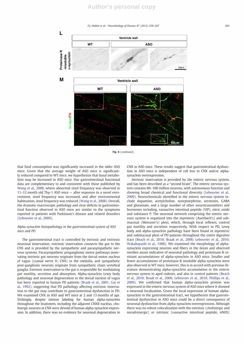
In order to determine whether alpha-synuclein induced patholog-ical changes could be observed in other peripheral organs in additionto the gastrointestinal tract, we also examined proteinase-K resistantalpha-synuclein immunostaining in cryosections of heart tissue fromASO and WT mice at 15 months of age (Figs. 4L–M). Proteinase-K re-sistant accumulations of alpha-synuclein resembling axonal swellingswere observed in the ventricle and atrial walls of ASO mice, whichwere not observed in WT littermate mice (Fig. 4L). Overexpressionof alpha-synuclein was confirmed in heart tissue lysates in ASOmice using western blot analysis (Fig. 4M), and immunostainingwith an antibody specific for the human form of alpha-synucleinshowed expression of the human alpha-synuclein protein in norad-renergic neuronal fibers.
During our analyses of human alpha-synuclein expression inperipheral organs of ASO mice, we also noticed that human alpha-synuclein is also expressed in the skin of these animals. Lewy bodypathology has been reported in the skin of patients with PD, and cu-taneous autonomic innervation, including that to sweat glands is de-creased in PD patients (Dabby et al., 2006; Jellinger, 2011). Furtheranalysis of potential PD-like pathology in cutaneous nerves in ASOmouse skin, and functional assessments of cutaneous innervation inthis model, are warranted.
Discussion
In summary, we show that the Thy1-ASO transgenic model ofalpha-synuclein overexpression displays pathological changes in pe-ripheral organ systems, including the gastrointestinal and cardiac sys-tems, and functional autonomic deficits, similar to the pathology anddebilitating clinical symptoms observed in patients with genetic andsporadic forms of PD. Specifically, the severely distended large intes-tine, decreased fecal water content and increased whole gut transittime in ASO mice are indicative of constipation, a symptom common-ly reported by PD patients. Axonal swellings and insoluble alpha-synuclein accumulations in heart and gut tissues, as revealed using
Fig. 3. Accumulation of alpha-synuclein in the myenteric plexus of ASO mice. (A) Immunostaining for alpha-synuclein in whole mount sections of colon myenteric plexus from15 month old ASO mice, using an antibody specific for the human form of the protein, reveals neuritic swellings. (B and C) Proteinase-K-resistant alpha-synuclein immunostainingin cryosections of ileum myenteric plexus from 15 month old ASO and WT mice, reveals accumulation of insoluble alpha-synuclein in mice overexpressing wildtype alpha-synuclein. Panel B shows staining after immunofluorescence detection, and panel C shows staining after peroxidase-based detection.
Fig. 2 (continued).
263P.J. Hallett et al. / Neurobiology of Disease 47 (2012) 258–267

Author's personal copy
proteinase-K resistant immunostaining for alpha-synuclein, are indic-ative of neurodegenerative processes.
Pathological and functional assessment of the gastrointestinal systemin ASO mice and PD
Gastrointestinal symptoms in PD include constipation, increasedgastrointestinal transit time and difficulty with defecation (Pfeiffer,2011), and can significantly impact quality of life for PD patients(Gallagher et al., 2010; Pfeiffer, 2010). Generally, constipation is asso-ciated with a higher risk of PD and incidental Lewy body disease(Abbott et al., 2001, 2003; Savica et al., 2009), and severe gastrointes-tinal symptoms and other non-motor PD symptoms can lead topatient morbidity (Zesiewicz et al., 2010). Our macroscopic patholog-ical assessment of the gastrointestinal tract in WT and ASO mice atdifferent ages, demonstrated that older (15 month) ASOmice showedsevere distension and blockage of the large intestine. Our data showdeficits in ASO mice as early as 2 months of age, with increased
retention of fecal matter throughout the gastrointestinal tract, and in-creased intestinal weight compared to WT littermates. Our functionalanalyses show that older ASO mice model constipation, by reducedfecal water content and increased whole gut transit time (WGTT). In-terestingly, although we found no overall significant difference inWGTT between ASO and WT mice at 2–4 months of age, 71% of ASOmice showed a longer WGTT than the average WGTT observed inthe WT littermate mice. At 12 months of age, WGTT in ASO micewas significantly longer than in WT mice, with 86% mice exhibitinga longer WGTT than the average WGTT in WT mice. Clinically, be-tween 20 and 89% PD patients report decreased bowel movement fre-quency, and greater disease severity appears to be associated withincreased severity of constipation (Pfeiffer, 2011). Indeed our dataon fecal water content and WGTT in ASO transgenic mice also indi-cates that gastrointestinal deficits increase in severity with increasingage. In order to ascertain that the gastrointestinal deficits in ASO micewere not a result of a reduced food intake, we measured food andwater intake in 2 and 12 month old mice. Interestingly we found
Fig. 4. Robust neuronal expression of human alpha-synuclein protein in the gastrointestinal myenteric plexus and in the heart of ASO mice. (A and B) Immunostaining for humanalpha-synuclein (green) using a specific antibody for the human form of the protein, and the pan-neuronal (intermediate filament) marker peripherin (red) in whole mounts ofcolonic myenteric plexi, shows a neuronal expression of human alpha-synuclein in neuronal fibers and in occasional cell bodies in ASO mice (B) but no expression of humanalpha-synuclein in WT mice. (C) Coimmunostaining for alpha-synuclein using a human specific antibody (green) and an antibody that detects both mouse and human alpha-synuclein (red) demonstrates a punctate localization of human alpha-synuclein, and shows that human alpha-synuclein is not overexpressed in all alpha-synuclein immunoreac-tive fibers. (D–K) Coexpression patterns of human alpha-synuclein with neuronal nitric oxide synthase (nNOS) (D and E), vasoactive intestinal peptide (VIP) (F and G), tyrosinehydroxylase (TH) (H and I) and vesicular acetylcholine transporter (VAChT) (J and K). Human alpha-synuclein puncta appear to surround and innervate nNOS immunoreactiveneurons, and show little colocalization with VIP, TH and VAChT immunoreactive neurons. Scale bar=50 μm (A, B), 20 μm (C–K). (L) Accumulation of insoluble alpha-synucleinin structures resembling axonal swellings is observed in the heart wall of 15 month-old ASO mice but not WT littermates. (M) Co-immunofluorescence labeling using an antibodythat recognizes human alpha-synuclein (green) and tyrosine hydroxylase (TH) (red) shows a punctate expression of human alpha-synuclein protein in many but not all TH-immunoreactive fibers, suggesting that in ASO mice, human alpha-synuclein is expressed in the sympathetic nervous system. Insets show higher magnification images of boxedareas.
264 P.J. Hallett et al. / Neurobiology of Disease 47 (2012) 258–267
Author's personal copy
that food consumption was significantly increased in the older ASOmice. Given that the average weight of ASO mice is significant-ly reduced compared to WT mice, we hypothesize that basal metabo-lism may be increased in ASO mice. Our gastrointestinal functionaldata are complementary to and consistent with those published byWang et al., 2008, where abnormal stool frequency was observed in11–12 month old Thy-1 ASO mice — after exposure to a novel envi-ronment, stool frequency was increased, and after environmentalhabituation, stool frequency was reduced (Wang et al., 2008). Overall,the dramatic macroscopic pathology and clear deficits in gastrointes-tinal function observed in ASO mice are similar to the symptomsreported in patients with Parkinson's disease and related disorders(Lebouvier et al., 2009).
Alpha-synuclein histopathology in the gastrointestinal system of ASOmice and PD
The gastrointestinal tract is controlled by intrinsic and extrinsicneuronal innervation; extrinsic innervation connects the gut to theCNS and is provided by the sympathetic and parasympathetic ner-vous systems. Parasympathetic preganglionic motor pathways inner-vating intrinsic gut neurons originate from the dorsal motor nucleusof vagus (cranial nerve X; CNX) in the medulla, and sympatheticpost-ganglionic neurons originate from sympathetic chain vertebralganglia. Extrinsic innervation to the gut is responsible for modulatinggut motility, secretion and absorption. Alpha-synuclein Lewy bodypathology and neuronal degeneration in the dorsal nucleus of vagushas been reported in human PD patients (Braak et al., 2001; Gai etal., 1992), suggesting that PD pathology affecting extrinsic innerva-tion to the gut may contribute to gastrointestinal dysfunction in PD.We examined CNX in ASO and WT mice at 2 and 13 months of age.Strikingly, despite intense labeling for human alpha-synucleinthroughout the brainstem, including the adjacent CNXII nucleus, cho-linergic neurons in CNXwere devoid of human alpha-synuclein expres-sion. In addition, there was no evidence for neuronal degeneration in
CNX in ASO mice. These results suggest that gastrointestinal dysfunc-tion in ASO mice is independent of cell loss in CNX and/or alpha-synuclein overexpression.
Intrinsic innervation is provided by the enteric nervous system,and has been described as a “second brain”. The enteric nervous sys-tem contains 80–100 million neurons, with autonomous function andshowing broad chemical and functional diversity (Lebouvier et al.,2009). Neurochemicals identified in the enteric nervous system in-clude dopamine, acetylcholine, norepinephrine, serotonin, GABAand glutamate, and a large number of other neurotransmitters andhormones including, vasoactive intestinal peptide (VIP), nitric oxideand substance P. The neuronal network comprising the enteric ner-vous system is organized into the myenteric (Auerbach's) and sub-mucosal (Meissner's) plexi, which, through local reflexes, controlgut motility and secretion respectively. With respect to PD, Lewybody and alpha-synuclein pathology have been found in myentericand submucosal plexi of PD patients throughout the entire digestivetract (Beach et al., 2010; Braak et al., 2006; Lebouvier et al., 2010;Wakabayashi et al., 1988). We examined the morphology of alpha-synuclein expressing neurons and fibers in the ileum and observedswollen axons indicative of neuronal pathology and proteinase-K re-sistant accumulations of alpha-synuclein in ASO mice. Smaller andfewer accumulations of proteinase-K insoluble alpha-synuclein werealso observed inWTmice, however, this is in accord with previous lit-erature demonstrating alpha-synuclein accumulation in the entericnervous system in aged rodents, and also in control patients (Beachet al., 2010; Braak et al., 2006; Lebouvier et al., 2010; Phillips et al.,2009). We confirmed that human alpha-synuclein protein wasexpressed in the enteric nervous system of ASOmice where it showeda neuronal localization. Given the local expression of human alpha-synuclein in the gastrointestinal tract, we hypothesize that gastroin-testinal dysfunction in ASO mice could be a direct consequence ofneuronal dysfunction from alpha-synuclein overexpression. Althoughthere was no robust colocalization with the extrinsic (cholinergic andnoradrenergic) or intrinsic (vasoactive intestinal peptide, nNOS)
Fig. 4 (continued).
265P.J. Hallett et al. / Neurobiology of Disease 47 (2012) 258–267
Author's personal copy
gastrointestinal neuronal populations examined, human alpha-synuclein was localized around nNOS-immunoreactive neurons, sug-gesting that human alpha-synuclein-overexpressing fibers innervatethis neuronal population. By inference we hypothesize that humanalpha-synuclein overexpression in ASOmice is localized in other entericneuron subpopulations, for example calbindin, somatostatin, tachyki-nin or serotonergic neurons; this warrants further investigation in fu-ture studies. Alternatively, overexpression of human alpha-synucleinmay induce a loss of neurotransmitter staining, as has been previouslyreported following axotomy (Lams et al., 1988). Our quantification ofperipherin immunoreactive neurons inmyenteric ganglia shows no sig-nificant change in neuronal number in 12 month old ASO mice com-pared to age-matched controls. Given our knowledge of the extensiveCNS pathology and behavioral abnormalities in ASOmice in the absenceof gross neuronal loss in the brain, our finding that myenteric ganglianeuronal number is not changed is consistent with the idea thatalpha-synuclein overexpression can cause neuronal dysfunction andneuronal pathology prior to, or in the absence of neuronal loss. Wehave previously shown that AAV-mediated overexpression of A53Tmu-tant alpha-synuclein in nigral dopamine neurons in rats causes a pro-gressive and protracted degeneration of dopamine neurons, that ispreceded by several predegenerative changes including axonal dystro-phy, disrupted neurotransmission, and changes in levels of proteins in-volved in synaptic transmission and cellular transport (Chung et al.,2009).
Histopathological analysis of the heart in ASO mice as a model of PD
Clinical symptoms of cardiovascular autonomic dysfunction inParkinson's disease patients include orthostatic hypotension and de-creased heart rate variability, and can result in serious complications(Goldstein, 2010; Ziemssen and Reichmann, 2010). Cardiac imagingstudies using scintigraphy and PET to assess sympathetic norad-renergic transmission, have shown cardiac sympathetic denervation(Braune et al., 1998; Goldstein et al., 2000, 2009; Mitsui et al., 2006;Orimo et al., 2007). In support of this are histological assessmentsof post-mortem tissue, which show neuronal loss in sympatheticganglia, reduced tyrosine hydroxylase innervation to the heart(Ghebremedhin et al., 2009; Mitsui et al., 2006; Orimo et al., 2008a,2008b), and alpha-synuclein immunoreactive Lewy body and neuritepathology in cardiac tissue (Fujishiro et al., 2008; Orimo et al., 2008b).It has previously been reported that heart weight inWT and ASOmiceis unchanged (Fleming et al., 2007), suggesting that there is no majorheart tissue atrophy or hypertrophy. We also found no obvious grosspathology of the heart in ASO or WT mice for the ages studied (2–15 months). However, our analyses of alpha-synuclein-induced path-ological changes in the heart of 15 month old ASO mice showed thepresence of swollen alpha-synuclein-immunoreactive fibers, indica-tive of axonal pathology, which were not present in WT mice.Human alpha-synuclein protein was expressed locally in the ventric-ular and atrial walls of ASO mice in a punctate pattern, where it waslocalized within TH-immunoreactive (noradrenergic) fibers. Thealpha-synuclein histopathological changes we have shown in hearttissue in ASO mice are consistent with functional cardiac abnormali-ties previously described in these mice, including altered barorecep-tor function and reduced heart rate variability (Fleming et al., 2007,2009) and also seen in PD patients (Goldstein, 2010). In addition wehave observed that ASOmice show an extreme sensitivity to anesthe-sia with xylazine (an alpha2 adrenergic agonist), which is suggestiveof general autonomic dysfunction.
General autonomic dysfunction associated with models ofalpha-synuclein pathology
During the course of this work, a parallel study described auto-nomic dysfunction combined with alpha-synuclein pathology in
alpha-synuclein PAC transgenic mice that have an insertion of the en-tire human SNCA gene on a null background for mouse alpha-synuclein (Kuo et al., 2010). In that study, gastrointestinal deficits(using similar assays to those performed in the current study) weredetected in mice overexpressing A53T and A30P mutant alpha-synuclein, as early as 3 months of age, but were not detected at anytime point in mice overexpressing WT alpha-synuclein. We believethat the autonomic nervous system pathology and gastrointestinalfunctional deficits described in the current study in ASO mice over-expressing WT human alpha-synuclein, and the lack of deficits/pathology in PAC transgenic mice overexpressing WT human alpha-synuclein, could be due to the differences in expression of thealpha-synuclein gene. Using the Thy1 promoter, alpha-synuclein ex-pression could be considered to be “spear-headed” within neuronalsystems, as compared to a more generalized expression using the ar-tificial chromosome (PAC).
Conclusion
These results emphasize that deficits and pathology in peripheralneuronal systems caused by the overexpression of human wildtypealpha-synuclein are similar to those that occur both in age-relatedsporadic PD and alpha-synuclein-mediated genetic forms of the dis-ease. In addition, our work demonstrates that overexpression of wild-type alpha-synuclein on the Thy1 promoter is sufficient to causeautonomic deficits and systemic pathology outside of the CNS. ASOmice offer a useful platform for the testing of mechanisms andnovel therapies for autonomic dysfunction in PD-related disorders.
Supplementary data to this article can be found online at http://dx.doi.org/10.1016/j.nbd.2012.04.009.
Acknowledgments
This work was conducted at McLean Hospital and was supportedby funds to O.I. from the National Institutes of Health/National Insti-tute of Neurological Disorders and Stroke P50 (Grant NS39793), TheConsolidated Anti-Aging Foundation, The Poul Hansen Family andthe Harold and Ronna Cooper Family. We thank Dr. Oliver Cooperfor experimental advice, and Tana Brown, Sarah Izen and YvetteLeung for technical assistance.
References
Abbott, R.D., et al., 2001. Frequency of bowel movements and the future risk of Parkin-son's disease. Neurology 57, 456–462.
Abbott, R.D., et al., 2003. Environmental, life-style, and physical precursors of clinicalParkinson's disease: recent findings from the Honolulu-Asia Aging Study. J. Neurol.250 (Suppl. 3), III30–III39.
Anderson, G., et al., 2007. Loss of enteric dopaminergic neurons and associated changesin colon motility in an MPTP mouse model of Parkinson's disease. Exp. Neurol. 207,4–12.
Beach, T.G., et al., 2010. Multi-organ distribution of phosphorylated alpha-synucleinhistopathology in subjects with Lewy body disorders. Acta Neuropathol. 119,689–702.
Braak, E., et al., 2001. Alpha-synuclein immunopositive Parkinson's disease-related in-clusion bodies in lower brain stem nuclei. Acta Neuropathol. 101, 195–201.
Braak, H., et al., 2006. Gastric alpha-synuclein immunoreactive inclusions in Meissner'sand Auerbach's plexuses in cases staged for Parkinson's disease-related brain pa-thology. Neurosci. Lett. 396, 67–72.
Braune, S., et al., 1998. Impaired cardiac uptake of meta-[123I]iodobenzylguanidine inParkinson's disease with autonomic failure. Acta Neurol. Scand. 97, 307–314.
Burre, J., et al., 2010. Alpha-synuclein promotes SNARE-complex assembly in vivo andin vitro. Science 329, 1663–1667.
Cannon, J.R., Greenamyre, J.T., 2010. Neurotoxic in vivo models of Parkinson's diseaserecent advances. Prog. Brain Res. 184, 17–33.
Chaudhuri, K.R., et al., 2006. Non-motor symptoms of Parkinson's disease: diagnosisand management. Lancet Neurol. 5, 235–245.
Chaumette, T., et al., 2009. Neurochemical plasticity in the enteric nervous system of aprimate animal model of experimental Parkinsonism. Neurogastroenterol. Motil.21, 215–222.
Chesselet, M.F., 2008. In vivo alpha-synuclein overexpression in rodents: a usefulmodel of Parkinson's disease? Exp. Neurol. 209, 22–27.
266 P.J. Hallett et al. / Neurobiology of Disease 47 (2012) 258–267
Author's personal copy
Chesselet, M.F., et al., 2008. Strengths and limitations of genetic mouse models of Par-kinson's disease. Parkinsonism Relat. Disord. 14 (Suppl. 2), S84–S87.
Chung, C.Y., et al., 2009. Dynamic changes in presynaptic and axonal transport proteinscombined with striatal neuroinflammation precede dopaminergic neuronal loss ina rat model of AAV alpha-synucleinopathy. J. Neurosci. 29, 3365–3373.
Cookson, M.R., Bandmann, O., 2010. Parkinson's disease: insights from pathways. Hum.Mol. Genet. 19, R21–R27.
Dabby, R., et al., 2006. Skin biopsy for assessment of autonomic denervation in Parkin-son's disease. J. Neural Transm. 113, 1169–1176.
Dawson, T.M., et al., 2010. Genetic animal models of Parkinson's disease. Neuron 66,646–661.
Drolet, R.E., et al., 2009. Chronic rotenone exposure reproduces Parkinson's diseasegastrointestinal neuropathology. Neurobiol. Dis. 36, 96–102.
Fernagut, P.O., et al., 2007. Behavioral and histopathological consequences of paraquatintoxication in mice: effects of alpha-synuclein over-expression. Synapse 61,991–1001.
Fleming, S.M., et al., 2004. Early and progressive sensorimotor anomalies in mice over-expressing wild-type human alpha-synuclein. J. Neurosci. 24, 9434–9440.
Fleming, S.M., et al., 2007. Alterations in baroreceptor function in transgenic mice over-expressing human alpha-synuclein. Program No. 50.9. 2007 Neuroscience MeetingPlanner. Society for Neuroscience, San Diego, CA. Online.
Fleming, S.M., et al., 2008. Olfactory deficits in mice overexpressing human wildtypealpha-synuclein. Eur. J. Neurosci. 28, 247–256.
Fleming, S.M., et al., 2009. Alterations in heart rate variability in transgenic mice over-expressing human wildtype alpha synuclein Program No. 531.9. 2009 Neurosci-ence Meeting Planner. Society for Neuroscience, Chicago, IL. Online.
Fujishiro, H., et al., 2008. Cardiac sympathetic denervation correlates with clinical andpathologic stages of Parkinson's disease. Mov. Disord. 23, 1085–1092.
Gai, W.P., et al., 1992. Age-related loss of dorsal vagal neurons in Parkinson's disease.Neurology 42, 2106–2111.
Gallagher, D.A., et al., 2010. What are the most important nonmotor symptoms in pa-tients with Parkinson's disease and are we missing them? Mov. Disord. 25,2493–2500.
Ghebremedhin, E., et al., 2009. Diminished tyrosine hydroxylase immunoreactivity inthe cardiac conduction system and myocardium in Parkinson's disease: an ana-tomical study. Acta Neuropathol. 118, 777–784.
Goldstein, D.S., 2010. Neuroscience and heart-brain medicine: the year in review.Cleve. Clin. J. Med. 77 (Suppl. 3), S34–S39.
Goldstein, D.S., et al., 2000. Cardiac sympathetic denervation in Parkinson disease. Ann.Intern. Med. 133, 338–347.
Goldstein, D.S., et al., 2009. Cardiac sympathetic denervation preceding motor signs inParkinson disease. Cleve. Clin. J. Med. 76 (Suppl. 2), S47–S50.
Greene, J.G., et al., 2009. Delayed gastric emptying and enteric nervous system dysfunc-tion in the rotenone model of Parkinson's disease. Exp. Neurol. 218, 154–161.
Greten-Harrison, B., et al., 2010. Alphabetagamma-Synuclein triple knockout mice re-veal age-dependent neuronal dysfunction. Proc. Natl. Acad. Sci. U. S. A. 107,19573–19578.
Jellinger, K.A., 2011. Synuclein deposition and non-motor symptoms in Parkinson dis-ease. J. Neurol. Sci. 310, 107–111.
Kuo, Y.M., et al., 2010. Extensive enteric nervous system abnormalities in mice trans-genic for artificial chromosomes containing Parkinson disease-associated alpha-synuclein gene mutations precede central nervous system changes. Hum. Mol.Genet. 19, 1633–1650.
Lams, B.E., et al., 1988. Loss of transmitter-associated enzyme staining following axot-omy does not indicate death of brainstem cholinergic neurons. Brain Res. 475,401–406.
Lebouvier, T., et al., 2009. The second brain and Parkinson's disease. Eur. J. Neurosci. 30,735–741.
Lebouvier, T., et al., 2010. Colonic biopsies to assess the neuropathology of Parkinson'sdisease and its relationship with symptoms. PLoS One 5, e12728.
Magen, I., Chesselet, M.F., 2010. Genetic mouse models of Parkinson's disease The stateof the art. Prog. Brain Res. 184, 53–87.
McLean, J.R., et al., 2012. Transcript expression levels of full-length alpha-synuclein andits three alternatively spliced variants in Parkinson's disease brain regions and in atransgenic mouse model of alpha-synuclein overexpression. Mol. Cell. Neurosci.49, 230–239.
Mitsui, J., et al., 2006. Pathology of the sympathetic nervous system corresponding tothe decreased cardiac uptake in 123I-metaiodobenzylguanidine (MIBG) scintigra-phy in a patient with Parkinson disease. J. Neurol. Sci. 243, 101–104.
Natale, G., et al., 2010. MPTP-induced parkinsonism extends to a subclass of TH-positive neurons in the gut. Brain Res. 1355, 195–206.
Nemani, V.M., et al., 2010. Increased expression of alpha-synuclein reduces neurotrans-mitter release by inhibiting synaptic vesicle reclustering after endocytosis. Neuron65, 66–79.
Neumann, M., et al., 2000. Alpha-synuclein accumulation in a case of neurodegenera-tion with brain iron accumulation type 1 (NBIA-1, formerly Hallervorden–Spatzsyndrome) with widespread cortical and brainstem-type Lewy bodies. Acta Neuro-pathol. 100, 568–574.
Orimo, S., et al., 2007. Degeneration of cardiac sympathetic nerve begins in the earlydisease process of Parkinson's disease. Brain Pathol. 17, 24–30.
Orimo, S., et al., 2008a. Cardiac sympathetic denervation in Parkinson's disease linkedto SNCA duplication. Acta Neuropathol. 116, 575–577.
Orimo, S., et al., 2008b. Axonal alpha-synuclein aggregates herald centripetal degener-ation of cardiac sympathetic nerve in Parkinson's disease. Brain 131, 642–650.
Pfeiffer, R.F., 2010. Gastrointestinal, urological, and sexual dysfunction in Parkinson'sdisease. Mov. Disord. 25 (Suppl. 1), S94–S97.
Pfeiffer, R.F., 2011. Gastrointestinal dysfunction in Parkinson's disease. ParkinsonismRelat. Disord. 17, 10–15.
Phillips, R.J., et al., 2009. Alpha-synuclein immunopositive aggregates in the myentericplexus of the aging Fischer 344 rat. Exp. Neurol. 220, 109–119.
Rockenstein, E., et al., 2002. Differential neuropathological alterations in transgenicmice expressing alpha-synuclein from the platelet-derived growth factor andThy-1 promoters. J. Neurosci. Res. 68, 568–578.
Savica, R., et al., 2009. Medical records documentation of constipation preceding Par-kinson disease: a case–control study. Neurology 73, 1752–1758.
Savica, R., et al., 2010. When does Parkinson disease start? Arch. Neurol. 67, 798–801.Song, D.D., et al., 2004. Enhanced substantia nigra mitochondrial pathology in human
alpha-synuclein transgenic mice after treatment with MPTP. Exp. Neurol. 186,158–172.
Taylor, T.N., et al., 2009. Nonmotor symptoms of Parkinson's disease revealed in an an-imal model with reduced monoamine storage capacity. J. Neurosci. 29, 8103–8113.
Tian, Y.M., et al., 2008. Alteration of dopaminergic markers in gastrointestinal tract ofdifferent rodent models of Parkinson's disease. Neuroscience 153, 634–644.
Ulusoy, A., et al., 2010. Viral vector-mediated overexpression of alpha-synuclein as aprogressive model of Parkinson's disease. Prog. Brain Res. 184, 89–111.
Wakabayashi, K., et al., 1988. Parkinson's disease: the presence of Lewy bodies in Auer-bach's and Meissner's plexuses. Acta Neuropathol. 76, 217–221.
Wang, L., et al., 2008. Abnormal colonic motility in mice overexpressing human wild-type alpha-synuclein. Neuroreport 19, 873–876.
Watson, J.B., et al., 2009. Alterations in corticostriatal synaptic plasticity in mice over-expressing human alpha-synuclein. Neuroscience 159, 501–513.
Zesiewicz, T.A., et al., 2010. Practice parameter: treatment of nonmotor symptoms ofParkinson disease: report of the Quality Standards Subcommittee of the AmericanAcademy of Neurology. Neurology 74, 924–931.
Ziemssen, T., Reichmann, H., 2010. Cardiovascular autonomic dysfunction in Parkin-son's disease. J. Neurol. Sci. 289, 74–80.
267P.J. Hallett et al. / Neurobiology of Disease 47 (2012) 258–267