author manuscript nih public access j. c. moore j. a ... · confocal stacks were generated using...
TRANSCRIPT

A genetic screen for vascular mutants in zebrafish revealsdynamic roles for Vegf/Plcg1 signaling during artery development
L. D. Covassin1, A. F. Siekmann1, M. C. Kacergis1, E. Laver1, J. C. Moore1, J. A.Villefranc1, B. M. Weinstein2, and N. D. Lawson1,*1 Program in Gene Function and Expression, University of Massachusetts Medical School,Worcester, MA2 Laboratory of Molecular Genetics, NICHD, NIH, Bethesda, MD
AbstractIn this work we describe a forward genetic approach to identify mutations that affect blood vesseldevelopment in the zebrafish. By applying a haploid screening strategy in a transgenic backgroundthat allows direct visualization of blood vessels, it was possible to identify several classes of mutantvascular phenotypes. Subsequent characterization of mutant lines revealed that defects in Vascularendothelial growth factor (Vegf) signaling specifically affected artery development. Comparison ofphenotypes associated with different mutations within a functional zebrafish Vegf receptor-2ortholog (referred to as kdr-like, kdrl) revealed surprisingly varied effects on vascular development.In parallel, we identified an allelic series of mutations in phospholipase c gamma 1 (plcg1). Togetherwith in vivo structure-function analysis, our results suggest a requirement for Plcg1 catalytic activitydownstream of receptor tyrosine kinases. We further find that embryos lacking both maternal andzygotic plcg1 display more severe defects in artery differentiation but are otherwise similar to zygoticmutants. Finally, we demonstrate through mosaic analysis that plcg1 functions autonomously inendothelial cells. Together our genetic analyses suggest that Vegf/Plcg1 signaling acts at multipletime points and in different signaling contexts to mediate distinct aspects of artery development.
IntroductionThe circulatory system provides oxygenation and waste removal for developing organs andtissues, while serving as a conduit for hormones and components of the immune system(Cleaver and Krieg, 1999). In addition, the endothelial cells lining nascent blood vessels alsoserve as a source for important inductive and guidance signals during organogenesis (Alt etal., 2006; Lammert et al., 2001). Accordingly, blood vessel function is essential for vertebrateembryonic development (Cleaver and Krieg, 1999). As organisms transition from embryonicto adult stages, new blood vessel formation (neovascularization) becomes more limited and inmost cases this process is associated with pathological conditions in the adult. As with normaltissue and organs during embryonic development, ischemic tissue or nascent tumors requireincreased vascularization and are known to produce and secrete numerous angiogenicmolecules that promote blood vessel growth (Semenza, 2003). In most cases, the signals that
*corresponding author: Nathan D. Lawson, Ph.D., Associate Professor, Program in Gene Function and Expression, University ofMassachusetts Medical School, Lazare Research Building, Room 617, 364 Plantation Street, Worcester, MA 01605, Phone: (508)856-1177, [email protected]'s Disclaimer: This is a PDF file of an unedited manuscript that has been accepted for publication. As a service to our customerswe are providing this early version of the manuscript. The manuscript will undergo copyediting, typesetting, and review of the resultingproof before it is published in its final citable form. Please note that during the production process errors may be discovered which couldaffect the content, and all legal disclaimers that apply to the journal pertain.
NIH Public AccessAuthor ManuscriptDev Biol. Author manuscript; available in PMC 2010 May 15.
Published in final edited form as:Dev Biol. 2009 May 15; 329(2): 212–226. doi:10.1016/j.ydbio.2009.02.031.
NIH
-PA Author Manuscript
NIH
-PA Author Manuscript
NIH
-PA Author Manuscript

promote neovascularization in these pathological settings in adults are the same that are usedby the embryo (Cleaver and Krieg, 1999). Furthermore, the signaling molecules and cellularmechanisms by which blood vessels form have been evolutionarily conserved (Weinstein andLawson, 2002). Accordingly, it is has been possible to gain significant insights on pathologicalblood vessel formation through the study of embryonic vascular development in vertebratemodel systems.
Among the important signaling molecules that are responsible for neovascularization in bothembryonic and adult settings are several receptor tyrosine kinase families. These includereceptors for vascular endothelial growth factors (Vegf), angiopoietins and the membrane-bound ephrin ligands (Yancopoulos et al., 2000). Vegf-A initiates and induces endothelial celldifferentiation at the earliest stages of blood vessel development (Ferrara et al., 2003). Vegf-A acts by binding Vegfr-2, an endothelial cell specific receptor tyrosine kinase (Quinn et al.,1993). Mice lacking either Vegf-A or Vegfr-2 display severely reduced numbers of endothelialcells and fail to form blood vessels (Carmeliet et al., 1996; Shalaby et al., 1995). Loss of specificVegf-A isoforms or endothelial autonomous ablation of Vegf signaling has revealed later rolesfor this signaling pathway in arterial endothelial cell differentiation and promoting endothelialcell survival, respectively (Carmeliet et al., 1999; Lawson et al., 2002; Lee et al., 2007;Stalmans et al., 2002). Furthermore, Vegf-A is also required for angiogenic sprouting and playsan important role in modulating vascular permeability in both adult and embryonic stages(Bates and Harper, 2002; Ferrara et al., 2003). In parallel to the Vegf signaling pathway, theangiopoietins and their receptor, Tie2, mediate subsequent sprouting and maturation of thevascular network at later stages of embryonic development. Angiopoietin/Tie2 signaling playsan important role in crosstalk between endothelial cells lining the blood vessels and associatedvascular smooth muscle cells during angiogenic sprouting (Davis et al., 1996; Maisonpierre etal., 1997; Suri et al., 1996; Suri et al., 1998). Similarly, the Eph receptor tyrosine kinases andtheir ligands, referred to as ephrins, mediate cell-cell signaling between distinct cell types inthe developing vascular system (Yancopoulos et al., 1998). Eph-ephrin signaling plays animportant role in mediating the interface and separation between arteries and veins (Gerety etal., 1999; Wang et al., 1998), as well as endothelial cell interaction with surroundingmesenchymal cells (Adams et al., 1999; Yancopoulos et al., 1998).
For the most part, the functional analysis of these and other signaling pathways during vasculardevelopment has proceeded largely through reverse genetic approaches using the mouse as amodel system. More recently, the zebrafish has emerged as an ideal model system for studyingembryonic vascular development (Roman and Weinstein, 2000). The transparency and externalfertilization of the zebrafish embryo, coupled with the use of endothelial cell-specificfluorescent protein transgenes have allowed an unprecedented view of blood vessel formationin a live organism (Beis and Stainier, 2006; Lawson and Weinstein, 2002; Roman et al.,2002; Siekmann et al., 2008). These particular aspects of the zebrafish embryo have made it atractable vertebrate model for forward genetic screening. Early screens relying on thestraightforward visualization of blood circulation in the zebrafish embryo led to theidentification of mutations that affected cardiovascular morphogenesis or function (Chen etal., 1996; Stainier et al., 1996). More recently, the use of transgene-assisted screens (Jin et al.,2007) has allowed more subtle characterization of defective vascular phenotypes in mutantzebrafish embryos. The ability to easily generate thousands of progeny embryos coupled withthe increasing quality of genomics tools has facilitated genetic mapping and identification ofgenes responsible for many of these phenotypes (Patton and Zon, 2001).
In this study, we describe a haploid transgenic screen in zebrafish embryos to identify mutantsthat perturb normal vascular development. This screening approach allowed robust and specificdetection of several different classes of mutant vascular phenotypes and the majority of thesewere subsequently observed in diploid embryos. Subsequent characterization and cloning of
Covassin et al. Page 2
Dev Biol. Author manuscript; available in PMC 2010 May 15.
NIH
-PA Author Manuscript
NIH
-PA Author Manuscript
NIH
-PA Author Manuscript

the genes responsible for these mutant phenotypes revealed that the zebrafish Vegf receptor,kdr-like (kdrl), and phospholipase c gamma 1 (plcg1) were essential for artery development.Interestingly, detailed phenotypic analysis of different kdrl alleles suggested diverse roles forthis receptor during development of blood vessels. Parallel analysis of plcg1 alleles, along within vivo structure/function experiments, indicated a crucial role for Plcg1 catalytic functiondownstream of receptor tyrosine kinases during artery development. Furthermore, analysis ofmaternal-zygotic plcg1 mutant embryos suggested an early role for Vegf/Plcg1 signalingduring arterial specification, while mosaic analysis revealed an endothelial autonomous rolefor plcg1 during vascular morpohgenesis. Taken together our results demonstrate the utility ofa transgenic haploid screening approach to identify mutants affecting vascular development inthe zebrafish and underscore the diverse roles of Vegf/Plcg1 signaling during arterydevelopment.
Materials and methodsFish handling and care
All zebrafish were maintained according to standard protocols (Westerfield, 1993) and inaccordance with University of Massachusetts Medical School IACUC guidelines.
Transgenic and mutant linesNomenclature for the zebrafish Vegf receptor orthologs was assigned based on recentlydescribed guidelines (Bussmann et al., 2008). The um19 allele is a 4 base pair deletion in exon2 of kdrl that was generated by targeted mutagenesis using zinc finger nucleases and isdescribed elsewhere (Meng et al., 2008). The Tg(fli1a:egfp)y1 line has been describedelsewhere (Lawson and Weinstein, 2002). To generate zebrafish embryos with red fluorescentblood vessels, we constructed a plasmid containing a chimeric endothelial enhancer-promoterfragment driving dsRed-Express via Gateway cloning into a Tol2 plasmid backbone (Villefrancet al., 2007). The resulting plasmid (pTolfli1ep:dsredex) was co-injected into 1-cell stage wildtype zebrafish embryos along with mRNA encoding the Tol2 Transposase (Kawakami et al.,2004). Embryos with the highest and least mosaic expression were grown to adulthood andfounders were identified by out-crossing. The Tg(fli1ep:dsredex)um13 line was derived from asingle founder.
Mutagenesis and screeningENU mutagenesis was performed based on previously described protocols (Solnica-Krezel etal., 1994). Briefly, we treated homozygous Tg(fli1a:egfp)y1 adult males with 3 mM N-ethyl-N-nitrosourea (ENU) for 1 hour once a week for a total of 4 weeks. Following a two-weekrecovery, fish were bred to wild type females to purge mutagenized gametes that usually harborgross chromosomal aberrations (Imai et al., 2000). ENU-treated males were then crossed tohomozygous Tg(fli1a:egfp)y1 females to generate an F1 generation. Beginning at 6 months ofage, F1 Tg(fli1a:egfp)y1 females were used to generate haploid embryos by performing invitro fertilization (Westerfield, 1993) using UV irradiated sperm. Each individual F1 Tg(fli1a:egfp)y1 female that successfully gave viable haploids was maintained in an individualtank. Subsequently, Tg(fli1a:egfp)y1 embryos were screened at 30 hours post fertilization andagain at 50 hpf for defects in segmental artery formation, as well as overall morphology usinga stereo dissection microscope equipped with epifluorescence. F1 Tg(fli1a:egfp)y1 females thatgave clutches of embryos in which fifty percent displayed a mutant vascular phenotype weresubsequently out-crossed to TL males to generate a map cross. Females that gave mutantphenotypes that exhibited general morphological defects or necrosis were not maintained.Genetic linkage mapping and candidate cloning of genes was carried out as described elsewhere(Lawson et al., 2003; Roman et al., 2002).
Covassin et al. Page 3
Dev Biol. Author manuscript; available in PMC 2010 May 15.
NIH
-PA Author Manuscript
NIH
-PA Author Manuscript
NIH
-PA Author Manuscript

Phenotypic analysis and microscopyAll diploid mutant embryos were analyzed at 30 hpf using a dissection microscope equippedwith epifluorescence (Leica MZFLIII, 1x PlanApo) to determine presence of segmentalarteries. Embryos were also observed with transmitted light at 55 hpf to observe circulatorypatterns and overall morphology. Vascular morphology defects were documented in moredetail using a Leica DMIRE2 microscope equipped with a TCS SP2 confocal laser scanningsystem (Objective: HC PL APO 20x/0.70 CS, dry). In most cases, vertical projections ofconfocal stacks were generated using Leica Confocal Software (LCS). Alternatively, verticalprojections were generated using the Imaris software package (Bitplane). Sprout length wasmeasured in 3-dimensional stacks using Imaris. For this purpose, 6 pairs of segmental arterysprouts were counted in at least 3 embryos from 3 different clutches. Length was measured asdistance of a perpindicular line from the dorsal wall of the dorsal aorta to the distal end of thesegmental artery. Overall morphology was documented by capturing digital images usingtransmitted light on a MZFLIII dissection microscope equipped with a Zeiss Axiocam mRCdigital camera using Axiovision software (Zeiss). More detailed morphological imaging wasperformed by differential interference contrast (DIC) microscopy using a Zeiss Axioskop2pluscompound microscope (Achroplan 20x/0.5 water; Achroplan 40x/0.8 water) equipped with aZeiss Axiocam hRC digital camera. All images were exported from Axiovision, LCS, or Imarisas TIF files and imported into Photoshop CS2 (Adobe) to generate figures.
RiboprobesAntisense riboprobes against ephrinb2a, dll4, and notch3, were generated as describedpreviously (Lawson et al., 2001; Siekmann and Lawson, 2007). Whole mount in situhybridization was performed as described elsewhere (Hauptmann and Gerster, 1994). DICimages of stained embryos were captured as described above.
Generation of germ cell plcg1 mutantsTo generate adults bearing plcg1 deficient germ cells, we utilized the germ-line replacementstrategy described elsewhere (Ciruna et al., 2002). Briefly, embryos from an incross of non-transgenic plcg1y18 carriers were injected with mRNA encoding EGFP with the 3′UTR fromthe nanos transcript. In parallel, wild type embryos were injected with a morpholino targetingthe dead end gene to ablate host germ cells. Donor cells from the margin of sphere stageembryos derived from putative plcg1y18 carriers were transplanted into the margin of host wildtype embryos. Donor and host embryos were then allowed to develop until 24 to 48 hpf. Hostembryos were grown to adulthood if the corresponding donor embryo was found to be mutantfor plcg1 by visual inspection and the host embryos displayed evidence of transplanted germcells as indicated by green fluorescence. Putative maternal (M) or paternal (P) mutant founderswere identified by crossing them to heterozygous plcg1y18 mutant carriers. In all cases,Mplcg1y18 and Pplcg1y18 carriers displayed fully penetrant plcg1 mutant phenotypes indicatingthat all germ cells were mutant.
Plcg1 rescue experimentsPreviously described mutations in the SH2 (Ji et al., 1999) and SH3 (Ye et al., 2002) domainsof Plcg1 were introduced into zebrafish plcg1 coding sequence by overlapping PCR and allconstructs were validated by sequencing. Mutant constructs were generated in the pCS-mtplcg1backbone (Lawson et al., 2003). All plasmids were linearized with Not I and mRNAsynthesized using the mMessage mMachine kit (Ambion) to drive transcription from the SP6promoter. We injected 400 pg of mRNA into 1-cell stage embryos derived from an incross ofplcg1y18 heterozygous carriers. As controls, embryos were injected with mRNA encoding wildtype Plcg1, or left uninjected. Embryos were scored at 30 hpf for segmental artery formation;embryos were classified as having no segmental arteries (SeA−), partial segmental arteries,
Covassin et al. Page 4
Dev Biol. Author manuscript; available in PMC 2010 May 15.
NIH
-PA Author Manuscript
NIH
-PA Author Manuscript
NIH
-PA Author Manuscript
with no dorsal longitudinal anastomotic vessel (SeA+DLAV−), or wild type (SeA+DLAV+).At 55 hpf, embryos were scored for blood circulation and classified with normal circulation(circ+), evidence of shunting between the dorsal aorta and posterior cardinal vein (partial), orno circulation (circ−). Following phenotypic analysis, all embryos were subjected to PCR-screening for genotyping.
To generate expression constructs for activated forms of Akt or MAP2K1, we first generatedGateway entry clones. A constitutively membrane-localized form of Akt was PCR-amplifiedfrom a plasmid template (kindly provided by Fumihiko Urano, UMass Medical School) usingthe following primers: 5′-GGGGACAAGTTTGTACAAAAAAGCAGGCTCCGCGTTTAGCTTATGGGGAG and5′-GGGGACCACTTTGTACAAGAAAGCTGGGTAGGCTGTGCCACTGGCTGAGT,where Akt coding sequence are underlined and the endogenous start codon is in bold. Anactivated form of human MAP2K1 was amplified from plasmid template (kindly provided byRoger Davis, UMass Medical School) using primers 5′-GGGGACAAGTTTGTACAAAAAAGCAGGCTccAAAATGCCCAAGAAGAAGC and5′-GGGGACCACTTTGTACAAGAAAGCTGGGTTTAGACGCCAGCAGCATGGGT;hMAP2K1 sequence is underlined, endogenous start codon is in bold. In both cases, the forwardprimer contains an attB1 site, while the reverse primer contains an attB2 site. PCR productswere subjected to BP cloning to generate pMEmAkt and pMEactMAP2K1 entry clones, whichwere sequence validated. Entry clones were then used in Gateway LR reactions to generateexpression clones. For actMAP2K1, we constructed N-terminal monomeric Cherry fusionproteins by recombining pMEactMAP2K1 with either pCSCherDest (to give pCS-mcher-actMAP2K1)or pTolfli1epCherDest (to give pTol-fli1ep:mcher-actMAP2K1) (Villefranc etal., 2007). For mAkt, we utilized multisite Gateway cloning to generate a tandem, unfusedmAkt-mCherry cassette using the viral 2A peptide sequence (Provost et al., 2007). For thispurpose, we recombined pMEmAkt with pCSDest2 (Villefranc et al., 2007) and p3E-2a-mcherry (kindly provided by Chi-Bin Chien, University of Utah) to give pCS-mAkt-2a-mcher. To generate an endothelial cell specific construct, we recombined pMEmAkt withp5Efli1ep, which contains a chimeric fragment of a fli1a enhancer and promoter (Villefranc etal., 2007), pDest Tol2pA (Kwan et al., 2007), and p3E-2a-mcherry to give pTol-fli1ep:mAkt-2a-mcherry. We utilized pCS-mCher-actMAP2K1 and pCS-mAkt-2a-mcherry formRNA synthesis as described elsewhere (Villefranc et al., 2007) and mRNA was injected at1- to 2-cell stage into wild type embryos. For pTol-fli1ep:mAkt-2a-mcher and pTol-fli1ep:mcher-actMAP2K1, we co-injected 25 pg of circular plasmid DNA with 25 pg Tol2transposase mRNA into 1-cell stage embryos derived from an incross of plcg1y13 heterozygouscarriers. Injected embryos were observed for segmental artery formation and circulation asdescribed above.
GenotypingThe plcg1y13 mutation eliminates a PstI site within the X-catalytic domain. PCR was performedusing primers 5′-CTGCGTTCAGCTATCCCATC and 5′-CCTCTTGAGCTGGTGTGGCGAand the resulting product was digested with PstI. The plcg1y16 mutation eliminates an EagI inthe N-terminal SH2 domain. For y16 genotyping, primers 5′-GTATGGACCAGCATGTGACGGAG and 5′-CTACAAAGGTCTCGCTCTCCCGC wereused for PCR and the resulting fragment was digested with EagI. For genotyping theplcg1y18 allele, we amplified a CA repeat within plcg1 (primers: 5′-TGTAGGATCTGAGGTTTGAT and 5′ TGAGATCCTTGAGCCTGGGG) that waspolymorphic in our background strain. Alternatively, we utilized a single nucleotidepolymorphism (SNP) within a StyI site in plcg1 in our crosses (5′-GCAACGAGTTCGAGATGAAGC and 5′-GTGTGGACGCATAGCCCTC). Genotyping of
Covassin et al. Page 5
Dev Biol. Author manuscript; available in PMC 2010 May 15.
NIH
-PA Author Manuscript
NIH
-PA Author Manuscript
NIH
-PA Author Manuscript
the kdrl alleles was performed as described elsewhere (Covassin et al., 2006; Meng et al.,2008).
Mosaic analysisFor mosaic analysis of plcg1 deficient cells in wild type vasculature, donor Tg(fli1a:egfp)y1
embryos were prepared by injecting with the lineage tracer Cascade Blue (Molecular Probes/Invitrogen) and 7.5 ng of a plcg1 morpohlino targeting the exon 1 splice donor site (Lawsonet al., 2003) at 1- to 2-cell stage. At sphere stage, cells were removed from donor embryos andtransferred to wild type Tg(fli1ep:dsredex)um13 hosts using a glass capillary. For analysis ofwild type cells in plcg1-deficient vasculature, donor Tg(fli1a:egfp)y1 embryos were injectedwith the lineage tracer Cascade Blue. Subsequently, donor cells were placed into Tg(fli1ep:dsredex)um13 embryos that had been injected at the 1-cell stage with the plcg1morpohlino. As a control, cells for Cascade Blue-injected wild type Tg(fli1a:egfp)y1 embryoswere transplanted into uninjected wild type Tg(fli1ep:dsredex)um13 host embryos. In all cases,embryos were grown to 26 to 28 hpf at which time position of GFP+ donor cells was determinedthrough visualization by confocal microscropy as described above. Emission from GFP,dsRedEx, and Cascade Blue was acquired by sequential scanning using a 488 nm, 561 nm and405 nm laser line, respectively. Subsequently, confocal stacks were used to generate verticalprojection and overlays using the included Leica software, Imaris, or Photoshop CS2.
ResultsA haploid transgenic screen to identify mutants that affect embryonic vascular development
Previous forward screening strategies to identify vascular mutants in zebrafish have relied onobservation of circulatory defects in F3 embryos as a primary screen (Chen et al., 1996; Jin etal., 2007; Stainier et al., 1996). However, the two-generation breeding scheme used in F3screens is labor- and cost-intensive and can take several years to screen sufficient numbers ofgenomes. In an effort to more easily screen for vascular mutants in zebrafish, we took advantageof the ability to generate haploid embryos to allow screening of progeny from F1 females.Additionally, we utilized Tg(fli1a:egfp)y1 zebrafish (Lawson and Weinstein, 2002) to allowdirect visualization of putative vascular phenotypes in developing haploid embryos derivedfrom F1 females.
To induce mutations in the male zebrafish germline, we treated homozygous Tg(fli1a:egfp)y1 males with N-ethyl-N-nitrosourea (ENU). Subsequently, we used ENU-treatedmales to generate F1 families (Figure 1A). To identify mutations that would lead to bloodvessel defects, we generated haploid embryos from individual Tg(fli1a:egfp)y1 F1 females andscreened for segmental artery formation at 1 or 2 days post fertilization. In general, we foundthat the overall morphology of haploid Tg(fli1:egfp)y1 zebrafish embryos was relativelynormal, although body length was shortened and somite formation, as well as headdevelopment was often mildly perturbed (Fig. 1B and data not shown). Despite these mildabnormalities, trunk blood vessel morphology and segmental artery formation was robust inwild type Tg(fli1:egfp)y1 haploid embryos allowing for straightforward identification of mutantvascular phenotypes (Fig. 1C–G). In total, we screened approximately 1200 mutagenizedgenomes and identified 17 females that produced clutches in which half of the haploid embryosdisplayed specific defects in vascular morphology. In all cases, general morphology of themutant embryos was comparable to their wild type siblings (data not shown). We were able toidentify 4 classes of mutant phenotypes in clutches of haploid embryos from F1 Tg(fli1:egfp)y1 females. Class I mutants such as y10, displayed a complete loss of segmental arteryformation (Figure 1D, Table 1) while the level of transgene expression was comparable to wildtype siblings indicating the presence of endothelial cells within the presumptive dorsal aortaand posterior cardinal vein. In Class II mutants, such as y16, we observed partial segmental
Covassin et al. Page 6
Dev Biol. Author manuscript; available in PMC 2010 May 15.
NIH
-PA Author Manuscript
NIH
-PA Author Manuscript
NIH
-PA Author Manuscript

artery formation (Figure 1E). A single Class III mutant, um7, displayed ectopic branching ofsegmental arteries (Figure 1F). The y11 mutant (Class IV) exhibited the most severe effect onvascular development with a complete loss of segmental arteries, defective formation of themajor trunk vessels (Figure 1G), as well as loss of cranial blood vessels (data not shown).
To confirm that the vascular defects observed in haploids would persist in diploid embryos andto facilitate genetic mapping, we out-crossed putative F1 female mutant carriers to a geneticallydiverse wild type line (TL; Figure 1A). We were able to obtain viable F2 families from 14 outof 17 females. In clutches of embryos from 10 out of the 14 families, we observed defectivevascular phenotypes that were similar or identical to those observed in mutant haploid embryos(see below). Subsequent bulk segregant analysis, fine genetic mapping and/or candidatecloning revealed 10 distinct alleles in 4 genes that were responsible for the observed vasculardefects. The y11 mutation lies in the endothelial cell specific transcription factor, ets-relatedprotein (etsrp), leading to global defects in endothelial gene expression, apoptosis ofendothelial cells, and defects in vascular morphogenesis (Pham et al., 2007). Consistent withthe ectopic sprouting defects observed in um7 mutants, we find that this mutation maps to theplexind1 locus (data not shown), a gene previously implicated in proper segmental arterypatterning in zebrafish (Torres-Vazquez et al., 2004). We found that Class I and II mutants thatdisplayed defects in segmental artery formation mapped to two genes implicated in Vegfsignaling, plcg1 and kdrl. Based on our previous findings concerning the role of Vegf in arterydevelopment, we focused on the detailed genetic and phenotypic characterization of thesemutants.
Mutations in a zebrafish Vegf receptor-2 ortholog cause defects in artery developmentWe have previously found that a mutation in a zebrafish Vegf receptor, referred to as kdr-like (previously referred to as flk1 or kdra) is responsible for partial segmental artery defectsassociated with the y17 mutant identified in our haploid screen (Table 1(Covassin et al.,2006)). Of the other segmental artery mutants identified in our screen, only um6 failed tocomplement y17 (data not shown), indicating that it also was a mutation in kdrl. Interestingly,the um6 mutation appeared to lead to more severe defects in segmental artery formation inhaploid embryos than those associated with y17 (Table 1). Therefore, to better evaluate theeffects of the kdrlum6 and kdrly17 mutations on vascular development, we compared them to anull kdrl allele that we generated through targeted mutagenesis using zinc finger nucleases(Meng et al., 2008). The um19 allele is a 4 base pair deletion in exon 2 of kdrl and introducesa frameshift that truncates the Kdrl receptor tyrosine kinase in the extracellular domain.
We first compared defects in vascular morphogenesis associated with each kdrl allele in mutantembryos bearing the Tg(fli1:egfp)y1 transgene. In wild type siblings at 30 hours postfertilization, a basic vascular network consisting of the dorsal aorta, segmental arteries and thedorsal longitudinal anastomotic vessel (DLAV) was evident (Figure 2A). The kdrlum6 mutationappeared to have the most severe effect on segmental artery formation leading to variableformation of partial segmental arteries in most embryos (Figure 2B, white arrows indicatepartial segmental arteries). In many cases, small filopodial extensions were observed at theusual site of segmental artery formation, but sprouts failed to emanate from the dorsal aorta(Figure 2B, red arrowheads). kdrlum6 mutant embryos also failed to display a distinctlyseparated dorsal aorta and posterior cardinal vein and instead a single major trunk vessel wasevident (Figure 2B, red bracket). We observed that defective segmental artery formation dueto the null kdrlum19 mutation was similar to kdrlum6 mutant embryos, although partial sproutswere consistently observed within every segment (Figure 2C). Interestingly, morphogenesisof the dorsal aorta and posterior cardinal vein was largely normal in kdrlum19 mutant embryos(Figure 2C). Embryos mutant for kdrly17 displayed the mildest segmental artery defects, withvariable formation of DLAVs and partial segmental artery formation evident (Figure 2D).
Covassin et al. Page 7
Dev Biol. Author manuscript; available in PMC 2010 May 15.
NIH
-PA Author Manuscript
NIH
-PA Author Manuscript
NIH
-PA Author Manuscript

However, dorsal aorta morphogenesis appeared abnormal as we observed its discontinuousformation in kdrly17 mutant embryos (Figure 2D, red arrowhead). This observation is consistentwith our previous finding of arteriovenous shunting between the dorsal aorta and posteriorcardinal vein in kdrly17 mutant embryos (Covassin et al., 2006). Quantification of segmentalartery length in each mutant confirmed that the um6 mutation most severely affected segmentalartery formation, while um19 and y17 had increasingly milder effects (Figure 2E). Subsequentmolecular analysis of the kdrl coding sequence in um6 mutant embryos revealed a T to Gtransversion resulting in a missense mutation that changed cysteine to glycine at amino acid1117 within the Kdrl cytoplasmic domain (Table 1). This cysteine residue is identical in therelated zebrafish vegf-receptor 2 ortholog, kdr, as well as related Vegf receptors in zebrafish,mouse, and human (Figure 2F).
Our analysis of vascular morphology in kdrl mutants revealed a surprisingly differential effecton angiogenic formation of segmental arteries and dorsal aorta morphogenesis. To furtherassess how these alleles affect vascular morphogenesis and function, we assayed circulatorydefects in kdrl mutant embryos. At 55 hpf, wild type embryos displayed a functional circulatorysystem that carries hemoglobinized red blood cells (Figure 3A and data not shown). Bycontrast, kdrlum6 mutant embryos failed to exhibit any active circulation and display pericardialedema (Figure 3B and data not shown). Closer inspection of embryos derived from kdrlum6
heterozygous carriers revealed that approximately five percent of all embryos with otherwisenormal circulatory flow displayed cranial hemorrhage (compare Figures 3C and D; Figure 3H).Subsequent genotyping of these embryos revealed that they were heterozygous for the T to Gtransversion (data not shown) suggesting that the kdrlum6 mutation can act as a mild dominantnegative. Despite the moderate severity of the segmental artery defects in kdrlum19 mutantembryos, this mutation had the mildest effect on circulatory function consistent with normaldorsal aorta morphogenesis in these embryos (Figure 2C). At 55 hpf all kdrlum19 mutantembryos displayed normal circulation throughout the vascular system with no evidence ofcardiac edema (Figure 3E, H). However, cranial hemorrhage was evident in approximatelyfifteen percent of all embryos, indicating a partially penetrant defect in vascular function inkdrlum19 mutants (Figure 3G, H). Consistent with our previous study, we observed variablecirculatory defects in kdrly17 mutant embryos, including some embryos with complete lack ofcirculation, presence of arteriovenous shunts between the dorsal aorta and posterior cardinalvein, and cranial hemorrhage (Figure 3H and data not shown). Taken together, our resultsindicate that these alleles differentially affect blood vessel formation in the developing embryo.
Mutations in phospholipase c gamma 1 perturb artery developmentOur previous work demonstrated that the y10 mutation (Figure 1C, Table 1) lies in a splicedonor site within the phospholipase c gamma 1 (plcg1) gene leading to a specific defect inartery development (Lawson et al., 2003). Plcg1 mediates signaling downstream of a numberof different receptor tyrosine kinases, including receptors for platelet derived growth factors(Pdgfs), fibroblast growth factors (Fgfs) and Vegf itself (Rhee, 2001; Wilde and Watson,2001). Plcg1 interacts with phosphorylated tyrosine residues on an activated receptor tyrosinekinase via its SH2 domains. Subsequent activation of Plcg1 occurs through phosphorylationof tyrosine residues by the upstream receptor tyrosine kinase. This results in induction of Plcg1catalytic activity leading to cleavage of phosphoinositide-4,5-bisphosphate (PIP2) to forminositol-3-phosphate (IP3) and diacylglycerol (DAG), second messengers that ultimatelyinduce calcium release and activation of protein kinase C isoforms, respectively (Rhee,2001). More recently, Plcg1 has been shown to transduce signals mediated by other upstreamactivators through interaction with its SH3 domain and can function in a catalytic domainindependent manner in this regard (Patterson et al., 2002; Ye et al., 2002).
Covassin et al. Page 8
Dev Biol. Author manuscript; available in PMC 2010 May 15.
NIH
-PA Author Manuscript
NIH
-PA Author Manuscript
NIH
-PA Author Manuscript

We found that five additional mutants failed to complement plcg1y10 or displayed geneticlinkage to plcg1 (data not shown and Table 1). The y13, y15, and y16 alleles are point mutationsin the plcg1 coding sequence that change conserved amino acid residues in relevant functionaldomains (Table 1; Fig. 4A and data not shown). Both plcg1y13 and plcg1y15 are in the X regionof the split catalytic domain essential for lipase activity (Rhee, 2001). In y13 mutant embryosC412 is mutated to R, while y15 changes I383 to N. In both cases, these residues are identicalin the catalytic domains of related phospholipases across a wide variety of species (Figure 4Aand data not shown). The plcg1y16 mutation changes R557 to H in the N terminal Src homology2 (SH2) domain. Based on crystal structure of the homologous SH2 domain in Src (Waksmanet al., 1993), this residue is important for direct binding to phosphorylated tyrosine residues.The y18 allele is a G to A transition in the consensus splice donor site (GT) for exonENSDARE00000761392 (ENSEMBL, Zv7) and leads to a deletion in the X catalytic domainand premature truncation of the coding sequence (Figure 4A and data not shown). The y19mutation results in a small deletion in the plcg1 coding sequence, although we have notidentified the genomic lesion in this mutant.
All plcg1 mutant embryos exhibited specific defects in artery development, but overall normalmorphology was normal (data not shown). Wild type sibling embryos displayed a fully formedsegmental artery network at 30 hpf (Figure 4B) while embryos mutant for plcg1y13 (Figure4C) or plcg1y18 (Figure 4E) failed to form segmental artery sprouts. In addition, plcg1y13 andplcg1y18 mutant embryos do not initiate blood circulation. Consistent with the observed haploidphenotype (see Figure 1E), the plcg1y16 mutation caused weaker defects in artery developmentas all mutant embryos display partial segmental artery sprouts at 30 hpf (Fig. 4D). Mostembryos mutant for plcg1y16 failed to initiate circulation, although we observed mutantembryos with arteriovenous shunts between the dorsal aorta and posterior cardinal vein (datanot shown).
Plcg1 can mediate Vegf signaling through direct interaction with Kdr in human endothelialcells (Takahashi et al., 2001) and can also interact with the related Vegf receptor, Flt4 (Borget al., 1995). Accordingly, we have found that combined loss of Kdrl and Flt4 recapitulatessegmental artery phenotype observed in plcg1 mutant zebrafish embryos(Covassin et al.,2006). However, these observations do not rule out the possibility that Plcg1 mediates signalingthrough other signaling molecules through interaction with its SH3 domain. Therefore, toinvestigate this possibility, we performed an in vivo rescue assay using forms of plcg1containing point mutations within the N-SH2 (R581K), C-SH2 (R689K) or SH3 (P837L)domains. Similar mutations in identical residues in mammalian plcg1 abrogate the function ofthese domains and prevent downstream signaling (Ji et al., 1999; Patterson et al., 2002; Ye etal., 2002). We injected mRNA encoding mutant forms of plcg1 into embryos derived from anincross of plcg1y18 heterozygous embryos (Figure 4F). We then assayed for segmental arteryformation at 30 hpf and circulation at 55 hpf, followed by PCR-based genotyping. Alluninjected plcg1y18 mutant embryos failed to exhibit segmental artery formation or circulation,while injection of mRNA encoding wild type Plcg1 rescued these defects in the majority ofmutant embryos (Figure 4G, H). Injection of mRNA encoding Plcg1 containing either SH2mutations failed to rescue segmental artery formation or circulation (Figure 4G, H). Bycontrast, mutant embryos injected with plcg1 mRNA containing the P837L mutation in theSH3 domain were rescued to a similar degree as wild type plcg1. These results demonstratethat the SH3 domain is dispensable for Plcg1 signaling during vascular development. Takentogether with the characterization of plcg1 mutants, our observations are consistent with anecessary role for Plcg1 catalytic activity downstream of receptor tyrosine kinases such as Kdrland Flt4 during vascular development.
Signaling molecules that can be activated as a result of Vegf signaling through Plcg1 includeAkt and components the MAP kinase cascade (Zachary and Gliki, 2001). Additionally, recent
Covassin et al. Page 9
Dev Biol. Author manuscript; available in PMC 2010 May 15.
NIH
-PA Author Manuscript
NIH
-PA Author Manuscript
NIH
-PA Author Manuscript

observations in zebrafish suggest that MAPK signaling is important for artery development(Hong et al., 2006). Therefore, we investigated whether activation of either of these pathwayscould rescue vascular defects downstream of plcg1. To activate the MAPK cascade, weexpressed a version of mitogen activated protein kinase kinase 1 (MAP2K1) containing aminoacid substitutions (Ser218>Glu and Ser222>Asp; referred to hereafter as actMAP2K1;Supplemental Figure 1) that lead to 20-fold increases in activity of its downstream substrate,MAPK1 (Mansour et al., 1994). Accordingly, embryos injected with mRNA encoding amCherrry fluorescent protein-actMAP2K1 fusion display overt defects in development as aconsequence of ubiquitous MAPK activation (Supplemental Figure 2C–H). To avoid theseearly developmental effects, we drove expression of mCherry-actMAP2K1 in endothelial cellsusing the fli1ep cis element in a Tol2-transposon backbone (Villefranc et al., 2007). Wild typeembryos expressing mCherry-actMAP2K1 displayed red fluorescence throughout the trunkvasculature (Figure 5A–C), including the segmental arteries. In some cases, segmental arteriesexpressing high levels of the transgene appeared to exhibit increased filopodial activity, whilethose expressing lower levels were normal (Figure 5A, B). In plcg1y13 mutant embryos, cellsexpressing fli1ep:mCherry-actMAP2K1 could be found within blood vessels (Figure 5D–F).However, mCher-actMAP2K1-positive cells did not form segmental arteries, despite theirability to contribute to the dorsal roof of the aorta in plcg1y13 mutant embryos (see arrows inFigure 5E, F). Similarly, we did not observe circulation in plcg1y13 mutant embryos expressingfli1ep-driven mCher-actMAP2K1, even in cases of low transgene mosaicism (data not shown).
To determine if Akt could rescue vascular defects downstream of plcg1, we expressed amyristoylated form of this kinase that leads to its constitutive activation (referred to as mAkt,(Kohn et al., 1996)). To monitor expression of mAkt, we generated a cassette containing theviral 2A sequence in between mAkt and monomeric Cherry fluorescent protein (mAkt-2A-mCher; Supplemental Figure 1), allowing expression of these two separate proteins from asingle transcript (Provost et al., 2007). As with activated MAP2K1, injection of mRNAencoding mAkt-2A-mCher led to overt defects on embryonic development (SupplementalFigure 2I–L). Therefore, we again used the fli1ep element to drive expression of mAkt-2A-mCher in endothelial cells. As with actMAPK21, we were able to observe mAkt-2A-mCher-expressing cells in the wild type vasculature of Tg(fli1a:egfp)y1 embryos (data not shown).Similarly, endothelial cells expressing mAkt-2A-mCher could localize to the aorta, but failedto initiate segmental artery sprout formation in plcg1y13 mutant embryos (Figure 5G–I) orrescue circulation (data not shown). Taken together, these results indicate that activation ofeither MAPK or Akt signaling alone does not appear to be sufficient downstream of plcg1 torescue vascular development.
We have previously reported that plcg1y10 mutant embryos display only partially penetrantdefects in arterial endothelial differentiation, while defects in circulation and segmental arteryformation are observed in all mutant embryos (Lawson et al., 2003). To determine if the otheralleles of plcg1 lead to similar arterial endothelial differentiation defects, we assayed arterymarker gene expression in embryos at 24 hpf, followed by genotypic analysis to identify mutantand wild type siblings. We observed that differences in artery marker gene expression couldbe separated into three phenotypic classes in embryos derived from plcg1 mutant heterozygousincrosses: normal (Figure 6A), weak (Figure 6B), or absent (Figure 6C). Following genotyping,we found that all wild type sibling embryos displayed normal artery specific expression of bothephrinb2a and notch3 (Figure 6D). By contrast more than half of plcg1y13 and plcg1y18 mutantembryos displayed complete loss of or weak artery marker gene expression, although theremaining mutant embryos expressed normal levels (Figure 6D). Consistent with the weakerartery defects in plcg1y16 mutants, we observed a higher proportion of ephrinb2a and notch3expression in these embryos (Fig. 6D).
Covassin et al. Page 10
Dev Biol. Author manuscript; available in PMC 2010 May 15.
NIH
-PA Author Manuscript
NIH
-PA Author Manuscript
NIH
-PA Author Manuscript

The partially penetrant defects in artery differentiation suggested to us that maternallydeposited plcg1 transcript (Tsang et al., 2004) could partially compensate for zygotic loss ofplcg1 in our mutants. To investigate this possibility, we performed germline replacement(Ciruna et al., 2002) to generate adult zebrafish that would produce plcg1 homozygous nullgametes. We then crossed maternal mutant (Mplcg1y18) and paternal mutant (Pplcg1y18)carriers and observed progeny embryos for defects in overall morphology, vascular function,and artery marker gene expression. Embryos derived from Mplcg1y18 x Pplcg1y18 crosses(referred to as MPZplcg1y18) displayed relatively normal morphology at 30 hpf (Fig. 6E).While collapsed brain ventricles (arrows in Figure 6E, F) were evident in MPZplcg1y18 mutantembryos, other morphological landmarks within the brain, such as the mid-brain hindbrainboundary were evident, suggesting that Fgf signaling was normal (Reifers et al., 2000).Incomplete filling of the ventricles has been observed in mutants lacking circulation(Loweryand Sive, 2005) and can be seen in kdrlum6 mutant embryos (see Figure 3B). Within the trunkof MPZplcg1y18 embryos, development of the neural tube, floor plate, and notochord, as wellas pigmentation, appeared normal (Fig. 6G). However, all embryos derived from Mplcg1y18
x Pplcg1y18 crosses failed to initiate circulation, displayed a disorganized dorsal aorta andposterior cardinal vein and exhibited cardiac edema at 55 hpf (data not shown). By contrast,embryos derived from a cross between a Mplcg1y18 female and a wild type Tg(fli1a:egfp)y1
male yielded all normal embryos that did not exhibit any vascular defects (Figure 6F, H, datanot shown). To determine if maternal plcg1 could rescue arterial specification in the absenceof zygotic gene function, we assayed artery maker gene expression (ephrinb2a and dll4) inMPZplcg1y18 embryos. Interestingly, we observed consistent variation in the penetrance ofartery differentiation defects between two different Mplcg1y18 female carriers. However, inboth cases loss of artery marker gene expression was more severe in MPZplcg1y18 embryosthan in zygotic plcg1y18 (Figure 6D). Embryos derived from one of the Mplcg1y18 femalecarrier failed to express either ephrinb2a or dll4, while nearly half of the embryos from thesecond Mplcg1y18 female carrier displayed weak artery marker gene expression (Figure 6D).We did not observe normal artery marker gene expression in any MPZplcg1y18 embryos. Theseresults suggest that maternal plcg1 contribution alone is largely dispensable for embryonicdevelopment. However, in the absence of zygotic plcg1, maternal contribution appears capableof providing a compensatory signal that can rescue artery differentiation.
Plcg1 can mediate Vegf receptor signaling in endothelial cell lines (Takahashi et al., 2001) andour previous work suggests that plcg1 is required for Vegf responsiveness in zebrafish embryos(Lawson et al., 2003). However, an endothelial cell autonomous function for plcg1 has notbeen demonstrated. Therefore, we performed mosaic analysis using cell transplantation inzebrafish embryos to investigate the requirement for plcg1 in endothelial cells. For this purpose,we utilized two endothelial cell specific transgenic lines. As a source of donor cells, we reliedon Tg(fli1a:egfp)y1 transgenic embryos. For host embryos, we used Tg(fli1ep:dsredex)um13
transgenic embryos in which the fli1ep element (Villefranc et al., 2007) drives expression ofthe red fluorescent protein, dsRedExpress, in all endothelial cells. As a general cell lineagetracer, we injected all donor embryos at the 1-cell stage with Cascade Blue. At sphere stage,cells were removed from Tg(fli1a:egfp)y1 donor embryos and transplanted into similarly stagedTg(fli1ep:dsredex)um13 recipients (Supplemental Figure 3). We then assayed for contributionof Egfp-positive donor cells in the red vasculature of host embryos between 25 to 28 hpf. Wefind that wild type Tg(fli1a:egfp)y1 donor cells are capable of contributing to all blood vesseltypes within the trunk vasculature when transplanted into wild type Tg(fli1ep:dsredex)um13
recipients (Figure 7A, Table 2). To determine if plcg1 was required autonomously inendothelial cells, we generated Plcg1-deficient host by injection of a plcg1 Morpholinofollowed by transplantation as described (Supplemental Figure 3). We observed that wild typeTg(fli1a:egfp)y1 cells could contribute to segmental arteries in embryos otherwise lackingplcg1, while host red fluorescent segmental arteries were not observed (Figure 7B). This resultindicates that Plcg1 functions autonomously within endothelial cells to mediate segmental
Covassin et al. Page 11
Dev Biol. Author manuscript; available in PMC 2010 May 15.
NIH
-PA Author Manuscript
NIH
-PA Author Manuscript
NIH
-PA Author Manuscript

artery sprouting. To determine if plcg1 deficient cells could contribute to wild type bloodvessels, we performed the converse transplantation experiment. In this case, we co-injectedPlcg1 MO and Cascade Blue to generate plcg1-deficient Tg(fli1a:egfp)y1 donor embryos(Supplemental Figure 3). In the resulting embryos, we failed to observe any plcg1 deficientEgfp-positive cells in segmental arteries (Table 2). However, plcg1-deficient cells were capableof contributing to other trunk blood vessels, including the dorsal aorta and posterior cardinalvein (Figure 7C, Table 2). Interestingly, we found that plcg1 deficient cells in dorsal aorta weremore likely to contribute to the ventral wall than to the dorsal wall of this blood vessel (Figure7D). Similarly, we observed that transplanted cells mutant for plcg1y13 could localize theventral wall of the aorta (Figure 7E, Table 2).
DiscussionIn this work we describe a forward genetic approach to identify vascular mutants using thezebrafish as a model system. Our screening strategy combined two particular aspects of thezebrafish model that proved beneficial: first, the use of a transgenic line expressing Egfp inendothelial cells allowing robust and direct visualization of blood vessel morphology in liveembryos (Lawson and Weinstein, 2002). Second, the ability to rapidly screen mutagenizedgenomes by generating haploid embryos from F1 females. Despite the haploid genotype in theembryos we were scoring, we found that this approach can reliably identify mutations affectingvascular development. We were able to identify several specific classes of vascular defects inhaploid Tg(fli1a:egfp)y1 embryos, including loss of segmental artery formation as well asectopic branching of these vessels. It was also possible to identify milder defects in segmentalartery sprouting and these were consistent with phenotypes associated with hypomorphicmutations in kdrl and plcg1 in diploid embryos. We observed that the majority of putativehaploid mutant phenotypes were recapitulated in diploid embryos. Furthermore, we eliminatedmany other vascular phenotypes during screening based on obvious morphological defects inother tissues. Consistently, the mutant phenotypes we observed in diploid Tg(fli1a:egfp)y1
embryos were specific to the vascular system. Thus, it is possible to utilize a transgenic haploidscreening strategy in zebrafish to reliably identify mutations that affect tissue-specificmorphogenetic processes. The increasing availability of cell type-specific transgenic linesshould allow this approach to be applied toward the genetic analysis of morphogenesis ordifferentiation in other organs and tissues in the developing zebrafish embryo.
Several groups have previously utilized the zebrafish to identify mutations affectingcardiovascular function and morphogenesis. In initial large-scale mutagenesis screens, twogroups working in parallel identified zebrafish mutants lacking normal circulation using simplevisual inspection by light microscopy (Chen et al., 1996; Stainier et al., 1996). By using a two-generation screening approach in which circulatory phenotypes were scored in the progeny ofF2 families (a so-called “F3 screen”), these groups identified approximately 70 loci thataffected cardiovascular function from nearly 6000 mutagenized genomes. More recently, Jinet al. have incorporated transgenic visualization of endothelial cells into an F3 screeningstrategy (Jin et al., 2007). In this case, circulatory defects were first screened in F3 progeny ofnon-transgenic fish, followed by visualization of the vascular system via alkaline phosphatasestaining. Identified F2 carriers were then crossed into the Tg(kdrl:egfp)s843 background (Jin etal., 2005) to visualize the vasculature in more detail. This approach led to the identification of30 loci that affected blood vessel formation out of approximately 4000 genomes screened. Ineach screen, the number of identified loci also included mutants that affected other tissues, inaddition to blood vessels. Given that our screen focused on blood vessel specific defects, thenumber of loci per mutagenized genome that we identified using the haploid transgenicapproach are similar.
Covassin et al. Page 12
Dev Biol. Author manuscript; available in PMC 2010 May 15.
NIH
-PA Author Manuscript
NIH
-PA Author Manuscript
NIH
-PA Author Manuscript
The central drawback to F3 screening approaches in zebrafish is that they are cost- and labor-intensive, require significant amounts of tank space, and generally take up to 5 years to screensufficient numbers of mutant genomes. The transgenic haploid screening approach we describehere can be performed by a small lab group within a much shorter time frame. Furthermore,confirmation of diploid phenotypes can be performed in a map-cross background, makingpreliminary genetic mapping much more rapid than standard F3 screens. An obvious drawbackof the haploid approach is the limitation on possible phenotypes that could be observed in ahaploid embryo. In addition, haploid embryos display overt developmental abnormalities andsome mutant phenotypes may be adversely affected by gene dosage. That being said, themajority of phenotypes we observed in haploid embryos were evident in diploids and we wereable to identify several different phenotypic classes indicating that our screening approach wasrobust and reliable.
Plcg1 has been implicated in a variety of signaling cascades in a number of different cell types(Rhee, 2001; Wilde and Watson, 2001). In most cases, Plcg1 functions downstream of receptortyrosine kinases through a canonical pathway that entails binding of the Plcg1 SH2 domainsto phosphorylated tyrosine residues within activated receptors, phosphorylation of tyrosineresidues on Plcg1 by the upstream kinase, and subsequent induction of Plcg1 catalytic activity(Rhee, 2001). More recently, several studies have revealed new signaling roles for Plcg1independent of its lipase activity (Patterson et al., 2002; Ye et al., 2002). However, the relevantmode of Plcg1 signaling in vivo and during embryonic development is not clear. Previously,we had found that plcg1 deficient zebrafish embryos displayed specific defects in arterydevelopment and failed to respond to exogenous Vegf (Lawson et al., 2003). Similar vasculardefects have been observed in plcg1 null mouse embryos (Liao et al., 2002) further suggestinga primary role for plcg1 during Vegf signaling and/or vascular development. The specificityof these developmental defects had been surprising based on the extensive range of signalingprocesses in which plcg1 was known to play a role. While maternal compensation could explainthe specificity of these embryonic defects, we find that this is not the case. Zebrafish embryoslacking maternal plcg1 develop normally and those that lack zygotic, maternal, and paternal,plcg1 proceed through gastrulation and are morphologically indistinguishable from zygoticmutants. We did not observe phenotypes associated with deficiency in other receptor tyrosinekinase signaling pathways that act early in development, such as Fgf (Reifers et al., 1998;Tsang et al., 2004) in any of these cases. These observations suggest that the primary role ofplcg1 during animal development is to mediate development of the vascular system.Furthermore, our allelic series and structure/function analysis indicate a canonical mode ofPlcg1 signaling downstream of Vegf receptors. Mutations within conserved residues of thePlcg1 catalytic domain result in similar defects associated with the putative null y18 allele,suggesting that catalytic activity is indispensable for Plcg1 function during vasculardevelopment. Furthermore, mutation of the N-SH2 domain leads to similar defects while Plcg1bearing mutations in either of the SH2 domains fails to rescue the plcg1 mutant phenotype.Consistent with its presumed role downstream of the Vegf receptors, we demonstrate thatplcg1 function is required autonomously within endothelial cells for proper segmental arterydevelopment and artery differentiation. Taken together with our previous observations thatplcg1 mutant embryos fail to respond to exogenous Vegf (Lawson et al., 2003), these resultsindicate that the primary role for plcg1 during development is to transduce Vegf signalingduring blood vessel formation.
Several lines of evidence indicate that the Vegf signaling pathway is providing distinct signalsto endothelial cells through Plcg1 at different points during vascular development. We havepreviously shown that kdrl and plcg1 are essential for artery differentiation or specification(Lawson et al., 2003; Lawson et al., 2002), a process that is known to occur during mid-somitogenesis stages prior to formation of the dorsal aorta (Zhong et al., 2001). Our findingsthat maternal plcg1 provides a compensatory signal for differentiation but not morphogenesis,
Covassin et al. Page 13
Dev Biol. Author manuscript; available in PMC 2010 May 15.
NIH
-PA Author Manuscript
NIH
-PA Author Manuscript
NIH
-PA Author Manuscript

which occurs during later stages, further supports the action of a Kdrl/Plcg1 signaling pathwayduring this earlier time point. During later segmental artery morphogenesis both kdrl and thezebrafish Vegf receptor-3 ortholog, flt4, appear to act through plcg1 (Covassin et al., 2006;Lawson et al., 2003) as combined loss of these receptors recapitulates the segmental artery andcirculatory defects observed in plcg1 mutant embryos. Whether the different combination ofactivating receptors (Kdrl versus Kdrl with Flt4) during differentiation and morphogenesiselicit quantitative or qualitative differences in signaling through Plcg1 is not known at thistime. The compensatory role of maternal Plcg1 at earlier time points may be most simplyexplained by a lower threshold of activation required for differentiation versus morphogenesis.In this case, Vegf signaling through Kdrl would lead to lower Plcg1 activity than that associatedwith both Kdrl and Flt4 during segmental artery sprouting (Figure 8A); in this case higherPlcg1 activity can promote active sprouting, and presumably concomitant differentiation, whilelower levels promote differentiation only. Alternatively, the formation of different signalingcomplexes between the receptors and Plcg1 in these two settings may lead to the activation ofdistinct downstream signaling molecules and corresponding cellular output (Figure 8B). Thispossibility would be consistent with the additional role of Plcg1 as an adaptor protein to mediatedownstream signaling molecules (Wilde and Watson, 2001). In either case, it is likely thatdistinct downstream effector molecules are subsequently acting to drive artery differentiationor morphogenesis downstream of Plcg1.
Our characterization of kdrl alleles would support the existence of qualitatively distinctsignaling outputs in these different developmental contexts. The kdrlum6 mutation leads to themost severe defects in both segmental artery formation and circulatory function, as well ascranial hemorrhage in some heterozygous carriers, suggesting that this mutation acts as a milddominant negative. The severity of the morphogenesis defects in these mutants as comparedto the null kdrlum19 allele suggests that the um6 mutant receptor may block signaling of multipleVegf receptors, including flt4, which can heterodimerize with Vegfr-2 in human endothelialcell lines (Alam et al., 2004). The milder effect of the kdrly17 mutation, which eliminates kinaseactivity (Covassin et al., 2006), is somewhat surprising in this regard since similar mutationsin related receptors yield dominant negative forms of these molecules (Dumont et al., 1994;Reith et al., 1993; Reith et al., 1990). Segmental artery formation is more robust in kdrly17
mutants when compared to the null phenotype raising the intriguing possibility that the Kdrlreceptor can function in a kinase independent manner during segmental artery sprouting.Despite the loss of kinase activity, the Kdrly17 receptor could still function as an adaptor orsubstrate for other kinases, such as Flt4, allowing sufficient, albeit compromised, activation ofplcg1 and relatively normal segmental artery formation. In this case, the dominant negativeum6 mutation would be predicted to block the interaction between Kdrl and Flt4, or preventinteraction with downstream effectors. This model would also be consistent with the effectsof y17 on dorsal aorta morphogenesis, which appear more severe than those associated withkdrlum19. While flt4 and kdrl are co-expressed in segmental artery tip cells, there is onlytransient co-expression of both transcripts in the dorsal aorta, followed by potent down-regulation of flt4 expression. Formation of the dorsal aorta may proceed through theindependent function of these receptors and therefore requires kinase activity of the Kdrlreceptor. Taken together, our results suggest that Vegf/Plcg1 signaling is acting in distinctsignaling contexts during artery differentiation, dorsal aorta morphogenesis and segmentalartery sprouting. Further identification of additional alleles in kdrl, as well as mutations inflt4, will undoubtedly be helpful to dissect the diverse genetic interactions of these receptorsand to identify downstream effectors that mediate each of these processes.
The zebrafish has proven to be an ideal model to study vascular development. The ability toperform forward genetic screens for vascular mutants has led to the identification of novelgenes and new insights on how blood vessels form during embryonic development. Ourdemonstration that haploid transgenic screening allows reliable identification of vascular
Covassin et al. Page 14
Dev Biol. Author manuscript; available in PMC 2010 May 15.
NIH
-PA Author Manuscript
NIH
-PA Author Manuscript
NIH
-PA Author Manuscript

mutants will facilitate future screening for new genes affecting this blood vessel formation. Byincorporating new transgenic lines that are more lineage-restricted within the vasculature, itwould be possible to further expand the range of phenotypic classes that could be identified.For example, use of artery or vein restricted transgenes may yield additional classes ofmutations that affect endothelial differentiation without overt defects in vascularmorphogenesis; these mutants would not have been easily identified in our screen. While theforward genetic approach has proven valuable, it is likely that new reverse genetic approacheswill begin to be more widely applied. The size of the zebrafish genome precludes saturationscreening and numerous mutants have now been repeatedly isolated from several differentscreens. The number of genes expressed in cardiovascular tissue in zebrafish embryos numberswell over 100, with many more likely to be identified. Moving forward, a concerted effort thatintegrates focused transgene-assisted forward genetic screens, along with reverse approachesthat take advantage of new technologies (e.g. zinc finger nucleases for genome modification)will allow for a comprehensive genetic analysis of endothelial cell development and vascularmorphogenesis.
Supplementary MaterialRefer to Web version on PubMed Central for supplementary material.
AcknowledgmentsWe would like to thank the members of the NIH and UMass Screen Teams: NIH – Brianne Lo, Josh Mugford, MichaelTsang, Neil Hukriede and Sue Lyons; UMass – Letitiah Etheridge, Seong-Kyu Choe, Kristen Alexa, Nicolas Hirschand Charles Sagerstrom. We thank John Polli for outstanding fish care. We thank Fumihiko Urano, Roger Davis, AlexSchier, Erez Raz, and Chi-Bin Chien for generously providing plasmids used in this study. This work was supportedby R01CA107454 (National Cancer Institute) awarded to N. D. L. and a Ruth Kirchstein Minority PredoctoralFellowship (F31HL081927; National Heart, Lung, and Blood Institute) awarded to J.A.V.
ReferencesAdams RH, Wilkinson GA, Weiss C, Diella F, Gale NW, Deutsch U, Risau W, Klein R. Roles of ephrinB
ligands and EphB receptors in cardiovascular development: demarcation of arterial/venous domains,vascular morphogenesis, and sprouting angiogenesis. Genes Dev 1999;13:295–306. [PubMed:9990854]
Alam A, Herault JP, Barron P, Favier B, Fons P, Delesque-Touchard N, Senegas I, Laboudie P, BonninJ, Cassan C, Savi P, Ruggeri B, Carmeliet P, Bono F, Herbert JM. Heterodimerization with vascularendothelial growth factor receptor-2 (VEGFR-2) is necessary for VEGFR-3 activity. Biochem BiophysRes Commun 2004;324:909–15. [PubMed: 15474514]
Alt B, Elsalini OA, Schrumpf P, Haufs N, Lawson ND, Schwabe GC, Mundlos S, Gruters A, Krude H,Rohr KB. Arteries define the position of the thyroid gland during its developmental relocalisation.Development 2006;133:3797–804. [PubMed: 16968815]
Bates DO, Harper SJ. Regulation of vascular permeability by vascular endothelial growth factors. VasculPharmacol 2002;39:225–37. [PubMed: 12747962]
Beis D, Stainier DY. In vivo cell biology: following the zebrafish trend. Trends Cell Biol 2006;16:105–12. [PubMed: 16406520]
Borg JP, deLapeyriere O, Noguchi T, Rottapel R, Dubreuil P, Birnbaum D. Biochemical characterizationof two isoforms of FLT4, a VEGF receptor- related tyrosine kinase. Oncogene 1995;10:973–84.[PubMed: 7898938]
Bussmann J, Lawson N, Zon L, Schulte-Merker S. Zebrafish VEGF receptors: a guideline tonomenclature. PLoS Genet 2008;4:e1000064. [PubMed: 18516225]
Carmeliet P, Ferreira V, Breier G, Pollefeyt S, Kieckens L, Gertsenstein M, Fahrig M, Vandenhoeck A,Harpal K, Eberhardt C, Declercq C, Pawling J, Moons L, Collen D, Risau W, Nagy A. Abnormal bloodvessel development and lethality in embryos lacking a single VEGF allele. Nature 1996;380:435–9.[PubMed: 8602241]
Covassin et al. Page 15
Dev Biol. Author manuscript; available in PMC 2010 May 15.
NIH
-PA Author Manuscript
NIH
-PA Author Manuscript
NIH
-PA Author Manuscript

Carmeliet P, Ng YS, Nuyens D, Theilmeier G, Brusselmans K, Cornelissen I, Ehler E, Kakkar VV,Stalmans I, Mattot V, Perriard JC, Dewerchin M, Flameng W, Nagy A, Lupu F, Moons L, Collen D,D’Amore PA, Shima DT. Impaired myocardial angiogenesis and ischemic cardiomyopathy in micelacking the vascular endothelial growth factor isoforms VEGF164 and VEGF188. Nat Med1999;5:495–502. [PubMed: 10229225]
Chen JN, Haffter P, Odenthal J, Vogelsang E, Brand M, van Eeden FJ, Furutani-Seiki M, Granato M,Hammerschmidt M, Heisenberg CP, Jiang YJ, Kane DA, Kelsh RN, Mullins MC, Nusslein-VolhardC. Mutations affecting the cardiovascular system and other internal organs in zebrafish. Development1996;123:293–302. [PubMed: 9007249]
Ciruna B, Weidinger G, Knaut H, Thisse B, Thisse C, Raz E, Schier AF. Production of maternal-zygoticmutant zebrafish by germ-line replacement. Proc Natl Acad Sci U S A 2002;99:14919–24. [PubMed:12397179]
Cleaver, O.; Krieg, PA. Molecular mechanisms of vascular development. In: Harvey, RP.; Rosenthal,N., editors. Heart Development. Academic Press; San Diego: 1999. p. 221-252.
Covassin LD, Villefranc JA, Kacergis MC, Weinstein BM, Lawson ND. Distinct genetic interactionsbetween multiple Vegf receptors are required for development of different blood vessel types inzebrafish. Proc Natl Acad Sci U S A 2006;103:6554–9. [PubMed: 16617120]
Davis S, Aldrich TH, Jones PF, Acheson A, Compton DL, Jain V, Ryan TE, Bruno J, Radziejewski C,Maisonpierre PC, Yancopoulos GD. Isolation of angiopoietin-1, a ligand for the TIE2 receptor, bysecretion-trap expression cloning. Cell 1996;87:1161–9. [PubMed: 8980223]
Dumont DJ, Gradwohl G, Fong GH, Puri MC, Gertsenstein M, Auerbach A, Breitman ML. Dominant-negative and targeted null mutations in the endothelial receptor tyrosine kinase, tek, reveal a criticalrole in vasculogenesis of the embryo. Genes Dev 1994;8:1897–909. [PubMed: 7958865]
Ferrara N, Gerber HP, LeCouter J. The biology of VEGF and its receptors. Nat Med 2003;9:669–76.[PubMed: 12778165]
Gerety SS, Wang HU, Chen ZF, Anderson DJ. Symmetrical mutant phenotypes of the receptor EphB4and its specific transmembrane ligand ephrin-B2 in cardiovascular development. Mol Cell1999;4:403–14. [PubMed: 10518221]
Hauptmann G, Gerster T. Two-color whole-mount in situ hybridization to vertebrate and Drosophilaembryos. Trends Genet 1994;10:266. [PubMed: 7940754]
Hong CC, Peterson QP, Hong JY, Peterson RT. Artery/vein specification is governed by opposingphosphatidylinositol-3 kinase and MAP kinase/ERK signaling. Curr Biol 2006;16:1366–72.[PubMed: 16824925]
Imai Y, Feldman B, Schier AF, Talbot WS. Analysis of chromosomal rearrangements induced bypostmeiotic mutagenesis with ethylnitrosourea in zebrafish. Genetics 2000;155:261–72. [PubMed:10790400]
Ji QS, Chattopadhyay A, Vecchi M, Carpenter G. Physiological requirement for both SH2 domains forphospholipase C-gamma1 function and interaction with platelet-derived growth factor receptors. MolCell Biol 1999;19:4961–70. [PubMed: 10373546]
Jin SW, Beis D, Mitchell T, Chen JN, Stainier DY. Cellular and molecular analyses of vascular tube andlumen formation in zebrafish. Development 2005;132:5199–209. [PubMed: 16251212]
Jin SW, Herzog W, Santoro MM, Mitchell TS, Frantsve J, Jungblut B, Beis D, Scott IC, D’Amico LA,Ober EA, Verkade H, Field HA, Chi NC, Wehman AM, Baier H, Stainier DY. A transgene-assistedgenetic screen identifies essential regulators of vascular development in vertebrate embryos. DevBiol 2007;307:29–42. [PubMed: 17531218]
Kawakami K, Takeda H, Kawakami N, Kobayashi M, Matsuda N, Mishina M. A transposon-mediatedgene trap approach identifies developmentally regulated genes in zebrafish. Dev Cell 2004;7:133–44. [PubMed: 15239961]
Kohn AD, Takeuchi F, Roth RA. Akt, a pleckstrin homology domain containing kinase, is activatedprimarily by phosphorylation. J Biol Chem 1996;271:21920–6. [PubMed: 8702995]
Kwan KM, Fujimoto E, Grabher C, Mangum BD, Hardy ME, Campbell DS, Parant JM, Yost HJ, KankiJP, Chien CB. The Tol2kit: a multisite gateway-based construction kit for Tol2 transposontransgenesis constructs. Dev Dyn 2007;236:3088–99. [PubMed: 17937395]
Covassin et al. Page 16
Dev Biol. Author manuscript; available in PMC 2010 May 15.
NIH
-PA Author Manuscript
NIH
-PA Author Manuscript
NIH
-PA Author Manuscript

Lammert E, Cleaver O, Melton D. Induction of pancreatic differentiation by signals from blood vessels.Science 2001;294:564–7. [PubMed: 11577200]
Lawson ND, Mugford JW, Diamond BA, Weinstein BM. phospholipase C gamma-1 is requireddownstream of vascular endothelial growth factor during arterial development. Genes Dev2003;17:1346–51. [PubMed: 12782653]
Lawson ND, Scheer N, Pham VN, Kim CH, Chitnis AB, Campos-Ortega JA, Weinstein BM. Notchsignaling is required for arterial-venous differentiation during embryonic vascular development.Development 2001;128:3675–83. [PubMed: 11585794]
Lawson ND, Vogel AM, Weinstein BM. sonic hedgehog and vascular endothelial growth factor actupstream of the Notch pathway during arterial endothelial differentiation. Dev Cell 2002;3:127–36.[PubMed: 12110173]
Lawson ND, Weinstein BM. In vivo imaging of embryonic vascular development using transgeniczebrafish. Dev Biol 2002;248:307–318. [PubMed: 12167406]
Lee S, Chen TT, Barber CL, Jordan MC, Murdock J, Desai S, Ferrara N, Nagy A, Roos KP, Iruela-ArispeML. Autocrine VEGF signaling is required for vascular homeostasis. Cell 2007;130:691–703.[PubMed: 17719546]
Liao HJ, Kume T, McKay C, Xu MJ, Ihle JN, Carpenter G. Absence of erythrogenesis and vasculogenesisin Plcg1-deficient mice. J Biol Chem 2002;277:9335–41. [PubMed: 11744703]
Lowery LA, Sive H. Initial formation of zebrafish brain ventricles occurs independently of circulationand requires the nagie oko and snakehead/atp1a1a.1 gene products. Development 2005;132:2057–67. [PubMed: 15788456]
Maisonpierre PC, Suri C, Jones PF, Bartunkova S, Wiegand SJ, Radziejewski C, Compton D, McClainJ, Aldrich TH, Papadopoulos N, Daly TJ, Davis S, Sato TN, Yancopoulos GD. Angiopoietin-2, anatural antagonist for Tie2 that disrupts in vivo angiogenesis. Science 1997;277:55–60. [PubMed:9204896]
Mansour SJ, Matten WT, Hermann AS, Candia JM, Rong S, Fukasawa K, Vande Woude GF, Ahn NG.Transformation of mammalian cells by constitutively active MAP kinase kinase. Science1994;265:966–70. [PubMed: 8052857]
Meng X, Noyes MB, Zhu LJ, Lawson ND, Wolfe SA. Targeted gene inactivation in zebrafish usingengineered zinc-finger nucleases. Nat Biotechnol 2008;26:695–701. [PubMed: 18500337]
Patterson RL, van Rossum DB, Ford DL, Hurt KJ, Bae SS, Suh PG, Kurosaki T, Snyder SH, Gill DL.Phospholipase C-gamma is required for agonist-induced Ca2+ entry. Cell 2002;111:529–41.[PubMed: 12437926]
Patton EE, Zon LI. The art and design of genetic screens: zebrafish. Nat Rev Genet 2001;2:956–66.[PubMed: 11733748]
Pham VN, Lawson ND, Mugford JW, Dye L, Castranova D, Lo B, Weinstein BM. Combinatorial functionof ETS transcription factors in the developing vasculature. Dev Biol 2007;303:772–83. [PubMed:17125762]
Provost E, Rhee J, Leach SD. Viral 2A peptides allow expression of multiple proteins from a single ORFin transgenic zebrafish embryos. Genesis 2007;45:625–9. [PubMed: 17941043]
Quinn TP, Peters KG, De Vries C, Ferrara N, Williams LT. Fetal liver kinase 1 is a receptor for vascularendothelial growth factor and is selectively expressed in vascular endothelium. Proc Natl Acad SciU S A 1993;90:7533–7. [PubMed: 8356051]
Reifers F, Bohli H, Walsh EC, Crossley PH, Stainier DY, Brand M. Fgf8 is mutated in zebrafishacerebellar (ace) mutants and is required for maintenance of midbrain-hindbrain boundarydevelopment and somitogenesis. Development 1998;125:2381–95. [PubMed: 9609821]
Reifers F, Walsh EC, Leger S, Stainier DY, Brand M. Induction and differentiation of the zebrafish heartrequires fibroblast growth factor 8 (fgf8/acerebellar). Development 2000;127:225–35. [PubMed:10603341]
Reith AD, Ellis C, Maroc N, Pawson T, Bernstein A, Dubreuil P. ‘W’ mutant forms of the Fms receptortyrosine kinase act in a dominant manner to suppress CSF-1 dependent cellular transformation.Oncogene 1993;8:45–53. [PubMed: 8380922]
Covassin et al. Page 17
Dev Biol. Author manuscript; available in PMC 2010 May 15.
NIH
-PA Author Manuscript
NIH
-PA Author Manuscript
NIH
-PA Author Manuscript

Reith AD, Rottapel R, Giddens E, Brady C, Forrester L, Bernstein A. W mutant mice with mild or severedevelopmental defects contain distinct point mutations in the kinase domain of the c-kit receptor.Genes Dev 1990;4:390–400. [PubMed: 1692559]
Rhee SG. Regulation of phosphoinositide-specific phospholipase C. Annu Rev Biochem 2001;70:281–312. [PubMed: 11395409]
Roman BL, Pham VN, Lawson ND, Kulik M, Childs S, Lekven AC, Garrity DM, Moon RT, FishmanMC, Lechleider RJ, Weinstein BM. Disruption of acvrl1 increases endothelial cell number inzebrafish cranial vessels. Development 2002;129:3009–19. [PubMed: 12050147]
Roman BL, Weinstein BM. Building the vertebrate vasculature: research is going swimmingly. Bioessays2000;22:882–93. [PubMed: 10984714]
Semenza GL. Angiogenesis in ischemic and neoplastic disorders. Annu Rev Med 2003;54:17–28.[PubMed: 12359828]
Shalaby F, Rossant J, Yamaguchi TP, Gertsenstein M, Wu XF, Breitman ML, Schuh AC. Failure ofblood-island formation and vasculogenesis in Flk-1-deficient mice. Nature 1995;376:62–6.[PubMed: 7596435]
Siekmann AF, Covassin L, Lawson ND. Modulation of VEGF signalling output by the Notch pathway.Bioessays 2008;30:303–13. [PubMed: 18348190]
Siekmann AF, Lawson ND. Notch signalling limits angiogenic cell behaviour in developing zebrafisharteries. Nature 2007;445:781–4. [PubMed: 17259972]
Solnica-Krezel L, Schier AF, Driever W. Efficient recovery of ENU-induced mutations from the zebrafishgermline. Genetics 1994;136:1401–20. [PubMed: 8013916]
Stainier DY, Fouquet B, Chen JN, Warren KS, Weinstein BM, Meiler SE, Mohideen MA, Neuhauss SC,Solnica-Krezel L, Schier AF, Zwartkruis F, Stemple DL, Malicki J, Driever W, Fishman MC.Mutations affecting the formation and function of the cardiovascular system in the zebrafish embryo.Development 1996;123:285–92. [PubMed: 9007248]
Stalmans I, Ng YS, Rohan R, Fruttiger M, Bouche A, Yuce A, Fujisawa H, Hermans B, Shani M, JansenS, Hicklin D, Anderson DJ, Gardiner T, Hammes HP, Moons L, Dewerchin M, Collen D, CarmelietP, D’Amore PA. Arteriolar and venular patterning in retinas of mice selectively expressing VEGFisoforms. J Clin Invest 2002;109:327–36. [PubMed: 11827992]
Suri C, Jones PF, Patan S, Bartunkova S, Maisonpierre PC, Davis S, Sato TN, Yancopoulos GD. Requisiterole of angiopoietin-1, a ligand for the TIE2 receptor, during embryonic angiogenesis. Cell1996;87:1171–80. [PubMed: 8980224]
Suri C, McClain J, Thurston G, McDonald DM, Zhou H, Oldmixon EH, Sato TN, Yancopoulos GD.Increased vascularization in mice overexpressing angiopoietin-1. Science 1998;282:468–71.[PubMed: 9774272]
Takahashi T, Yamaguchi S, Chida K, Shibuya M. A single autophosphorylation site on KDR/Flk-1 isessential for VEGF-A- dependent activation of PLC-gamma and DNA synthesis in vascularendothelial cells. Embo J 2001;20:2768–78. [PubMed: 11387210]
Torres-Vazquez J, Gitler AD, Fraser SD, Berk JD, Van NP, Fishman MC, Childs S, Epstein JA, WeinsteinBM. Semaphorin-plexin signaling guides patterning of the developing vasculature. Dev Cell2004;7:117–23. [PubMed: 15239959]
Tsang M, Maegawa S, Kiang A, Habas R, Weinberg E, Dawid IB. A role for MKP3 in axial patterningof the zebrafish embryo. Development 2004;131:2769–79. [PubMed: 15142973]
Villefranc JA, Amigo J, Lawson ND. Gateway compatible vectors for analysis of gene function in thezebrafish. Dev Dyn 2007;236:3077–87. [PubMed: 17948311]
Waksman G, Shoelson SE, Pant N, Cowburn D, Kuriyan J. Binding of a high affinity phosphotyrosylpeptide to the Src SH2 domain: crystal structures of the complexed and peptide-free forms. Cell1993;72:779–90. [PubMed: 7680960]
Wang HU, Chen ZF, Anderson DJ. Molecular distinction and angiogenic interaction between embryonicarteries and veins revealed by ephrin-B2 and its receptor Eph-B4. Cell 1998;93:741–53. [PubMed:9630219]
Weinstein BM, Lawson ND. Arteries, veins, Notch, and VEGF. Cold Spring Harb Symp Quant Biol2002;67:155–62. [PubMed: 12858536]
Westerfield, M. The Zebrafish Book. University of Oregon Press; Eugene, Oregon: 1993.
Covassin et al. Page 18
Dev Biol. Author manuscript; available in PMC 2010 May 15.
NIH
-PA Author Manuscript
NIH
-PA Author Manuscript
NIH
-PA Author Manuscript

Wilde JI, Watson SP. Regulation of phospholipase C gamma isoforms in haematopoietic cells: why one,not the other? Cell Signal 2001;13:691–701. [PubMed: 11602179]
Yancopoulos GD, Davis S, Gale NW, Rudge JS, Wiegand SJ, Holash J. Vascular-specific growth factorsand blood vessel formation. Nature 2000;407:242–8. [PubMed: 11001067]
Yancopoulos GD, Klagsbrun M, Folkman J. Vasculogenesis, angiogenesis, and growth factors: ephrinsenter the fray at the border. Cell 1998;93:661–4. [PubMed: 9630209]
Ye K, Aghdasi B, Luo HR, Moriarity JL, Wu FY, Hong JJ, Hurt KJ, Bae SS, Suh PG, Snyder SH.Phospholipase C gamma 1 is a physiological guanine nucleotide exchange factor for the nuclearGTPase PIKE. Nature 2002;415:541–4. [PubMed: 11823862]
Zachary I, Gliki G. Signaling transduction mechanisms mediating biological actions of the vascularendothelial growth factor family. Cardiovasc Res 2001;49:568–81. [PubMed: 11166270]
Zhong TP, Childs S, Leu JP, Fishman MC. Gridlock signalling pathway fashions the first embryonicartery. Nature 2001;414:216–20. [PubMed: 11700560]
Covassin et al. Page 19
Dev Biol. Author manuscript; available in PMC 2010 May 15.
NIH
-PA Author Manuscript
NIH
-PA Author Manuscript
NIH
-PA Author Manuscript

Figure 1.A transgenic haploid screen for vascular mutants. A. Breeding strategy for transgenic haploidscreen. B. Transmitted light image of a Tg(fli1a:egfp)y1 haploid embryo. C. Segmental arteries(red arrowheads) in a wild type Tg(fli1a:egfp)y1 haploid embryo. Red bracket denotes dorsalaorta. D. Loss of segmental arteries in a y10 mutant haploid embryo. E. Partial segmental arterysprouts (red arrowheads) in a y16 mutant haploid embryo. F. Excessive segmental arterybranching in a um7 mutant haploid embryo. G. Loss of segmental artery formation and failureto form the dorsal aorta in a y11 haploid mutant embryo. B–G. Lateral views, anterior to theleft, dorsal is up. C, D, E G. Confocal fluorescent micrographs. F. Epifluorescent image. B.Scale bar is 250 μM. C–G. Scale bar is 50 μM.
Covassin et al. Page 20
Dev Biol. Author manuscript; available in PMC 2010 May 15.
NIH
-PA Author Manuscript
NIH
-PA Author Manuscript
NIH
-PA Author Manuscript

Figure 2.Mutations in kdrl affect segmental artery formation. A.-D. Confocal fluorescent micrographsof zebrafish embryonic trunk blood vessels at 30 hours post fertilization. Lateral views, anteriorto the left, dorsal is up. Scale bar is 50 μM A. Wild type Tg(fli1a:egfp)y1 diploid sibling embryo.Arrowheads indicate DLAV. Arrows denote segmental arteries and dorsal aorta (DA) isindicated by a red bracket. B. Segmental arteries in a kdrlum6;Tg(fli1a:egfp)y1 diploid mutantembryo. Partial segmental arteries indicated by white arrows. Red arrowheads indicate somiteboundaries at which segmental arteries failed to sprout. Red bracket indicates a trunk vesselwith a single lumen. C. Partial segmental artery formation (white arrows) in a kdrlum19;Tg(fli1a:egfp)y1 diploid mutant embryo. A well-formed dorsal aorta (red bracket) is apparent inthis embryo. D. A kdrly17;Tg(fli1a:egfp)y1 diploid mutant embryo with both fully formed(indicated by asterisks) and partial segmental arteries (white arrows). The red arrow indicatesregion of the dorsal aorta that is discontinuous. E. Quantification of sprout length in kdrl mutantembryos and their wild type siblings. F. Location of the um6 mutation within the zebrafishKdrl cytoplasmic domain. ClustalW alignment of conserved regions from Vegf-receptor-2/Kdr and Vegf-receptor-3/Flt4 from zebrafish (Dr), human (Hs), rat (Rn), and mouse.
Covassin et al. Page 21
Dev Biol. Author manuscript; available in PMC 2010 May 15.
NIH
-PA Author Manuscript
NIH
-PA Author Manuscript
NIH
-PA Author Manuscript
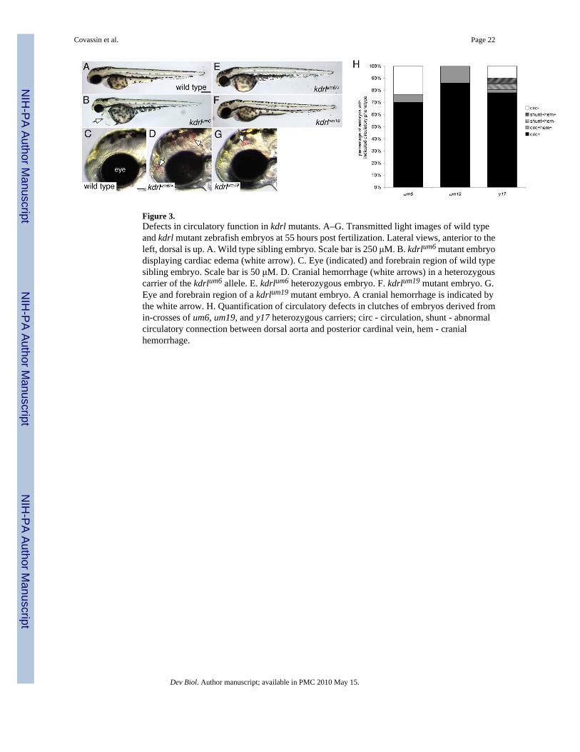
Figure 3.Defects in circulatory function in kdrl mutants. A–G. Transmitted light images of wild typeand kdrl mutant zebrafish embryos at 55 hours post fertilization. Lateral views, anterior to theleft, dorsal is up. A. Wild type sibling embryo. Scale bar is 250 μM. B. kdrlum6 mutant embryodisplaying cardiac edema (white arrow). C. Eye (indicated) and forebrain region of wild typesibling embryo. Scale bar is 50 μM. D. Cranial hemorrhage (white arrows) in a heterozygouscarrier of the kdrlum6 allele. E. kdrlum6 heterozygous embryo. F. kdrlum19 mutant embryo. G.Eye and forebrain region of a kdrlum19 mutant embryo. A cranial hemorrhage is indicated bythe white arrow. H. Quantification of circulatory defects in clutches of embryos derived fromin-crosses of um6, um19, and y17 heterozygous carriers; circ - circulation, shunt - abnormalcirculatory connection between dorsal aorta and posterior cardinal vein, hem - cranialhemorrhage.
Covassin et al. Page 22
Dev Biol. Author manuscript; available in PMC 2010 May 15.
NIH
-PA Author Manuscript
NIH
-PA Author Manuscript
NIH
-PA Author Manuscript

Figure 4.plcg1 is required for segmental artery formation. A. Functional domains within Plcg1. PH –pleckstrin homology domain; X, Y – split catalytic domains; SH2 – src homology-2 domains;SH3 – src homology 3 domain. Residues affected by the y13 and y16 mutations are outlinedin red and aligned with conserved sequences from other species (Dr – zebrafish, Hs – human,Dm – Drosophila, Mm – mouse). The y18 allele leads to a truncation in the X-catalytic domain.B–E. Confocal fluorescent micrographs of trunk blood vessels at 30 hours post fertilization.Lateral views, anterior to the left, dorsal is up. B. Wild type sibling embryo. Scale bar is 50μM. C. Loss of segmental arteries in a plcg1y13 mutant embryo. D. Partial segmental arteries(white arrows) in a plcg1y16 mutant embryo. E. plcg1y18 mutant embryo. F. Position of pointmutations engineered into rescue constructs in the Plcg1 SH2 and SH3 domains. G. Graphindicating proportion of embryos displaying segmental artery or DLAV formation followinginjection with mRNA encoding the indicated Plcg1 construct. H. Graph indicating proportionof embryos displaying partial or complete circulation following injection with mRNA encodingthe indicated Plcg1 construct; SeA - segmental arteries, DLAV - dorsal longitudinalanastamotic vessel, circ - circulation, partial - shunt between dorsal aorta and posterior cardinalvein.
Covassin et al. Page 23
Dev Biol. Author manuscript; available in PMC 2010 May 15.
NIH
-PA Author Manuscript
NIH
-PA Author Manuscript
NIH
-PA Author Manuscript
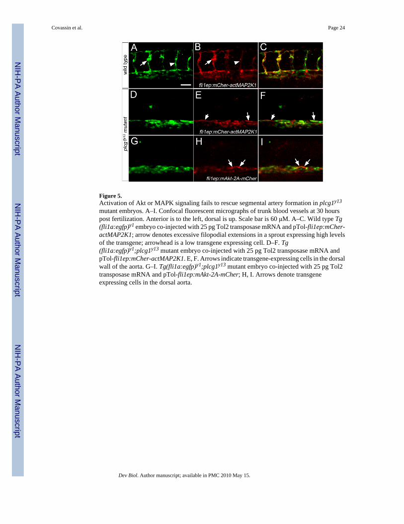
Figure 5.Activation of Akt or MAPK signaling fails to rescue segmental artery formation in plcg1y13
mutant embryos. A–I. Confocal fluorescent micrographs of trunk blood vessels at 30 hourspost fertilization. Anterior is to the left, dorsal is up. Scale bar is 60 μM. A–C. Wild type Tg(fli1a:egfp)y1 embryo co-injected with 25 pg Tol2 transposase mRNA and pTol-fli1ep:mCher-actMAP2K1; arrow denotes excessive filopodial extensions in a sprout expressing high levelsof the transgene; arrowhead is a low transgene expressing cell. D–F. Tg(fli1a:egfp)y1;plcg1y13 mutant embryo co-injected with 25 pg Tol2 transposase mRNA andpTol-fli1ep:mCher-actMAP2K1. E, F. Arrows indicate transgene-expressing cells in the dorsalwall of the aorta. G–I. Tg(fli1a:egfp)y1;plcg1y13 mutant embryo co-injected with 25 pg Tol2transposase mRNA and pTol-fli1ep:mAkt-2A-mCher; H, I. Arrows denote transgeneexpressing cells in the dorsal aorta.
Covassin et al. Page 24
Dev Biol. Author manuscript; available in PMC 2010 May 15.
NIH
-PA Author Manuscript
NIH
-PA Author Manuscript
NIH
-PA Author Manuscript

Figure 6.Artery differentiation defects in plcg1 mutants. A–C. Classification schema to categorize levelof artery marker gene expression in plcg1 mutant embryos. Differential interference contrast(DIC) images of zebrafish embryo trunks following whole mount in situ hybridization to detectephrinb2a (efnb2a). A. Normal ephrinb2a expression in the dorsal aorta (white arrow). B.Weak ephrinb2a in distinct patches within the dorsal aorta (white arrows). C. Loss of dorsalephrinb2a expression classified as “absent”. D. Quantification of artery marker geneexpression in y13, y16, y18 and MPZy18 mutant embryos. E, F. Transmitted light images ofembryos at 30 hpf. Scale bar is 250 μM. G, H. DIC images of embryos at 30 hpf. E–H., Lateralviews, anterior to the left, dorsal is up. E, G. MPZplcg1y18 mutant embryo. Scale bar is 30μM; nt – neural tube, fp – floor plate, nc – notochord. Asterisked bracket denoted absence ofdefined dorsal aorta or posterior cardinal vein. F, H. Mplcg1y18; Tg(fli1a:egfp)y1 mutantembryo; DA – dorsal aorta, indicated by a bracket.
Covassin et al. Page 25
Dev Biol. Author manuscript; available in PMC 2010 May 15.
NIH
-PA Author Manuscript
NIH
-PA Author Manuscript
NIH
-PA Author Manuscript

Figure 7.Plcg1 is required autonomously in endothelial cells for segmental artery sprouting. A.-E.Confocal fluorescent micrographs of trunk blood vessels at approximately 27 hpf. Lateralviews, anterior to the left, dorsal is up. In all images, host cells are expressing dsRedEx (red),while donor cells are expressing enhanced green fluorescent protein (green); in A–C, donorcells are also labeled with Cascade Blue (blue). A. Control embryo derived from transplantationof wild type donor Tg(fli1a:egfp)y1 cells into a Tg(fli1ep:dsredex)um13 host. Donor contributionto a developing segmental artery (white arrow) and the caudal vein (white arrowhead) isindicated. B. Contribution of donor Tg(fli1a:egfp)y1 cells to a segmental artery (white arrow)in a plcg1 MO injected Tg(fli1ep:dsredex)um13 host. C. Donor plcg1-MO injected Tg(fli1a:egfp)y1 cells (white arrows) within the posterior cardinal vein (lumen indicated by awhite bracket) in a wild type Tg(fli1ep:dsredex)um13 host. D. Donor plcg1-MO injected Tg(fli1a:egfp)y1 cells (white arrowheads) within the ventral wall of the dorsal aorta (DA, lumendenoted by a white bracket) in a wild type Tg(fli1ep:dsredex)um13 host. E. Donor Tg(fli1a:egfp)y1;plcg1y13 mutant cells (white arrowheads) within the ventral wall of the dorsalaorta (DA, lumen denoted by a white bracket) in a wild type Tg(fli1ep:dsredex)um13 host. A–C. Scale bar is 50 μM. D, E. Scale bar is 30 μM.
Covassin et al. Page 26
Dev Biol. Author manuscript; available in PMC 2010 May 15.
NIH
-PA Author Manuscript
NIH
-PA Author Manuscript
NIH
-PA Author Manuscript
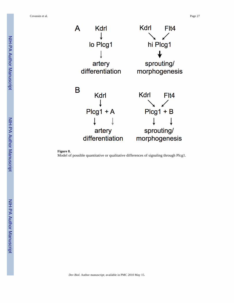
Figure 8.Model of possible quantitative or qualitative differences of signaling through Plcg1.
Covassin et al. Page 27
Dev Biol. Author manuscript; available in PMC 2010 May 15.
NIH
-PA Author Manuscript
NIH
-PA Author Manuscript
NIH
-PA Author Manuscript

NIH
-PA Author Manuscript
NIH
-PA Author Manuscript
NIH
-PA Author Manuscript
Covassin et al. Page 28Ta
ble
1
Segm
enta
l arte
ry m
utan
ts id
entif
ied
by h
aplo
id tr
ansg
enic
scre
enin
g
Cla
ssge
neal
lele
mut
atio
naf
fect
ed d
omai
nSe
AR
efer
ence
Ikd
rlum
6C
1117→
Gcy
topl
asm
icno
neM
eng
et a
l.; th
is st
udy
plcg
1y1
0SA
PHno
neLa
wso
n et
al.
y13
C41
2→R
X-c
atal
ytic
none
this
stud
yy1
5I 3
83→
NX
-cat
alyt
icno
ne-
y18
SDal
lno
neC
ovas
sin
et a
l.; th
is st
udy
y19
ND
all
none
-II
kdrl
y17
L 846→
RA
TP b
indi
ngpa
rtial
Cov
assi
n et
al.;
this
stud
ypl
cg1
y16
R55
7→H
N-S
H2
parti
alth
is st
udy
III
plex
inD
1um
7N
DN
Dec
topi
c-
IVet
srp
y11
inse
rtion
DN
A b
indi
ngpa
rtial
Pham
et a
l.
SA –
splic
e ac
cept
or; S
D –
splic
e do
nor;
ND
– n
ot d
eter
min
ed.
Dev Biol. Author manuscript; available in PMC 2010 May 15.
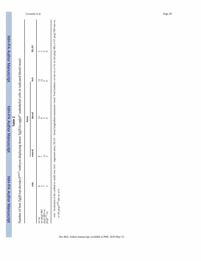
NIH
-PA Author Manuscript
NIH
-PA Author Manuscript
NIH
-PA Author Manuscript
Covassin et al. Page 29Ta
ble
2
Num
ber o
f hos
t Tg(
fli1e
p:ds
rede
x)um
13 e
mbr
yos d
ispl
ayin
g do
nor T
g(fli
1a:e
gfp)
y1 e
ndot
helia
l cel
ls in
indi
cate
d bl
ood
vess
el.
Aor
ta
vein
vent
ral
dors
alSe
AD
LA
V
wt>
wt
88
1313
5w
t>pl
cg1
MO
57
815
5pl
cg1
MO
>wt
711
30
0pl
cg1y1
3 >wt
23
00
0
vein
– lo
caliz
atio
n in
the
card
inal
or c
auda
l vei
n; S
eA –
segm
enta
l arte
ry; D
LAV
– d
orsa
l lon
gitu
dina
l ana
stam
otic
ves
sel.
Tota
l num
bers
: wt i
nto
wt,
n=15
; wt i
nto
plcg
1 M
O, n
=27;
plc
g1 M
O in
to w
t,
n=19
; plc
g1y1
3 in
to w
t, n=
3.
Dev Biol. Author manuscript; available in PMC 2010 May 15.