australian and new zealand college of veterinary...
TRANSCRIPT
Australian and New Zealand
College of Veterinary Scientists
Dermatology Chapter Science Week
Proceedings 12th/13th July 2013
Musings from a Master
and various other topics

2
THANK YOU The Dermatology Chapter of the Australian
and New Zealand College of Veterinary
Scientists would like to thank Merial for
their generous sponsorship of Chapter
scientific programs and social events in
2013-14.
Merial GOLD LEVEL SPONSOR
Science Week, Gold Coast 2013

3
THANK YOU The Dermatology Chapter of the Australian
and New Zealand College of Veterinary
Scientists would like to thank Elanco for
their generous sponsorship of Chapter
scientific programs and social events in
2013-14.
Elanco GOLD LEVEL SPONSOR
Science Week, Gold Coast 2013
4
10th Annual Dermatology Chapter Meeting of the Australian New Zealand College of Veterinary Scientists After a one-year absence, for the World Congress in Veterinary Dermatology in Vancouver, the Dermatology Chapter is again holding its annual meeting during Science week at the Gold Coast, Queensland. Science week continues to provide an important forum for the discussion and presentation of recent developments, research and the most up-to-date knowledge over a wide range of veterinary specialties. This year we are privileged to have Danny Scott, one of the world’s best-known and most entertaining presenters in Veterinary Dermatology, as our principle speaker. He will be joined by an array of local speakers. As in previous years the chapter will have two full days of presentations. The morning of the first day is a joint collaboration with the Veterinary Ophthalmology Chapter. The focus of the presentations in the morning session will be on the eyelids and the ocular surface. The afternoon of the first day sees the program return dermatology only. Our major speaker, Danny Scott, will be discussing two challenging parasitic conditions of dogs, dermodicosis and scabies. The parasitic theme continues with a presentation from Linda Vogelnest on skin diseases and ticks. The day will conclude with David Robson’s intriguingly titled discussion, Yoda and the art of dog shampooing. We return again to dermatology on day two where our major speaker, Danny Scott, will be presenting on a number of topics covering conditions rare and not so rare. He will be joined by two local speakers to discuss some of the new ideas and treatments for atopic dermatitis. We are also pleased to see three presentations by our local dermatology residents, covering feline pemphigus and two presentations on MRSP. The program this year is very wide-ranging and diverse and should interest not just those starting out in dermatology but also those who have many years experience in the field. The chapter is very privileged to have Danny Scott join us. It is a rare opportunity to be able to learn from someone of his calibre. With Thanks Peter Hill Karyn Wesselingh Andrew Carter (Dermatology Chapter Science Week Coordinators)

5
Table of Contents
Speakers 7 Programme Friday 12th July Morning Combined Ophthalmology and dermatology session Eyelid anatomy and congenital defects
Cameron Whittaker 9 Developmental abnormalities of eyelids and surgical correction Jeff Smith 11 Meibomian glands and their abnormalities
Robin Stanley 14 Uveodermatologic syndrome (Vogt-Koyanagi-Harada-like syndrome) Mike Bernays 18 Non-neoplastic dermatosis of the eyelids of cats and dogs 20 Danny Scott Inflammatory and immune-mediated diseases of the ocular surface
Kelly Caruso 23 Allergic conjunctivitis and allergy testing
Greg Burton 25 Afternoon – Dermatology session Canine demodicosis
Danny Scott 30 Canine scabies – lessons from 350 dogs
Danny Scott 33 Skin diseases caused by ticks
Linda Vogelnest 35 Yoda and the art of dog shampooing
David Robson 40

6
Saturday 13th July Topical treatment of atopic dermatitis
Mandy Burrows 47 Novel allergens in canine allergic skin disease
Ken Mason 51 Feline atopic dermatitis
Danny Scott 55 Allergic skin diseases in the cat: macroscopic to microscopic
Danny Scott 57 Pursuing anecdotes in canine dermatology
Danny Scott 60 Pursuing anecdotes in feline dermatology
Danny Scott 62 Case reports and research projects Resident project – MRSP
Philippa Ravens 64 Characterisation of methicillin –susceptible and methicillin-resistant isolates of Staphylococcus pseudintermedius from the skin of Australian dogs
Meng Siak 69 Resident project – Feline pemphigus
Debbie Simpson 73

7
Speakers Mandy Burrows Mandy is a Fellow of the Australian College of Veterinary Scientists in Veterinary Dermatology and a registered specialist in veterinary dermatology. She is a consultant in veterinary dermatology and has two dermatology practices in Perth, Western Australia that provide secondary and tertiary referral advice for skin, ear and allergy problems in dogs, cats and horses. She lectures in dermatology at Murdoch University Veterinary Hospital and she teaches undergraduate veterinary students and the dermatology unit of the Masters in Veterinary Medicine at both Murdoch and Massey University, New Zealand. She is currently the Chief Examiner and serves on the Council and the Board of Examiners of the Australian and New Zealand College of Veterinary Scientists and is a member of the Advisory Committee for the Registration of Veterinary Specialists. She is a member of the Australian Advisory Board for Infectious Diseases in companion animals and is the current Australian and New Zealand representative and the Secretary of the World Association for Veterinary Dermatology. She has extensive experience with clinical dermatology in companion animals and she enjoys teaching dermatology to veterinary undergraduate and postgraduate students. Greg Burton Greg graduated from the University of Queensland in 1983. After spending several years in general practice he passed his Membership of the Australian College of Veterinary Scientists in Small Animal Surgery. Greg became a Fellow of the College in Veterinary Dermatology in 1997. From 1999 to now Greg has been a director of the Skin, Ear and Allergy Service at the Melbourne Veterinary Specialist Centre, In 2002, he became the Principle Fellow in Dermatology at the University of Melbourne in addition to taking responsibility for teaching of under-graduate dermatology in the veterinary faculty. Greg has authored and co-authored numerous articles in both Australian and international journals. Ken Mason Ken is a veterinarian of 40 years standing. He lives in Brisbane and is a self-confessed cat person. Currently he is involved with research into a superficial science. Philippa Ravens Philippa graduated from the University of Sydney in 2004 and worked in small animal general practice in Sydney for five years. She accepted a residency in veterinary dermatology with Linda Vogelnest in August of 2009. She became a Member of the Australian College of Veterinary Scientists in Canine and Feline medicine and completed a Masters degree in Veterinary Clinical Studies through Murdoch University in 2011. David Robson David graduated in 1992 from the University of Queensland, He worked as an associated at Chermside Veterinary Hospital, Brisbane, for six years and gained Membership of the Australian College of Veterinary Scientists in Medicine of Dogs in 1999. Later that year he took up residency at the Animal Skin Ear and Allergy service at the Melbourne Veterinary Referral Centre, Glen Waverley, under the supervision of Greg Burton, and passed his ACVS Fellowship examination in dermatology in 2005. His major publications include a two-part review on cyclosporine and its role in dermatology, the cytology of the normal anal sac and the longest case series to date of Demodex injai demodicosis. He has practiced clinical dermatology and lectured both in Australia and overseas.

8
Danny Scott Dr. Scott is a 1971 graduate of the University of California, Davis. He is currently Professor of Medicine and Co-Chief of the Dermatology Service at Cornell University. He is a Diplomate of the American College of Veterinary Dermatology and the American College of Veterinary Pathologists (Honorary). Dr. Scott’s duties include clinical service; teaching veterinary students and residents in dermatology and pathology; diagnostic dermatopathology; clinical and dermatopathologic research; consultations for area veterinarians; various committees. He has authored or co-authored over 580 publications and given over 400 continuing education presentations around the world. Meng Siak Dr. Siak graduated from Murdoch University Veterinary School, Western Australia, in 2006 with honours. He then spent one year in private practice in tropical far North Queensland, Australia, before returning to Murdoch University to commence an internship in veterinary dermatology. At the end of the internship in 2009, he successfully applied for a veterinary dermatology resident position recognised by the Australia and New Zealand College of Veterinary Scientists. Debbie Simpson Debbie is a resident at the Melbourne Veterinary Specialist Centre. She graduated from Massey University, New Zealand in 2008 and did a one-year internship at the Veterinary Specialist Group in Auckland, followed by a one year internship at Veterinary Specialist Services in Brisbane. She is due to complete her 3 year residency in dermatology in 2013. Linda Vogelnest Linda graduated from Sydney University in 1984 and worked in private and university small animal practice in Australia and the UK for over ten years before following a long time interest in dermatology. Linda achieved Membership of the ACVSc in Feline Medicine in 1997, undertook a Dermatology residency program from 1997-2001 based partly in Sydney and Melbourne, and achieved Fellowship of the ACVSc in Veterinary Dermatology in 2003. She ran a small and large animal dermatology service at the University of Sydney from 1999-2012, before moving to private practice at the Small Animal Specialist Hospital in 2013. Linda continues to teach dermatology to veterinary students in pre-clinical years, and consults at Agnes Banks Equine Hospital monthly. Linda’s special interests include individualized patient care in atopic dermatitis, ear diseases, dermatohistopathology, skin surface cytology sampling, and dermatology in exotics and horses.

9
Eyelid anatomy and congenital malformations
Cameron Whittaker BVSc, DVCS, Dip ACVO, MACVSc
Kelly Caruso BSc, VMD, Dip ACVO, MACVSc Eye Clinic for Animals
64 Atchison Street, Crows Nest. 2065 250 Parramatta Rd, Homebush West. 2140
Anatomy The eyelids are very specialised structures and as ophthalmologists we will usually try to preserve their function and anatomic placement as much as possible. Having said this, the eyelids do have many similarities to normal skin and for the most part can be considered “adapted skin”. The eyelid is made up of several layers: from superficial to deep these are: skin, subcutaneous tissue, orbicularis oculi, orbital septum and tarsal plates, and palpebral conjunctiva. Lying within the tarsal plate is the Meibomian gland, which secretes modified sebum, which contributes to the tear film. The hairline stops about 2mm from the eyelid margin, and there are eyelashes (cilia) on the upper eyelid but not the lower eyelid. With the exception of the prepuce and labia minora, the eyelid has the thinnest skin in the whole body. The skin of the eyelid contains glands and hairs, although the exact nature of the glands is poorly understood. (R.Dubielzig pers comm). The eyelids are supplied with blood by 2 arches on each upper and lower lid. The arches are formed by anastomoses of the lateral palpebral arteries and medial palpebral arteries, branching off from the lacrimal artery and ophthalmic artery, respectively. The sensory nerves to the upper eyelids are from the infratrochlear, supratrochlear, supraorbital and lacrimal nerves from the ophthalmic branch of the trigeminal nerve. The skin of the lower eyelid is supplied by branches of the infratrochlear at the medial angle, the rest is supplied by branches of the infraorbital nerve of the maxillary branch of the trigeminal nerve. Motor supply is provided by the auriculopalpebral branch of the facial nerve to the orbicularis oculi muscle, with contributions from the oculomotor nerve and sympathetic supply to Muller’s muscle. Muscles of the eyelid include the orbicularis oculi muscle (innervated by the facial nerve), levator palpebrae both dorsal and ventral (innervated by the oculomotor nerve), and Muller’s muscle (innervated by the sympathetic nerve). Glandular structures in the eyelids of dogs are somewhat controversial. The most prominent gland is the Meibomian gland, a modified sebaceous gland, with accessory lacrimal gland (the Glands of Kraus – in the fornix, and Gland of Wolfring – just above the tarsal plate) being contentious. The gland of Zeis is also somewhat contentious BUT there is a gland of Moll (a ciliary associated gland). The conjunctiva lines the inside of the eyelids and covers the sclera, and both sides of the third eyelid. It is composed of non–keratinised, stratified columnar epithelium with goblet cells. The conjunctiva is a mucous membrane in part responsible for components of the tear film, and also facilitating the smooth movement of the eyelids over the corneal surface. It is also responsible for immune surveillance. The epithelial layer contains blood vessels, fibrous tissue, and lymphatic vessels. Additional cells in the conjunctival epithelium include melanocytes, T and B lymphocytes. Sensory innervation of the conjunctiva is divided into 4 parts: the dorsal aspect is supplied by the supraorbital, supratrochlear and infratrochlear nerves, the ventral aspect is supplied by the infraorbital nerve, the lateral aspect is supplied by the lacrimal nerve (with a contribution from the zygomaticofacial nerve), and circumcorneal innervation is via the long ciliary nerves. The third eyelid is an accessory eyelid, vestigial in humans, which contributes to the ocular health of the eye. It does this by producing 30% of the tear film via the gland at its base, and by acting as a “windscreen wiper” of the cornea to mechanically wipe away dirt and debris from the corneal surface. It has a T shaped cartilage providing structural support, and the base of it has a relatively high density of goblet cells. The goblet cells produce mucus, which contains mucin, immunoglobulins, urea, salts, glucose, leucocytes, cellular debris, and enzymes. Mucins have been actively investigated of late. There are currently over a dozen mucin genes, and are either secretory or membrane bound. The secretory mucins (MUC 1, MUC 4, MUC 16) are soluble in tears
10
and distributed over cornea in blinking, and then shunted to nasolacrimal duct. They act to bind debris, hold fluid in place (are hydrophilic) and harbour defense molecules. The membrane bound mucins (MUC 2, MUC5AC, MUC 5B) act to lubricate the ocular surface, and prevent pathogen adherence. Congenital eyelid abnormalities It is not normal for the eyelids to have any hairs at all coming out of the Meibomian gland opening at the grey line. If hairs do grow out of the openings we call them distichiae, and if they erupt perpendicularly through the palpebral conjunctiva they are termed ectopic cilia. Surgery is usually curative, and can include the use of cryotherapy, excision or electrolysis. Each modality has its pros and con, but for the most part all therapies enjoy a reasonably high success rate. Dermoids can be seen in all aspects of the eye. They typically involve the cornea and/or the conjunctiva but certainly can be seen in the eyelids. The deleterious effects of dermoids in the eye mainly derives from the corneal contact with the hairs produced by the dermoid. This results in chronic irritation. Surgical excision is usually curative, with or without some form of eyelid reformation procedure, or tissue transposition surgery in the case of the cornea. The third eyelid can develop a scroll in the cartilage – probably more developmental per se than congenital but still an anatomic, likely genetically related, defect. This is particularly seen in Giant Breeds of dogs, such as Mastiffs, Great Danes St Bernards etc. Surgery involves removing a piece of the vertical aspect of the T shaped cartilage. Eyelid agenesis is a congenital problem seen in both domestic and wild cats. In essence, a variable aspect of the upper and/or lower eyelid is not present. Affected animals usually also have an absence of the lacrimal gland in the dorsolateral aspect of the eye. Damage occurs as a result of chronic exposure and a poor to absent tear production. Numerous techniques have been described to treat these cases, from cryosurgery to remove the trichiasis hairs, to various advancement flaps. Recently a novel technique transposing the lip commissure to the eyelid has been shown to create an excellent substitute for the absence of eyelid margin, and in some cases may even allow some recovery of the new eyelids to blink. Affected animals can do surprisingly well with some of the surgical techniques available, and be left with a comfortable eye with excellent vision. References: Gelatt, KN. Veterinary Ophthalmology 4th Edition. 2007. Wiley, John and Sons.

11
Developmental abnormalities of eyelids and their surgical correction
Jeffrey S Smith BVSc, FACVSc, Dip ACVO Eye Clinic for Animals
64 Atchison Street,Crows Nest. 2065 250 Parramatta Rd,Homebush West. 2140
The topics of this paper are the congenital eyelid abnormalities encountered in animals, the clinical progression of affected individuals and the surgical techniques available to restore comfort and function. Eyelid development The eyelids need to conform to the cornea in order to protect the cornea by adequate blinking and spreading of the tear film. Maldirection of eyelid hair with corneal contact will cause discomfort, discharge and ulceration. The development of eyelids, in utero, is completed late in gestation. In most small animals, the eyelids are still fused, as in utero, at birth and open by a combination of cellular apoptosis and epithelial keratinization along the lid margins. Guinea pigs, cattle, sheep and horses are normally born with eyelids separated and open. Eyelids open normally in rabbits and cats at approximately 10 days and a few days later in puppies. Premature opening is unusual but can lead to the cornea being insufficiently lubricated as the lacrimal glands are not yet functional. Prolonged eyelid fusion is more common. As a result of prolonged closure, infection can occur beneath the eyelids in the conjunctiva and cornea (conjunctivitis neonatorum). Often more than one in the litter is affected. Organisms include herpes virus in kittens, acquired from the genital tract of the queen. This is the most common pathogen in affected kittens. Mixed bacteria infect puppies. There is pronounced swelling of the eye and permanent damage to the cornea, including rupture, can occur unless the exudates are drained. Eyelid separation is not difficult and can be achieved with hemostats or scissors via the medial canthal area. Post-operative irrigation and appropriate antimicrobials should be used. These must include antiviral drugs for cats - Idoxuridine 0.1% eye drops 5-6 x daily after hot compress cleaning. These drops are available from a compounding pharmacy. Do not use topical steroids or non-steroidals in these cases. Classification of developmental abnormalities Incomplete development Partial dysgenesis or total agenesis can occur. The upper eyelid is almost always the lid affected. In practice, the most commonly affected patient is a young kitten rescued from poor surroundings. The degree of deformity is variable and involves primarily the lateral half to two-thirds of the upper lid. There is an absence of lid margin with facial hair contacting the cornea (trichiasis). At the time of presentation, the cornea may be showing signs of low-grade keratitis. Ulceration is unusual and pain seldom complicates the problem. Vision is usually adequate. Coloboma Another form of incomplete development, coloboma, involves the presence of a notch or fissure in the eyelid. This creates a localised failure to cover the cornea when blinking. The problem is rare and repair is not urgent. Dermoid The presence of normal skin structures in an abnormal site. The cornea and conjunctiva are affected primarily, but the lateral eyelid canthal area is sometimes affected. German Shepherds seem predisposed in Australia, at least. The plaque produces hair of variable length, which streams across the cornea causing minor irritation.

12
Small palpebral fissure usually conceals an underdeveloped eye (microphthalmos). The problems is seen most commonly in Collies. The microphthalmic eye is usually non-visual. Oversized eyelids or macro palpebral fissure is seen in large breeds of dog. Problems do not usually manifest in infancy. Later, entropion/ectropion complex problems arise. Entropion This problem is seen most commonly in large animals wherein eyelids at birth can be floppy, lacking tone. The lower lid turns in causing spasm. Some breeds of dog, notably the Sharpei, exhibit upper and lower entropion seen after the lids open. A procedure to temporarily relieve the lid contact is often necessary. As the animal grows, normal anatomy is usually re established in large animals and occasionally in small animals. Surgical techniques The following points should be taken into account: 1) Can the selected procedure wait or is it necessary to operate as soon as possible? Pain is the determining factor. Delaying surgery until the animal is more mature is a poor option. Apart from pain, permanent corneal damage can occur. 2) Will the surgery be curative or will further surgery be likely? Many procedures performed in the first weeks or months will be of temporary benefit only. 3) Are the necessary instruments and suture materials available? You will need Adson toothed forceps, both fine and medium sized; scissors to cut skin and sturdy iris scissors; Castroviejo needle holders with sturdy tips and needle holders of your choice; and PDS 6/0 and 5/0, and 4/0 monofilament should be available. 4) Aftercare considerations including drug therapy. Hot compress cleaning 1-2x daily + topical Cloxacillin eye ointment (Orbenin, Pfizer Australia*) 1-2 x daily is my usual recommendation. 5) Signs indicative of surgery failure. Continuation of spasm beyond 1-2 days and signs of hair-corneal contact persisting. Inability to successfully blink after surgery will create complications. Specific procedures1,2 Procedure for the temporary relief of lower and upper entropions Several vertical mattress sutures of non-absorbable material, eg 4/0 monofilament nylon, are placed perpendicular to the inturning eyelid. The more proximal of the two bites taken needs to be placed quite close to the lid margin. The procedure can be performed under local anesthesia with sedation if necessary eg Medetomidine. This drug is ideal in Sharpei puppies in which this "tacking" procedure is often necessary. Procedure for more permanent cure of lower entropion This would seldom be selected in infants. The standard procedure is the Celsus technique, well described in text books.1,2 It is important to remove a strip of skin close to, but not impinging upon, the lid margin and following the line of the eyelid. Absorbable sutures such as 6/0 PDS are inserted with all knots pulled away from the margin. Correction of upper lid dysgenesis Several choices are available to the surgeon. The defect is seldom painful and corneal damage is chronic. Temporary relief of hair contact can be achieved by epilation of facial hair as needed. The re establishment of a lid margin can be achieved by a lip-to-lid transposition (see Dr Whittaker's notes). The transposition of a strip of skin from nearby the defect is well described but does not solve the issue of locally maldirected hair.1
Correction of megaloblepharon is necessary early in life only where entropion is part of the problem. A combination entropion / ectropion procedure is selected. This involves eyelid skin excision, tightening and narrowing the palpebral fissure. Tightening is performed by wedge excision of a portion of eyelid, usually laterally. Notches or gaping areas are eliminated in this fashion. Closure of such surgical defects is often achieved in two layers after local lid splitting.
13
Lid splitting is necessary when the tension of bringing two sides together for closure is high. The procedure is used to mobilise the skin of the eyelid and the palpebral conjunctiva in two halves. The split is achieved with a 64 Beaver blade run along the line of the Meibomian gland openings centrally positioned within the lid margin. The lid is split into two halves. The depth of the split depends upon the size of the surgical defect. The inner layer is sutured, then the skin layer is closed. There is no tension on the wound. "When in trouble, split the lid" (Dr Rowan Blogg, pers comm. circa 1980). Eyelid Dermoid excision The surgical excision involves removal of the plaque of skin and reconstructing the defect in the lid. The lateral canthal angle must be re created carefully so that the eyelids close comfortably. It will not matter if the palpebral fissure is slightly smaller on the affected side. Stretching open will take place over time. *Orbenin eye ointment. Pfizer Australia. Warf Rd. West Ryde. NSW 2114 References: 1. Bistner,S.I, Aguirre,G and Batik,G. Atlas of Veterinary Ophthalmology,1977.WB.Saunders Company 2. Gelatt,K.N, Veternary Ophthalmology 4th Ed. 2007.Wiley,John and Sons

14
Meibomian glands and abnormalities
Robin Stanley, BVSc(Hons), FANZCVSc Animal Eye Care
181 Darling RdEast Malvern, Victoria, 3145
Anatomy and function The meibomian glands are an important component of the eyelids, and are important in contributing to ocular surface health. Diseases involving the meibomian glands can also result in corneal and or conjunctival disease.1 The openings of these glands can be seen right on the eyelid edge as small spots. When the eyelid edge is squeezed, the secretions from the meibomian glands can be seen. This is normally a white, waxy, lipid substance (meibomium) that is an important part of the pre-corneal tear film. The lipid layer secreted by the meibomian glands is the outermost layer of the pre-corneal tear film. This creates a surface tension-like effect that helps keep the tears on the cornea, and this effect also helps to stop the tears from running over the edges of the eyelids. 2,3 The meibomian glands in the eyelid edge are modified sebaceous glands. They run vertically in the tarsal plate and exit into the lid margin. Clinically some breeds of dogs are affected with distichiasis (extra eyelashes) when the meibomian glands grow lashes.2,4 The eyelids also contain the gland of Zeis, which are modified sebaceous glands, and the ciliary glands (of Moll), which are modified sweat glands. Both these glands may contribute to the tear film. Clinically, disease of the eyelids, e.g. infection or inflammation, may result in keratoconjunctivitis sicca (dry eye) because the quality of the tear film is reduced.5 These animals may have a normal Schirmer tear test (STT) as this measures the quantity of the tears, but will have reduced tear quality, measured with tear break up times (TBUT).1,3,5 Clinical Conditions involving the Meibomian Glands Keratoconjunctivitis Sicca (dry eye) due to meibomian gland dysfunction (MGD) The meibomian glands provide the lipid layer of the pre-corneal tear film (PCTF). This lipid layer is the outermost layer of the PCTF. Its main functions are to reduce evaporation of the PCTF, and reduce the runoff of the PCTF.1,6 MGD will lead to inflammation of the meibomian glands, and this primarily results in an increase in meibomian gland secretions. 7 This may be seen as increased oily secretions on the eyelid margin. The increased oil secretions are accompanied by bacterial colonization. This can result in keratoconjunctivitis sicca (KCS). Diseases of the eyelids, e.g. Staphylococcal infections, may reduce or alter the Meibomian gland secretions reducing the quality of the tears. 7 In such cases the Schirmer Tear Test (STT) will be normal, as the amount of the aqueous component of the PCTF is unchanged, but its quality is reduced so that it runs off the cornea, resulting in signs of KCS. With MGD the KCS diagnosis can be made by the eye looking dry, e.g. having mucoid discharge with conjunctival hyperaemia, with or without vascular keratitis; and also by performing a tear break up time (TBUT). MGD is one of the most common causes of KCS in humans. 7 MGD is a much less commonly recognised cause of KCS in the veterinary species. Treatment of MGD induced dry eye is aimed at restoring normal meibomian gland function, and also symptomatic treatment with topical immunomodulators such as cyclosporin or tacrolimus, and artificial tears, and or topical viscous preparations.

15
In humans MGD (also called posterior blepharitis) is the most common form of eyelid disease with up to 40% of eye cases having some form of MGD. Initially humans are asymptomatic, but with time may develop irritation associated with the inadequate tear film resulting in corneal disease. 7 Primary meibomianitis in humans is associated with inspissation of the meibomian glands, their secretions becoming thicker, more difficult to express resulting in engorgement of the glands. The anterior eyelid is normal, hence the term posterior blepharitis. In humans this primary meibomianitis may be associated with acne rosacea, acne vulgaris and seborrhoeic dermatitis. 7 In some cases the inflammation spreads and then secondary bacterial involvement is seen. It is unclear whether the bacteria cause the thickening of the meibomian gland secretions or if the Staph simply create more inflammation. 7 Staphylococcus epidermidis and Staphylococcus aureus produce lipases that hydrolyse sebaceous gland lipids, resulting in free fatty acids and other by-products that irritate the cornea and conjunctiva and cause tear film instability.8 In human patients with seborrhoeic dermatitis the meibomian glands will produce more secretions. This lipid material will react with tear film proteins resulting in saponification. This is a cause of intense irritation in most human patients with obvious lid disease.7 Eyelid blepharitis Affected dogs will show swollen and erythematous eyelids. Usually multiple eyelids are affected. The initial presentation may be swelling of the eyelid; they sometimes ulcerate and discharge purulent material. Affected animals may be pruritic. 2 In our practice Rottweilers seem to be a breed predisposed to blepharitis. Like canine bacterial pydoerma, the normal bacterial flora becomes pathogenic when the normal ecosystem is altered. We see this most commonly with allergies. This is why it is so important to treat any underlying aetiology. Anything that can cause irritation to the eyelid can result in blepharitis. Conditions such as eyelid tumours, extra eyelashes, ectopic cilia, prominent nasal folds, fungal or parasitic infections, bacterial folliculitis, allergies and immune-mediated skin diseases may contribute to the development of eyelid blepharitis. The meibomian glands are predisposed to bacterial infections, as are normal hair follicles. Like canine bacterial pyoderma, the meibomian gland is infected, and in most cases we see the meibomian gland is ruptured resulting in a deep pyoderma. The gland rupture may lead to a foreign body type reaction against the lipid material secreted normal secreted externally by the meibomian glands. This deep reaction may result in draining tracts and sinus formation. Persians, and other feline breeds with lagophthalmos will be predisposed to blepharitis. In cats Feline Herpes Virus has also been implicated as a cause of eyelid disease. Diagnostic approach includes skin scraping for cytology and to check for demodecosis, aspirating the pustules, culture and sensitivity, and in some cases biopsy of the affected eyelid. Treatment often needs to be prolonged. Any underlying cause needs to be addressed. Part of the problem is infection. In canine bacterial pyoderma Staphylococcus intermedius is the implicated in most cases. I commence treatment with oral doxycycline 5mg/kg BID to help control the infection component. Therapy must be given for a minimum of 21 days, and for at least 2 weeks after the resolution of clinical signs. If the response is poor I then usually give oral cephalosporin antibiotics. Oral doxycycline inhibits bacterial lipase production. This reduces free fatty acids, which helps to lessen eyelid blepharitis and to reduce the signs of irritation. Doxycycline also inhibits keratinisation of the meibomian glands, and in humans has been shown to reduce the number of Staphylococcus organisms.9,10 In a human study comparing low and high dose doxycycline regimes, patient symptoms were equally improved in both dosage groups.9 The author has found that some of these Rottweilers require pulse antibiotic therapy, i.e. 2 days of antibiotics a week long term. Recently in humans a topical azithromycin preparation has been used for the management of chronic cases of blepharitis. 10

16
The other part of this condition is inflammation to the infection. Oral prednisolone is required to control this component of the problem. The author usually prescribes oral prednisolone 1 mg/kg once daily for five days, reducing to 1 mg/kg every second day. Topical therapy is very useful in the treatment of canine bacterial pyoderma. Obviously most shampoos, particularly those containing antibiotics cannot be used on the eyelids for fear of damaging the cornea. Johnson’s “no more tears” baby shampoo can be diluted 1 part to 10 parts water to cleanse off the scabs, and to assist drainage of the purulent material. In humans, warm compresses are considered important for the treatment of blepharitis. Perhaps we should pay more attention to cleaning affecting eyelids. Applying a warm cleanser or compress will reduce the viscosity of the meibomian secretion, allowing a more normal function. Topical preparations such as Panolog, Amacin or Tricin can be applied. These are well tolerated by the cornea. In dogs an E collar may be needed to prevent self trauma. Omega fatty acids found in fish oils and flaxseed have been used in humans with keratoconjunctivitis sicca and/or MGD.11 Humans with KCS treated with topical cyclosporin had fewer meibomian gland inclusions and less lid margin injection.12 Topical cyclosporine was also effective in human patients with acne rosacea and meibomian gland dysfunction.13 Extra Eyelashes – Distichia As previously discussed, the meibomian glands are modified hair follicles. In numerous breeds, the meibomian glands can produce hairs, these are the extra eyelashes.2 Breeds commonly seen at Animal Eye Care with distichia are Staffordshire Bull Terriers, Poodles, and Cocker Spaniels. If the extra eyelashes are small, and soft they will then float in the tear film, and not cause any clinical signs, then surgery is not required. If the extra eyelashes are large and stiff, they will irritate the cornea resulting in corneal scarring, either vascular or pigmentary, and/or ulceration. Surgery is required if the extra eyelashes are causing clinical signs. Extra eyelashes can be removed by a number of techniques. Transconjunctival excision 2,4 and cryoepilation 2,14 are the surgical techniques preferred by most ophthalmologists. At any one time some 10 to 15 % of extra eyelash follicles are inactive. If the follicle is inactive then this follicle is likely to regrow later, usually within 3 months of the first surgery. It is the author’s experience that of distichia cases requiring surgery, approximately 80% require one surgery, 15% require 2 surgeries, and less than 5% require 3 or more surgeries. Ectopic cilia are small hairs that grow through the conjunctival surface and result in severe irritation to the corneal surface, resulting in linear corneal ulceration, and severe blepharospasm. 2,15 These ectopic cilia arise from normal eyelid hairs that grow the wrong way. These do not arise from the meibomian glands. References 1. Carter R, Colitz CM. The causes, diagnosis, and treatment of canine keratoconjunctivitis sicca. Veterinary Medicine 2002; 97:683-94. 2. Stades FC, van der Woerdt. Diseases ans Surgery of the Canine Eyelids. In Veterinary Ophthalmology. 5th Ed. John Wiley & Sons, 2013: 832-904. 3. Moore CP, Norman J, Wilsmaat EV, Nordheim LJ, Majors S, Collier LL. Density and Distribution of Canine Conjunctival Goblet Cells. Investigative Ophthalmology and Visual Sciences 1987; 28: 1925-32. 4. Long RD. Treatment of distichiasis by conjunctival resection Journal of Small Animal Practice 1991; 32: 146-8 5. Kaswan, RL, Salisbury MA. A new perspective on canine keratoconjunctivitis sicca. Treatment with ophthalmic cyclosporine. The Veterinary Clinics of North America. Small Animal Practice 1990; 20: 583-99. 6. Ofri R, Orgad K, Kass PH, Dikstein L. Canine meibometry: establishing baseline values for meibomian gland secretions in dogs.The Veterinary Journal 2007; 174: 536-540.

17
7. Jill Pitts, O.D., and Christopher Lievens, O.D.Put the Squeeze on Meibomian Gland Disease. 2009 http://www.revoptom.com/content/c/15811/ 8. McCulley JP, Shine WE. Changing concepts in the diagnosis and management of blepharitis. Cornea. 2000;19(5):650-8. 9. Yoo SE, Lee DC, Chang MH. The effect of low-dose doxycycline therapy in chronic meibomian gland dysfunction. Korean Journal of Ophthalmology. 2005; 19(4):258-63. 10. Ianaro A, Ialenti A, Maffia P, et al. Anti-inflammatory activity of macrolide antibiotics. Journal of Pharmacology and Experimential Therapeutic. 2000;292(1):156-63. 11. Paranjpe DR, Foulks GN. Therapy for meibomian gland disease. Ophthalmology Clinics of North America. 2003;16(1):37-42. 12. Perry HD, Donnenfeld ED. Topical 0.05% cyclosporine in the treatment of dry eye. Expert Opinions in Pharmacotherapy. 2004;5(10):2099-107. 13. Schechter BA, Katz RS, Friedman LS. Efficacy of topical cyclosporine for the treatment of ocular rosacea. Advances in Therapeutics. 2009;26(6):651-9. 14 Chambers ED, Slatter DH. Cryotherapy (N2O) of canine distichiasis and trichiasis: an experimental and clinical report. Journal of Small Animal Practice. 1984; 25(11): 647-659. 15. D’Anna N, Sapienza JS, Guandalini A, Guerriero A. Use of a dermal biopsy punch for removal of ectopic cilia in dogs: 19 cases. Veterinary ophthalmology. 2007; 10: 65-67.
18
Vogt-Koyanagi-Harada (uveodermatologic) syndrome
Michael Bernays BVSc, FANZCVSc Animal Eye Services
Shop 14, The Springwood Centre Cnr Lexington & Logan Rds
Underwood 4119 Overview
Vogt-Koyanagi-Harada (VKH) syndrome is an autoimmune disease directed against melanocytes. In man the disease syndrome was defined over a 20 year period by Drs Vogt, Koyanagi and Harada, who independently described patients with bilateral uveitis, exudative retinal detachments, neurologic and skin abnormalities. In animals the disease was first described in Akitas in Japan in 1977. It has many similarities to the companion disease in humans however the term uveodermatologic syndrome (UDS) has wider usage in veterinary medicine because the canine syndrome is rarely associated with neurological signs. Pathogenesis and immunology The pathogenesis of UDS is not completely understood but the cell-mediated immune response appears to be directed against a tyrosinase related protein which is expressed specifically in melanocytes. There is an apparent breed predisposition in Akitas, Samoyeds, Siberian Huskies and Shetland Sheepdogs. Isolated cases have been reported sporadically in other breeds. An immunohistochemical study in two Akitas showed lymphocytes in the eyes of affected dogs were mainly B cells and macrophages (suggesting a Th2 type response) while the skin lesions were mainly T cells and macrophages (Th1 type response). An increased frequency of certain DLA class alleles have been identified in Akitas in the USA and these alleles seem to carry a higher relative risk for UDS. The disease has been reported in a Husky with one non affected blue eye (no uveal pigment). Ophthalmic disease In canine UDS, ocular signs are characterised by a panuveitis ie inflammation affecting the whole of the uvea. The degree to which the anterior uvea (iritis) and the posterior uvea (chorioretinitis) are affected can be variable in individual affected dogs. Ocular findings typically will include bilateral anterior uveitis, chorioretinitis, iris or choroidal depigmentation and bullous retinal detachment. Cataract, posterior synechia of the iris to the lens, iris bombe and secondary glaucoma can occur with chronicity. Some end stage globes can end up normotensive as ciliary body inflammation finally destroys the secretory epithelium. Dermatologic disease The disease causes poliosis, vitiligo and alopecia. Most commonly depigmentation and inflammation of the facial mucocutaneous junctions - eyelid margins, lips and nasal planum is noted but occasionally the scrotum and footpads can be affected. Histopathology Histologically the ocular changes are typically characterised by a granulomatous panuveitis with prominent perivascular lymphoid aggregates, pigment containing macrophages and melanophages. The anterior chamber may contain freely floating lymphocytes and plasma cells. The secondary architectural changes may also be seen in enucleated globes. These can include retinal detachment, destruction of the retinal pigment epithelium, choroidal scarring and subretinal neovascularisation and signs of secondary glaucoma (posterior synechia, iris bombe, anterior chamber shallowing, iridocorneal angle closure, ganglion cell layer loss and optic disc cupping).

19
With respect to the skin, histopathology usually shows an interface dermatitis with a lichenoid pattern with large histiocytic cells, plasma cells, melanophages and small mononuclear cells typically seen. Diagnosis Diagnosis is based on a consideration of breed combined with the typical ocular and dermatologic signs. General physical examination is usually normal outside of the ophthalmic and the dermatological signs. Immune function tests and serological investigations for typical infectious causes of uveitis are negative. The disease can be confirmed with a higher level of certainty by biopsies of skin around mucocutaneous junctions with confirmation of the typical histopathology seen in UDS. Treatment The key to successful treatment is early recognition of the disease and aggressive treatment with corticosteroids. This is especially important with respect to the eye where the effects of poorly controlled inflammation can be very destructive to normal intraocular architecture, resulting in posterior synechia, pupil block glaucoma and vision loss. The usual recommendation is Prednisolone 1-2mg/kg BID tapering as inflammation is controlled. Topical corticosteroids such as Prednisolone Acetate 1% drops (Prednefrin Forte, Allergan laboratories) can be used, initially up to 4-6 times daily to assist in controlling the inflammation in the anterior segment. Atropine 1% drops can be used to reduce the risk of synechia if the uveitis is detected in the early stages, however it should be used with caution in the later stages of the disease as it can exacerbate the secondary intraocular hypertension caused by obstruction of aqueous drainage. Flare ups can occur if the corticosteroids are stopped too early. In man, standard recommendations require treatment for greater than 6 months before tapering back to alternate day maintenance therapy. Immunomodulatory treatment with drugs such as cyclosporine, tacrolimus, mycophenolate mofetil, azathioprine or cyclophosphamide can be considered in patients who are not responsive to, or who suffer from adverse effects of high-dose systemic corticosteroids. Comparison with human disease In man the disease is usually characterised by a bilateral granulomatous panuveitis with exudative retinal detachments and ocular depigmentation, with associated cutaneous (alopecia, poliosis and vitilogo), neurological (headache, nausea, stiffness of the neck and back) and auditory (hearing disturbances, vertigo and tinnitus) manifestations. The neurological manifestations are associated with the irritation of meninges. VKH disease occurs more commonly in certain ethnic groups with increased skin pigmentation, including those from Asian, Middle Eastern, Hispanic, and Native American backgrounds. Interestingly it does not seem to be common in people with black African backgrounds so skin pigmentation is not the only risk factor, although it is much less common in white people where the signs seen are mainly ocular. Several human leukocyte antigen (HLA) associations have been found in patients with VKH syndrome, including HLA-DR4, HLA-DR53, and HLA-DQ4. In human patients there seems to be a spectrum of disease presentations – human classifications of the disease by the International Committee on Nomenclature have defined criteria for the diagnosis of VKH disease, dividing it into complete, incomplete or probable VKH disease. Inflammation and loss of melanocytes has been described in a number of tissues, including the skin, inner ear, meninges, and uvea.

20
Non-neoplastic dermatoses of the eyelids in cats and dogs
Danny W. Scott, DVM, DACVD, DACVP(Hon) Department of Clinical Sciences College of Veterinary Medicine
Cornell University Ithaca, New York 14853 USA
To quote the ophthalmologic “Bible”, “Eyelid diseases in dogs are frequent, of considerable significance, and represent an important part of the practitioner’s and eye specialist’s ophthalmic case load”.1 Even though non-neoplastic dermatoses of feline and canine eyelids are believed to be common, they are only discussed in textbook chapters,1-3 and in review articles.4-6 I could not find a single study devoted to the prevalences of the various causes of non-neoplastic diseases of canine or feline eyelids. Given that eyelid skin is very similar to that found elsewhere on the body, the same disease processes are identified (Table 1).1-3 In addition, the bacteria and fungi isolated from normal eyelid skin are similar to those found elsewhere on the skin.7-9 Non-neoplastic dermatoses rarely affect only the eyelids.1-3 As is the case in all of dermatology, a carefully developed medical history and thorough physical examination are critical to the development of a prioritized differential diagnosis. There is great value in recognizing symmetrical versus asymmetrical disease, and determining if it is “an itch that rashes or a rash that itches”. Diagnostic tests are those used in dermatology: scrapings, trichography, cytology, culture, biopsy, and response to treatment. Therapy may be specific, symptomatic, or both of these. References 1. Stades FC, Gelatt KN. Diseases and surgery of the canine eyelid. In: Gelatt KN, ed. Veterinary Ophthalmology, 4th edn., Vol. II. Ames: Blackwell Publishing, 2007: 563-617. 2. Stiles J, Townsend WM. Feline ophthalmology. In: Gelatt KN, ed. Veterinary Ophthalmology, 4th edn., Vol. II. Ames: Blackwell Publishing, 2007: 1095-1164. 3. Miller WH Jr, Griffin CE, Campbell KL. Diseases of eyelids, claws, anal sacs, and ears. In: Muller & Kirk’s Small Animal Dermatology, 7th edn. St. Louis: Elsevier, 2013: 724-31. 4. Johnson BW, Campbell KL. Dermatoses of the canine eyelid. Compendium of Continuing Education 1989; 11: 385-94. 5. de Geyer G. Dermatologie des paupières du chien et du chat. Première partie: Étude générale. Pratique Médicale et Chirurgicale de l’Animal de Compagnie 1993; 28: 605-11. 6. de Geyer G. Dermatologie des paupières du chien et du chat. Deuxième partie: Étude spéciale. Pratique Médicale et Chirurgicale de l’Animal de Compagnie 1993; 28: 613-25. 7. Kakoma I, Gerding PA, Weisiger R, Cormany K. Identification of staphylococci and micrococci from eyes of clinically normal dogs: relative frequency of isolation, β-lactamase production, and selected antimicrobial sensitivity profiles. Canine Practice 1993; 18: 11-7. 8. Gerding PA Jr, Cormany K, Sweisiger R, Kakoma I. Survey and topographic distribution of bacterial and fungal microorganismis in the eyes of clinically normal cats. Feline Practice 1993; 21: 20-3. 9. Espinola MD, Lilenbaum W. Prevalence of bacteria in the conjunctival sac and on the eyelid margin of clinically normal cats. Journal of Small Animal Practice 1996; 37: 64-6.

21
Table 1. Reported causes of non-neoplastic feline and canine eyelid dermatoses1-3
Bacterial
* Folliculitis/furunculosis (usually staphylococci)
* Mucocutaneous pyoderma (especially German shepherd)
Mycobacterial infections
Fungal
* Dermatophytosis
* Malassezia dermatitis
Systemic mycoses
* Miscellaneous mycoses (e.g. sporotrichosis, phaeohyphomycosis, aspergillosis, candidiasis)
Parasitic
* Demodicosis
* Scabies (feline and canine)
Trombiculosis
Ticks
Stick-tight fleas (Echidnohaga gallinacea)
Myiasis (especially Cuterebra spp.)
* Dirofilariasis
Viral
Feline cowpox
* Feline herpesvirus
* Feline leukemia-associated giant cell dermatitis
Protozoal
Leishmaniosis
Neosporosis
Allergic
* Atopic dermatitis
* Food allergy
* Mosquito-bite allergy
* Insect/arachnid allergy (dog)
Contact allergy

22
Table 1(cont). Reported causes of non-neoplastic feline and canine eyelid dermatoses
Autoimmune
* Pemphigus (foliaceus, erythematosus, vulgaris)
* Pemphigoid (bullous, mucous membrane)
Linear IgA bullous dermatosis
Epidermolysis bullosa acquisita
* Lupus erythematosus (systemic, discoid, vesicular cutaneous)
* Uveodermatologic syndrome (VKH-like syndrome)
* Alopecia areata
Immune-mediated
* Adverse cutaneous drug reaction
Ulcerative blepharitis of the medial canthus (especially German shepherd)
Miscellaneous
* Sterile granuloma/pyogranuloma syndrome
* Cutaneous reactive histiocytosis
* Zinc-responsive dermatosis
* Contact dermatitis
* Feline eosinophilic granuloma complex
* Necrolytic migratory erythema
Actinic dermatitis
* Dermatomyositis
* Vitiligo
* Feline periocular leukotrichia
* Endocrinopathy (especially hypothyroidism, hyperadrenocorticism)
* Hypotrichoses/ectodermal dysplasias/follicular dysplasias
*Stuff I actually see!

23
Autoimmune diseases of the ocular surface in dogs and cats
Kelly Caruso BSc, VMD, Dip ACVO, MACVSc
Cameron Whittaker BVSc, DVCS, Dip ACVO, MACVSc Eye Clinic for Animals
64 Atchison Street,Crows Nest. 2065 250 Parramatta Rd,Homebush West. 2140
Autoimmune disease of the eyelids Pyogranulomatous blepharitis Pyogranulomatous blepharitis is an autoimmune disease of the eyelids which does not seem to have any age, breed or sex predilection. We suspect that it is a hypersensitivity reaction to the Staphylococcal antigens in the Meibomian (tarsal) glands of the eyelid margins. Affected animals typically have multifocal peri-meibomian swellings with discrete nodules/abscess like abnormalities. Secondary alopecia, excoriation and associated bleeding with open wounds are not uncommon in more severe cases. Treatment for pyogranulomatous blepharitis involves using systemic immunosuppresants. Medications which have been tried previously include oral steroids, azothioprim, and drugs which are T cell modulators such as cyclosporine and tacrolimus. This disease can be controlled remarkably well and quickly with oral anti inflammatories, but usually maintenance therapy is required. Oral antibiotics have been used, such as amoxicillin with clavulanic acid, as well as doxycycline. In the case of doxycycline the immunomodulatory effect is more important than the antibiotic effect. Desensitisation to the Staphylococcal antigens has been used in the past but has not proven to be particularly effective. Autoimmune disease of the eyelid glands Keratoconjunctivitis sicca (KCS) While KCS may be result from various things including congenital, iatrogenic and acquired causes, autoimmune disease is the most common cause of KCS. The autoimmune component is likely to be a delayed type hypersensitivity reaction to the lacrimal epithelium antigens. Epidemiologically females tend to be more affected than males, with a higher incidence in lactating bitches. This suggests that testosterone likely acts as an immunomodulator. Furthermore there is a strong breed predisposition, with breeds such as beagles, bulldogs, cocker spaniels, pugs, west highland terriers and shih tzus being over represented. Treatment for KCS essentially revolves around replacing the tear quantity as near to possible whilst attempting to stimulate endogenous tear production. Various tear replacement treatments are used, with variations in the length of time and viscosity of the various tear substitutes. In essence, symptomatic therapy is particularly important to the overall health of the eye, until such time as the eye can naturally produce its own tears, if at all. Treatment to stimulate natural tear production is achieved by the use of immunomodulators, such as T cell modulators. The role of such medications is to alter the ratio of T helper to T suppressor cells. The use of such immunomodulators in the last 30 years has revolutionized the treatment of KCS. The efficacy of immunomodulation is a function of the Schirmer Tear Test (STT) reading at the time of initiation of treatment. If the STT is over 5 mm/minute then high success rates can be expected. If there is little or no tear production, then the chances of success may be only 50% to “kick start” the lacrimal gland. In a limited number of cases, immunomodulators will not work, in which case parotid duct transplants may be used. This surgery usually has a high success rate to establish tear flow, but the use of saliva as a tear substitute can cause mineral deposits to precipitate onto the cornea and result in pain. Other complications such as duct blockage, and sialoliths are also possible. Autoimmune disease of the cornea and third eyelid Chronic superficial keratitis (CSK) / Uberreiter’s syndrome / pannus / plasmoma

24
CSK is an autoimmune disease primarily affecting the cornea, but also often affecting the third eyelid. There is also a strong breed disposition to the disease, with German shepherd breeds, Belgian shepherds and greyhounds being overly represented. The exact cause is not known, but genetics likely plays some role in the development however no genetic link has been documented. Environmental factors likely play a role, with increased exposure to UV radiation a suggested instigating cause. An increased incidence of the disease is also seen at higher altitudes suggesting oxygen levels may play a role as well. Typical presentation is of a vascularisation of the cornea, usually from the lateral limbal area, with vessel extension into the central cornea, with variable degrees of pigmentation associated. Associated corneal mineralisation may be associated, as may inflammation of the third eyelid as well. Cytology of the areas reveals a preponderance of plasma cells. Treatment involves topical immunosuppressants in combination with topical steroids initially, and then tapered over time. This may include drugs such as prednisolone acetate and dexamethasone sulphate. T cell modulators such as cyclosporine, tacrolimus and pimecrolimus are also a mainstay of therapy. Treatment is for life but can usually be tapered to once daily treatments. Throughout the course of the year treatment may need to be altered, probably as a consequence of variations in the UV levels. Eosinophilic keratitis in cats In yet another bizarre disease that only cats seem to get, eosinophilic keratitis is an immune mediated corneal disease typified by granular, chalky white lesions on the cornea that are very responsive to topical immunomodulatory therapy. There is the suggestion that herpes virus may play a role in some way, as affected cats often have a history of herpes virus infection. Whether the corneal antigenicity is in some way altered to promote dystrophic corneal changes is not fully understood. Cytology is diagnostic, and affected cats have eosinophils on corneal scrapings. Treatment is usually achieved with topical steroids and/or T cell modulators. Like most of the immune based diseases of the eye, immunomodulatory therapy is usually required for life, but can be quite successful in controlling the underlying disease. Other corneal abnormalities responsive to immunomodulators There are a number of poorly understood corneal diseases that do seem to respond to immunomodulation topically. Whilst they do not fall easily into a given class of disease, they represent a not insignificant cluster of cases seen in private practice. The exact antigenicity of the canine cornea has not been significantly researched. Suffice to say that in humans there are documented autoimmune primary corneal diseases resulting in deposition of various abnormal materials such as lipids, cholesterol or calcium in the various layers of the cornea. In animals anecdotally a response of such a cluster of clinical signs to immunomodulators at least hints at the possibility that similar disease causations exist within animals. Autoimmune disease of the sclera and episclera Nodular granulomatous episcleritis (NGE) / fibrous histiocytoma NGE is an autoimmune disease of the scleral/episcleral antigens. Affected animals have a fleshy coloured, often vascularised mass entering into the cornea often, but predominantly affecting the perilimbal areas. There may be some breed predisposition in breeds such as the maltese terrier but no definitive genetic link has been established. The most effective treatment includes topical, and intralesional therapy immunomodulators, with the use of systemic doxycycline, and niacinamide having been reported as well – albeit with variable results. References: Gelatt, KN. Veterinary Ophthalmology 4th Edition. 2007. Wiley, John and Sons.

25
Allergic Conjunctivitis.
Greg Burton BVSc FANZCVSc Animal Skin Ear and Allergy Service
Melbourne Veterinary Specialist Centre 70 Blackburn Rd, Glen Waverley 3150
Allergic conjunctivitis is classified as inflammation triggered by conjunctival inoculation of allergens to which the patient has been sensitised. In people 75% of allergic conjunctivitis sufferers claim the symptoms are intrusive enough to reduce quality of life. More severe forms of atopic kerato-conjunctivitis can be vision threatening. Conjunctivitis is also reported as a clinical manifestation of canine atopic dermatitis affecting approximately 21% of the atopic population. As dermatologists we may be guilty of not focusing enough on this rather unpleasant symptom. Our validated scoring systems the Canine Atopic Dermatitis Extent and Severity Index (CADESI-03) and the Canine Atopic Dermatitis Lesion Index (CADLI) do not include ocular scores. This makes reviewing clinical outcomes of allergic conjunctivitis challenging. So the aim of today is to focus a little more attention on allergic conjunctivitis as an entity, look at what we do know, what we don’t know, what we need to know and how dermatology and ophthalmology may better collaborate to better manage patients with ocular manifestations of allergy. Clinical manifestations in people. 25% of people with atopic dermatitis have concurrent conjunctivitis. Allergic or atopic conjunctivitis is characterised by marked pruritus, chemosis, hyperaemia and periorbital oedema. The onset is usually hyperacute and self limiting, resolving within 24 hours. Repeated episodes are seen mostly through Spring and Autumn, often associated with respiratory and /or cutaneous symptoms and conjunctival papillae (small elevations due to inflammatory infiltrates) may be visible. Perennial disease can occur with sensitivities to dust mites and animal danders. Vernal keratoconjunctivitis (VKC) is a distinct clinical form affecting young children and young adults. It shows seasonal variation, is more severe in arid climates and is characterised by photophobia and pruritus, ropy mucoid discharge, “cobblestoning” of the superior palpebral eyelid and white spots called ‘Trantas dots’ at the limbus (eosinophilic inflammatory infiltrates). The cornea may be involved with resultant “shield” ulceration (due to the characteristic shape) that scars and threatens vision. The cause of the ulceration may be mechanical due to the roughened palpebral conjunctiva. Atopic keratoconjunctivitis (AKC) has a later age of onset between 20 and 60 years of age. Skin lesions precede eye disease. Ocular pruritus is chronic, usually perennial and there is erythema and scaling of the periocular skin. Secondary infection with Staphylococcus aureus is common and may cause infection of the eyelash follicles. The conjunctiva shows mild papillary reaction with the inferior conjunctiva more affected that the superior conjunctiva. The conjunctiva may be swollen, red and with a tenacious clear or pus containing discharge. The condition can persist for many years and causes severe photophobia, weeping and itching. Chronic inflammation results in defects in the tear film with secondary corneal involvement (keratitis, ulceration and vision threatening scarring). There is also associated with this condition a tendency for cataract formation , keratoconus and herpetic keratitis. Cataract formation can be 8 to 10% of patients and is independent of steroid therapy. Pathogenesis: Conjunctival epithelium expresses Toll-Like receptors (TLRs) and are part of the innate immune response. TLRs are triggered by microbial peptides to upregulate the conjunctival response to antigens. As distinct to the skin that becomes hyperplastic in chronic inflammation the conjunctiva thins. This increases the risk of bulbar exposure to antigens. TLR expression is altered with inflammation with AKC showing increased TLR2 expression. Conjunctival epithelia are not bystanders in allergic inflammation. As well as TLR 2 upregulation (increased response to bacterial pathogen associated molecular patterns) they express receptors for multiple TH1 and TH2 cytokines and can produce ,on activation, pro-inflammatory cytokines and cytokines that play a role in active recruitment and retention of inflammatory cells.

26
Conjunctival goblet cells express all 4 histamine receptors and histamine upregulates mucous secretion 1.7 x . Leukotrienes increase goblet cell secretion between 2.6 and 4.5 X basal. Goblet cell mucous increases in response to conj and corneal sensory triggers to increase mucous flow. The aim is to coat and protect the cornea, entrap microbes and antigens and allow for removal via the nasolacrimal duct. The conjunctival mucosa is rich in dendritic cells, lymphocytes and mast cells and is the most exposed mucosal surface. Conjunctival dendritic cells (DC) play a pivotal role to deviate the antigen response down the allergy pathway but the exact mechanisms are not fully elucidated. Allergen is captured by immature DC. These up-regulate CCR7 expression as they mature, increased CCR7 expression results in interaction with CCR7 ligands and directed migration to the lymph node paracortex where activation of Th 2 T- cells occurs. Th2 cytokines IL4, IL5 and IL13 promote B cell maturation and isotype switching to IgE. IgE is then tethered to mast cells via the high affinity IgE receptor. Experimentally blockade of CCR7 at the ocular surface prevents the development of allergic conjunctivitis. The allergens that trigger IgE mediated events are often easily solubilised in the tear film. Cross linking of IgE of mast cells of a sensitised patient results in degranulation, release of preformed mediators (such as histamine) and production and release of proteases and eicosanoids, cytokines and chemokines with resultant itch and inflammatory recruitment. Prostaglandin D(2)(PGD(2)), released from mast cells, is present in allergic conjunctival tears and may elicit classical allergic responses via interaction with the high-affinity DP2 receptor (chemoattractant receptor-homologous molecule expressed on Th2 cells, CRTh2). Antagonism of DP2 receptor inhibits eosinophil recruitment and Th2 cytokine production. CCR7 from mast cells also mediates dendritic – T cell interactions on the conjunctiva with memory T-cell, NK cells, eosionophil and monocyte recruitment. CCR7 antagonism inhibits inflammation in allergic conjunctivitis models so plays a role in development and expression of allergic inflammation. AKC and VKC are more complex with marked T –cell, eosinophilic and mastocytic inflammation and type I and IV mediated events . Tissue destruction and fibrosis can occur with the severe forms of these conditions. Corneal immune rejection in AKC correlates with Th2 IL4 levels. Treatment: (in people) Topical antihistamines + astringents: Visine Allergy® (naphalozine and pheniramine) - useful only for very mild disease. Mast cell stabilisers: Olopatadine and ketotifen (Zaditen) ® are mast cell stabilisers, H1 antagonists and anti-inflammatory effect. Mast cell inhibition takes 1 to 2 weeks of use to achieve. These drugs are safe and well tolerated long term and are first line choices in people. Require continued useage throughout the allergy season. Non-steroidals: Ketorolac trometromol (Acular) reduce inflammation but clinical efficacy limited and may cause worsening keratitis in patients with atopic keratoconjunctivitis and compromised ocular surface. Appears to be the most effective of the NSAIDS and can be useful with mast cell stabilisers and AHs in patients where glucocorticoids ( GCs ) contraindicated. Best not to use if there is corneal involvement. Glucocorticoid drops: These are indicated in the severe forms (vernal and atopic keratoconjunctivitis) but risk versus benefit ratio suggests they have no role in allergic conjunctivitis due to risk of glaucoma, cataract, melting cornea and viral keratitis (in people). Subconjunctival glucocorticoids can be useful in vernal and atopic keratoconjunctivitis. Preferred steroids are ester base steroids eg loteprednol etabonate 0.2% (Lotemax ®).Prednefrin forte is 1% pred acetate and Maxidex ® is 0.1% dexamethasone sodium phosphate. Gel forms preferred due to lower levels or preservatives and combination moisturisers and reduce “blur” post applicaton (mucoadhesive technology). Flouromethalone 0.1% is very effective with low intraocular penetration and no increase in IOP (intraocular pressure). The recommendation for people is that IOP be checked after 10 days of starting topical GCs. The “softer”ester based steroids have less risk of increasing IOP but the risk is increased with the wearing of contact lenses. 2% ciclosporin topically reported to be effective in allergic conjunctivitis in people, including VKC. Oral antihistamines can be effective but anti-cholinergic effects can be seen and not indicated in patients with glaucoma risk. Histamine may do more in allergic inflammation then the traditional role of

27
an acute phase mediator. Histamine, via it’s 4 receptors also plays a role in dendritic cell maturation, T-cell maturation and migration and endothelial cell proliferation. Second generation antihistamines like ceterizine and 3rd generation like fexofenadine and desloratidine have been shown to inhibit Th2 cell cytokine formation. Doses required are often 3 to5 times higher than that first generation AHs but lower lipophilicity reduces blood brain entry. Can consider for chronic disease. Sublingual immunotherapy (SLIT) A systemic literature review was performed in 2011. Scoring outcomes included total ocular symptom scores an individual ocular symptom scores (such as itchy eyes, red eyes, watery eyes, swollen eyes) and ocular medication scores and conjunctival immediate allergen sensitivity (CIAS). Forty-two trials (n = 3958 total participants; n= 2011 SLIT and n = 1947 placebo) had available data to evaluate the efficacy of SLIT on allergic conjunctivitis and were included in the meta-analyses. Sublingual immunotherapy induced a significant reduction in both total ocular symptom scores and individual ocular symptom scores for red eyes, itchy eyes and watery eyes compared to placebo. Those participants having active treatment showed an increase in the threshold dose for the conjunctival allergen provocation test. No significant reduction was observed in ocular eye drops use. The author’s concluded SLIT is moderately effective in reducing total and individual ocular symptom scores in participants with AC. The quality of the evidence was moderate and they concluded further studies are required.
What do we know in dogs? (not much!)
Conjunctivitis has been reported to occur in 20.8% of the canine atopic population. A more recent study of 60 atopic dogs evaluated for ocular involvement showed hyperaemia (90%), pruritus (73%) , chemosis 70%, ocular discharge 60%, epiphora 57% and corneal neovascularisation was seen in 5 dogs , 2 of which had superficial punctuate keratitis. Conjunctival provocation test (CPT) performed on Dermatophagoides sensitised dogs had a postive predictive value of 91.7% and 90.9% and a negative predictive value of 95.8 and 92% for D farinae and D pteronyssinus respectively. CPT was a reliable diagnostic tool. There was significant correlation between CADESI 03 head scores and ocular involvement. Atopic dogs WITHOUT clinical evidence of ocular disease have been shown to have increased numbers of bacteria (S. pseudintermedius), lymphocytes and eosinophils (via conjunctival scrapings) when compared to age matched controls. There was no correlation between skin lesions and bacterial colonisation of the conjuctival sac suggesting the presence of bacteria was not transference from the skin. 5/21 atopic dogs had Malassezia spp identified on conjunctival scrapings. The diagnosis of allergic conjunctivitis (atopic conjunctivitis) is made by exclusion. Seasonality and concurrent atopic dermatitis are strong indicators but anatomical triggers and tear abnormalities must be ruled out. Serum IgE testing may NOT be a sensitive test for allergic conjunctivitis. Sensitised beagles to ovalbumen show conjunctival (and skin IgE) reactions on provocation testing AFTER serum IgE becomes negative. I could find any published studies comparing treatment outcomes for atopic conjunctivitis in dogs. Neither CADESI 03 or CADLI include a score for ocular lesions. I could find no published outcomes for ASIT or SLIT for ocular allergy in the dog. Retrospective observations. This data will include percentage of atopic dogs diagnosed at MVSC (Melbourne Veterinary Specialist Centre) between March 2011 and March 2012 that had concurrent ocular disease (conjunctivitis and keratoconjunctivitis) and 12 month outcomes. Specific focus will be the response of ocular symptoms and drug reliance after 12 months on allergen specific immunotherapy. This data will be presented on the day and was not available at the time of printing. On current trends it would suggest that approximately 1/3 of dogs referred to MVSC for suspected atopic dermatitis have a history of ocular involvement. Mucoid discharge, watery eyes, red eyes reported more often than itchy eyes. Ocular involvement appears to correlate with face and ear involvement moreso than contact distribution. Success of ASIT hard to guage as in many cases inadequate documentation of ocular symptoms post treatment. Corneal involvement tends to persist even in dogs whose cutaneous symptoms have responded to ASIT.

28
GBs treatment recommendations (based on rampant anecdote) Step up approach for suspected atopic conjunctivitis � Antimicrobial trial (Conoptal bid + Nizoral 5mg/kg bid) 7 days and assess response. Microbial colonisation of conjunctiva documented in atopic dogs. Microbial PAMPs may increase inflammation (increased TLR 2 expression in atopic conjunctivitis in people). �Residual inflammation post antimicrobials treated with ZADITEN (mast cell stabiliser) bid and Prednefrin forte (frequency determined by severity of inflammation). Discontinue the steroid drops after 2 to3 weeks and try and maintain with Zaditen. Acular in place of Pred forte IF no corneal involvement and increase risk of steroids adverse events eg (small dogs, exopthalmic breeds, breeds with recurrent corneal erosion syndrome) �ASIT or SLIT concurrent with above �If there is corneal involvement then Zaditen, Optimmune +/- glucocorticoid drops depending on severity. May also require additional treatment to improve tear film (including eyelid surgery). These dogs require ophthalmologist examination (dermatologists need to know their limitations!) Prospective study needed A prospective study is needed to assess the true frequency of ocular involvement in canine atopic dermatitis, particularly corneal involvement and vision threatening sequelae. We also need to know whether dermatologists (ASIT or SLIT) have a role in the management of these patients. A secondary aim would be to assess the sensitivity and specificity of conjunctival provocation testing for pollen sensitised dogs. Inclusion criteria; Clinical diagnosis of atopic dermatitis based on standard accepted criteria PLUS positive intradermal skin tests or serum IgE PLUS clinical diagnosis of allergic conjunctivitis or keratoconjunctivitis by an ophthalmologist. The animals would be co-managed by a dermatologist and an ophthalmologist. Dermatological scoring would include CADLI (canine atopic dermatitis lesion index) and VAS (pruritus visual analogue scale) and drug reliance scores. Ophthalmologist scores would include ocular scoring 0 to 4 for tearing, redness, pruritus, corneal neovascularisation/interstitial infiltration, conjunctival follicles and drug reliance. Individual ocular symptom scores and global score recorded. Scoring would be performed at 0, 3 , 6 and 12 months on ASITor SLIT. Whether this was performed as a multicentre study would depend on the enthusiasm of the assembled group. I think the results would be useful for both groups. Recommended reading Altan A. Ozcan, MD, T. Reha Ersoz, MD, and Erol Dulger, MD. Management of Severe Allergic Conjunctivitis With Topical Cyclosporin A 0.05% Eyedrops. Cornea. 2007 Volume 26, Number 9 Calderon MA, Penagos M, Sheikh A, Canonica GW, Durham S. Cochrane Database Syst Rev. Sublingual immunotherapy for treating allergic conjunctivitis. 2011 Jul 6;(7) DeVivo AJ and Sheid T . Anti-inflammatories in Ocular Allergy Treatment. Review of Optometry . p52 Feb 2013 Favrot C et al. A prospective study on the clinical features of chronic atopic dermatitis and its diagnosis. Vet Derm (2010) 21, 23-31 Laurenco –Martins A M et al . Allergic conjunctivitis and conjunctival provocation tests in atopic dogs. Veterinary Ophthalmology (2011) 14, 248–256 Ledbetter E. Clinical approach to conjunctivitis. ASAVA 2013 Maloney G and mcClusky R .Classifying and managing allergic conjunctivitis. Medicine Today Nov 2007, vol 8 no 11 pg 16 Plant J et al. Development and validation of the Canine Atopic Dermatitis Lesion Index, a scale for rapid scoring of lesions severity for canine atopic dermatitis. Vet Derm 2012 , 23, 515- Saban D et al. New twists to an old story: Novel Concepts in the pathogenesis of allergic eye disease. Current Eye research 2013, 38 (3), 317-330 Stebbins KJ et al: DP2 (CRTh2) antagonism reduces ocular inflammation induced by allergen challenge and respiratory syncytial virus. Int Arch Allergy Immunol. 2012 ;157 (3):259-68

29
Wilhem S, Kovalik M and Favrot C. Breed-associated phenotypes in canine atopic dermatitis . Vet Derm 2010, 22,143-9 Zeman b etal. Allergic piulmonary and ocular tissue responses in the absence of serum IgE in an allergic dog model. Vet Immunol and immunopathol. 87 (2002) 373-8
30
Canine demodicosis
Danny W. Scott, DVM, DACVD, DACVP(Hon) Department of Clinical Sciences College of Veterinary Medicine
Cornell University Ithaca, New York 14853 USA
Demodicosis (demodectic mange, red mange, follicular mange) is an inflammatory parasitic disease of dogs characterized by the presence of larger than normal numbers of demodectic mites.1 The initial proliferation of mites may be due to genetic and/or immunologic disorder. For this presentation, I will mostly limit my comments to information published since the previous edition of Small Animal Dermatology in 2001. It has been believed that Demodex (D.) canis is part of the normal resident flora of dogs. However, the mites are very difficult to demonstrate by skin scrapings or trichography.2 However, D. canis DNA was detected in all healthy dogs evaluated.3,4 Previous literature had suggested that dogs harbor three Demodex species: D. canis (~275 µm length), D. injai (“long-bodied”; ~350 µm), and “D. cornei” (unofficial; “short-bodied”; ~120 µm). However, recent genetic testing indicated that D. canis and D. injai are distinct species, but that “D. cornei” is a morphologic variant of D. canis.5 The gold standard for diagnosis remains the skin scraping.1 Trichography and exudate cytology can be useful ‒ especially in difficult-to-scrape areas ‒ but are inferior to skin scrapings and not useful for determining response to therapy.1,6-8 The pathogenesis of canine demodicosis remains poorly understood, although genetics and the immune system seem to be very important.1 A very large study conducted in the United States demonstrated very strong breed predilections in American Staffordshire terrier, Staffordshire bull terrier, Chinese Shar-pei, and French bulldog.9 Associations between generalized demodicosis and some alleles of the DLA system were reported.10 MHC II expression was upregulated on antigen-presenting cells, T lymphocytes, and follicular keratinocytes of skin-biopsy specimens from dogs with demodicosis.11 Dogs with demodicosis have decreased numbers of CD4+ T lymphocytes and decreased CD4+/CD8+ T lymphocyte ratios in peripheral blood.12,13 A significant increase in apoptosis of peripheral blood leukocytes and increased TGF-β expression by peripheral blood mononuclear cells of dogs with demodicosis were reported.14,15 Dogs with demodicosis were reported to have higher erythrocyte lipid peroxides and superoxide dismutase activity, and reduced glutathione and catalase activities.16 These authors suggested that antioxidant supplementation may be beneficial in this state of oxidative stress.16 Although demodicosis is frequently mentioned as a cause of canine otitis externa in textbooks and such, well-documented cases of same are extremely rare. Otitis externa as the sole manifestation of demodicosis was diagnosed in one dog (0.01%) of the canine dermatology cases seen over an 11-year period.17 Studies on the treatment of canine demodicosis continue to be difficult to interpret. Firstly, spontaneous resolution of generalized demodicosis is known to occur.1,18,19 Secondly, there is “no scientific basis for a clear and consistent differentiation between localized and generalized demodicosis”.8 Current therapeutic options include amitraz dips (500 ppm every week),1,8 ivermectin orally (0.3 to 0.6 mg/kg every day),1,8 milbemycin orally (1 to 2 mg/kg every day),1,8 moxidectin orally (0.2 to 0.5 mg/kg every day),1,8 moxidectin spot-on every week,1,8,20,21 and doramectin subcutaneously or orally (0.6 mg/kg every week).1,8,22,23 Amitraz collars, closantel, deltamethrin, vitamin E, herbal and homeopathic remedies, muramyldepeptide-parapoxvirus, Propionibacterium acnes, phoxime, pour-on or weekly subcutaneous injections of ivermectin, oral selamectin, lufenuron, thiabendazole, and levamisole cannot be recommended.1,8 The skin disease with D. injai is typically much different than that seen with D. canis.1,24-27 The dermatosis is typically dorsal and/or facial in distribution, with greasiness and considerable pruritus.

31
Mite numbers are typically small. Therapy is identical to that described for D. canis. References 1. Miller WH Jr, Griffin CE, Campbell KL. Demodicosis. In: Muller & Kirk’s Small Animal Dermatology, 7th edn. St. Louis: Elsevier, 2013: 304-13. 2. Fondati Am DeLucia M, Furiani N, Monaco M, Ordeix L, Scarampella F. Prevalence of Demodex canis-positive healthy dogs at trichoscopic examination. Veterinary Dermatology 2010; 21: 46-51. 3. Ravera I, Altet L, Francino O. Development of a real-time PCR to detect Demodex canis DNA in different tissue samples. Parasitology Research 2011; 108: 305-8. 4. Ravera I, Altet L, Francino O, Sánchez A, Roldán W, Villanueva S, Bardagi M, Ferrer L. Small Demodex populations colonize most parts of the skin of healthy dogs. Veterinary Dermatology 2013; 24: 168-72. 5. Sastre N, Ravera I, Villanueva S, Altet L, Bardagi M, Sánchez A, Francino O, Ferrer L. Phylogenetic relationships in three species of canine Demodex mite based on partial sequences of mitochondrial 16S rDNA. Veterinary Dermatology 2012; 23: 509-14. 6. Beco L, Fontaine J, Bergvall K, Favrot C. Comparison of skin scrapes and hair plucks for detecting Demodex mites in canine demodicosis: a multicentre prospective study. Veterinary Dermatology 2007; 18(abstract): 381. 7. Saridomichelakis MN, Koutinas AF, Farmaki R, Leontides LS, Kasabalis D. Relative sensitivity of hair pluckings and exudate microscopy for the diagnosis of canine demodicosis. Veterinary Dermatology 2007; 18: 138-40. 8. Mueller RS, Bensignor E, Ferrer L, Holm B, Lemarie S, Paradis M, Shipstone MA. Treatment of demodicosis on dogs: 2011 clinical practices guidelines. Veterinary Dermatology 2012: 23: 86-96. 9. Plant JD, Lund EM, Yang M. A case-control study of the risk factors for canine juvenile-onset generalized demodicosis in the USA. Veterinary Dermatology 2010; 20: 67-72. 10. It V, Barrientos L, López-Gappa J. Association of canine juvenile generalized demodicosis with the dog leukocyte antigen system. Tissue Antigens 2010; 76: 67-70. 11. Huisinga M, Failing K, Reinacher M. MHC class II expression by follicular keratinocytes in canine demodicosis ‒ an immunohistochemical study. Veterinary Immunology and Immunopathology 2007; 118: 210-20. 12. Fukata T, Aoki S, Yoshikaw H, Kambayashi Y, Hitoh K, Kitigawa H. Significance of CD4/CD8 lymphocytes ratio in dogs suffering from demodicosis. Journal of the Japanese Veterinary Medical Association 2005; 58: 113-16. 13. Singh SK, Dimri U, Sharma MC, Sharma B, Saxena M. Determination of CD4+ and CD8+ T cells in the peripheral blood of dogs with demodicosis. Parasitology 2010; 137: 1921-4. 14. Singh SK, Dimri U, Sharma MC, Swarap D, Sharma B, Pandey HO, Kumari P. The role of apoptosis in immunosuppression of dogs with demodicosis. Veterinary Immunology and Immunopathology 2011; 144: 487-92. 15. Tani K, Morimoto M, Hayashi T, Inokuma H, Ohnishi T, Hayashiya S, Nomura T, Une S, Nakaichi M, Taura Y. Evaluation of cytokine messenger RNA expression in peripheral blood mononuclear cells from dogs with canine demodicosis. Journal of Veterinary Medical Science 2002; 64: 513-18. 16. Dimri U, Ranjan R, Kumar N, Sharma MC, Swarup D, Sharma B, Kataria M. Changes in oxidative stress indices, zinc and copper concentrations in blood in canine demodicosis. Veterinary Parasitology 2008: 154: 98-102. 17. Scott DW. Otitis externa as the only manifestation of canine demodicosis. Japanese Journal of Veterinary Dermatology 2012; 18: 245. 18. Bruzinskaite-Schmidhalter R, Nette-Mettler CS. Spontaneous remission of canine demodicosis ‒ predisposing factors. Veterinary Dermatology 2011; 22(abstract): 301. 19. Scott DW. Untreated generalized demodicosis in young dogs. Veterinary Dermatology 2012; 23: 174-5. 20. Mueller RS, Meyer D, Bensignor E, Sauter-Louis C. Treatment of canine generalized demodicosis with a ‘spot-on’ formulation containing 10% moxidectin and 2.5% imidacloprid (Advocate®, Bayer Health Care). Veterinary Dermatology 2009; 20: 441-6. 21. Paterson TE, Halliwell RE, Fields PJ, Louw ML, Louw JP, Ball GS, Pinckney RD, McKibben JS. Treatment of canine generalized demodicosis: a blind, randomized clinical trial comparing the efficacy of Advocate® (Bayer Animal Health) with ivermectin. Veterinary Dermatology 2009; 20: 447-55. 22. Johnstone IP. Doramectin as a treatment for canine and feline demodicosis. Australian Veterinary Practitioner 2002; 32: 98-103. 23. Murayama N, Shibata K, Nagata M. Efficacy of weekly oral doramectin treatment in canine demodicosis. Veterinary Record 2010; 167: 63-4. 24. Hillier A, Desch CE. Large-bodied Demodex mite infestation in 4 dogs. Journal of the American Veterinary Medical Association 2002; 220: 623-7.

32
25. Robson DC, Barton GG, Bassett HR, Shipstone M, Mueller RS. Eight cases of demodicosis caused by a long-bodied Demodex species (1997-2002). Australian Veterinary Practitioner 2003; 33: 64-72. 26. Bensignor E, Guaguère E, Prélaud P. Demodicosis due to Demodex injai in dogs: eight cases. Veterinary Dermatology 2006; 17(abstract): 356-7. 27. Ordeix L, Bardagi M, Scarampella F, Ferrer L, Fondati A. Demodex injai infestation and dorsal greasy skin and hairs in eight wirehaired fox terrier dogs. Veterinary Dermatology 2009; 20: 267-72.

33
Canine scabies
Danny W. Scott, DVM, DACVD, DACVP(Hon) Department of Clinical Sciences College of Veterinary Medicine
Cornell University Ithaca, New York 14853 USA
Canine scabies (sarcoptic mange) is a contagious and zoonotic ectoparasitic skin disease caused by infestation with the burrowing mite, Sarcoptes scabiei var canis, and characterized by intense nonseasonal pruritus and a typical distribution of papulocrustous lesions.1,2 Molecular analyses and genetic epidemiology support the conspecificity of Sarcoptes mites and indicated that this genus consists of a single heterogeneous species in animals. Sarcoptes mites also cause disease in foxes, coyotes, wolves, cats, and humans. Canine scabies tends to affect dogs that frequent dog parks, doggie day care centers, grooming parlors, veterinary clinics, and wild canid territory. Erythematous crusted papules are most commonly seen in a more-or-less symmetrical pattern affecting the pinnae, face, ventrum, elbows, hocks, legs, and paws. Thick yellowish-gray-to-honey-colored crusts accumulate on the margins of the pinnae, elbows, and hocks. The dorsum is usually spared. Scabies is one of the most pruritic canine skin diseases, and affected dogs frequently continue to scratch in the examination room. Pruritus worsens with increased heat, warm baths, and continues at night. The major differential diagnoses for canine scabies are atopic dermatitis, food allergy, contact dermatitis, Malassezia dermatitis, pemphigus foliaceus, and adverse cutaneous drug reaction. The diagnosis of canine scabies is strongly suggested by the historical and clinical findings. The pruritus of canine scabies is often poorly-responsive to systemic glucocorticoids (e.g., 1 mg/kg prednisolone or prednisone given orally every 24 hours). A positive pinnal-pedal reflex has been reported to be strongly indicative of scabies, although the methodologies for performing this examination vary greatly. In one large study, a positive pinnal-pedal reflex had a specificity of 93.8% and a sensitivity of 81.8% for a diagnosis of canine scabies.3 The definitive diagnosis of canine scabies requires demonstrating mites and/or mite eggs in skin scrapings. However, skin scrapings are reported to be negative in 30 to 80% of the dogs with scabies. In two studies, mites and/or mite eggs could be found in fecal flotations in 10 to 27% of the dogs with scabies. Because mites and/or mite eggs are so difficult to demonstrate on dogs with scabies, skin scrapings are never used to exclude the diagnosis. Hence, a therapeutic trial with reliable acaricidal products must be performed whenever canine scabies is suspected. Products reported to be effective for the treatment of canine scabies include 2 to 4% lime sulfur dips, 0.09% phosmet dips, 4% malathion dips, 250 to 500 ppm amitraz dips, deltamethrin spray, fipronil spray, ivermectin pour-on, moxidectin spot-on, amitraz spot-on, fipronil spot-on, selamectin spot-on, pyriprole spot-on, moxidectin orally or subcutaneously, doramectin orally or subcutaneously, milbemycin orally, and ivermectin orally or subcutaneously.1,2 We conducted a retrospective study of 350 dogs suspected of having scabies based on historical and clinical findings, and on 1,345 dogs with various dermatoses affecting the pinnae wherein the pinnal-pedal reflex was negative.2 Skin scrapings were positive in 29% of the dogs that responded to miticidal therapy. The pinnal-pedal reflex was positive in 78.4% of the dogs with confirmed (mites found) and presumed (mites not found; cured with miticidal therapy) scabies, and all of these had lesions on the pinnal margins. In contrast, only 1 to 12% of the dogs with other pinnal diseases had a positive pinnal-pedal reflex. Eighty-three percent of the dogs suspected of having scabies, but with negative skin scrapings, were cured with miticidal therapy. Dogs suspected of having scabies, but not responding to miticidal therapy, most commonly had atopic dermatitis or food allergy. Scabies accounted for 3.8% of the canine dermatology cases, and no age, breed, or sex predilections were observed.

34
References 1. Miller WH Jr, Griffin CE, Campbell KL. Canine scabies. In: Muller & Kirk’s Small Animal Dermatology, 7th edn. St. Louis: Elsevier, 2013: 315-319. 2. Scott DW, Miller WH Jr. A retrospective study of 350 dogs suspected of having scabies (1988-1998). Japanese Journal of Veterinary Dermatology 2013; 19 (in press). 3. Mueller RS, Bettenay SV, Shipstone M. Value of the pinnal-pedal reflex in the diagnosis of canine scabies. Veterinary Record 2001; 148: 621-23.

35
Skin diseases caused by ticks
Dr Linda Vogelnest BVSc MANZCVSc FANZCVSc Small Animal Specialist Hospital
1/1 Richardson Place North Ryde, Sydney, Australia
Ticks are part of the diverse animal group ‘arthropods’ (invertebrates with exoskeleton, segmented body, jointed appendages) that make up ~90% of the animal kingdom and include insects (3 pairs of legs, wings), crustaceans (branching limbs), and arachnids (4 pairs of legs, fused head/thorax). Arachnids include spiders, scorpions, harvestman, and the order acarina (fused head, thorax, abdomen) that consist of mites (small; some parasitic) and ticks (larger leathery bodies; all parasitic), which are both very resilient arthropods, exploiting ecological niches worldwide.1,2,3,4 As parasites, all ticks are exclusively blood feeders (on birds, mammals, reptiles, or amphibians), and although most are not host-specific, many have strong host preferences.1,2 There are 4 life cycle stages (egg, larva, nymph, adult), which must detach from each host after feeding to enable moulting. Larvae, nymphs and adults all undergo 'questing' behaviour in search of new hosts; where they climb to the top of nearest vegetation (usually no higher than 50cm), wave their forelegs to and fro slowly, and wait for passing hosts. Ticks typically require 3 different hosts, ideally of varied size, to complete the life cycle.1,2 After accessing a host, ticks may migrate for up to 2 hours before attaching.3,4 There are 3 families of ticks:1-6
1. One unique species from southern Africa (Nuttalliella namaqua) 2. Soft ticks (Argasid; 193 species): primitive with soft wrinkled surface; feed rapidly, some have
extreme host specificity, many on birds; often nocturnal, reside in nests/burrows/resting areas; several nymph stages; adults can survive for several years without feeding
3. Hard ticks (Ixodid; >700 species): more specialised and highly parasitic; have scutum (hard shield) and elongated mouthparts with rows of backward pointing teeth (cement for firmer attachment in some species); remain attached to hosts for 4-14 days for feeding; 1-3 host species
Ticks and associated diseases Worldwide, ticks are of greatest importance as vectors of disease in man and animals; second to mosquitos for transmission of human pathogens and the major vectors for cattle pathogens.2,3,5,6 Their ability for prolonged host attachment (days to weeks) enhances their ability as vectors.5 Pathogens transmitted by ticks include bacteria (Rickettsia, Ehrlichia, Borrelia), viruses (tick-borne encephalitis virus, flaviviruses), and protozoa (Babesia, Theileria).1,2,5 Lyme disease, caused by Borrelia spp., is the most common tick-borne disease in the northern hemisphere, occurring in focal clusters in temperate climates, with ixodid ticks are the most common vectors.1,2,5,7 Animal infestations are more severe than humans, particularly in tropical and sub-tropical areas, and in addition to disease transmission, may cause economically relevant hide damage and/or severe anaemia.6 Tick paralysis is also encountered worldwide in dogs, sheep, cattle and occasionally humans associated with a range of ticks; most severely Ixodes holocyclus in Australia. Cutaneous disease may occur as features of transmitted infections, or local tick site reactions; and anaphylaxis occurs rarely in humans.2,3,8 Ticks exotic to Australia Argasid (soft) ticks Spinose ear tick (Otobius megnini) - soft tick; USA, Africa, India. Mainly parasitise cattle, horses, cats, dogs; occasionally humans; report in horses in WA. Only larval and nymphal stages are parasitic. Associated with acute otitis externa, and may cause anaemia.1,9,10
Dermacentor spp. - occur on all continents except Australia. American dog tick (Dermacentor variabilis) - well distributed worldwide, especially Atlantic coast areas. Usually 3-host tick: typically dogs (adults), and field mice. Main vector for Rickettsia rickettsia (Rocky Mt spotted fever; humans, dogs), Francisella tularensis (tularemia; humans, rabbits), and Anaplasma spp. (anaplasmosis; ruminants). Accessory vector for viral encephalitis (St Louis encephalitis; humans; more often mosquito borne). Also associated with tick paralysis.1,2

36
Ixodes spp. - largest genus of hard ticks; widely distributed; many species inhabit nests or burrows; relatively few parasitise larger mammals; mainly 3-host ticks, many indiscriminate feeders facilitating role as vectors. Main vectors for lyme disease (Borrelia burgdorferi, other Borrelia spp; humans, rarely dogs) and babesiosis (Babesia microti [USA], B. divergens [Europe]); domestic mammals; uncommon humans). Important species exotic to Australia include: • Ixodes scapularis [deer/black legged tick] and I. pacificus [western black legged tick] in USA • I. ricinus (sheep/castor bean tick) in Europe, and I. persulcatus (taiga tick) in China.2 Amblyomma spp. - widespread in tropical/subtropical areas; 3-host ticks, adults often parasitising livestock, especially cattle.2 Exotic species include: • Gulf Coast tick (Amblyomma maculatum) - SE America; larvae/nymphs feed on small
mammals/ground dwelling birds; adults primarily on ears of large mammals (deer, wildlife). Economic pest of cattle.2
• Lone Star tick (Amblyomma americanum) - SE USA. Reservoirs: deer, birds. Main vector for E. chaffeensis, E. ewingii (ehrlichiosis; humans, livestock; in USA); and vector for Francisella tularensis (tularemia; humans, rabbits).2 Associated with larval tick dermatitis in humans.11
Australian ticks There are ~ 75 species of ticks in Australia; the majority are hard ticks with native animal natural hosts (bandicoot, kangaroo, koala, possum, birds, reptiles). Tick paralysis in domestic dogs/cats, and hide damage/anaemia are more frequent disease associations; with relatively minor role as vectors.3,12-14 Argasid (soft) ticks
• Kangaroo soft tick (Ornithodoros gurneyi) - one of largest Australian ticks; Qld, NT, WA, SA, NSW. Dwell in shaded areas in soil below kangaroo squats; rapid day feeders. Associated with painful bite sites in humans, and reports of temporary blindness.3
• Seabird & fairy penguin tick (Ornithodoros capensis) - widespread in Australia and worldwide; dwell in nesting areas. May bite humans and domestic birds; may produce localised dermatitis in humans, and may carry arboviruses (arthropod-borne viruses) in Qld.3
Paralysis ticks • Paralysis tick (Ixodes holocyclus) - major paralysis tick in Australia, roughly confined to 20km band
on east coast (Qld Gulf to Gippsland Vic); dwell in moist leaf litter, wet sclerophyll and temperate forests; main host is bandicoot; also possum, koala, kangaroo.3,4,13 Associated with life threatening paralysis (10,000 dog/cat cases p/a with 5% mortality; occasional mortality in humans (more than for red-back/funnel web spider bites); allergic urticarial to severe anaphylactic reactions (humans),8,13,14 and larval tick dermatitis (dogs, cats;15 humans).16,17 Major vector for Rickettsia australis (Qld tick typhus; humans).14
o Adults most abundant spring/early summer; larvae mid-to-late summer; nymphs winter o Females feed up to 10 days, lay up to 3,000 eggs off hosts over several weeks o Males rarely feed on hosts; parasitise female ticks (3-4 males can parasitise one female)
• Tasmanian paralysis tick (Ixodes cornuatus) - coastal SE NSW, central/eastern Vic, Tas. May cause tick paralysis: less severe; more restricted range.3,13 Minor vector for Qld tick typhus.14
• Marsupial tick (Ixodes hirsti) - NSW, Vic, Tas, SA. May cause tick paralysis: less severe disease.13 Dog ticks Brown dog tick (Rhipicephalus sanguineus) - found worldwide, warmer climates; inland Australia: mainly northern, Qld, WA, NSW, Vic. 3-host tick; dog preferred for all stages. Major vector of dog diseases worldwide: ehrlichiosis (Ehrlicia canis [USA]; humans, dogs; exotic to Australia), babesiosis (Babesia canis vogeli [Australia]), Rocky Mt spotted fever (Rickettsia rickettsia; exotic to Australia).2,3,5 Cattle ticks (introduced) • Cattle tick (Boophilus microplus) - common in tropical areas worldwide; Qld, NT, WA, NE NSW).
One-host tick, associated with hide damage and anaplasmosis (Anaplasma marginale) in cattle. • Scrub/bush/NZ cattle tick (Haemaphysalis longicornis) - SE Qld, coastal NSW, NE Vic (esp.
Murray valley).12-14 Associated with larval tick dermatitis in dogs and cats.15 Marsupial ticks
• Common marsupial tick (Ixodes tasmani) - all states of Aust. Hosts include possum, bandicoot, glider, wallaby, quoll, koala, native rat, wombat; parasitise rabbits, dogs, cats, horses.12-14 Major

37
vector for Rickettsia honei (Flinders Island tick typhus; humans), and minor vector for Rickettsia australis (Qld tick typhus; humans).14 Associated with larval tick dermatitis in dogs and cats.15
• Possum tick (Ixodes trichosuri) - NSW, Vic Tas. Principal host is brush-tailed possum. Associated with larval tick dermatitis in dogs and cats.15
• Ornate kangaroo tick (Amblyomma triguttatum) - WA, Qld, Northern NSW; subspp. triguttatum associated with Coxiella burnettii (Q fever).12,13
• Numbat tick (Ixodes vestitus) - WA; Wallaby tick (Haemaphysalis bancrofti) - coastal Qld & NSW to Nowra, and KI; Wombat tick (Aponomma auruginans) - SE Australia; Ixodes australiensis - reported on marsupials and dogs in WA, Tas.12,13
Reptile ticks
• Aponomma hydrosauri (lizards, snakes) - Vic, Flinders Island SA. Associated with Flinders Island spotted fever (Rickettsia honei).12,14
Tick-related skin disease Localised inflammation Any tick bite can cause a non-specific erythematous papular or urticarial lesion appearing in the first 48 hours. A wide range of tick species have been implicated, however most reactions in humans and animals are mild, resolving within days after tick detachment.1,7,9,10,18,19 Local ulceration progressing to nodules that may persist for months will occur occasionally.18 Pain and severe pruritus have been associated with soft tick bites in humans (Argas brumpti;2 kangaroo soft tick [Ornithodoros gurneyi] in Australia3). Eosinophil and basophil infiltrates occur at bite sites from a variety of ticks, including Ixodes spp., in cattle/laboratory animals with acquired tick resistance.18,20 Delayed Th1 basophil-mediated hypersensitivity is proposed, with histamine and inflammatory mediator release theorised to evoke irritation and subsequent self-grooming, facilitating host removal of ticks.20
Dermatoses associated with transmitted diseases Tick-borne diseases are often associated with fever/malaise, and may have transient cutaneous reactions that are often distinctive and facilitate early diagnosis. Lyme disease in humans has a spectrum of cutaneous changes, including acute ‘erythema migrans’ (expanding round-to-oval sharply demarcated red to bluish-red lesions, typically 2 weeks after a tick bite, allowing definite diagnosis in >90% cases); typically solitary ‘borrelia lymphocytoma’ (non-painful, soft, bluish red nodules or plaques; common in Europe; rare in USA); and chronic ‘acrodermatitis chronica atrophicans’ (bluish red discoluration progressing to thin wrinkling atrophic skin, multiple telangiectasias, months to years later.7 Cutaneous lesions have not been confirmed in dogs with lyme disease, although erythematous target lesions are anecdotally reported.18 Tick-borne rickettsial diseases in humans also often have distinctive cutaneous manifestations (e.g. Rocky Mt spotted fever: classical papular to nodular rash). Approximately 20% of dogs with Rocky Mt spotted fever have cutaneous signs, varying from mild erythema, to oedema, to marked necrosis;7,10,18 purported to relate to cutaneous vasculitis.10
Co-dermatoses Severe dermatophilosis in cattle in Africa is associated with Amblyomma variegatum infestations.2 Allergic reactions Local urticarial and regional angioedema occurs rarely at human tick bite sites, and occasional life-threatening anaphylaxis is reported (may immediately follow tick removal).2,3,8 IgE-mediation has been confirmed.8 Reactions can occur to larvae, nymphs, or adults; in contrast to tick paralysis where adult ticks dominate. Two major I. holocyclus allergens (digestive enzymes in saliva) have been identified.8 Larval/nymphal tick dermatoses ‘Scrub itch’ or ‘seed tick itch’ in humans Sporadic reports link larval tick forms to pruritic dermatitis in man. I. holocyclus larvae were implicated in a highly seasonal form of ‘scrub-itch’ in rural SE Qld, as distinct to the common trombiculid-associated ‘scrub-itch’ (Eutrombicula hirsti) in this area; peaking in late summer/autumn. Larvae were not associated with lesions in one series of 50 patients; both I. holocyclus and Haemaphysalis longicornis larvae were active in this region. H. longicornis larvae were

38
implicated in acute pruritic dermatitis in the northern hemisphere (Soviet Union).16 A similar papular presentation is reported from one person in NZ, recently travelled from Australia, associated with numerous larval I. holocyclus.17
In USA, multiple erythematous papules in people, with partially burrowed larvae, have been associated with lone star tick larvae (Amblyomma americanum).11 Larval tick dermatitis in dogs, cats Papular dermatitis associated with imbedded larval ticks, is reported in dogs and cats in Sydney, with 14 dogs (88%) and 4 cats (25%) presenting with severe pruritus/pain. Ticks were putatively identified as larval (96%) or nymphal (4%) Ixodes holocyclus (25%), I. trichosuri (57%), I. tasmani (2 ticks), and Haemaphysalis longicornis (1 tick); multiple species were present on some animals. Infestations occurred in late summer/autumn, peaking in February, consistent with expected peak larval incidence. A hypersensitivity response was proposed to explain the variation in host response from asymptomatic to severe irritation and distress.15 Spinose ear tick in dogs, cats - larval and nymphal stages of Otobius megnini are the parasitic stage; infestations vary from asymptomatic to severely irritating.1,9,10 Differential diagnoses for larval/nymphal tick dermatitis The recognition of tick larvae or nymphs with papular lesions, facilitated by magnification and microscopic examination, particularly in areas/seasons known to be frequented by ticks, readily confirms a diagnosis.15 The absence of immature ticks as in some human presentations,16 makes diagnosis less certain. A range of mites may cause a similar pruritic papular dermatitis: Environmental mites - cause episodic pruritic dermatoses in humans and animals1,3,21,22
• Straw itch mite (Pyemotes tritici, P. ventricosus) - worldwide distribution. Associated with epidemics of pruritic dermatitis classically through manipulation of infested grains/hay/straw;22 also feed off other arthropods: used for biological control of some species (e.g. fire ants).3
• Trombiculids/chigger/harvest/red mites (Eutrombicula hirsti [Qld]; associated with ‘scrub-itch’; E. samboni [SE Aust]3, E. alfreddugesi, E. splendidus [USA]1,22
• Grass-itch mite (Odontacarus australiensis) - Qld, NSW. Associated with ‘scrub-itch’.3 • Whirligig mites (Anystis spp.)3 - large red mites in soil/plants. Associated with painful bites. • Red-spider/gardener mites (Paratetranychus insularis; Tetranychus urticae)3 - common on plants. Bird mites • Starling/bird/tropical fowl mite (Ornithonyssus bursa) - widespread in warm to tropical regions;
exposure from bird nests under eaves/window ledges/awnings in spring/early summer when young fledge; often misnamed ‘bird lice’.3
• Chicken mite (Dermanyssus gallinae).1,3 Food storage mites - do not feed on humans; *presumed contact reactions21,22
• Mould mite (Tyrophagus putrescentiae) - infest high protein/fat foods. Associated with grocers itch/copra itch,* and allergic reactions.
• House/furniture mite (Glycyphagus domesticus) - found in houses, barns, hay/straw, beehives, birds’ nests. Associated with grocers itch.*
• Flour mite (Acarus siro) - temperate regions; infest grain/flour, bird nests. Associated with bakers itch,* and allergic reactions.
Preventative therapies Unlike flea control where numerous recent pharmaceutical advances have occurred, tick control options have remained fairly static in recent years, and in particular reproduction control is limited.23,24 In addition, many ticks require 3 hosts, often including wildlife, so complete host tick control is rarely possible.2,23,24 Tick prevalence is known to vary across seasons, reflecting climatic conditions; most tick species are present in low numbers in the environment for most of the year, reach peak populations for short periods when climatic conditions are optimal, and may be absent for some periods. During the off-host period, ticks are especially vulnerable to desiccation and require a high relative humidity (>85%) for survival. Hydration is also a crucial factor determining questing activity.2,4 Regardless, ticks are clearly great survivors, with a range of adaptations that facilitate survival.20
Veterinary acaracides with high efficacy are available (e.g. imidocloprid-permethrin, fipronil-methoprene). However, as efficacy does not reach 100% and environmental sources and wildlife hosts largely remain untreated, on-going environmental exposure under natural conditions can be expected. Environmental acaracidal treatment may occasionally be helpful for localised outbreaks, but is rarely feasible.23,24 To limit most disease associations with ticks, including vector actions, irritant/allergic reactions, and paralysis, acaracides with high tick repellent and quick effective kill rates

39
are optimal.25 Human products like DEET (N,N-diethyl-3-methyl benzamide) have high repellency effect, but limited residual activity requiring frequent application for efficacy. Some products such as fipronil have no repellent activity, and although high killing efficacy, speed of kill is slow.25 Synthetic pyrethroids (e.g. permethrin) have faster toxic effect but low repellency activity; although new products (e.g. flumethrin) have greater repellency and look promising.26 The fact that tick-related diseases remain prevalent and problematic worldwide indicates effect control options are currently limited.23-26
References 1. Miller WH, Griffin CE, Campbell KL. Arthropod Parasites. In: Muller and Kirk’s Small Animal Dermatology, 7th edn. Missouri: Elsevier, 2013: 294–336. 2. 11.Jongejan F, Uilenberg G. The global importance of ticks. Parasitology 2004; 129: S3-S14. 3. Southcott R. Some harmful Australian arthropods. Scorpions, mites, ticks and myriapods. Medical Journal of Australia 1986; 145: 590-595. 4. Ticks. Department of Medical Entomology, University of Sydney 2003; www.medent.usyd.edu.au/fact/ticks.htm 5. Kaufman WR. Ticks: physiological aspects with implications for pathogen transmission. Ticks and Tick-borne Diseases 2010; 1: 11-22. 6. Manzano-Roman R, Diaz-Martin V, de la Fuente J et al. Soft ticks as pathogen vectors: distribution, surveillance and control. In: Shah MM ed. Parasitology. InTech: 2012; 125-162. 7. Mullegger E, Glatz M. Skin manifestations of lyme borreliosis. Diagnosis and management. American Journal of Clinical Dermatology 2008; 9 (6); 355-368. 8. Brown AF, Hamilton DL. Tick bite anaphylaxis in Australia. Emergency Medicine Journal 1998; 15: 111-113. 9. Sosna CB. The clinical signs and diagnosis of external parasite infestation. Veterinary Medicine 1992; 87(6): 548-564. 10. Merchant SR. Dermatologic aspects of tick bites and tick-transmitted diseases. Veterinary Clinics of North America. Small animal practice. 1991; 21(1): 145-155. 11. Fisher EJ, Mo J, Lucky AW. Multiple pruritic papules from lone star tick larvae bites. Archives of Dermatology 2006; 142(4): 491-494. 12. Roberts FH. The Australian species of Aponoma and Amblyomma (Ixodoidea). Australian Journal of Zoology 1953; 1(1); 111-163. 13. Nicholson GM, Graudins A, Wilson HI et al. Arachnid toxinology in Australia: from clinical toxicology to potential implications. Toxicon 2006; 48: 872-898. 14. Spratt DM. Australian ecosystems, capricious food chains and parasitic consequences for people. International Journal for Parasitology 2005; 35: 717-724. 15. Baxter CG, Vogelnest LJ, Doggett SL. Dermatoses caused by infestations of immature Ixodes spp. on dogs and cats in Sydney, Australia. Australian Veterinary Journal 2009; 87(5): 182-187. 16. Sutherst RW, Moorhouse DE. Ixodes holocyclus larvae and ‘scrub-itch’ in southeast Queensland. Southeast Asian journal of tropical medicine and public health 1971; 2(1): 82-83. 17. Heath AC, Hardwick S. The role of humans in the importation of ticks to New Zealand: a threat to public health and biosecurity. Journal of the NZ Medical Association 2011; 124(1339): 67-82. 18. Schwartz RA, Steen CJ. Arthropod bites and stings. In: Fitzpatrick’s Dermatology in General Medicine, 8th edn. 2012; Vol 2: 2604-2609. 19. Moriello KA. Zoonotic skin diseases of dogs and cats. Animal health research reviews 2004; 4 (2); 157-168. 20. Brossard M, Wikel SK. Tick immunobiology. Parasitology 2004; 129: S161-176. 21. Halliday RB. Health and safety issues related to mites in stored grain. Proceedings of the Australian Postharvest Technical Conference, CSIRO Stored Grain Research Laboratory, Canberra, 2003; 116-118. 22. Burkhart CN, Burkhart CG. Scabies, other mites, and pediculosis. In: Fitzpatrick’s Dermatology in General Medicine, 8th edn. 2012; Vol 2: 2569-2577. 23. Dryden MW. Flea and tick control in the 21st century: challenges and opportunities. Veterinary Dermatology 2009; 20: 435-440. 24. Piesman J, Eisen L. Prevention of tick-borne diseases. Annual review of entomology 2008; 53: 323-343. 25. Holos L, Baneth G, Beugnet F et al. Defining the concept of ‘tick repellency’ in veterinary medicine. Parasitology 2012; 139(4):419-423. 26. Stanneck D, Kruedewagen EM, Fourie JJ et al. Efficacy of an imidacloprin/flumethrin collar against fleas and ticks on cats/dogs. Parasites and Vectors 2012; 5: 82/102.

40
Yoda and the art of dog shampooing
D Robson Animal Skin Ear and Allergy Clinic, Melbourne Veterinary Specialist Centre,
70 Blackburn Rd, Glen Waverley Vic 3150
Introduction The idea for this talk began when I saw yet another dog with a history of use of a medicated shampoo every 3-4 weeks. It seemed that reasonable goals for any shampoo at any stage were going to be cleansing of the skin, removal of odour and a cosmetically acceptable outcome without causing irritation. So while a medicated shampoo may do a better job of odour removal if there is infection present this improvement it is likely only going to be very transient with monthly bathing because there is no evidence to suggest the antimicrobial effects are going to last anywhere near this length of time. Why not just use a normal cleansing shampoo and save the medicated shampoo for usage where it is actually going to make a difference to the clinical outcome? What I didn’t want was a situation where a client was disappointed in the performance of an effective shampoo simply because it was not being used as often as was needed. It started me thinking of several things: 1. What are we trying to achieve with shampooing in each case? Sole treatment, adjunctive therapy or preventative therapy for a dermatological disease? 2. If a shampoo is acting as a drug delivery system for an active ingredient how often does it need to be delivered before we are underdosing with respect to frequency and not achieving the outcomes we wished to achieve? 3. What evidence do we have to support these usage patterns? The aim of this review then is to try and answer these questions for the three most common dermatological conditions that topical shampoo therapy is used for i.e. Malassezia dermatitis, bacterial pyoderma and atopic dermatitis, focusing on the shampoos commonly used in Australia. The fine details on how to shampoo a dog which is extremely important to the outcome of therapy, are beyond the scope of this talk and these notes. One current recommended dose for shampoo is 1-2ml per kg depending on the coat and the dog.1 Methods The published literature was searched for the keywords “shampoo” AND “dog” using a meta search engine covering multiple databases including the University of Melbourne Catalogue, CAB abstracts, Web of Science, Medline, Scopus, Pubmed and Current Contents Connect. Proceedings of most (but not all were available) conferences of the ECVD, ACVD and WAVD since 1996 were manually searched for relevant references. Google was also useful for tracking down specific posters for which only abstracts were available in the published literature, and also the occasional unpublished study. Lastly, Virbac, Dermcare and Blackmores were contacted directly to see if there was any additional literature, published or unpublished that may be useful in assessing evidence based efficacy. Malassezia Dermatitis The goal of shampoo therapy in the management of Malassezia dermatitis is typically either as a treatment, or as a preventative. Use of shampoos as a sole treatment for Malassezia dermatitis Multiple studies have demonstrated the efficacy of a combination of 2% chlorhexidine and 2% miconazole (Malaseb, Sebolyse; Dermcare) twice weekly for three weeks in the treatment of Malassezia dermatitis with one evidence based review leaving it the only recommendation with good evidence for treatment of this disease.2
41
Pyoderm S has been trialled for the treatment of Malassezia dermatitis. In one study the shampoo was used “twice weekly for three weeks with two successive applications (10 minutes pause) and thorough final rinse.” 26/28 dogs reached a zero score for Malassezia based on cytology and significant clinical improvement.3 A second study compared a different 3% chlorhexidine shampoo by the same manufacturer (Microbex, Virbac) with Malaseb in a prospective, randomised, single-blinded, field clinical trial in the treatment of Malassezia dermatitis. The chlorhexidine shampoo was used three times weekly for the first 2 weeks, then if necessary twice weekly for two weeks, then if necessary once weekly. The Malaseb was used twice weekly up to 6 weeks. Both had a 10 minutes contact time. No significant cytological or clinical differences were noted between groups. 4/22 dogs showed minor transient adverse events during 3% chlorhexidine shampoo treatment, including acute pododermatitis in a forelimb (one case), exfoliation and scaling (two cases), and increased pruritus (two cases). Such events lasted a few days and resolved spontaneously without treatment cessation. No adverse event was reported with Malaseb. 1 Douxo Chlorhex shampoo (3% chlorhexidine gluconate, 0.5% climbazole; Sogeval) was used in a blinded comparative culture-based study with Malaseb (Dermcare) in 16 basset hounds. The Malaseb group showed 65% reduction in Malassezia CFUs (colony-forming units) at 48h and 75% reduction at 96h. The Douxo Chlorhex group showed 68% and 63% improvement at the same time points. Both groups showed clinical improvement at both time points. No significant difference in any parameter was found at any time point between groups.5 Piroctone olamine is the active ingredient in Mediderm (0.721% piroctone olamine; Blackmores). No studies have been published with this product and only a single open clinical study supplied showing significant improvement cytologically in 2/2 cases with significant yeast overgrowth following once or twice weekly use for an unreported duration of time.6 There are few studies showing efficacy of other shampoos with this active in dogs. 0.5% piroctone olamine and ammonium lactate shampoo (Sebomild; Virbac) showed efficacy over at least 4 days, with progressive reduction in Malassezia pachydermatis CFUs (colony forming units) on quantitative culture.7 Sebomild was also used every 3 days for 3 weeks as a sole therapy in seborrhoea including dogs with Malassezia and bacterial pyoderma. 13/16 dogs showed significant cytological improvement at the end of the study and 8/16 dogs showed <1 Malassezia and coccus per 1000x field.8 This same shampoo when used weekly in combination with a leave-on lotion with similar actives twice weekly showed significant improvement in clinical scores and mean 75% reduction in yeast counts at d21.9 The cure rate was not stated for either of these studies. No studies were found or supplied on the efficacy of Sebazole (Virbac). While econazole has been reported to have in vitro efficacy against Malassezia,10 one report found “no relationship between in vitro activity and commercial formulations of the antimicrobials”.11 Use of shampoos in combination with antibiotics as a treatment for Malassezia dermatitis Only one study has examined the response of Malassezia dermatitis to topical and systemic therapies. The conclusion of that study in 30 dogs was that while twice weekly Malaseb and ketoconazole 10mg/kg q24h were both effective clinically and cytologically for treatment of Malassezia dermatitis, the use of both together produced a statistically better clinical and cytological outcome compared with either treatment alone, moreso after 6 weeks.12 Use of shampoos as a preventative of Malassezia dermatitis No published clinical studies have examined the use of shampoos as a preventative for relapsing Malassezia dermatitis.

42
Bacterial Pyoderma Use of shampoos as a sole treatment for bacterial pyoderma Multiple studies have been performed previously to assess efficacy of various shampoos in killing Staphylococcus bacteria on the skin surface as sole therapy. Disease Shampoo Frequency Time
left on Duration Response Reference
Superficial bacterial pyoderma
3% chlorhexidine gluconate
Twice weekly
10 min 3 weeks 3/10 cured, 5/10 improved
13
Bacterial overgrowth 3% chlorhexidine gluconate
Twice weekly
10 min Up to 6 weeks
14/18 cytological cure 14
Superficial bacterial pyoderma
2% chlorhexidine gluconate or acetate
Twice weekly
5 min 1 week 10/10 improved, none cured
15
Superficial bacterial pyoderma (cephalexin resistant)
2% chlorhexidine acetate
EOD 5 min 2 weeks 5/8 cured, 3/8 improved 16
Impetigo Pyoderm S Twice weekly
NR 3 weeks 12/15 cured, 2/15 improved
17
Mixed Malassezia / staphylococcal dermatitis
Malaseb q4d 10 min 2 weeks 35/66 cured cytologically, 30/66 improved
18
Of the tested shampoos, active ingredients available in the Australian market in 2013 are chlorhexidine gluconate 3%, chlorhexidine 2% / miconazole 2% and chlorhexidine acetate 2%. A dose titration study showed chlorhexidine gluconate 3% the ideal concentration for anti-staphylococcal activity when used as a sole active in a shampoo base19 though clearly this does not stop other formulations being effective. Comparison of studies of efficacy of different shampoos is difficult because of different active ingredients, shampoo formulations, diseases under treatment and testing protocols, however, some conclusions can be drawn. • The minimum frequency of application of shampoo as a sole therapy should be twice weekly for at
least 2 weeks and up to 6 weeks to have a chance of curing superficial or surface skin infections. Increasing the frequency to every second day may improve success rates if this is tolerated by the dog.
• Topical chlorhexidine therapy can be effective as a sole therapy in methicillin resistant staphylococcal pyoderma
• 10 minutes application time is likely to provide best results, but much of the antibacterial activity may have occurred by 2-5 minutes.
• Some cases (be they methicillin resistant or sensitive) will not respond to topical therapies as a sole therapy. It is not possible to assess based on current evidence if some of these are actually compliance failures.
The only evidence for efficacy of Mediderm (Blackmore’s; active ingredient piroctone olamine) is a small unpublished study in which 4 dogs with bacterial pyoderma showed improvement with weekly or twice weekly use of the shampoo. 2/4 dogs showed cytological improvement to less than 3 cocci per high power field (HPF), and 2/4 dogs showed complete cytological resolution. The duration of therapy was not noted.6 No studies were found or supplied on the efficacy of Sebazole (Virbac). Use of shampoos in combination with antibiotics as a treatment for bacterial pyoderma Only one study has compared disease response to antibiotics compared with cephalexin with shampoo concurrently. In that study use of oral cephalexin with concurrent 10% ethyl lactate shampoo twice weekly lead to a better cosmetic, lesion and cytological outcome compared with cephalexin alone.19a However, as evidence for efficacy of ethyl lactate as a topical antimicrobial is conflicting,20 it is possible that the action of shampooing alone may improve outcomes as it has been shown in one study that weekly use of shampoo vehicle alone also showed decrease in surface bacterial counts up to 5 days post usage.21

43
A second study using chlorhexidine-based products was an unpublished randomised controlled open study of 54 dogs examining response of superficial bacterial pyoderma to cephalexin compared with cephalexin combined with Douxo chlorhexidine shampoo and spray (Sogeval). The topical treatments were shampoos on d0, 5 and 7 followed by sprays on d9, d10, d12, d15, d18 and d21. Of the 53 dogs that completed the study there was no significant difference in lesion score between the two groups at the beginning or end of the treatment period at d21, though the global and coat improvement scores were significantly higher in the shampoo treatment group.22 While use of shampoos seems to lead to a better cosmetic outcome in the treatment of pyoderma, it is unclear based on these studies of there is significant clinical advantage in concurrent use of topical therapies over use of effective oral antibiotics alone. In the case of suspected antibiotic resistant infections it is possible that concurrent use of antibacterial shampoos, particularly chlorhexidine 2-3%, may improve clinical outcomes by leading to clinical response in some cases where antibiotics as a sole therapy may underperform. Use of shampoos as a preventative of bacterial pyoderma The duration of residual activity of antimicrobial shampoos post bathing on the skin has been assessed up to 24 hours post bathing by two studies. Kwochka and Kowalski (1991) showed a 0.5% chlorhexidine shampoo to have 65% reduction in bacterial CFUs compared with control 1 hour post bathing following bacterial challenge with Staphylococcus intermedius.23 A second study using a similar design using Pyohex (3% chlorhexidine) showed some reduction (detail not present in abstract) in bacterial CFUs 24 hours post bathing.19 Another recent study examined the persistence of antibacterial activity on hair and found Malaseb (Dermcare), Pyohex (Dermcare) with and without Pyohex Lotion (Dermcare) bacterial growth still inhibited in vitro for at least 7 days post bathing following twice weekly bathing for 2 weeks.24 It was not noted if the antibacterial activity extended into the follicular segment of the hair. The clinical relevance of this persistence of activity on the hair in the treatment or prevention of bacterial infection of the epidermis or follicles is unknown. The only clinical study examining prevention of infection that the author was able to find was the previously mentioned unpublished randomised controlled open study of 54 dogs which had a second leg examining relapse rates of the pyoderma up to d49 following cessation of cephalexin therapy at d21. By d49 24% of dogs in the cephalexin only group (6.5 dogs according to a graph) had relapsed with pyoderma compared with 0% in the chlorhexidine group where the spray had continued weekly from d21 to d49. This was found to be significant.22 In an unpublished study for registration, Pyohex leave-on Conditioner (Dermcare) was found to reduce bacterial CFUs on the skin following bacterial challenge after application and two weeks after application colony counts were approximately three times less in dogs treated with Pyohex shampoo and conditioner compared with shampoo alone. However, there was no untreated control in the study to assess benefits over no treatment at all, and there was no evidence to suggest whether this reduction in skin bacteria was adequate to reduce the frequency of clinical relapses of pyoderma.25 This study also supported claims made on the Dermcare website for 4 day residual activity though the lack of a negative control makes this claim hard to substantiate on this data alone. There are no other published data to support this claim on canine skin for this product.25a There is no evidence for any clinically relevant preventative effect of medicated shampooing on the recurrence rate of pyoderma beyond twice weekly application. There is at least some evidence to suggestion that leave on topical chlorhexidine sprays or lotions applied weekly to at most every 14 days may have some preventative effect on bacterial pyoderma relapse rates. Atopic Dermatitis Use of shampoos as a primary treatment for Atopic Dermatitis (AD) The 2010 Practice Guidelines for evidence based treatment of canine atopic dermatitis26 suggested that bathing dogs with an acute flare of AD might reduce their pruritus (itch) manifestations and that benefit appears to lie in the mechanical action of washing the pet. For dogs with chronic AD, weekly bathing with a mild non-irritating shampoo and lukewarm water was likely to be beneficial for a direct soothing effect to the skin, the physical removal of surface allergens and microbes and an increase in

44
skin hydration. However, there are limited studies (and many of these are present in abstract only) to back this recommendation and comparison between them is difficult. Schilling and Mueller (2012) found in a double blinded placebo controlled study of 27 dogs with mild to moderate allergic pruritus bathed twice weekly for 4 weeks in a test shampoo (containing chlorhexidine, lactoferin, piroctone olamine, chitosan and essential fatty acids) or placebo vehicle only found no difference in CADESI between groups or pre- and post treatment, but did find pruritus (as per Rybnicek et al)26a significantly reduced in both treatment (d0 5.6, d21 3.7) and placebo (d0 4.85, d21 3.25) groups, with no difference between the groups. Interestingly, the owners of four dogs reported the development of a poorer coat quality and a drier skin after four weeks of shampooing which was confirmed by the evaluating veterinarians. All of these animals were in the placebo group. No similar observation was made for any of the dogs in the treatment group.27 In another study examining the treatment effects of weekly Allermyl shampoo, Allermyl shampoo in a whirlpool bath and whirlpool bathing with water alone, it was found significant improvement in pruritus 24h post bathing in the first 2 groups (25-38.7% showed >50% improvement in pruritus, 6.3-16.1% showed >90% improvement in pruritus) but not whirlpool bathing with water alone. After 4 weeks though there was no significant different in pruritus confirming the short lasting effect of topical shampoo therapy.27a Interestingly, in the initial presentation of this data there was a significant difference between shampoo therapy in the whirlpool and conventional shampoo therapy starting on day 5 and there was no difference between conventional shampoo therapy and the control (whirlpool only) group.28 This conflicting conclusion was a consequence of different statistical analysis of the same data. A further study using Allermyl shampoo and lotion when used alternately every 3 days in an open study of 35 atopic dogs showed significant improvement in lesions and pruritus over 3 weeks.29 Allermyl was again used in dogs with AD initially twice weekly in the first week then once weekly in combination with Megaderm (Virbac) daily. Improvement in lesions and pruritus at 2 and 4 weeks was half to two thirds of that of prednisolone 0.5mg/kg q24h for 7d then eod at the same time period. Clinical signs between the groups were not significantly different at 6 weeks.30 One last study compared Allermyl etd with Douxo PS shampoo etd and Douxo PS shampoo etd for 3 applications then Douxo PS spray etd for 3 weeks total. CADESI and pruritus improved significantly at the d10 check but not at d20.31 In contrast to some of these studies, a single blinded randomised cross-over study with 11 pruritic dogs without parasitic disease or infection found that weekly Allermyl shampoo treatment with tap water (but not ultrapure soft water) did not improve pruritus.32 These studies support the recommendation of the 2010 guidelines, and suggest that bathing 1-2 times weekly may be beneficial in some cases of atopic dermatitis, with a better response expected in cases of mild to moderate pruritus. There is more evidence to suggest efficacy for Allermyl (Virbac) in particular though this shampoo is not available in Australia. Having said that, there is neither evidence to suggest superiority of one shampoo over another, nor superiority of any particular actives over another for the treatment of the uninfected AD patient. More studies, not surprisingly, are warranted to answer these questions. The Elephant in the Room: Shampoo Reactions
Up to 40-50% of women and 30% of men with atopic eczema report sensitive skin with increased susceptibility to irritants and secondary infection.33 Studies have reported clinical improvement with soap-free cleansers in combination with topical treatments. In children with scalp scaling with atopic dermatitis improvement was seen with less frequent shampooing and topical corticosteroids.34 Over the last decade, the MVSC dermatologists have seen multiple cases of dogs, typically atopic, that get acutely pruritic after bathing in certain shampoos, and usually this reaction extends to multiple shampoos. Transient increase in scaling and pruritus post shampooing has been reported occasionally in at least one other report also.4 Some cases appear to have more chronic reactions leading to greasiness in the coat that improves with reduced frequency of shampooing. We have found that some will tolerate the ‘milder’ veterinary shampoos, some only the human soap-free cleansers (e.g. Pinetarsol, QV cleanser, Cetaphil Gentle Cleanser) and very rarely, water only. Chronic changes following twice-weekly bathing have also been reported.26 For these cases in particular, less frequent bathing seems to result in a better clinical outcome than more frequent bathing.

45
A search was performed for these cases in the MVSC database but this was inadequate in finding all the cases because of a lack of consistent terminology used in the records. With this in mind, a prospective study has commenced and I hope to have some data on the frequency of this phenomenon in our practice in about two years. Conclusion There is little clinical evidence for any tangible benefits of shampoo therapy beyond cosmetic effects when performed less than weekly, unless used in tandem with specific leave on lotions. That is not to say that shampooing is not potentially beneficial when done less frequently, just that there is no current evidence to support this. Certainly more studies are required on the potential preventative effects of shampoo therapy with medicated shampoos on recurrent infections to determine their efficacy and ideal frequency of use, and also on the potential for shampoos to cause adverse reactions, especially in dogs with atopic dermatitis. Based on the current evidence Yoda (1980) largely had it right with respect to shampooing when he reported “Do or do not. There is no try.” 35 References 1. Maynard L, Rème CA, Viaud S. Comparison of two shampoos for the treatment of canine Malassezia dermatitis: a randomised controlled trial. J Small Anim Pract. 2011;52(11):566-72. 2. Negre A, Bensignor E & Guillot J. Evidence-based veterinary dermatology: a systematic review of interventions for Malassezia dermatitis in dogs. Veterinary Dermatol. 2009 20(1): 1-12 3. Jasmin P, Schroeder H, Briggs M, Last R & Sanquer A. Assessment of the efficacy of a 3% chlorhexidine shampoo in the control of elevated cutaneous Malassezia populations and associated clinical signs (Malassezia dermatitis) in dogs. . Proceedings ESVD/ECVD Congress September 4-6th Tenerife, Spain. Available at: http://www.vetcontact.com/presentations/p_virbac7/p_virbac7.pdf . [Accessed 29 April 2013] 5. Bourdeau P, Bruet V, Roussel A et al. Comparative efficacy of two shampoos containing an azole derivative to decrease clinical signs and Malassezia pachydermatis counts in basset hounds: a comparative blinded, randomized trial. Veterinary Dermatology 2011; 22: 467 (Abstract) 6. Anon. Clinical review of the efficacy of PawDerm Gentle Medicated Shampoo for the prevention and/or treatment of superficial dermatitis in 8 dogs. Unpublished. Dated 4th August 2008. Supplied by Blackmores. 7. Bourdeau P, Blumstein P, Marchand A-M, Gardey L, Jasmin P & Gattoc H. An in vivo procedure to evaluate antifungals agents on Malassezia pachydermatis in dogs: example with a piroctone olamine containing shampoo. Journal de Mycologie Médicale 2006 ; 16(1): 9–15 8. Reme CA, Schroeder H, Briggs M. Efficacy of an ammonium lactate-piroctone olamine shampoo for the management of dry scaly seborrheic disorders in dogs. Vet Dermatol 2005; 16: 358 (abstract). Available at: http://www.vetcontact.com/presentations/p_virbac13/p_virbac13.pdf . [Accessed 29 April 2013] 9. Reme C, Cadot P, Holzapfel G and Jasmin P. Efficacy of combined topical therapy with keratoregulating shampoo and lotion in the management of keratoseborrhoeic disorders associated with Malassezia proliferation in dogs. Proceedings ESVD/ECVD Congress September 26-28, 2002, Nice France. Available at : http://www.vetcontact.com/presentations/p_virbac8/p_virbac8.pdf . [Accessed 29 April 2013] 10. Hammer KA, Carson CF, Riley TV. In vitro activities of ketoconazole, econazole, miconazole, and Melaleuca alternifolia (tea tree) oil against Malassezia species. Antimicrob Agents Chemother. 2000 Feb;44(2):467-9. 11. Lorenzini R, Mercantini R, De Bernardis F. In vitro sensitivity of Malassezia spp. to various antimycotics. Drugs Exp Clin Res. 1985;11(6):393-5. 12. Bensignor E, Hahn H and Guillot J. Topical vs. systemic treatment of Malassezia dermatitis in dogs: a comparative, blinded, randomized trial. Vet Dermatol 2012; 23 (suppl. 1): 84 (abstract). 13. Loeffler A, Cobb MA, Bond R. Comparison of a chlorhexidine and a benzoyl peroxide shampoo as sole treatment in canine superficial pyoderma. Vet Rec. 2011 Sep 3;169(10):249 14. Viaud S, Maynard L, Sanquer A. Comparison of two shampoos as sole treatment for canine bacterial overgrowth syndrome. Vet Rec. 2012 Jun 30;170(26):675

46
15. Murayama N, Nagata M, Terada Y et al. Comparison of two formulations of chlorhexidine for treating canine superficial pyoderma. Vet Rec 2010; 167: 532–533. 16.Murayama N, Terada Y, Nagata M. Dose assessment of 2% chlorhexidine acetate for canine superficial pyoderma. Vet Dermatol 2011; 22: 449–453 17. Reme CA, Schroeder H, Briggs M. Efficacy of a 3% chlorhexidine shampoo for the resolution of canine juvenile impetigo. Vet Dermatol 2005; 16: 358 (abstract). Available at: http://www.vetcontact.com/presentations/p_virbac12/p_virbac12.pdf . [Accessed 29 April 2013] 18. Murayama N Nagata M Efficacy of Malaseb containing 2% miconazole nitrate and 2% chlorhexidine gluconate in topical management of Malassezia dermatitis : A Randomized, investigator-blinded, controlled study. 2010 Japan J Vet Dermatol 16(3) 125-132 19. Mason KV, Frost A, O'Boyle D & McKinnon L. A dose response study to determine the optimal concentration of chlorhexidine in a shampoo base. Vet Dermatol. 2000 11 (supp 1): 20 (Abstract) 19a. de Jaham C. Effects of an ethyl lactate shampoo in conjunction with a systemic antibiotic in the treatment of canine superficial bacterial pyoderma in an open-label, nonplacebo-controlled study. Vet Ther. 2003 Spring;4(1):94-100 20. Mueller RS, Bergvall K, Bensignor E, Bond R. A review of topical therapy for skin infections with bacteria and yeast. Vet Dermatol. 2012 Aug;23(4):330-41 21. Stroh A, Werckenthin C, Luis CS, Mueller RS. Influence of a phytosphingosine-containing chlorhexidine shampoo on superficial bacterial counts and bacterial adherence to canine keratinocytes.. Vet Microbiol. 2010 Feb 24;141(1-2):190-3 22. Anon. Clinical efficacy of the DOUXO® Chlorhexidine PS Shampoo + DOUXO® Chlorhexidine PS Micro-emulsion Spray protocol in controlling superficial pyoderma in dogs. DOUXO. 2013. DOUXO. [ONLINE] Available at: http://douxo.us/dermatology/douxo-dermatology-dogs-pyodermia-shampoo.html. [Accessed 29 April 2013]. 23. Kwochka KW, Kowalski JJ. Prophylactic efficacy of four antibacterial shampoos against Staphylococcus intermedius in dogs. Am J Vet Res. 1991 Jan;52(1):115-8. 24. Kloos I, Straubinger RK, Werckenthin C, Mueller RS. Residual antibacterial activity of dog hairs after therapy with antimicrobial shampoos. Vet Dermatol. 2013 Apr;24(2):250-e54 25. McKinnon, L. and Roberts, L. 2000. An in-vivo study to determine the maximum residual activity of chlorhexidine gluconate in a medicated lotion. Dermcare-Vet Pty Ltd, Springwood Australia 25a. Anon. Pyohex Conditioner. Available at: http://www.dermcare.com.au/products/2-uncategorised/57-pyohex-conditioner [Accessed 2 July 2013] 26. Olivry T, DeBoer DJ, Favrot C, Jackson HA, Mueller RS, Nuttall T, Prélaud P; International Task Force on Canine Atopic Dermatitis. Treatment of canine atopic dermatitis: 2010 clinical practice guidelines from the International Task Force on Canine Atopic Dermatitis. Vet Dermatol. 2010 Jun;21(3):233-48. Available at: http://www3.interscience.wiley.com/cgi-bin/fulltext/123371643/PDFSTART [Accessed 29 April 2013] 26a. Rybnícek, J., Lau-Gillard, P. J., Harvey, R. & Hill, P. B. (2009) Further validation of a pruritus severity scale for use in dogs. Veterinary Dermatol. 2009; 20: 115–122 27. Schilling J, Mueller RS. Double-blinded, placebo-controlled study to evaluate an antipruritic shampoo for dogs with allergic pruritus. Vet Rec. 2012 Jul 28;171(4):97 27a. Löflath A, von Voigts-Rhetz A, Jaeger K, Schmid M, Kuechenhoff H, Mueller RS. The efficacy of a commercial shampoo and whirlpooling in the treatment of canine pruritus - a double-blinded, randomized, placebo-controlled study. Vet Dermatol. 2007;18(6):427-31 28. Löflath A, von Voigts-Rhetz A, Mueller RS. The use of a whirlpool in topical antipruritic therapy – a double blinded, randomized, cross-over study. Vet Dermatol. 2006;17(3):215 (abst) 29. Reme CA, Mondon A, Calmon JP, Poisson L, Jasmin P and Carlotti DN. Efficacy of combined topical therapy with antiallergic shampoo and lotion for the control of signs associated with atopic dermatitis in dogs. Veterinary Dermatol. 2004; 15 (Suppl. s1): 33 (abstract) 30. Reme CA, Lloyd DH, Burrows A, Heineking-Ehlers M, Schutz W, Stechmann K and T. Iwasaki T. Anti-allergic shampoo and oral essential fatty acid combination therapy to relieve signs of atopic dermatitis in dogs: a blinded, prednisolone-controlled trial. Vet Dermatol 2005; 16: 355 (abstract) 31. Bourdeau P., Bruet V and Gremillet C. Evaluation of phytosphingosine containing shampoo and microemulsion spray in the clinical control of allergic dermatoses in dogs: preliminary results of a multicentre study. Vet Dermatol. 2007; 18: 177 (abstract) 32. Ohmori K, Tanaka A, Makita Y, Takai M, Yoshinari Y, Matsuda H. Pilot evaluation of the efficacy of shampoo treatment with ultrapure soft water for canine pruritus. Vet Dermatol. 2010;21(5):477-83 33. Cheong WK. Gentle cleansing and moisturizing for patients with atopic dermatitis and sensitive skin. Am J Clin Dermatol. 2009;10 Suppl 1:13-7 34. Allen HB, Honig PJ. Scaling scalp diseases in children. Clin Pediatr (Phila). 1983; 22(5): 374-7 35. Yoda M. The Empire Strikes Back 1980, motion picture, Lucasfilm LLC, San Francisco

47
Topical treatment of atopic dermatitis (AD) Amanda Burrows, FANZCVS (Dermatology),
Animal Dermatology Clinic-Perth
Introduction During the past 10 years, concepts regarding the pathogenesis of atopic dermatitis (AD) have evolved substantially, including mechanisms involved in the primary disease and the role of secondary cofactors. These new findings have profound effects on the present approach to AD diagnosis and treatment. Management of AD now requires that we view the large number of available treatment options as tools, the challenge being to select which combination of tools will provide the best long term control for an individual patient. This lecture will focus on the role of topical therapy in the management of atopic dermatitis in the dog and cat. A. Reduction of pruritus and skin lesions with pharmacological agents
Short-term treatment with a topical glucocorticoid:
Topical corticosteroids remain the first-line symptomatic treatment for human AD. Topical glucocorticoids are used less frequently in dogs and cats for management of atopic dermatitis but can be valuable adjuncts to therapy in veterinary patients. Topical glucocorticoids deliver drug directly to the site of inflammation, are effective at decreasing pruritus, and generally cause less side-effects than oral glucocorticoids. There is good evidence for the high efficacy of the medium potency glucocorticoid spray 0.0584% hydrocortisone aceponate (HCA: Cortavance ®) spray for reduction of skin lesions and pruritus in canine AD. Unlike conventional topical glucocorticoids, HCA is metabolised within the skin into a largely inactive moieties within the skin allowing it to maintain local potency without the risk of systemic side effects. There is some evidence for the efficacy of this product for reduction of skin lesions and pruritus in feline HD. Such treatment is most suitable for localised skin lesions and for a short duration in dogs and cats of suitable temperament with a capable owner. In cats that do not tolerate the application of the spray, the product can be wiped on lesional skin using cotton wool. Other medium potency topical corticosteroids that may be useful include products that are not licensed for veterinary use including 0.1% mometasone (Elocon®) lotion, cream or ointment or 0.1% methylprednisolone aceponate (Advantan ®) fatty ointment, ointment, cream or lotion. Clinicians must tailor the frequency and duration of application to the severity of clinical signs and should note that these treatments are intended for use only over a limited period; caution is advised with long-term use, as adverse effects are likely to occur. A typical treatment protocol may involve daily application for an initial seven to ten day period and then tapering the frequency to an alternate daily application and then once or twice a week. Ointments are favoured in areas of dry or thickened skin and cream for larger areas. Carbomer gels are preferred for exudative and hyperhidrotic (moist) lesions. In humans with AD, there is evidence of high benefit, cost effectiveness and low risk of proactive intermittent applications of topical glucocorticoids and tacrolimus to skin areas repeatedly affected during flares of AD. Such intermittent application of potent anti-inflammatory drugs onto healed skin appears to delay or prevent flares of AD skin lesions.
Whether or not a similar strategy would be equally effective in dogs with AD has not been established at this time, but because of the possible benefit, low risk and low cost, such interventions are worth considering in dogs with recurrent moderate or severe AD.
48
The most common and important adverse events following the prolonged application of a potent topical glucocorticoid on the same area are thinning of the skin (cutaneous atrophy), comedones and superficial follicular cysts (milia), even though the risk of skin atrophy appears low with the new diester glucocorticoids such as hydrocortisone aceponate (Cortavance ® spray). Because of such atrophogenic effect, however, topical glucocorticoids might be temporarily indicated to induce a thinning of lichenified chronic skin lesions. Topical corticosteroids are often used on a maintenance basis for some cases such as twice a week, but cutaneous side effects must still be monitored. Once the inflammation has been controlled, treatment for barrier function is important.
Topical glucocorticoids should be used with caution in dogs that have diabetes mellitus, systemic infections, or are pregnant. They should also be avoided in dogs with demodicosis. Topical application of more potent glucocorticoids (such as dexamethasone and betamethasone) are known to suppress the HPA axis rapidly after application. These more potent corticosteroids should be used for short-term treatment and avoided if possible for long term maintenance. Owners can absorb glucocorticoids through the skin and develop side-effects so they should always wear gloves when applying topical treatments, and wash their hands after use.
Short-term treatment with topical calcineurin inhibitors As an alternative to topical glucocorticoids, 0.1% tacrolimus ointment (Protopic®) has been shown to be effective, especially in dogs with localised AD. The efficacy of tacrolimus ointment appears highest when used twice daily for one week with ensuing reduced frequency of application as needed to control clinical signs. As in humans with AD, the application of tacrolimus might be followed by signs suggesting mild irritation. The relatively slow onset of clinical benefit of tacrolimus ointment suggests that this formulation may not be suitable to treat acute flares of canine AD. Tacrolimus is not available in Australia or New Zealand but clinicians can substitute 1% pimecrolimus (Elidel ®).
B. Improve Barrier Function Bathing with a non-irritating shampoo: There is moderate evidence that shampoo therapy can reduce pruritus. A small double blinded randomised controlled trial showed that a weekly bath with a 10 min application of a shampoo containing lipids, complex sugars and antiseptics (Allermyl ®) led to a 50% reduction of pruritus scores within 24 h in 25% of treated dogs. When this shampoo was used in a whirlpool, the antipruritic effect was more pronounced. Interestingly, the use of the whirlpool without shampooing had a similar antipruritic benefit in one of five dogs. There is currently no evidence of any superior benefit of any specific anti-pruritic ingredient such as oatmeal, pramoxine, antihistamine, lipids or glucocorticoids in shampoo or conditioning agents. If the skin is greasy and scaly, anti-seborrheic shampoos are indicated. If infections are deemed to contribute the clinical signs, then antiseptic shampoo therapies are indicated. It is important to note that frequent shampooing might further dry and irritate the skin, especially with antiseborrhoeic or antimicrobial products, and owners should be reminded to report any exacerbation following bathing so that a different shampoo might be prescribed. In some cases, moisturisers are indicated to alleviate any skin dryness after bathing.. In summary, weekly bathing with a mild non irritating shampoo and lukewarm water is likely to be beneficial for a direct soothing effect to the skin, the physical removal of surface allergens and microbes and an increase in skin hydration. The application of undiluted shampoos should be avoided. The use of low sulphate shampoos makes theoretical sense based on human studies but veterinary studies are lacking at this point. Study findings to date suggest that the benefit from bathing might lie primarily in the action of washing the pet rather than the selection of any specific active ingredient. When dogs have very inflamed skin it makes sense to consider a whirlpool bath with no shampoo until the inflammation has reduced. Shampoo products that may be of value that are available in Australia and New Zealand include Pure Animal Wellbeing ® shampoo and conditioner range or in dogs that can tolerate topical oatmeal Episoothe® and Aloveen® products.. Moisturising products can be applied after bathing and applied to lightly haired contact regions between baths on daily to alternate daily basis

49
Topical lipid formulations There is insufficient evidence supporting the use of topical formulations containing essential fatty acids (EFA), essential oils, or complex lipid mixtures for improvement of coat quality, barrier function or any other clinically relevant benefit in dogs with AD. Some lipid-based topical emollient products appear effective in human AD, however and several products are under development and evaluation in the veterinary arena. In recent studies, ceramides, free fatty acids and cholesterol have all been found to be lower in the skin of untreated dogs with atopic dermatitis than in normal dogs. Topical treatment with sphingolipid emulsion resulted in significant increase in values for cutaneous ceramides detectable after just three weeks of topical therapy in atopic dogs. The normalisation of lipids showed also a relationship with normalisation in ultrastructure. The ultrastructural and chemical benefits with the topical application of sphingolipids are reflected in early clinical trials. An open study in dogs with atopic dermatitis showed clinical improvement with twice weekly application of a combination of ceramides and fatty acids for 12 weeks with significant improvement of clinical scores and erythema noticeable as early after six weeks. Another double blinded, placebo controlled study applying the same emulsion three times a week for four weeks reported a significant decrease in clinical signs when compared to the control group. Thus topical application of ceramides can be beneficial in allergic patients and requires multiple applications per week. It is important to stress that the benefits are not immediate and may be best used in combination with other treatment modalities. Essential fatty acid (EFA) therapy has been considered for skin barrier repair. An open study that used a spot-on once a week and a spray (once) daily containing essential oils and unsaturated fatty acids for eight weeks showed decreased clinical scores and pruritus significantly in both groups with no difference between groups. Products that may be of value that are available in Australia and New Zealand include Essential 6®. There are many other products on the market labelled for skin barrier repair ranging from ceramide precursors (phytosphingosines) to combinations of humectants. Although the theory behind the use of these products is reasonable, there is very little evidence to guide the clinician in terms of what is really effective. Very few studies have been completed and most information is anecdotal. Conclusion In summary, the treatment of AD must be individualised for each patient. Treatment of chronic AD is most challenging and should identify flare factors, elimination of these factors (if feasible), optimisation of skin care, reduction of skin lesions and pruritus and prevention of recurrence of signs after remission. Not all topical treatments will be suitable for every patient; topical drugs will not be equally effective for, or tolerated by, every dog or cat. As veterinarians we should try to abide by evidence-based medical principles while considering cost and ease of the various treatment options and the quality of life of each patient.
50
Recommended reading 1. Bensignor E, Olivry T. Treatment of localized lesions of canine atopic dermatitis with tacrolimus ointment: a blinded randomized controlled trial. Veterinary Dermatology 2005; 16: 52–60 2. Loflath A, von Voigts-Rhetz A, Jaeger K et al. The efficacy of a commercial shampoo and whirlpooling in the treatment of canine pruritus – a double-blinded, randomized, placebo controlled study. Veterinary Dermatology 2007; 18: 427–31. 3. Marsella R, Nicklin CF, Saglio S et al. Investigation on the clinical efficacy and safety of 0.1% tacrolimus ointment (protopic) in canine atopic dermatitis: a randomized, double-blinded, placebo controlled, cross-over study. Veterinary Dermatology 2004; 15: 294–303. 4. Marsella, R. (2013), Fixing the skin barrier: past, present and future – man and dog compared. Veterinary Dermatology, 24: 73–e18. doi: 10.1111/j.1365-3164.2012.01073.x 5. Nuttall T, Mueller R, Bensignor E et al. Efficacy of a 0.0584% hydrocortisone aceponate spray in the management of canine atopic dermatitis: a randomised, double blind, placebo-controlled trial. Veterinary Dermatology 2009; 20: 191–8. 6. Olivry T, DeBoer DJ, Favrot C et al (2010) Treatment of canine atopic dermatitis: 2010 clinical practice guidelines from the International Task Force on Canine Atopic Dermatitis. Veterinary Dermatology 21: 233-248 7. Piekutowska A, Pin D, Rème CA et al. Effects of a topically applied preparation of epidermal lipids on the stratum corneum barrier of atopic dogs. Journal of Comparative Pathology 2008; 138: 197–203.

51
New Allergens in Canine Atopic Disease - or is it Contact Atopy? Adjunct Professor Ken Mason BSc, MVSc, FACVSc, & Dr Merja Ruutu MSc., PhD
The University of Queensland. Dermcare vet Pty Ltd 22 Aranda Street, Springwood, QLD, 4127
Introduction I have previously spoken at this forum about new IgE reacting allergens we were seeing causing canine allergic dermatitis. Many years ago I spoke at the American Academy of Allergy co conference with the ACVD, in the mid 1980’s. At the latter I graphed the frequency of allergens seen with presumed Atopic dogs. It seemed that the frequency was a binomial pattern, or is it biphasic or like an uneven two humped camel? Some animals had 1 to 5 IDST positive reactions, which consisted exclusively of house dust, including house dust mites and various insects, including fleas but no pollens. Those with greater numbers often ranged from 10 and more often to 30 or more. In this group the IDST positives were mainly pollens of trees, grasses and weeds. The latter group I felt was consistent with Canine Allergic Inhalation Dermatitis, as Atopy was then known. I concluded those with just a few environmental allergens seemed to be a separate disease but not Atopy. With the latest quest to better understand this syndrome, there are new names and better definitions. Today Canine Atopic Dermatitis (cAD) is a common pruritic skin disease defined as: 1. A genetically predisposed inflammatory and pruritic skin disease with characteristic clinical
features and IgE antibodies, to (multiple) environmental (principle pollen) allergens.
2. A parallel condition termed ‘atopic-like dermatitis’ (ALD) has no discernable IgE response to environmental or other allergens. (Or as reported here, IgE to plant leaf allergens nominally labeled Contact Atopy?)
3. The proposed pathogenesis of canine AD has IgE-mediated early and late-phase hypersensitivity reactions to airborne allergens. However there is a conceptual proposal and evidence to suggest that epidermal barrier defects contribute to disease pathogenesis.
Why have I repeated this new mantra, which we all know and have absorbed with great gusto? The words in brackets and bold are my additions that I believe are critical and missing. Personally I have not been a believer in Canine Atopy until very recently. This is despite and because of my training in Florida under the great omnipotent Guru himself (R W H). My opinion changed very little. However I had new skills and in destroying some of Richards’ eloquent experiments I like to think I contributed to his embracing and redefying the new order, i.e. R W Halliwell ‘Revised nomenclature for veterinary allergy. Veterinary Immunology and Immunopathology 2006; 114: 207–8.i Am I happy now? Possibly yes with the shake up and recent progress however I still had questions which have consumed the last 15 years and I hope to publish the results before I retire. The aim was to go where no person (PCT)
has gone before (1,2) and prove that the allergen identified caused the clinical signs the patient has. Material and Methods The parameters of this prospect research were developed from a study of retrospective (pre 1999) case material of Atopic dogs and their success or failure to immunotherapy. From which I then developed a prospective study in the new millennium. All prospective cases of pruritic canines that were consistent with the diagnosis of canine atopy underwent a robust assessment including signalment, history and clinical examination for a cause of pruritus. Because of the complexity, cases were cleared of secondary infection and fleas before entering the study. Cases of simple flea or food allergy were not included. Relevant new questions from the retrospective study were added and an extensive contact allergy testing was incorporate into the protocol.

52
The definition of canine atopy has changed over time however the old judgement of “it’s hard to define but you know it when you see it” (3) seems to fit. All prospective cases were personally handled by the author. Initially, a visit to the home where the dog resided was made to sample the plant material for identification and making scratch testing material. Later, clients were asked to bring in samples of plants for testing Any blood samples obtained were stored frozen for laboratory analysis. The western blot analysis was used to identify allergens using plant extracts, dog serum and anti-canine IgE. Greer extracts were used when available. Intradermal skin tests for typical air and environmental allergens were sourced from Greer Lenoir North Carolina. A few pollens not available were prepared in a laboratory at Dermcare-Vet. A scratch test was performed with extract of home lawn grass and weed samples. These were crushed with phenol saline in a mortar with a pestle and applied to a scratch on the canine skin. The scratch was made with a 25-gauge needle, without causing bleeding. A saline and histamine control was included and the test was read at 20 minutes and some had observations at 8 hours and 24 hours. All dogs with Atopy signs were isolated off the yard lawn and any plants, after a bath, for 24 to 36 hours. In some instances this was accomplished by keeping in the house but in a few instances this was accomplished by fencing a patch of yard off where the lawn was removed, or in a cement pen. The dogs were then allowed on the lawn typically for a morning elimination run. Clients were asked to report if signs reoccurred and how long after reintroduction this happened. Results The results revealed that cAD dogs could be divided into 3 groups based on similar clinical signs, history and results. For the purpose of clarity, cases are referred to as Inhaled Atopy, Contact Atopy and a Combination of both. The designation Inhaled Atopy is a convenience and is not to be construed as the point of allergen entry into the body. This may or may not be proven to be the case in the future.
Contact Atopy Seventeen cases were recorded. The average age on onset was 8.4 months. The average age at first consultation was 2.3 years. Of these 17 dogs the following information was recorded. a) Were signs seasonal?
Seasonal x 2 (11.765%), Non-seasonal x 13 (76.47%), Unknown x 2 (11.765%). In the unknown cases, dogs were presented very young, less than 12 months of age so seasonality could not be determined.
b) Breed or type? Short-haired breeds x 13 (76.47%), Med-long haired breeds x 4 (23.53%).
c) Does the animal avoid walking on wet lawn? Yes x 10 (58.82%), No x 2 (11.765%) Unknown x 5 (29.41%). In the unknown cases, owners were either unsure or question was not asked during consultation.
d) Posterior carpus/tarsus affected? Yes x 13 (76.47%), No x 4 (23.53%).
e) Ventral abdomen affected? Yes x 16 (94.12%), No x 1 (5.88%).
f) Papules present? Yes x 10 (58.82%), No x 7 (41.18%).
g) Isolation test results? Positive (100% improvement) x 10 (58.82%), Incomplete graded as (significant improvement) x 5 (29.415%), Negative (no improvement) x 0, Isolation test not performed x 2 (11.765%). In these cases there was a positive history of resolving at kennels or in another yard.
h) Time to react after grass challenge? Immediate x 2 (11.765%), up to 8 hours x 4 (23.53%), 8 to 24 hours x 0, more than 24 hours x 1 (5.88%) Other x 10 (58.82%). These cases were either not recorded, owners were non-compliant in follow up, or isolation test was not performed.

53
Inhaled Atopy Thirty-four cases were recorded. The average age on onset was 1.83 years of age (note that in 5 cases the age of onset was unknown). The average age at seeking the first consultation was 4.16 years. Of these 34 dogs the following information was recorded:
a) Were signs seasonal? Seasonal x 4 (11.76%), Non-seasonal x 20 (58.82%), Unknown x 10 (29.42%). In the unknown cases, dogs were either adopted rescue dogs so no previous records available, or owners were unsure.
b) Breed or type: Short-haired breeds x 7 (20.58%) Med-long haired breeds x 27 (79.42%).
c) Does the animal avoid walking on wet lawn? Yes x 4 (11.76%), No x 15 (44.12%), Unknown x 15 (44.12%). In the unknown cases, owners were either unsure or question was not asked during consultation.
d) Posterior carpus/tarsus affected? Yes x 5 (14.70%), No x 29 (85.30%).
e) Ventral abdomen affected? Yes x 11 (32.35%), No x 23 (67.65%).
f) Papules present? Yes x 9 (26.47%), No x 25 (73.53%).
g) Isolation test results: Isolation tests were not performed except in three cases, all of which were negative.
h) Time to react after grass challenge? This was not recorded since isolation test was either not performed or was negative.
Combination of Contact Atopy and Inhaled Atopy Twenty-nine cases were recorded. The average age on onset was 20.27 months. The average age at first consultation was 3.9 years. Of these 29 dogs the following information was recorded: a) Were signs seasonal?
Seasonal x 4 (13.79%), Non-seasonal x 23 (79.31%), Unknown x 2 (6.90%). In the unknown cases, owners were not sure of the seasonality.
b) Breed or type: Short-haired breeds x 16 (55.17%), Med-long haired breeds x 13 (44.83%)
c) Does the animal avoid walking on wet lawn? Yes x 11 (37.93%), No x 3 (10.35%), Unknown x 15 (51.72%). In the unknown cases, owners were either unsure or the question was not asked during consultation.
d) Posterior carpus/tarsus affected? Yes x 14 (48.28%), No x 15 (51.72%).
e) Ventral abdomen affected? Yes x 19 (65.52%), No x 10 (34.48%).
f) Papules present? Yes x 13 (44.82%), No x 16 (55.18%).
g) Isolation test results: Positive (100% improvement) x 10 (34.48%), partially positive (partial improvement) x 11 (37.93%), Negative (no improvement) x 1 (3.45%), Isolation test not performed x 7 (24.14%): the history indicated noticeable improvement on boarding at kennels or another yard.

54
h) Time to react after grass challenge? Immediate x 1 (3.45%), Up to 8 hours x 4 (13.79%), 8 to 24 hours x 2 (6.90%), more than 24 hours x 2 (6.90%), other x 20 (68.96%). These other cases were either not recorded, owners were non-compliant in follow up, or isolation test was not performed.
Western blots were supportive of the scratch test made from yard or lawn items. It also confirmed that there were IgE binding to extracts of yard plants leaves. Western blots of inhaled Atopic serums revealed that allergens in pollen of grass and weeds used in the intradermal skin test did not overlap with allergens of the leaves of the same lawn plants. Conclusion From the clinical and laboratory data there is support for some of the ALD cases were Canine Contact Atopy. The results of this study raise suspicion that cases with IDST results showing mainly house dust mites and various insect extracts and not a range of pollens may have been contact atopy and the causative allergens missed. The contact allergy cases had more in common with the experimental canine atopy (4) than inhaled atopy. Immunoblotting results supported the view that leaf material can induce IgE-specific allergen response. These results strongly suggest that allergens in pollen and leaf differ from each other, indicating the involvement of leaf material as an independent allergen.
References
1. William Shatner narrated every episode of Star Trek with “Where no man has gone before" It refers to the mission of the Starship Enterprise.
2. Captain James Cook declared to go “farther than any man has been before me”, in his ship, the Endeavour, which lent its name to the last space shuttles.
3. Potter Stewart Justice. The supreme court of the USA. Concurring opinion in Jacobellis v. Ohio 378 U.S. 184 (1964), regarding possible obscenity.
4. Marsella R, Olivry T, Nicklin C, Lopez J (2006c) Pilot investigation of a model for canine atopic dermatitis: environmental house dust mite challenge of high-IgE-producing Beagles, mite hypersensitive dogs with atopic dermatitis and normal dogs. Vet Dermatol 17:24–35
• PCT politically correct term.

55
Feline atopic dermatitis
Danny W. Scott, DVM, DACVD, DACVP(Hon) Department of Clinical Sciences College of Veterinary Medicine
Cornell University Ithaca, New York 14853 USA
Feline atopic dermatitis (FAD; feline atopy) owes its birth and current level of interest and investigation to Dr. Lloyd Reedy, whose landmark article on intradermal testing and allergen-specific immunotherapy (ASIT; hyposensitization) was published in 1982.1 FAD is now a widely accepted clinical and therapeutic reality.2,3 Atopic dermatitis is defined as a genetically-predisposed inflammatory and pruritic allergic skin disease with characteristic clinical features that is associated with IgE most commonly directed against environmental allergens.2 It is currently believed to be a multifactorial disease most likely result from a complex interaction between host and environment. The genetic aspects of atopic dermatitis in cats have not undergone the depth of investigation that they have in humans and dogs. However, atopic dermatitis has been reported in related cats and littermates.2 The immunopathogenesis of atopic dermatitis is similar to that described in humans and dogs.2 We recently completed a retrospective study (1988-2003) of 194 cases of FAD, which accounted for 13.8% of the feline dermatology cases and 0.9% of all cats examined at our university clinic.3 No sex predilection was evident. Although 51.5% of the cats were <3 years old at the onset of clinical signs, no age group was over-represented. Abyssinians, Himalayans, and Persians were over-represented. Clinical signs were nonseasonal in 67.5% of the cats, and seasonal in 32.5%. The most common cutaneous reaction patterns – in descending order – were initially lesionless symmetrical pruritus (ILSP; 51% of cats; sole pattern in 21.6%), self-induced hair loss (SIHL; 45.8% of cats; sole pattern in 19.5%), miliary dermatitis (MD; 30.9% of cats; sole pattern in 17%) and eosinophilic granuloma complex (EGC; 13.4% of cats, sole pattern in 5.7%). Two or more cutaneous reaction patterns were present in 36.2% of the cats. The ILSP pattern commonly involved the face, ears, and neck. The SIHL pattern commonly involved the abdomen, back, and legs. The MD pattern commonly involved the back and neck. The EGC pattern commonly involved the lips, abdomen, and medial thighs. Secondary bacterial infections (folliculitis/furunculosis, otitis externa) were present in 18.6% of the cats. Secondary yeast infections (dermatitis, otitis externa) were present in 6.6% of the cats. Only 4.6% of the cats had concurrent food allergy. Allergy testing (intradermal, serological, or both of these) was performed in 24.7% of the cats. Positive reactions were seen to at least one pollen and one mold in 89.6% and 81.3%, respectively, of the tests. Nine of the 10 most common reactions were to molds and pollens. Positive reactions to house dust mites were seen in 41.7% of the cats. Medical management was successful in 78.9% of the cats. Systemic glucocorticoids were most commonly used: methylprednisolone acetate (20 mg/cat; 1 to 4 times/year; effective in 100% of cats treated; follow-ups up to 10 years); dexamethasone(0.2 mg/orally q24h for induction and 0.05 to 0.1 mg/kg q2-7d for maintenance; effective in 100% of cats treated; follow-ups up to 7 years); prednisone (2 mg/kg orally q24h for induction, <0.05 mg/kg q48h for maintenance; effective in 56.3% of cats treated; follow-ups up to 7 years); prednisolone (2 mg/kg orally q24h for induction and <0.5 mg/kg q24h for maintenance; effective in 83.7% of cats treated; follow-ups up to 3 years). Annual physical examinations, hemograms, and biochemistry panels detected no significant adverse reactions. Antihistamines (chlorpheniramine 2 mg/cat orally q12h; clemastine 0.67 mg/cat orally q12h; cyproheptadine 2 mg/cat orally q12h) provided satisfactory control in 30.6% of the cats treated, with follow-ups of up to 9 years. A commercial omega-6/omega-3 fatty-acid supplement provided satisfactory control in 16.2% of the cats treated; with follow-ups up to 8 years. In cats that had not responded to chlorpheniramine or the commercial fatty-acid product as single agents, 66.7% were satisfactorily controlled when both medicaments were administered.

56
Twenty-seven cats received ASIT. In the 20 cats that received ASIT for at least 1 year, 90% were satisfactorily controlled. Two cats failed ASIT and seven cats were lost to follow-up. In 19 cases, owners decided that ASIT would not be possible due to their cat’s personalities or concerns over damaging the pet-owner bond. Wherein ASIT was successful, house dust mites were in the prescriptions in only 44.4% of the cases, but all prescriptions contained molds and pollens. ASIT injections were required every 2 to 3 weeks, no adverse effects were reported, and success was not determined by cutaneous reaction pattern or allergy testing method. References 1. Reedy LM. Results of allergy testing and hyposensitization on selected feline skin diseases. Journal of the American Animal Hospital Association 1982; 18: 618-23. 2. Miller WH Jr, Griffin CE, Campbell KL. Feline atopic dermatitis. In: Muller & Kirk’s Small Animal Dermatology, 7th edn. St. Louis, Elsevier, 2013: 388-92. 3. Scott DW, Miller WH Jr. Feline atopic dermatitis: a retrospective study of 194 cases (1988-2003). Submitted 2013.
57
Allergic skin diseases in the cat: macroscopic to microscopic
Danny W. Scott, DVM, DACVD, DACVP(Hon) Department of Clinical Sciences College of Veterinary Medicine
Cornell University Ithaca, New York 14853 USA
Allergic skin diseases are very common in the cat, accounting for 32.7% of our feline dermatology cases over a 15-year period.1 The classic feline allergic skin diseases are listed in Table 1. The classic cutaneous reactions patterns of the allergic cat are:2 1. Symmetrical initially lesionless pruritus, which almost always results in rapid excoriation: most commonly affects the face, ears, and neck; the most common pattern. 2. Self-induced hair loss without visible skin lesions: most commonly affects the abdomen, back, and legs; second most common pattern. 3. Papulocrustous (“miliary”) dermatitis: most commonly affects the back and neck; third most common pattern. 4. Eosinophilic granuloma complex: least common pattern; includes:
a. Indolent (“rodent”) ulcer: well-circumscribed, red-brown, alopecic, glistening, raised border, nonpruritic; especially upper lips. b. Eosinophilic plaque: well-circumscribed, raised, flat-topped, erythematous, oozing, eroded-to-ulcerated, very pruritic; especially abdomen and medial thighs. c. Eosinophilic (“linear”) granuloma: well-circumscribed, raised, firm, papulonodular-to-linear, yellow-pink-to-erythematous, variably pruritic; especially lips and caudal thighs.
Many cats have two or more of these reaction patterns present at the same time. Very rarely, allergic cats may present with urticaria, angioedema, exfoliative dermatitis, or plasma cell pododermatitis. It is essential to remember that these reaction patterns (a) do not indicate a specific allergic disease, and (b) can be produced by nonallergic diseases (Tables 2, 3 and 4).2 Hence, the diagnostic approach includes a thorough history and physical examination, and various combinations of: 1. Skin scrapings. 2. Combings. 3. Fecal. 4. Cytology. 5. Trichography. 6. Culture. 7. Stopping current drug(s). 8. Response to therapy. 9. Novel diet. 10. “Allergy testing” (blood, skin). 11. Skin biopsy. Histopathologic findings vary with the morphology of the gross lesion(s).2 The symmetrical initially lesionless pruritus and papulocrustous dermatitis reaction patterns are typified by superficial and deep, perivascular-to-interstitial, eosinophilic dermatitis.3 The self-induced hair loss reaction pattern is characterized by subtle superficial perivascular dermatitis with lymphocytes or mast cells predominating.2 The eosinophilic granuloma complex lesions have unique microscopic findings.2 Other microscopic findings in allergic cats may include infiltrative lymphocytic mural folliculitis,4 eosinophilic folliculitis and furunculosis,5 epithelial mucinosis,2 and intraepithelial mast cells.6

58
Table 1. Allergic skin diseases of the cat
Atopic dermatitis
Food allergy
Flea-bite allergy
Mosquito-bite allergy
Adverse cutaneous drug reaction
Contact allergy
Intestinal parasite hypersensitivity
Table 2. Differential diagnosis for symmetrical initially lesionless pruritus
Atopic dermatitis
Food allergy
Adverse cutaneous drug reaction
Otodectic mange
Trombiculosis
Table 3. Differential diagnosis for self-induced hair loss
Allergy
Atopic dermatitis
Food allergy
Flea-bite allergy
Adverse cutaneous drug reaction
Ectoparasitism
Cheyletiella
Otodectes
Lynxacarus
Trombiculosis
Pediculosis
Infection
Bacterial folliculitis
Dermatophytosis

59
Table 4. Differential diagnosis for eosinophilic granuloma complex
Atopic dermatitis
Food allergy
Insect allergy (flea, mosquito)
Adverse cutaneous drug reaction
Staphylococcal infection
Foreign bodies
Idiopathic
References 1. Scott DW, Miller WH Jr, Erb HN. Feline dermatology at Cornell University: 1407 cases (1988-2003). Journal of Feline Medicine and Surgery 2012; 15: 307-16. 2. Miller WH Jr, Griffin CE, Campbell KL. Hypersensitivity disorders. In: Muller & Kirk’s Small Animal Dermatology, 7th edn. St. Louis, Elsevier, 2013: 363-431. 3. Scott DW. Superficial and deep dermal eosinophilic inflammation: a retrospective study of skin-biopsy specimens from cats with atopic dermatitis, food hypersensitivity, or flea-bite hypersensitivity. Japanese Journal of Veterinary Dermatology 2011; 17: 73-7. 4. Rosenberg AS, Scott DW, Erb HN, McDonough SP. Infiltrative lymphocytic mural folliculitis: a histopathologic reaction pattern in skin-biopsy specimens from cats with allergic skin disease. Journal of Feline Medicine and Surgery 2010; 12: 80-5. 5. Scott DW, Miller WH Jr, Shanley KJ. Sterile eosinophilic folliculitis in the cat: an unusual manifestation of feline allergic skin disease? Companion Animal Practice 1989; 19: 6-11. 6. Scott DW. Epidermal mast cells in the cat. Veterinary Dermatology 1990; 1: 65-9.

60
Pursuing anecdotes in canine dermatology
Danny W. Scott, DVM, DACVD, DACVP(Hon) Department of Clinical Sciences College of Veterinary Medicine
Cornell University Ithaca, New York 14853 USA
Residents in veterinary dermatology are often surprised to find out how much of what we say and do in our specialty is anecdotal. Let me share some recent anecdote hunts. Juvenile impetigo A retrospective study was conducted on 65 dogs with juvenile impetigo examined over a 19-year period.1 The dogs were 5 weeks to 10 months of age when the condition was first recognized. No breed predilections were apparent, but females were more frequently affected. Skin lesions were present on the abdomen, medial thighs, and axillae. Pruritus was not associated with the lesions. In 70% of the dogs, owners had not noticed the skin lesions. Hygiene, nutrition, and general health were excellent for all dogs. Contagion and zoonosis were not documented. All dogs recovered within 0.5 to 6 weeks, with or without treatment, and there was no indication that therapy hastened recovery. No relapses were reported. Localized demodicosis Localized demodicosis was diagnosed in 46 dogs accounting for 0.6% of the canine dermatology cases and 0.1% of the canine hospital population over an 11-year period.2 Seventy-two percent of the dogs were less than 12 months old when examined. Rottweillers, collies, and German shepherds appeared to be over-represented. Lesions were most commonly seen on the periocular region, face, chin, and lip, but occurred in a variety of body sites. Lesions were asymptomatic and the dogs were otherwise healthy. No dog received miticidal therapy. Wherein follow-up was available (85% of the cases), all dogs spontaneously recovered and did not relapse. Post-clipping hair follicle arrest A retrospective study was conducted on 14 dogs with post-clipping hair follicle arrest over an 11-year period.3 These dogs accounted for 0.2% of the canine dermatology cases and 0.04% of the canine hospital population. The dogs varied from 0.5 to 10 years of age when the close clipping occurred, with no apparent sex predilection. Close clipping occurred in all seasons of the year. Sled dogs, plush-coated dogs, golden retrievers, and American cocker spaniels appeared to be over-represented. All dogs regrew a normal hair coat after 7 to 30 months. Schnauzer comedone syndrome The Schnauzer comedone syndrome is a visually distinctive dermatosis of miniature schnauzers. The syndrome was diagnosed in 16 dogs, accounting for 0.2% of the canine dermatology cases and 0.04% of the canine hospital population over an 11-year period.4 Interestingly, only two of the dogs were presented for just the syndrome, and 12 (75%) of the owners had not previously noticed the condition. Follow-up information was available for 10 (62%) of the dogs, and the syndrome was unchanged for 3 months to 9 years. Idiopathic nasodigital hyperkeratosis Idiopathic nasodigital hyperkeratosis is a visually distinctive disorder in dogs with a typical history and that are otherwise healthy.5 The condition was diagnosed in 35 dogs, accounting for 0.4% of the

61
canine dermatology cases and 0.1% of the canine hospital population over an 11-year period. English bulldogs, miniature poodles, miniature schnauzers, American cocker spaniels, and Doberman pinschers may be predisposed. Most dogs (71.4%) had only nasal involvement. The condition is usually asymptomatic, stable over time, and not reported to spontaneously resolve. Fly-bite dermatitis Fly-bite dermatitis was diagnosed in 35 dogs, accounting for 0.4% of the canine dermatology cases and 0.1% of the canine hospital population over an 11-year period.6 Labrador retrievers appeared to be over-represented. Three different clinical presentations were recognized. Type I presentation was characterized by annular, macular, 1 to 3 cm diameter erythematous targetoid lesions on the abdomen, axillae, lateral surfaces of the pinnae, or combinations of these. This presentation occurred from April to June, which is blackfly (Simulium spp.) season. Type 2 presentation was characterized by asymptomatic, 2 to 4 mm crusted/hemorrhagic papules on the abdomen or lateral surfaces of the pinnae. This presentation occurred from June to November, which is deerfly (Chrysops spp.) and stablefly (Stomoxys spp.) season. Type 3 presentation was characterized by variably symptomatic hemorrhagic crusts and ulcers on the tips or folds of the pinnae. This presentation occurred from July to September (deerfly, stablefly). The dermatoses occur during fly season in the dogs that go outdoors. References 1. Scott DW, Miller WH. Juvenile impetigo in dogs: a retrospective study of 65 cases (1976-2005). Journal of Veterinary Clinical Science 2008; 1: 5-10. 2. Scott DW, Miller WH. Localized demodicosis in dogs: a retrospective study of 46 cases (1988-1998). Japanese Journal of Veterinary Dermatology 2012; 18: 87-91. 3. Scott DW, Miller WH. Retrospective record review of canine post-clipping hair follicle arrest. Veterinary Dermatology 2012; 23: 248-49. 4. Scott DW, Miller WH. Schnauzer comedone syndrome: a retrospective study of 16 cases (1988-1998). Japanese Journal of Veterinary Dermatology 2012; 189: 165-67. 5. Scott DW, Miller WH. Idiopathic nasodigital hyperkeratosis in dogs: a retrospective analysis of 35 cases (1988-1998). Japanese Journal of Veterinary Dermatology 2012; 189: 169-70. 6. Scott DW, Miller WH. Fly-bite dermatitis in dogs: a retrospective study of 35 cases (1988-1998). Japanese Journal of Veterinary Dermatology 2012; 18: 239-43.

62
Pursuing anecdotes in feline dermatology
Danny W. Scott, DVM, DACVD, DACVP(Hon) Department of Clinical Sciences College of Veterinary Medicine
Cornell University Ithaca, New York 14853 USA
Residents in veterinary dermatology are often surprised to find out how much of what we say and do in our specialty is anecdotal. The following is a potpourri of feline dermatological anecdote pursuits. Cetirizine for cats with allergic dermatitis First generation (traditional H1- antagonists) have antihistaminic, anticholinergic, and sedative effects. Second generation (nonsedating) H1- antagonists cause minimal side effects. Only four previously published clinical trials using antihistamines for the management of allergic pruritus in cats were available.1-4 The antihistamines evaluated were all first-generation: chlorpheniramine, clemastine, cyproheptadine, and oxatomide. Cetirizine is a second-generation antihistamine known to affect eosinophil function, and eosinophils are usually prominent in skin-biopsy specimens from allergic cats. A recent pharmacokinetic study of cetirizine in normal cats indicated that once-daily dosing (1 mg/kg) was appropriate, and side effects were not seen.5 We have recently completed a clinical trial with cetirizine in 32 cats with allergic skin disease: 14 with atopic dermatitis, 16 with allergic dermatitis of undetermined cause (atopic dermatitis and/or food allergy), 2 with atopic dermatitis and food allergy.6 Cutaneous reaction patterns included self-induced alopecia, initially nonlesional pruritus, and eosinophilic granuloma complex. In our study, 41% of the cats realized mild-to-moderate reduction in pruritus which was repeatable and sustainable. There was no association between cutaneous reaction pattern, age, or severity of pruritus and response to cetirizine. Skin-biopsy findings in cats with allergic dermatitis The significance of two histopathologic reaction patterns in cats with allergic dermatitis has been recently evaluated. The prevalence of infiltrative lymphocytic mural folliculitis (ILMF) was evaluated in skin-biopsy specimens from 354 cats with various inflammatory dermatoses and from 33 cats with normal skin.7 Although ILMF was present in 33/47 dermatoses studied, the prevalence of ILMF in allergic dermatoses was significantly greater than in nonallergic dermatoses. ILMF was not observed in normal skin. The depth of perivascular-to-interstitial eosinophilic inflammation was evaluated in skin-biopsy specimens from cats with atopic dermatitis, food allergy, and flea-bite allergy.8 All cats had the “miliary dermatitis” or “initially nonlesional symmetrical pruritus” cutaneous reaction patterns. Dermal inflammation was both superficial and deep in 93% of the cats. There was no difference in histopathological reaction pattern based on clinical diagnosis or clinical cutaneous reaction pattern. Feline acne Feline acne is an uncommon disorder of cats.9 There is no apparent age, breed, or sex predilection. Owners are often not aware that their cat has acne. The etiopathogenesis of feline acne is unknown, and triggering factors such as stress, viral infections, allergies, lifestyle, and food/water bowl contact have not been corroborated.

63
About 58% of affected cats present with the asymptomatic comedone stage, and 42% present with secondary bacterial folliculitis/furunculosis. Cats with the asymptomatic comedonal stage can be observed or treated with various topicals. Cats with secondary bacterial folliculitis/furunculosis require topical and/or systemic antibiotic therapy. The comedonal stage persists for life. Idiopathic eosinophilic granuloma in cats In most instances, feline eosinophilic granulomas are associated with allergies, especially atopic dermatitis, food allergy, flea-bite allergy, and mosquito-bite allergy. Small numbers of cases have been attributed to ectoparasites (Cheyletiella, Notoedres, Otodectes), staphylococcal infection, foreign bodies (cactus tines, insect parts), allergic contact dermatitis, and idiopathy. A retrospective study was conducted on 55 cats with idiopathic eosinophilic granuloma.10 Ninety-three percent of the cats had an age of onset of <4 years old. Lesions occurred most commonly on the lips, caudal thighs, and chin, and were usually asymptomatic. Papular-to-nodular or linear lesions were seen in 70% and 30% of the cats, respectively. Seventy-eight percent of the cats received no treatment and – where follow-up information was available (67% of cases) – underwent spontaneous remissions with no relapses recorded. References 1. Miller WH, Scott DW. Efficacy of chlorpheniramine maleate for the management of pruritus in cats. Journal of the American Veterinary Medical Association 1990; 197: 67-70. 2. Prost C. Les dermatoses allergiques du chat. Pratique Médicale et Chirurgicale de l’Animal de Compagnie 1992; 28: 151-64. 3. Miller WH, Scott DW. Clemastine fumarate as an antipruritic agent in pruritic cats: results of an open clinical trial. Canadian Veterinary Journal 1994; 35: 502-4. 4. Scott DW, Rothstein E, Beningo KE, Miller WH Jr. Observations on the use of cyproheptadine hydrochloride as an antipruritic agent in allergic cats. Canadian Veterinary Journal 1998; 39: 634-37. 5. Papich MG, Schooley EK, Reinero CR.: Pharmacokinetics of cetirizine in healthy cats. American Journal of Veterinary Research 2008; 69: 670-74. 6. Griffin JS, Scott DW, Miller WH Jr, Tranchina MM. An open clinical trial on the efficacy of cetirizine hydrochloride in the management of allergic pruritus in cats. Canadian Veterinary Journal 2012; 53: 47-50. 7. Rosenberg AS, Scott DW, Erb HN, McDonough SP. Infiltrative lymphocytic mural folliculitis: a histopathological reaction pattern in skin-biopsy specimens from cats with allergic skin disease. Journal of Feline Medicine and Surgery 2010; 12: 80-5. 8. Scott DW. Superficial and deep dermal eosinophilic inflammation: a retrospective study of skin-biopsy specimens from cats with atopic dermatitis, food hypersensitivity, or flea-bite hypersensitivity. Japanese Journal of Veterinary Dermatology 2011; 17: 73-7. 9. Scott DW, Miller WH Jr. Feline acne: a retrospective study of 74 cases (1988-2003). Japanese Journal of Veterinary Dermatology 2010; 16: 203-9. 10. Scott DW, Miller WH Jr. Idiopathic eosinophilic granuloma in cats: a retrospective study of 55 cases (1988-2003). Japanese Journal of Veterinary Dermatology 20121; 18: 13-8.

64
Canine superficial pyoderma: screening for antimicrobial resistance in causal Staphylococcus isolates, and comparison of culture
sampling methods.
PA Ravensa, LJ Vogelnesta, E Ewen, KL Bosward and JM Norris Faculty of Veterinary Science, The University of Sydney, NSW 2006, Australia
a Current address: Small Animal Specialist Hospital, Level 1, Richardson Place, North Ryde,
NSW 2113, Australia Introduction Superficial bacterial pyoderma (SBP) is a common secondary complication of a range of canine skin diseases.1 The most frequently identified causal bacterium is Staphylococcus pseudintermedius (previously known as S. intermedius)2, a gram-positive facultatively anaerobic coccus that forms part of the normal cutaneous and mucosal microflora of dogs.3 Other coagulase positive and some coagulase negative staphylococcal species have also been implicated in some instances.4,5,6 The diagnosis of SBP is based upon the presence of typical gross cutaneous lesions, positive findings on microscopic examination of surface cytology, and less commonly by histolopathologic findings. Treatment of SBP frequently involves topical and/or systemic antibiotics. Staphylococcal species have long been known to be resistant to penicillins as a consequence of β-lactamase production, however S. pseudintermedius has historically been considered slow to acquire resistance to other antimicrobial groups, with high levels of sensitivity to common empirically chosen antimicrobials such as first generation cephalosporins and potentiated penicillins.1 Meticillin resistant S. pseudintermedius (MRSP) isolates were first reported in 19997 and numerous reports of MRSP have emerged subsequent to this worldwide, apparently with increased frequency.8 MRSP are generally considered resistant to all other β-lactams, including cephalosporins and amoxicillin–clavulanate, irrespective of susceptibility test results. MRSP also hold a wide range of antibiotic resistance genes conferring resistance to many other classes of commonly used antimicrobial drugs.9
The prevalence of MRSP infection in dogs with SBP varies, with recent reports as high as 66.5% of isolates displaying resistance.10 It appears that a limited number of MRSP clones are disseminated worldwide, with a distinct geographical distribution.11 The cause of the increasing frequency of MRSP identification has not been identified with certainty, although prior hospitalisation and antimicrobial therapy have been suggested as potential risk factors.2,12,13
A potential consequence of MRS emergence is that empirically chosen antibiotics may not be effective in the treatment of canine staphylococcal pyoderma. In regions where MRSP is recognised, therapeutic choices may ideally be based upon culture and sensitivity testing. Sample collection from SBP for microbial culture is not straightforward, however, and is potentially impacted by the presence of normal cutaneous flora. Intact pustules are recognised as an ideal lesion to sample, but will only be present in a subset of SBP presentations. Despite a plethora of studies on Staphylococcus spp and other microbes involved in canine SBP, an optimal method of sample collection for microbial culture from non-pustular presentations of SBP has not as yet been elucidated. Standardisation of technique, together with validity and repeatability of results may be important for accurate identification of causal organisms and antimicrobial resistance profiles.
The purpose of this study was to compare three easily performed methods of skin surface sampling for bacterial culture from dogs diagnosed with SBPa, and to use all three methods to screen for staphylococcal antimicrobial resistance in canine SBP in the Sydney region. Materials and methods Sample population Samples were obtained from dogs diagnosed with SBP at two university hospitals in Sydney and a semi-rural location in Camden. Inclusion criteria were clinical signs consistent with SBP and intracellular cocci and neutrophils on surface cytology. Dogs were excluded from the study if they had been treated with antimicrobial medications in the preceding two weeks.

65
Sample collection methods A sterile metal ring with a 2cm central diameter was placed over the area of skin identified as being affected by superficial pyoderma (consistent skin lesions and surface cytology). The central part of the sterile ring was used as a guide to demarcate a consistently sized area of skin to be sampled each time. A new sterilised ring was used for each sample. Samples were collected from each patient in an area of clinical disease by the following three methods: a) Sterile dry swab: rubbed vigorously over sampling area for 5 seconds, then placed in a sterile bijou bottle containing 2ml sterile phosphate buffered saline (PBS), and the swab tip aseptically broken off into the bijou bottle prior to closure and shaking. b) Sterile swab moistened with sterile PBS: rubbed vigorously on another sampling area for 5 seconds. The swab was then placed in a sterile bijou bottle containing 2ml sterile PBS, then placed in a sterile bijou bottle containing 2ml PBS, and the swab tip aseptically broken off into the bottle prior to closure and shaking. c) Skin scraping: a sterile size 10 scalpel blade was dipped into sterile PBS gently scraped over another sampling area for 5 seconds collecting surface scale without causing haemorrhage, then partly submerged into a sterile bijou bottle containing 2mls PBS, jiggled vigorously, and scraped against the inside rim of the bottle to liberate the accumulated sample into the PBS. A 100µl aliquot of inoculated saline from each of the three bijou bottles were then plated onto three separate 5% sheep blood agar plates (9cm diameter) for qualitative culture and incubated at 37oC aerobically for 24-36 hours. Five representative bacterial colonies from each sample collection method (total 15 colonies) were subcultured onto 5% sheep blood agar and grown for a further 24-36 hours aerobically. Each of the 15 subcultured isolates were then Gram stained and routine biochemical testing performed (catalase test, coagulation test (tube and slide)).
A formalin-fixed 8mm punch biopsy of affected skin was taken where possible.
Characterisation of bacterial isolates Isolates were initially identified as Staphylococcus on the basis of their colony morphology and a range of standard phenotypic tests including Gram stain, positive catalase test, positive slide and tube coagulase test using rabbit plasma (Remel, Oxoid), positive Voges Proskauer reaction, their ability or not to ferment mannitol and maltose, and their susceptibility to polymyxin B.3 All isolates were stored for future characterization in 50% glycerol/brain heart infusion broth at -80°C. Antimicrobial susceptibility testing Using CLSI disk diffusion protocols and interpretive guidelines M31-A3,14 isolates underwent antimicrobial susceptibility testing against 14 antimicrobial drugs (Table 1). All isolates were tested on two separate occasions.
Histological examination and FISH Formalin-fixed, paraffin-embedded 5 µm sections of the skin biopsies were stained using haematoxylin and eosin (H&E), Gram Twort and Giemsa. To assess for Staphylococcus species within the tissue, formalin-fixed, paraffin-embedded histologic sections (4 µm) from each tissue were mounted on silane coated slides and evaluated by fluorescence in situ hybridisation (FISH) using a eubacterial probe (EUB-338; GCTGCCTCCCGTAGGAGT) by a previously described technique.15 Slides were examined under a fluorescent microscope (Olympus BX60F-3) at 400x. Control samples were formalin-fixed, paraffin-embedded tissue prepared by forming a ‘bacterial sandwich’ using lung tissue. Control bacteria in these preparations were S. pseudintermedius and Pseudomonas aeruginosa. Statistical analysis The number of antimicrobial resistant strains in each sampling method group and within each dog were compared using the Chi-squared test (p value < 0.05 considered statistically significant) and a Monte-Carlo approximation of Fisher's exact test. Results Sample population Samples were obtained from 27 dogs that met the inclusion criteria for the study with a final diagnosis of SBP. A total of 78 samples were taken; (27 moist swabs, 25 dry swabs, and 26 scrape samples) from which a total of 226 isolates were obtained (79 from moistened swabs, 70 from dry swabs, and 77 from scrape samples). Species identification

66
Representative isolates from each of the 27 dogs underwent phenotypic characterisation. 24 were identified as belonging to SIG), three were identified as as S. schleiferi. Antimicrobial susceptibility testing The antimicrobial susceptibilities of isolates are shown in Table 1. Table 1. Antimicrobial susceptibility of Staphylococcal isolates from canine SBP SIG S. schleiferi Total S I R S I R S I R Antimicrobial Amoxycillin clavulanate
198 (95.7) 0 9(4.3) 20 (100) 0 0 218 (96.0) 0 9 (4.0)
Cefovecin 186 (89.9) 7 (3.4) 14 (6.8) 16 (80) 0 4 (20) 202 (89.0) 7 (3.1) 18 (7.9) Cefoxatin 207 (100) 0 0 20 (100) 0 0 227 (100) 0 0 Cephalexin 30 (93.8) 1 (3.1) 1 (3.1) 3 (100) 0 0 33 (94.2) 1 (2.9) 1 (2.9) Cephalothin 200 (96.6) 4 (1.9) 3 (1.4) 20 (100) 0 0 220 (96.9) 4 (1.8) 3 (1.3) Chloramphenicol 198 (95.7) 0 9 (4.3) 20 (100) 0 0 218 (96.0) 0 9 (4.0) Clindamycin 176 (85.0) 9 (4.3) 22 (10.6) 19 (95.0) 1 (5.0) 0 195 (85.9) 10 (4.4) 22 (9.7) Doxycycline 158 (76.3) 46 (22.2) 3 (1.4) 20 (100) 0 0 178 (78.4) 46 (20.3) 3 (1.3) Enrofloxacin 198 (95.7) 9 (4.3) 0 20 (100) 0 0 218 (96.0) 9 (4.0) 0 Erythromycin 175 (84.5) 0 32 (15.5) 19 (95.0) 0 1 (5.0) 194 (85.5) 0 33 (14.5) Gentamicin 28 (96.6) 1 (3.4) 0 3 (100) 0 0 31 (96.9) 1 (3.1) 0 Oxacillin 195 (94.2) 3 (1.4) 9 (4.3) 18 (90) 1 (5) 1 (5) 213 (93.8) 4 (1.8) 10 (4.4) Penicillin 30 (14.5) 0 177 (85.5) 11 (55) 0 9 (45) 41 (18.1) 0 186 (81.9) Sulfamethoxazole trimethoprim
198 (95.7) 0 9 (4.3) 20 (100) 0 0 218 (96.0) 0 9 (4.0)
SIG: Staphylococcus intermedius group; S: susceptible; I: intermediate; R: resistant. S, I and R were classified according to Clinical and Laboratory Standards Institute guidelines. Meticillin (oxacillin) resistance was encountered in two (7.4%) of 27 dogs. Meticillin resistance was present in all nine of nine isolates (SIG) from one dog. Only one of seven isolates (S. schleiferi) from the other dog was meticillin resistant. In total, 10 (4.4%) of the 227 isolates tested were meticillin resistant. Multidrug resistance (MDR) was encountered in one (7.4%) of 27 dogs. This dog was also meticillin resistant. Resistance was highest to penicillin (81.9% of isolates) and erythromycin (14.5%). Resistance to all other antimicrobials tested was less than 10%. Histological examination and FISH Histological sections were examined from 26 of the 27 dogs in order to determine the location of bacteria. Locality of coccoid bacteria was similar amongst all samples, with all bacteria restricted to the stratum corneum. Discussion The results of this susceptibility study indicate the effectiveness of commonly used first-line antibiotics such as cephalosporins, fluoroquinolones and amoxycillin/clavulanic acid in staphylococcal canine SBP in the Sydney region. In line with other studies, a high proportion of isolates were resistant to penicillin, indicating that this is a poor choice for treatment of dogs with SBP. Although resistance to doxycycline was uncommonly encountered, sensitivity to this drug was also low relative to other antimicrobials tested due to the high number of isolates with intermediate susceptibility. This suggests that while doxycycline may be effective for treatment of SBP, it may be best used when cultures indicate susceptibility rather than as an empirical choice. Susceptibility to to clindamycin, erythromycin and cefovecin was evident in fewer than 90% of isolates suggesting that while these antimicrobials may be useful for treatment of canine SBP, other antimicrobials may be more suitable in situations of empirical therapy. Greater than 95% of isolates were sensitive to chloramphenicol, sulfamethoxazole trimethoprim and gentamicin. While these antimicrobials may be effective for the treatment of SBP, their use is limited by the potential for serious, albeit uncommon, adverse effects.16 Results of the present study indicate a low frequency of MR and MDR staphylococcal isolates. This is in contrast with other recent studies with frequencies of 10.4% (Spain),17 17.6% (Korea),18 27.3-38.2% (USA)19 and 55- 66.5% (Japan).10,20

67
The reasons behind this comparatively low rate of MR are not known. Risk factors for acquisition of MRS in dogs include the previous use of antimicrobial drugs,2.13,17,19 residence in an urban rather than rural environment,17 and prior hospitalisation of the patient.13 Furthermore, MRSP has been detected within the veterinary clinic environment,20 suggesting that nosocomial transmission may be important. The population of dogs sampled in this study may have had a lower rate of previous antimicrobial administration, exposure to veterinary clinics or urban habitation than those of other studies to account for the low rate of MRSP detection, although these factors were not assessed in this study. In order to determine the precise location of bacteria within the skin of dogs affected by SBP, biopsy samples were examined histologically. Bacteria were present only within the most superficial layers of the epidermis, indicating that the use of surface sampling techniques (such as swab and scrapings) are appropriate to obtain causative organisms in cases of SBP. Antimicrobial resistance profiles appeared similar amongst the three methods of surface sampling (moistened swab, dry swab and surface scrape), indicating that any of the three easily performed techniques were likely to yield similar results. In conclusion, low levels of antibiotic resistance indicate that use of empirically chosen antimicrobials (with the exception of non-potentiated penicillins) for treatment of canine SBP is a reasonable approach in this region. Despite this, consideration should be given to the possibility of MRS involvement in cases of refractory SBP that have been treated with appropriate doses and durations of empirically chosen antibiotics. In these cases, culture and susceptibility profiles ay be used to guide therapy, which may be facilitated by any of the three methods tested in this study. References
1. Miller WH, Griffin CG, Campbell KL: Muller and Kirk's Small Animal Dermatology. 7th edn. Elsevier, Missouri 2013:195. 2. Sasaki T, Kikuchi K, Tanaka Y et al. Methicillin-resistant Staphylococcus pseudintermedius in a veterinary teaching hospital. J Clin Microbiol 2007;45:1118–1125. 3. Bannoehr J, Guardabassi L. Staphylococcus pseudintermedius in the dog: taxonomy, diagnostics, ecology, epidemiology and pathogenicity. Vet Dermatol 2012;23:253-352 4. Frank LA, Kania SA, Hnilica KA et al. Isolation of Staphylococcus schleiferi from dogs with pyoderma. J Am Vet Med Assoc 2003;222:451– 454. 5. Kania SA, Williamson NL, Frank LA et al. Methicillin resistance of staphylococci isolated from the skin of dogs with pyoderma. Am J Vet Res 2004;65:1265–1268. 6. May ER, Hnilica KA, Frank LA et al. Isolation of Staphylococcus schleiferi from healthy dogs and dogs with otitis, pyoderma, or both. J Am Vet Med Assoc 2005;227:928-931. 7. Gortel K, Campbell KL, Kakoma I et al. Methicillin resistance among staphylococci isolated from dogs. Am J Vet Res 1999;60:1526–1530. 8. van Duijkeren E, Catry B, Greko C et al. Review on methicillin-resistant Staphylococcus pseudintermedius. J Antimicrob Chemoth 2011;66:2705-2714. 9. Perreten V, Kadlec K, Schwarz S et al. Clonal spread of methicillin-resistant Staphylococcus pseudintermedius in Europe and North America: an international multicentre study. J Antimicrob Chemoth 2010;65:1145–1154. 10. Kawakami T, Shibata S, Murayama N. Antimicrobial susceptibility and methicillin resistance in Staphylococcus pseudintermedius and Staphylococcus schleiferi subsp. coagulans isolated from dogs with pyoderma in Japan. J Vet Med Sci 2010;72:1615–1619. 11. Ruscher C, Lubke-Becker A, Semmler T et al. Widespread rapid emergence of a distinct methicillin- and multidrug-resistant Staphylococcus pseudintermedius (MRSP) genetic lineage in Europe. Vet Microbiol 2010;144:340–346. 12. Weese J, Frank LA, Reynolds LM et al. Retrospective study of methicillin-resistant and methicillin-susceptible Staphylococcus pseudintermedius infections in dogs. In: Abstracts of the First ASM/ ESCMID Conference on Methicillin-Resistant Staphylococci in Animals: Veterinary and Public Health Implications, American Society for Microbiology, Washington, DC, USA, 2009: Abstract 43A. 13. Nienhoff U, Kadlec K, Chaberny IF et al. Methicillin-resistant Staphylococcus pseudintermedius among dogs admitted to a small animal hospital. Vet Microbiol 2011;150:191–197. 14. CLSI, 2008 Performance Standards for Antimicrobial Disk and Dilution Susceptibility Tests for Bacteria Isolated from Animals; Approved standard -Third Edition 15. Recordati C, Radaelli E, Simpson KW et al. A simple method for the production of bacterial controls for immunohistochemistry and fluorescent in situ hybridisation. J Mol Hist 2008;39:459-462.

68
16. Frank L, Loeffler A. Meticillin-resistant Staphylococcus pseudintermedius: clinical challenge and treatment options. Vet Dermatol 2012;23:283-e56. 17. Huerta B, Maldonado A, Ginel PJ et al. Risk factors associated with the antimicrobial resistance of staphylococci in canine pyoderma. Vet Microbiol 2011;150:302–308. 18. Yoon JW, Lee KJ, Chae M. Antibiotic resistance profiles of Staphylococcus pseudintermedius isolates from canine patients in Korea. J Microbiol Biotechnol 2010;20:1764–1768. 19. Eckholm NG, Outerbridge CA, White SD, Sykes JE. Prevalence of and risk factors for isolation of meticillin-resistant Staphylococcus spp. from dogs with pyoderma in northern California, USA. Vet Dermatol 2013;24:154-e34. 20. van Duijkeren E, Kamphuis M, van der Mije IC. Transmission of methicillin-resistant Staphylococcus pseudintermedius between infected dogs and cats and contact pets, humans and the environment in households and veterinary clinics. Vet Microbiol 2011;150:338-343. The Study was funded by a grant from the Canine Research Foundation

69
Characterisation of methicillin-resistant Staphylococcus
pseudintermedius from cases of canine pyoderma in Australia
Meng Siak,1 Amanda K Burrows,1 Geoffrey Coombs,2 Manouchehr Khazandi,3 Sam Abraham,3 Jacqueline M. Norris,4 J. Scott Weese5 and Darren J. Trott3
1 Animal Dermatology Clinic-Perth, Murdoch Veterinary Hospital, School of Veterinary and Biomedical Science, Murdoch University, Murdoch WA 6150, Australia 2 Department of Microbiology and Infectious Diseases, Pathwest Laboratory Medicine, Western Australia, Royal Perth Hospital, Wellington Street, Perth WA 6000, Australia 3 School of Animal and Veterinary Sciences, The University of Adelaide, Roseworthy Campus, Mudla Wirra Rd, Roseworthy SA 5371, Australia 4 Faculty of Veterinary Science, The University of Sydney, Sydney NSW 2006, Australia 5 Department of Pathobiology and Centre for Public Health and Zoonoses, Ontario Veterinary College, University of Guelph, Guelph, Ontario N1G 2W1, Canada This study is funded by a grant from the dermatology chapter of the Australia and New Zealand College of Veterinary Scientists Introduction Methicillin-resistant Staphylococcus pseudintermedius (MRSP) is an emerging pathogen in veterinary companion animal practice, affecting dogs, cats and horses. 1,2_ENREF_1 Furthermore, there is evidence of transmission of MRSP to owners and veterinary personnel caring for infected pets 1,3 leading to current concerns that MRSP strains could adapt to become resident commensal organisms in humans with subsequent horizontal transmission between individuals. 4 The two most commonly reported MRSP clones in the literature are multilocus sequence type (MLST) ST71, which is reported to be dominant amongst European and Japanese MRSP isolates and ST68, which appears to have a comparatively higher prevalence in North America. 5 Recently, sequence analysis of the mec-associated direct repeat unit (dru typing) has been described as a more simple, rapid and cost effective technique for typing methicillin-resistant staphylococci compared to MLST. 6 Furthermore, dru typing has been recently applied to a MRSP collection, with the predominant dru clusters 9a and 11a associated with ST71 and ST68, respectively. 7 In this study we report the isolation of MRSP from cases of pyoderma in dogs referred to a veterinary dermatology clinic in Perth, Western Australia and their genetic relatedness to internationally disseminated MRSP clones. Methods Bacterial strains and identification Twelve MRSP isolates were obtained from diseased dogs of various ages, sex and dermatological conditions that presented to the Animal Dermatology Clinic, Perth, Western Australia between February 2011 and November 2012. Signalment, site and sample type and clinical history for each isolate are shown in Table 1. Antibiogram phenotyping Antimicrobial susceptibility profiles for the 12 MRSP isolates were determined on Mueller-Hinton Agar using Clinical Laboratory Standards Institute (CLSI) disk diffusion standards against cefoxitin and oxacillin. CLSI interpretive criteria were used. Additionally, oxacillin minimum inhibitory concentration (MIC) testing was performed using E-test strips (Biomérieux). Screening for mecA and pulsed field gel electrophoresis The presence of the mecA gene in the isolates was identified by real-time PCR. Genetic relatedness of the isolates was determined by pulsed-field gel electrophoresis (PFGE) using SmaI DNA digestion. Dru typing Sequence analysis of the mec-associated dru region was performed by real-time PCR.

70
Results Antimicrobial resistance phenotypes All 12 isolates were resistant to oxacillin indicating that these strains were also likely to be methicillin-resistant. These 12 isolates had oxacillin MICs that ranged from 1.5 - >256 µg/mL. The cefoxitin zone sizes for these isolates were all ≤ 29 mm. Molecular characterisation All 12 isolates were mecA-positive, confirming their identification as methicillin-resistant strains. For nine of the MRSP isolates, PFGE using SmaI did not result in resolvable DNA restriction bands. The three remaining MRSP isolates that could be typed by PFGE each belonged to distinct pulsotypes. Sequence analysis of the mec-associated dru region identified all nine of the MRSP isolates (SP1-SP9) that did not cut using SmaI as dt11cb, indicating that these isolates may represent a clonal population. The three remaining MRSP isolates that could be resolved by PFGE were identified as dt11af (SP10), dt11a (SP11), and dt10h (SP12). All of the dru types except for dt10h fall within the 11a dru cluster, which is associated with the internationally disseminated ST68 clonal lineage. 7 Discussion We report the first isolation of MRSP from cases of recurrent canine pyoderma in Australia. The dru typing results and molecular comparison by PFGE confirmed that 11 of the 12 isolates belong to dru cluster 11a, which corresponds with the internationally distributed ST68 MRSP clonal cluster predominant in the US and to a lesser extent Canada. 7 Rather than emerging from methicillin-sensitive indigenous strains, Australian isolates of MRSP are therefore likely to have originated from North America. This is conceivable given that over 7000 dogs are imported into Australia annually following appropriate vaccination and quarantine procedures and the majority of these animals originate from the US, and recent evidence of the potential for long term MRSP colonization in dogs. In this study, MRSP isolates were definitively identified using a combination of in vitro resistance to oxacillin and detection of the mecA gene by real-time PCR. However, many diagnostic laboratories in Australia do not routinely include oxacillin in their antimicrobial susceptibility testing. Cefoxitin susceptibility has been used as a substitute for oxacillin for phenotypic identification of MRSA. 8 However, cefoxitin disk diffusion tests using interpretive guidelines recommended for human isolates of MRSA and coagulase negative staphylococci are unreliable for identifying MRSP in companion animals, as supported by previous studies. 9-12 A cefoxitin breakpoint resistance of ≤30mm=resistant and ≥31mm=susceptible has been proposed. 13 This study is in agreement that this breakpoint may be more reliable in predicting methicillin-resistant Staphylococcus pseudintermedius. In conclusion, we report the isolation of MRSP from dogs with chronic recurrent pyoderma referred to a specialist dermatology practice in Perth, Western Australia. The majority of isolates belong to ST68 indicating that the strains may have originally been derived from North American clones. The first nine isolates appeared to be clonally related and possessed an extreme drug resistance phenotype. It remains to be determined how widespread these MRSP strains have become in the remainder of Australia.
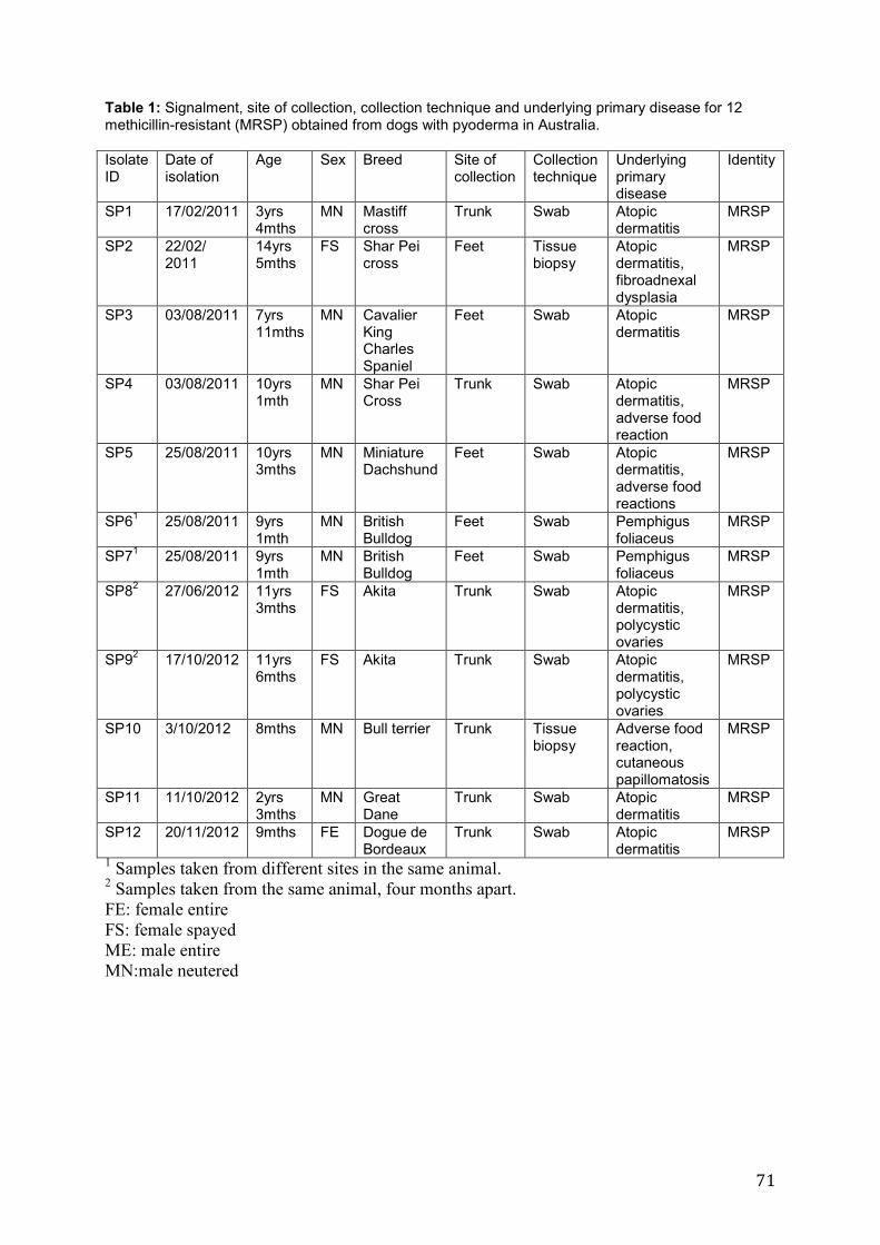
71
Table 1: Signalment, site of collection, collection technique and underlying primary disease for 12 methicillin-resistant (MRSP) obtained from dogs with pyoderma in Australia. Isolate ID
Date of isolation
Age Sex Breed Site of collection
Collection technique
Underlying primary disease
Identity
SP1
17/02/2011 3yrs 4mths
MN Mastiff cross
Trunk Swab Atopic dermatitis
MRSP
SP2 22/02/ 2011
14yrs 5mths
FS Shar Pei cross
Feet Tissue biopsy
Atopic dermatitis, fibroadnexal dysplasia
MRSP
SP3 03/08/2011 7yrs 11mths
MN Cavalier King Charles Spaniel
Feet Swab Atopic dermatitis
MRSP
SP4 03/08/2011 10yrs 1mth
MN Shar Pei Cross
Trunk Swab Atopic dermatitis, adverse food reaction
MRSP
SP5 25/08/2011 10yrs 3mths
MN Miniature Dachshund
Feet Swab Atopic dermatitis, adverse food reactions
MRSP
SP61 25/08/2011 9yrs 1mth
MN British Bulldog
Feet Swab Pemphigus foliaceus
MRSP
SP71 25/08/2011 9yrs 1mth
MN British Bulldog
Feet Swab Pemphigus foliaceus
MRSP
SP82 27/06/2012 11yrs 3mths
FS Akita Trunk Swab Atopic dermatitis, polycystic ovaries
MRSP
SP92 17/10/2012 11yrs 6mths
FS Akita Trunk Swab Atopic dermatitis, polycystic ovaries
MRSP
SP10 3/10/2012 8mths MN Bull terrier Trunk Tissue biopsy
Adverse food reaction, cutaneous papillomatosis
MRSP
SP11 11/10/2012 2yrs 3mths
MN Great Dane
Trunk Swab Atopic dermatitis
MRSP
SP12 20/11/2012 9mths FE Dogue de Bordeaux
Trunk Swab Atopic dermatitis
MRSP
1 Samples taken from different sites in the same animal. 2 Samples taken from the same animal, four months apart.
FE: female entire
FS: female spayed
ME: male entire
MN:male neutered

72
References 1. Morris DO, Boston RC, O'Shea K, et al. The prevalence of carriage of meticillin-resistant staphylococci by veterinary dermatology practice staff and their respective pets. Veterinary Dermatology 2010; 21: 400-407. 2. Bannoehr J, Guardabassi L. Staphylococcus pseudintermedius in the dog: taxonomy, diagnostics, ecology, epidemiology and pathogenicity. Veterinary Dermatology 2012; 23: 253-266 3. van Duijkeren E, Kamphuis M, van der Mije IC, et al. Transmission of methicillin-resistant Staphylococcus pseudintermedius between infected dogs and cats and contact pets, humans and the environment in households and veterinary clinics. Veterinary Microbiology 2011; 150: 338-343. 4. Weese JS. Staphylococcal control in the veterinary hospital. Veterinary Dermatology 2012; 23: 292-298 5. Perreten V, Kadlec K, Schwarz S, et al. Clonal spread of methicillin-resistant Staphylococcus pseudintermedius in Europe and North America: an international multicentre study. The Journal of antimicrobial chemotherapy 2010; 65: 1145-1154. 6. Goering RV, Morrison D, Al-Doori Z, et al. Usefulness of mec-associated direct repeat unit (dru) typing in the epidemiological analysis of highly clonal methicillin-resistant Staphylococcus aureus in Scotland. Clinical Microbiology and Infection 2008; 14: 964-969. 7. Weese JS, Sweetman K, Edson H, et al. Evaluation of minocycline susceptibility of methicillin-resistant Staphylococcus pseudintermedius. Veterinary microbiology 2013; 162: 968-971. 8. Broekema NM, Van TT, Monson TA, et al. Comparison of cefoxitin and oxacillin disk diffusion methods for detection of mecA-mediated resistance in Staphylococcus aureus in a large-scale study. Journal of clinical microbiology 2009; 47: 217-219. 9. Bemis DA, Jones RD, Frank LA, et al. Evaluation of susceptibility test breakpoints used to predict mecA-mediated resistance in Staphylococcus Pseudintermedius Isolated from Dogs. Journal of Veterinary Diagnostic Investigation 2009; 21: 53-58. 10. Bemis DA, Jones RD, Hiatt LE, et al. Comparison of tests to detect oxacillin resistance in Staphylococcus intermedius, Staphylococcus schleiferi, and Staphylococcus aureus isolates from canine hosts. Journal of Clinical Microbiology 2006; 44: 3374-3376. 11. Weese JS, Brisson, B.A. Infection with methicillin-resistant Staphylococcus pseuintermedius masquerading as cefoxitin susceptible in a dog. Journal of the American Veterinary Medical Association 2009; 235: 1064-1066. 12. Schissler JR, Hillier A, Daniels JB, et al. Evaluation of clinical laboratory standards institute interpretive criteria for methicillin-resistant Staphylococcus pseudintermedius Isolated from dogs. Journal of Veterinary Diagnostic Investigation 2009; 21: 684-688. 13. Bemis DA, Jones RD, Videla R, et al. Evaluation of cefoxitin disk diffusion breakpoint for detection of methicillin resistance in Staphylococcus pseudintermedius isolates from dogs. Journal of veterinary diagnostic investigation : official publication of the American Association of Veterinary Laboratory Diagnosticians, Inc 2012; 24: 964-967. A full version of this manuscript has been submitted to the Journal Of Medical Microbiology

73
Use of prednisolone as monotherapy in the treatment of feline pemphigus foliaceus: a retrospective study of 37 cats
Debbie Simpson, Animal Skin Ear and Allergy Clinic, Melbourne Veterinary Specialist Centre,
70 Blackburn Rd, Glen Waverley Vic 3150
Background Prednisone doses of up to 8mg/kg/day have been used to treat feline pemphigus foliaceus (PF). Oral prednisolone has more favourable pharmacokinetics in cats than prednisone therefore lower doses of prednisolone may be effective in treatment of feline PF. Hypothesis/Objectives To assess the dose of prednisolone effective in inducing and maintaining remission of PF in cats. A secondary aim was to assess clinical presentation and epidemiological information regarding feline PF and to compare this with the published veterinary literature. Animals 37 client-owned cats with PF treated with prednisolone monotherapy for induction of remission were included in the study. Methods Retrospective analysis of records of a private veterinary dermatology referral practice between the years of 1995 and 2013 was performed. History, clinical signs, cytological and/or histopathological findings, lack of response to antimicrobials, absence of fungal hyphae on PAS staining and/or negative fungal culture and clear response to immunosuppressive therapy were used to confirm the diagnosis. Cats were included in the study if prednisolone was used as a monotherapy induction protocol. Results 36/37 cats achieved complete remission on prednisolone alone within 8 weeks. The most common induction dose was 1mg/kg twice a day. 21 cases were maintained on prednisolone only at a mean total weekly dose of 2.8mg/kg. 14% of cats discontinued medication completely. Conclusions and clinical importance – Prednisolone at 1mg/kg twice daily induced remission of PF in 97% of cats. Clawfold inflammation and exudate was a common presenting sign. Secondary bacterial overgrowth in clawfolds resolved with immunosuppressive rather than antimicrobial therapy.